Rapid Modulation of PSMA Expression by AndrogenDeprivation: Serial 68Ga-PSMA-11 PET in Men withHormone-Sensitive and Castrate-Resistant ProstateCancer Commencing Androgen Blockade
Louise Emmett1,2, Charlotte Yin1, Megan Crumbaker2, George Hruby3,4, Andrew Kneebone3,4, Richard Epstein1,Quoc Nguyen2, Adam Hickey1, Noah Ihsheish1, Gordon O’Neill5, Lisa Horvath2,6, Venu Chalasani4, Phillip Stricker2,6,and Anthony M. Joshua1,2
1St. Vincent’s Hospital, Sydney, Australia; 2Garvan Institute for Medical Research, Sydney, Australia; 3GenesisCare, Sydney,Australia; 4Royal North Sahore Hospital, Sydney, Australia; 5St. Vincent’s Clinic, Sydney, Australia; and 6Chris O’Brien Life House,Sydney, Australia
Prostate-specific membrane antigen (PSMA) may be targeted for
both diagnostic and therapeutic purposes in the management ofprostate cancer (PCa). In preclinical models, androgen blockade
(AB) increases expression of PSMA in both hormone-sensitive and
castrate-resistant xenotypes. The aim of this study was to evaluate
the effect of AB treatment on 68Ga-PSMA-11 PET imaging in hor-mone-naive (luteinizing hormone-releasing hormone [LHRH] ±bicalutamide) and in castrate-resistant men (enzalutamide or
abiraterone) with metastatic PCa. Methods: Serial 68Ga-PSMA-11PET was prospectively performed at baseline and on days 9, 18,
and 28 in 8 men with measurable metastatic hormone-sensitive PCa
commencing LHRH ± bicalutamide (cohort 1) and 7 men with cas-
trate-resistant PCa commencing either enzalutamide or abiraterone(cohort 2). Gleason score, age, time since diagnosis, and prior treat-
ments were documented. Testosterone and prostate-specific anti-
gen (PSA) were measured at baseline and all imaging time points.
PET/CT was quantitatively analyzed for SUVmax, SUVmean, and totaltumor volume. Results: In cohort 1, a median 30% (interquartile
range [IQR], 5–61) reduction in SUVmax was recorded by day 9 after
AB. A reduction from baseline SUVmax occurred in 86.5% (6/7) menby day 9 (P , 0.04), with an associated PSA response in 100% men
(P , 0.03). Total tumor volume reduced in all men by 74.5% (IQR,
27–97) (P , 0.02). After day 9, PSMA response heterogeneity was
noted, with persistently high or increasing SUVmax in 37.5% (3/8)and marked reduction in 62.5% (5/8). In cohort 2, a median 45%
(IQR, 12.7–66) increase in intensity of PSMA SUV was recorded by
day 9 after AB. All men demonstrated an increase in SUVmax and
SUVmean on PSMA PET compared with baseline (P , 0.04). Thisincrease at day 9 plateaued by day 28. PSA responses were more
delayed in cohort 2 (−15% [IQR, 70−138]), with 2 of 7 men demon-
strating PSA progression. Conclusion: There is rapid dichotomous
response on 68Ga-PSMA PET imaging to AB-dependent on thepresence of a hormone-sensitive or castrate-resistant PCa pheno-
type. This has important implications for interpretation of PSMA
PET, and in the timing and sequencing of PSMA-targeted therapy.
J Nucl Med 2019; 60:950–954DOI: 10.2967/jnumed.118.223099
Prostate-specific membrane antigen (PSMA) is a transmem-brane glycoprotein on the cell surface of prostate cancers (PCas) thatcan be effectively targeted for both PET diagnostic and therapeuticpurposes in the management of PCa. The PSMA receptor has anoncogenic signaling role in the PCa cell, acting on glutamate recep-tors and activating the Pi3K and Akt growth pathways (1–3). Andro-gen blockade (AB) appears to increase expression of PSMA in cellline and mouse models in both hormone-sensitive and castrate-re-sistant states (4). However, there is a paucity of information on theeffect of AB on PSMA PET intensity in vivo, the impact of this effecton the PET imaging results of men commencing androgen depriva-tion therapy, and the timing of these changes. The aim of this studywas to prospectively evaluate the impact of commencing AB on68Ga-PSMA-11 PET quantitative parameters in men with metastaticdisease in the hormone-sensitive setting, or with enzalutamide orabiraterone in men with castrate-resistant PCa.
MATERIALS AND METHODS
Patient Selection
Between August 2017 and July 2018, 15 men with histologically
confirmed, Gleason score (GS) 7–10 PCa with metastatic disease onconventional imaging were prospectively consented and enrolled in the
study. Men were recruited to 1 of 2 cohorts: cohort 1 (n 5 8) includedthose with newly diagnosed hormone-sensitive PCa commencing treat-
ment (luteinizing hormone-releasing hormone [LHRH]6 bicalutamide).Cohort 2 (n 5 7) included men with castrate-resistant PCa commencing
AB with either enzalutamide or abiraterone. Castrate-resistant diseasewas defined as disease progression despite castrate levels of testosterone
(,1.7 nmol/L). Demographic and clinical features including patient age,GS, time since diagnosis, and prior lines of treatment were documented.
Prostate-specific antigen (PSA) and testosterone levels were measured atbaseline and at all imaging time points. Written informed consent was
obtained from all patients before enrolment, and this prospective trial was
Received Nov. 6, 2018; revision accepted Nov. 28, 2018.For correspondence or reprints contact: Louise Emmett, Department of
approved by the St. Vincent’s Hospital Human Research and Ethics
Committee (HREC/17/SVH/25).
PSMA PET/CT
Serial 68Ga-PSMA PET scans were obtained at baseline and ondays 9, 18, and 28 after commencing AB. 68Ga-HBEDD-CC
PSMA-11 (68Ga-PSMA) was produced on-site compliant with goodlaboratory practice procedure using a TRASIS automated radiopharmacy
cassette. Radiopharmacy quality control was undertaken using a high-pressure liquid chromatography method. Patients were injected with 2.0
MBq of 68GaPSMA per kilogram, with imaging parameters (dose, timeafter injection, and imaging protocols) repeated identically at each time
point for each patient enrolled. All PET CT imaging was undertaken
using a Phillips Ingenuity TOF-PET/64 slice CT scanner. A non–con-
trast-enhanced CT scan was acquired 60 min after tracer injection usingthe following CT parameters: 2 mm slice thickness soft-tissue reconstruc-
tion kernel, 120 keV and 50 mAs, pitch of 0.828, 600 mm field of view,and a 512 matrix. Immediately after CT scanning, a whole-body PET
scan was acquired for 2 min per bed position. The emission data werecorrected for randoms, scatter, and decay using the Phillips Body-dynam-
ic.xml and Body.xml reconstruction protocol.
Image Quantitation
Visual and quantitative image analyses was undertaken using MIMMaestro software at each time point. The total number of PSMA-avid
metastatic lesions, intensity, and total metabolic tumor volume weremeasured. SUVmax and mean SUVmean were recorded for each lesion,
and on whole-body assessment to obtain a total-body SUVmax, SUVmean
and total metabolic tumor volume (mLs). Salivary gland SUVmax on
PSMA PET was documented at each time point for each patient.
Statistical Analysis
Descriptive statistics were used to summarize baseline characteristics
and outcomes of interest. The Wilcoxon signed-rank test was used forcomparison of nonparametric data. All tests were 2-sided, and a P value
of 0.05 or less was considered statistically significant. Statistical analysiswas performed with SPSS Statistics, version 24.0 (SPSS Inc.).
RESULTS
Baseline Characteristics
In total, 15 men with histologically confirmed PCa underwentserial 68Ga-PSMA PET scans after commencement of AB. Base-line clinical characteristics and previous treatments are summa-rized in Table 1.
Cohort 1 (Hormone-Sensitive PCa)
LHRH alone was commenced in 1 of 8 and LHRH1 bicalutamide(50 mg daily) in 7 of 8 men (Table 1). All men completed baselinescans before commencing AB. Eighty-seven percent (7/8) of mencompleted the days 9 and 18 scans and 100% the day 28 scan. Allscans were undertaken within 5 d of the predetermined time points.Molecular Imaging Response. There was a median 30%
(interquartile range [IQR], 5–61) reduction in SUVmax by day 9from baseline (P , 0.04) (Table 2). A reduction from baselineSUVmax occurred in 86.5% (6/7) of men by day 9 (Fig. 1). Onepatient had an increase in SUVmax at day 9, with subsequent re-duction by days 18 and 28 (Fig. 2). After day 9, 68Ga-PSMA
TABLE 1Patient Characteristics
Characteristic Cohort 1 Cohort 2
Age (y) 69 (59–82) 72 (59–81)
Time from diagnosis
(mo)
2.3 (0.7–2.7) 24 (11.3–147.4)
Gleason score
7 4/8 2/7
8 1/8 1/7
9–10 3/8 4/7
Baseline PSA 80 (15–148) 9.2 (4.9–490)
Baseline testosterone
(nmol/L)
11.6 (6.5–27.7) 0.3 (0.3–1.6)
Prior treatment
None 8/8 —
LHRH — 7/7
Docetaxel — 2/7
Treatment on trial
LHRH 1 bicalutamide 7/8 —
LHRH 1/8 7/7
Abiraterone — 1/7
Enzalutamide — 6/7
Data in parentheses are IQRs.
TABLE 2Comparison of Clinical and Imaging Parameters between Baseline, Day 9, and Day 28 After Commencing AB in both
Hormone-Sensitive (C1) and Castrate-Resistant (C2) Disease States
Variable Baseline (C1) Day 9 (C1) Day 28 (C1) Baseline (C2) Day 9 (C2) Day 28 (C2)
EFFECT OF ANDROGEN BLOCKADE ON PSMA PET INTENSITY • Emmett et al. 951
response heterogeneity was noted, with persistently high or in-creasing SUVmax in 37.5% (3/8) and marked reduction in 62.5%(5/8) (Figs. 2 and 3). Total body tumor volume reduced significantlyover the 28 d of the study in all men (–74.5% [IQR, –27%–97%])(P , 0.02).PSA Response. There was a marked reduction in PSA in
response to commencing AB by day 28 (291% [IQR, –97.7%–57%]) (Fig. 4). This biochemical response was also observed inthose men with an increase or stable 68Ga-PSMA SUVmax in re-sponse to AB (90% PSA reduction in these 2 men). Testosteronedropped rapidly and was ,1.0 nmol/mL by day 28 in all men.
Cohort 2 (Castrate-Resistant PCa)
All men in cohort 2 had failed previous treatment with LHRHand had not yet received a second-generation androgen-signalinginhibitor. Two (of 7) had received docetaxel. All men commencedtreatment with either enzalutamide (6/7) or abiraterone (1/7) 1 dafter baseline 68Ga-PSMA PET and continued treatment through-out the trial. All men in cohort 2 underwent baseline and days 18and 28 imaging. Six (of 7) completed day 9 imaging.Molecular Imaging Response. A median 45% (IQR, 12.7–66)
increase in SUVmax was recorded by day 9 after AB (Table 2).All men demonstrated an increase in SUVmax and mean on
68Ga-PSMA PET at day 9 compared with baseline (P , 0.04).This increase in SUVmax plateaued by days 18 and 28 (Fig. 5).PSA Response. Median baseline PSA in this cohort was 9.2 (IQR,
4.9–490). PSA response to treatment was more delayed in cohort 2(Fig. 5) compared with cohort 1. There was no change in PSA by day9, but a median drop in PSA of 15% (IQR,270%–1138%) by day28. Two men in cohort 2 demonstrated PSA progression while onstudy.Salivary gland activity was measured in all men at all time points
and was not significantly different from baseline in either cohort.
DISCUSSION
PSMA is a transmembrane glycoprotein that is overexpressedon nearly all PCa cells and that has become an attractive target forboth the imaging and the therapy of PCa at many stages of thisdisease (5–10). Cell line and small-animal work has demonstratedthat the PSMA receptor is manipulable with receptor density influ-enced by androgen receptor blockade (4,11–14). The fact thatexternal factors such as AB are capable of influencing PSMAreceptor density has a considerable impact on 68Ga-PSMA PET’svalue as a diagnostic agent, its ability to monitor treatmentresponse, and its use as a therapeutic target. This study confirms
that 68Ga-PSMA PET is significantly influ-enced by the presence of AB, that thismanipulation of the68Ga-PSMA intensityoccurs early after commencing treatment,and that the effect of AB on PSMA PET ishighly dependent on the castration sensitiv-ity of the PCa cells at the time of imaging.PSMA expression on the PCa cell
surface has repeatedly been reported toincrease with androgen receptor inhibitionin vitro (10–12). Evans et al. demonstratedthat PSMA expression is increased withboth orchidectomy and enzalutamide inmice bearing PSMA-expressing xenografts(11). Hope et al. confirmed that PSMAintensity increased significantly with AB
FIGURE 1. Metastatic bone disease demonstrated marked reduction in PSMA SUVmax from baseline ([A] SUVmax 8) to day 9 ([B] SUVmax 3)
(red arrow), in response to LHRH 1 bicalutamide (patient C in Fig. 2).
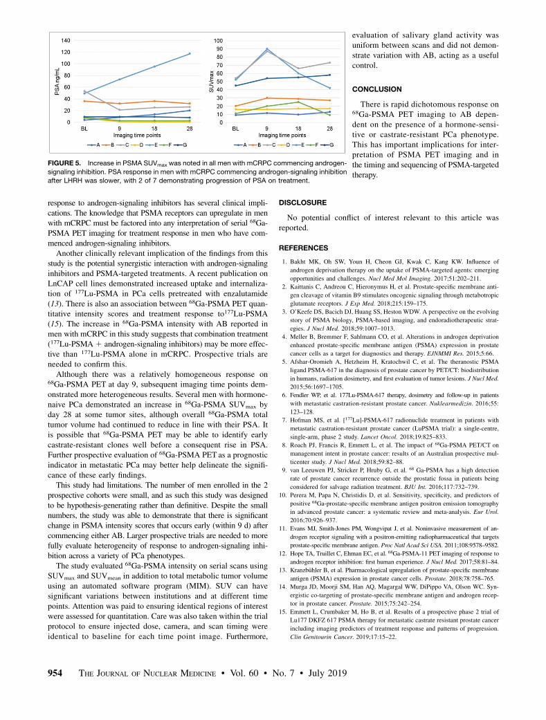
FIGURE 2. Serial-time-point PSMA PET imaging measuring SUVmax (A) and serial serum PSA
(B) in men with hormone-sensitive PCa commencing LHRH ± bicalutamide.
952 THE JOURNAL OF NUCLEAR MEDICINE • Vol. 60 • No. 7 • July 2019
in LNCaP-AR xenograft mouse models, and demonstrated an in-crease in PSMA in a man with PCa despite a concomitant re-duction in PSA in response to 4 wk of AB (12). An increase inPSMA messenger RNA was measured with antiandrogen treat-ment in castrate-sensitive, castrate-resistant, and abiraterone-tolerant castrate-resistant cell lines (4). These previous findingsare quite different from what we found in vivo using serial 68Ga-PSMA PET in hormone-naive men. This study demonstrated that asignificant reduction in 68Ga-PSMA intensity occurred in 86% ofmen as early as day 9 after commencing initial treatment with AB.This finding was unexpected, given the in vitro findings and isprobably multifactorial. A reduction in PSMA intensity in mencommencing first-line AB is likely explained by the strong anti-proliferative effect of AB on most PCa phenotypes in the early(hormone-naive) stages of disease. The marked reduction in 68Ga-PSMA intensity echoed the marked reduction in PSA levels inthese men. Although there may have been a cellular increase inPSMA receptor activity in some cells, the marked involutory re-sponse of hormone-naive PCa cells to AB has overshadowed
this, with a resultant overall rapid reduction in 68Ga-PSMA PETintensity. Furthermore, it may be that the mouse xenotypes andcell lines used previously have represented a more aggressive, lesstreatment-responsive PCa phenotype than those of the men en-rolled in this study.This rapid reduction in 68Ga-PSMA PET intensity identified in
men with hormone-sensitive disease commencing AB has impor-tant clinical considerations. PSMA PET imaging may significantlyunderestimate the volume of metastatic disease in hormone-naivemen who have commenced on AB even within 9 d. Accuratestaging with PSMA PET must therefore occur before commencingAB to ensure adequate imaging sensitivity.In contrast, all men with metastatic castration-resistant prostate
cancer (mCRPC) commencing androgen-signaling inhibition dem-onstrated an increase in intensity of 68Ga-PSMA PET by day 9compared with baseline. This finding is consistent with the pre-clinical results seen in mouse and cell line work, likely due to theslower treatment response of PCa to second- (or third-) line treat-ment. This increase in 68Ga-PSMA quantitative intensity scores in
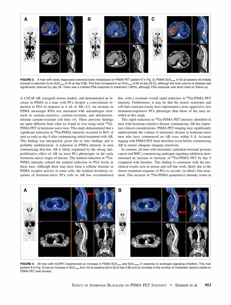
FIGURE 3. A man with newly diagnosed extensive bone metastases on PSMA PET (patient E in Fig. 2). PSMA SUVmax of 35 at baseline (A) initially
showed a reduction to an SUVmax of 20 at day 9 (B). This then increased to an SUVmax of 65 at day 28 (C), although the total volume of disease was
significantly reduced by day 28. There was a marked PSA response to treatment (−90%), although PSA response was short lived on follow-up.
FIGURE 4. All men with mCRPC experienced an increase in PSMA SUVmax and SUVmean in response to androgen-signaling inhibition. This man
(patient B in Fig. 5) had an increase in SUVmax from 20 at baseline (A) to 30 at day 9 (B) and an increase in the number of metastatic lesions visible on
PSMA PET (red arrows).
EFFECT OF ANDROGEN BLOCKADE ON PSMA PET INTENSITY • Emmett et al. 953
response to androgen-signaling inhibitors has several clinical impli-cations. The knowledge that PSMA receptors can upregulate in menwith mCRPC must be factored into any interpretation of serial 68Ga-PSMA PET imaging for treatment response in men who have com-menced androgen-signaling inhibitors.Another clinically relevant implication of the findings from this
study is the potential synergistic interaction with androgen-signalinginhibitors and PSMA-targeted treatments. A recent publication onLnCAP cell lines demonstrated increased uptake and internaliza-tion of 177Lu-PSMA in PCa cells pretreated with enzalutamide(13). There is also an association between 68Ga-PSMA PET quan-titative intensity scores and treatment response to177Lu-PSMA(15). The increase in 68Ga-PSMA intensity with AB reported inmen with mCRPC in this study suggests that combination treatment(177Lu-PSMA1 androgen-signaling inhibitors) may be more effec-tive than 177Lu-PSMA alone in mCRPC. Prospective trials areneeded to confirm this.Although there was a relatively homogeneous response on
68Ga-PSMA PET at day 9, subsequent imaging time points dem-onstrated more heterogeneous results. Several men with hormone-naive PCa demonstrated an increase in 68Ga-PSMA SUVmax byday 28 at some tumor sites, although overall 68Ga-PSMA totaltumor volume had continued to reduce in line with their PSA. Itis possible that 68Ga-PSMA PET may be able to identify earlycastrate-resistant clones well before a consequent rise in PSA.Further prospective evaluation of 68Ga-PSMA PET as a prognosticindicator in metastatic PCa may better help delineate the signifi-cance of these early findings.This study had limitations. The number of men enrolled in the 2
prospective cohorts were small, and as such this study was designedto be hypothesis-generating rather than definitive. Despite the smallnumbers, the study was able to demonstrate that there is significantchange in PSMA intensity scores that occurs early (within 9 d) aftercommencing either AB. Larger prospective trials are needed to morefully evaluate heterogeneity of response to androgen-signaling inhi-bition across a variety of PCa phenotypes.The study evaluated 68Ga-PSMA intensity on serial scans using
SUVmax and SUVmean in addition to total metabolic tumor volumeusing an automated software program (MIM). SUV can havesignificant variations between institutions and at different timepoints. Attention was paid to ensuring identical regions of interestwere assessed for quantitation. Care was also taken within the trialprotocol to ensure injected dose, camera, and scan timing wereidentical to baseline for each time point image. Furthermore,
evaluation of salivary gland activity wasuniform between scans and did not demon-strate variation with AB, acting as a usefulcontrol.
CONCLUSION
There is rapid dichotomous response on68Ga-PSMA PET imaging to AB depen-dent on the presence of a hormone-sensi-tive or castrate-resistant PCa phenotype.This has important implications for inter-pretation of PSMA PET imaging and inthe timing and sequencing of PSMA-targetedtherapy.
DISCLOSURE
No potential conflict of interest relevant to this article wasreported.
REFERENCES
1. Bakht MK, Oh SW, Youn H, Cheon GJ, Kwak C, Kang KW. Influence of
androgen deprivation therapy on the uptake of PSMA-targeted agents: emerging
opportunities and challenges. Nucl Med Mol Imaging. 2017;51:202–211.