Cell Signaling DOI: 10.1002/anie.201301219 Rapidly Reversible Manipulation of Molecular Activity with Dual Chemical Dimerizers** Yu-Chun Lin,* Yuta Nihongaki, Tzu-Yu Liu, Shiva Razavi, Moritoshi Sato, and Takanari Inoue* The four main characteristics of cellular signaling events are that they are rapid, local, specific, and reversible. With these features, cells spatiotemporally choreograph dynamic signal- ing. In particular, reversibility enables cells to adjust the duration of a signaling event and efficiently utilize their finite resources. This characteristic is exemplified by small GTPases (enzymes that hydrolyze guanosine triphosphate, GTP) and phosphatidylinositol lipids, which trigger diverse cellular processes, including proliferation, transformation, migration, and apoptosis. [1] To generate the precise command for each function, these signaling molecules are tightly regulated by a pair of enzymes that switch their activity on or off: guanine nucleotide exchange factors (GEFs) and GTPase-activating proteins for small GTPases, and phosphatidylinositol kinases and phosphatases for phosphatidylinositol lipids. [1b, 2] The chemically inducible dimerization (CID) technique has been widely used to rapidly manipulate molecular activities. [3] In a CID system, a chemical dimerizer, such as rapamycin (Scheme 1 a), induces the dimerization of two proteins: FK506 binding protein (FKBP) and the FKBP12– rapamycin binding protein (FRB). When FKBP is prelocal- ized to the plasma membrane and the FRB-fused protein of interest (FRB–POI) to the cytosol, rapamycin-induced dime- rization results in the relocation of cytosolic FRB–POI to the plasma membrane (left and middle panels in Scheme 1 b). [4] The accumulation of the POI at the plasma membrane subsequently triggers a biological effect that is pertinent to the specific POI molecule. The entire process can be induced on a timescale of seconds in intact living cells. Owing to the rapid, local, and specific induction of signaling, the CID technique has proven powerful and versatile as an exper- imental perturbation tool. To fulfill the fourth characteristic of signaling, namely, reversibility, one may consider washing rapamycin out to dissociate the dimerized complex. However, the clearance of rapamycin from cells is extremely slow. [5] Furthermore, the binding affinity between rapamycin and FKBP is extremely high (200 pm). [3b,c, 6] Accordingly, once rapamycin-induced manipulation has been turned on, it is challenging to turn it off on a comparable timescale. [3b,c, 7] Nevertheless, the rapamycin-dissociation kinetics should be a function of experimental conditions, such as the washout protocol, cell type, dimerizer concentration, and the protein configuration and expression level of both the FKBP and FRB constructs. Therefore, we began to evaluate the rever- sibility of CID by using a series of CID probes previously developed in our laboratory. Specifically, we co-transfected COS-7 cells with fluorescently tagged FKBP and FRB proteins that each reside in a distinct compartment within the cell: CFP–FRB (CFP = cyan fluorescent protein) is cyotosolic, whereas YFP–FKBP (YFP = yellow fluorescent Scheme 1. a) Structure of the dimerizers rapamycin (Rapa) and GA 3 - AM used in this study. b) Schematic representation of the rapid, local, specific, and reversible modulation of molecular activity by dual CID systems: Rapamycin binds to FKBP and traps FRB, and thus causes the relocation of CFP–FRB–POI from the cytosol to the GAIs–YFP– FKBP–C2(LACT)-labeled plasma membrane and the activation of the POI-dependent signaling event at the plasma membrane (as indicated by “ON”). The subsequent addition of GA 3 -AM induces dimerization between the GAIs and GID1 and thus results in relocation of the GAIs–YFP–FKBP–C2(LACT)/rapamycin/CFP–FRB–POI complex as a whole from the plasma membrane to the Tom20–mCherry–GID1- labeled mitochondria and termination of the POI-dependent signaling event at the plasma membrane (as indicated by “OFF”). [*] Dr. Y.-C. Lin, T.-Y. Liu, S. Razavi, Prof. T. Inoue Department of Cell Biology, Center for Cell Dynamics Johns Hopkins University, Baltimore, MD 21205 (USA) E-mail: [email protected][email protected]Homepage: http://www.jhu.edu/inouelab Y. Nihongaki, Prof. M. Sato Department of General Systems Studies Graduate School of Arts and Sciences, University of Tokyo (Japan) T.-Y. Liu Emory College of Arts and Sciences, Atlanta, GA 30322 (USA) S. Razavi Department of Biomedical Engineering, Johns Hopkins University Baltimore, MD 21205 (USA) Prof. T. Inoue JST, 4-1-8 Honcho Kawaguchi, Saitama 332-0012 (Japan) [**] We thank Dr. Jun Liu for FK506. We also thank Siew Cheng Phua for careful proofreading of the manuscript. This study was supported in part by the National Institutes of Health (NIH) (grant GM092930 to T.I.) and by JST (10216). Supporting information for this article is available on the WWW under http://dx.doi.org/10.1002/anie.201301219. A ngewandte Chemi e 1 Angew. Chem. Int. Ed. 2013, 52,1–6 # 2013 Wiley-VCH Verlag GmbH & Co. KGaA, Weinheim These are not the final page numbers! Ü Ü
Transcript
Cell SignalingDOI: 10.1002/anie.201301219
Rapidly Reversible Manipulation of Molecular Activity with DualChemical Dimerizers**Yu-Chun Lin,* Yuta Nihongaki, Tzu-Yu Liu, Shiva Razavi, Moritoshi Sato, and Takanari Inoue*
The four main characteristics of cellular signaling events arethat they are rapid, local, specific, and reversible. With thesefeatures, cells spatiotemporally choreograph dynamic signal-ing. In particular, reversibility enables cells to adjust theduration of a signaling event and efficiently utilize their finiteresources. This characteristic is exemplified by small GTPases(enzymes that hydrolyze guanosine triphosphate, GTP) andphosphatidylinositol lipids, which trigger diverse cellularprocesses, including proliferation, transformation, migration,and apoptosis.[1] To generate the precise command for eachfunction, these signaling molecules are tightly regulated bya pair of enzymes that switch their activity on or off: guaninenucleotide exchange factors (GEFs) and GTPase-activatingproteins for small GTPases, and phosphatidylinositol kinasesand phosphatases for phosphatidylinositol lipids.[1b, 2]
The chemically inducible dimerization (CID) techniquehas been widely used to rapidly manipulate molecularactivities.[3] In a CID system, a chemical dimerizer, such asrapamycin (Scheme 1a), induces the dimerization of twoproteins: FK506 binding protein (FKBP) and the FKBP12–rapamycin binding protein (FRB). When FKBP is prelocal-ized to the plasma membrane and the FRB-fused protein ofinterest (FRB–POI) to the cytosol, rapamycin-induced dime-rization results in the relocation of cytosolic FRB–POI to theplasma membrane (left and middle panels in Scheme 1b).[4]
The accumulation of the POI at the plasma membranesubsequently triggers a biological effect that is pertinent tothe specific POI molecule. The entire process can be induced
on a timescale of seconds in intact living cells. Owing to therapid, local, and specific induction of signaling, the CIDtechnique has proven powerful and versatile as an exper-imental perturbation tool. To fulfill the fourth characteristicof signaling, namely, reversibility, one may consider washingrapamycin out to dissociate the dimerized complex. However,the clearance of rapamycin from cells is extremely slow.[5]
Furthermore, the binding affinity between rapamycin andFKBP is extremely high (200 pm).[3b,c,6] Accordingly, oncerapamycin-induced manipulation has been turned on, it ischallenging to turn it off on a comparable timescale.[3b,c,7]
Nevertheless, the rapamycin-dissociation kinetics shouldbe a function of experimental conditions, such as the washoutprotocol, cell type, dimerizer concentration, and the proteinconfiguration and expression level of both the FKBP andFRB constructs. Therefore, we began to evaluate the rever-sibility of CID by using a series of CID probes previouslydeveloped in our laboratory. Specifically, we co-transfectedCOS-7 cells with fluorescently tagged FKBP and FRBproteins that each reside in a distinct compartment withinthe cell: CFP–FRB (CFP = cyan fluorescent protein) iscyotosolic, whereas YFP–FKBP (YFP = yellow fluorescent
Scheme 1. a) Structure of the dimerizers rapamycin (Rapa) and GA3-AM used in this study. b) Schematic representation of the rapid, local,specific, and reversible modulation of molecular activity by dual CIDsystems: Rapamycin binds to FKBP and traps FRB, and thus causesthe relocation of CFP–FRB–POI from the cytosol to the GAIs–YFP–FKBP–C2(LACT)-labeled plasma membrane and the activation of thePOI-dependent signaling event at the plasma membrane (as indicatedby “ON”). The subsequent addition of GA3-AM induces dimerizationbetween the GAIs and GID1 and thus results in relocation of theGAIs–YFP–FKBP–C2(LACT)/rapamycin/CFP–FRB–POI complex asa whole from the plasma membrane to the Tom20–mCherry–GID1-labeled mitochondria and termination of the POI-dependent signalingevent at the plasma membrane (as indicated by “OFF”).
[*] Dr. Y.-C. Lin, T.-Y. Liu, S. Razavi, Prof. T. InoueDepartment of Cell Biology, Center for Cell DynamicsJohns Hopkins University, Baltimore, MD 21205 (USA)E-mail: [email protected]
Y. Nihongaki, Prof. M. SatoDepartment of General Systems StudiesGraduate School of Arts and Sciences, University of Tokyo (Japan)
T.-Y. LiuEmory College of Arts and Sciences, Atlanta, GA 30322 (USA)
S. RazaviDepartment of Biomedical Engineering, Johns Hopkins UniversityBaltimore, MD 21205 (USA)
Prof. T. InoueJST, 4-1-8 Honcho Kawaguchi, Saitama 332-0012 (Japan)
[**] We thank Dr. Jun Liu for FK506. We also thank Siew Cheng Phua forcareful proofreading of the manuscript. This study was supported inpart by the National Institutes of Health (NIH) (grant GM092930 toT.I.) and by JST (10216).
Supporting information for this article is available on the WWWunder http://dx.doi.org/10.1002/anie.201301219.
protein) is expressed at the plasma membrane once fused withthe Lyn N-terminal signal sequence (Lyn–YFP–FKBP). Wethen added rapamycin to induce the relocation of CFP–FRBto the plasma membrane (see Figure S1 a in the SupportingInformation). To evaluate the dissociation kinetics of theinduced dimerization complex, we then washed away therapamycin-containing medium ten times. In each washingstep, the washout efficiency was over 95%; thus, extracellularrapamycin was diluted theoretically by a factor of 1015. Even3 h after the extensive washout procedure, CFP–FRBremained primarily at the plasma membrane (see Fig-ure S1b). We then took a more proactive approach and usedchemical competitors of rapamycin.[8] In one study, FK506reversed rapamycin-induced dimerization reasonably well ona timescale of 10 min in HEK-293T cells.[9] To test theefficiency of this competition strategy in our dimerizationsystem, we first treated cells with rapamycin to induce therapid relocation of CFP–FRB from the cytosol to the Lyn–YFP–FKBP-expressing plasma membrane. Subsequently, weadded FK506 at increasing concentrations to compete withthe interaction between FKBP and rapamycin. However,even treatment for 1 h with FK506 failed to dislodge CFP–FRB from the plasma membrane (see Figure S2). Otherrapamycin competitors, such as FK506M and synthetic ligandof FKBP (SLF) also had little to no effect (data not shown).Thus, neither extensive washout nor an excess of a competitorcould reverse the rapamycin-induced dimerization on a time-scale of seconds. These results highlight the inherent limi-tations of these approaches.
We therefore explored a different strategy to induce therapid reversal of dimerization-induced cellular effects. Wereasoned that the sequestration of a whole dimerized complexcontaining a POI from the plasma membrane could deacti-vate the POI-triggered effect. We recently developed a novelCID system that is completely orthogonal to the existingrapamycin system.[4b] This new system employs GA3-AM(Scheme 1a) as a chemical dimerizer that induces the rapiddimerization of GAIs (N-terminal 92 amino acids fromgibberellin-insensitive) and GID1 (gibberellin-insensitivedwarf 1).[4b] We believed it might be possible to induce therelocation of the POI to the plasma membrane withrapamycin, and then to relocate the rapamycin-induceddimerization complex as a whole to other places in cells,such as mitochondria, by the use of GA3-AM (Scheme 1b,middle and right panels). With this double-relocation strategy,it is crucial that FKBP is localized to the plasma membranefirmly enough that the FRB–POI fusion protein is accumu-lated for the induction of biological effects, but looselyenough to be dissociated from the plasma membrane:a delicate criterion that the Lyn sequence does not satisfy asa membrane anchor. We therefore chose to use a lipid-bindingdomain, such as the C2 domain from lactadherin (C2-(LACT)), as the membrane anchor.[10] C2(LACT) binds tophosphatidylserine and primarily localizes to the inner leafletof the plasma membrane, yet shuttles between membrane andcytosol with biased retention at the membrane.[11]
We first evaluated C2(LACT) as a targeting motif for thedouble-relocation strategy. To this end, we introduced threeconstructs into the COS-7 cells: CFP–FRB, GAIs–YFP–
FKBP–C2(LACT), and Tom20–mCherry–GID1 (Tom20 isa mitochondrial transmembrane protein; mCherry is a redfluorescent protein). These fusion proteins localized to thecytosol, the plasma membrane, and the mitochondria, respec-tively (Figure 1a). The addition of rapamycin induced rapidaccumulation of CFP–FRB at the plasma membrane (Fig-ure 1a; see also Movie S1 in the Supporting Information).Upon the subsequent addition of a second dimerizer, GA3-AM, both CFP and YFP fluorescence decreased at the plasmamembrane, with a concomitant increase at the mitochondria(Figure 1a,b; see also Movie S1); these observations indi-cated that the rapamycin-induced dimerization complex (i.e.,CFP–FRB and GAIs–YFP–FKBP–C2(LACT)) had beensuccessfully sequestered to the mitochondria through GA3-
Figure 1. Double relocation of a cytosolic protein by dual CID systems.a) GAIs–YFP–FKBP–C2(LACT) (green), CFP–FRB (blue), and Tom20–mCherry–GID1 (red) were expressed in COS-7 cells. Treatment withrapamycin (100 nm) induced the relocation of CFP–FRB to the plasmamembrane. Subsequent treatment with GA3-AM (10 mm) inducedrelocation of the GAI–YFP–FKBP–C2(LACT)/rapamycin/CFP–FRB com-plex to the mitochondria. Insets show enlarged views of the regions inthe dashed boxes with a higher contrast. The rapamycin-inducedrelocation of CFP–FRB to the plasma membrane is highlighted by redarrowheads. Scale bar: 10 mm. b) The fluorescence intensity of CFP–FRB in the regions of interest was measured at the indicated timepoints. The values indicate fluorescence intensity divided by the initialfluorescence. Error bars represent the standard error of the mean(SEM; n= 10 cells from three independent experiments).
AM-induced dimerization with Tom20–mCherry–GID1. Thekinetics of the CFP–FRB relocations were quantified bycalculating the time required for half-maximal accumulation(t1/2) at the plasma membrane ((26.7� 4.6) s for the initialrelocation) and the mitochondria ((97.8� 17.3) s for thesecond relocation; Figure 1b). Whereas the rate of the initialrelocation to the plasma membrane reflects rapamycin-induced dimerization alone, that of the second relocationfrom the plasma membrane is a function of two factors: thedissociation of C2(LACT) from the plasma membrane andthe dimerization of the GAIs and GID1. In control experi-ments, we replaced one of the chemical dimerizers withdimethyl sulfoxide and observed a single relocation event forCFP–FRB (see Figure S3 a–c). We also confirmed that theFRB domain is indispensable for the dual relocation strategy(see Figure S3d,e). These results clearly demonstrate thatdual CID systems can rapidly sequester the initial dimerizedcomplex as a whole.
To test whether this double-relocation strategy can beused to rapidly turn on and then off molecular activity, we firstchose the small GTPase Rac as a model. Previously, weshowed that the recruitment of a Rac-specific GEF, Tiam1, tothe plasma membrane activates endogenous Rac and inducesmembrane ruffling in a “sustained” manner.[4a, 12] For “tran-sient” Rac activation, we generated CFP–FRB–Tiam1, whichwas co-transfected into cells with GAIs–YFP–FKBP–C2-(LACT), Tom20–mCherry–GID1, and mCherry–CAAX (aplasma-membrane marker). Treatment with rapamycin trig-gered the accumulation of CFP–FRB–Tiam1 at the plasmamembrane (Figure 2a; see also Movie S2). Upon the subse-quent addition of GA3-AM, the rapamycin-induced dimeri-zation complex (i.e., CFP–FRB–Tiam1 and GAIs–YFP–FKBP–C2(LACT)) was rapidly recruited from the plasmamembrane to the mitochondria (Figure 2a; see alsoMovie S2). Quantitative analysis of mCherry fluorescence inthe plasma-membrane region indicated that the initialdimerization significantly increased membrane ruffling, andthe subsequent dimerization decreased this ruffling to thebasal level (Figure 2 b–d). These results clearly demonstratethat the double-relocation strategy can be used to rapidly turnon and off the Rac signaling pathway.
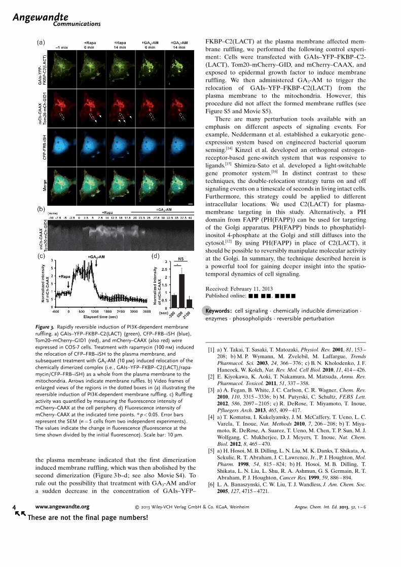
We next determined whether this strategy could beapplied to other signaling molecules. We showed previouslythat the recruitment of an inter-SH2 domain (iSH) froma regulatory PI3K subunit, p85, to the plasma membraneactivates phosphatidylinositol 3-kinase (PI3K) and producesphosphatidylinositol 3,4,5-trisphosphate (PIP3), which resultsin membrane ruffling.[13] We therefore constructed CFP–FRB–iSH, which was co-transfected with Lyn–mCherry–FKBP and the YFP-labeled PH domain from AKT (YFP–PH(AKT)) as a PIP3 biosensor. The accumulation of CFP–FRB–iSH at the plasma membrane upon the addition ofrapamycin was followed by the accumulation of YFP–PH-(AKT) at the plasma membrane and the “sustained” induc-tion of membrane ruffles (see Figure S4 and Movie S3). Wethen co-transfected CFP–FRB–iSH with GAIs–YFP–FKBP–C2(LACT), Tom20–mCherry–GID1, and mCherry–CAAXfor reversibility experiments. After the rapamycin-induceddimerization of the FKBP and FRB fusion proteins, we added
GA3-AM, which triggered the relocation of the dimerizedcomplex of CFP–FRB–iSH and GAIs–YFP–FKBP–C2-(LACT) from the plasma membrane to the mitochondria(Figure 3a). Quantitative analysis of mCherry fluorescence at
Figure 2. Rapidly reversible induction of Rac-dependent membraneruffling. a) GAIs–YFP–FKBP–C2(LACT) (green), CFP–FRB–Tiam1(blue), Tom20–mCherry–GID1 (red), and mCherry–CAAX (also red)were expressed in COS-7 cells. After serum starvation for 2 h, theaddition of rapamycin (100 nm) induced the relocation of CFP–FRB–Tiam1 to the plasma membrane, and subsequent treatment with GA3-AM (10 mm) induced the relocation of the chemically dimerizedcomplex (i.e., GAIs–YFP–FKBP–C2(LACT)/rapamycin/CFP–FRB–Tiam1) as a whole from the plasma membrane to the mitochondria.Arrows indicate membrane ruffles. b) Video frames of enlarged viewsof the regions in the dotted boxes in (a) illustrating the reversibleinduction of Rac-dependent membrane ruffling. c) Ruffling activity wasquantified by measuring the fluorescence intensity of mCherry–CAAXat the cell periphery. d) Fluorescence intensity of mCherry–CAAX at theindicated time points. ** p<0.01. Error bars represent the SEM (n =8cells from three independent experiments). The values indicate thechange in fluorescence (fluorescence at the time shown divided by theinitial fluorescence). Scale bar: 10 mm.
the plasma membrane indicated that the first dimerizationinduced membrane ruffling, which was then abolished by thesecond dimerization (Figure 3b–d; see also Movie S4). Torule out the possibility that treatment with GA3-AM and/ora sudden decrease in the concentration of GAIs–YFP–
FKBP–C2(LACT) at the plasma membrane affected mem-brane ruffling, we performed the following control experi-ment: Cells were transfected with GAIs–YFP–FKBP–C2-(LACT), Tom20–mCherry–GID, and mCherry–CAAX, andexposed to epidermal growth factor to induce membraneruffling. We then administered GA3-AM to trigger therelocation of GAIs–YFP–FKBP–C2(LACT) from theplasma membrane to the mitochondria. However, thisprocedure did not affect the formed membrane ruffles (seeFigure S5 and Movie S5).
There are many perturbation tools available with anemphasis on different aspects of signaling events. Forexample, Neddermann et al. established a eukaryotic gene-expression system based on engineered bacterial quorumsensing.[14] Kinzel et al. developed an orthogonal estrogen-receptor-based gene-switch system that was responsive toligands.[15] Shimizu-Sato et al. developed a light-switchablegene promoter system.[16] In distinct contrast to thesetechniques, the double-relocation strategy turns on and offsignaling events on a timescale of seconds in living intact cells.Furthermore, this strategy could be applied to differentintracellular locations. We used C2(LACT) for plasma-membrane targeting in this study. Alternatively, a PHdomain from FAPP (PH(FAPP)) can be used for targetingof the Golgi apparatus. PH(FAPP) binds to phosphatidyl-inositol 4-phosphate at the Golgi and still diffuses into thecytosol.[12] By using PH(FAPP) in place of C2(LACT), itshould be possible to reversibly manipulate molecular activityat the Golgi. In summary, the technique described herein isa powerful tool for gaining deeper insight into the spatio-temporal dynamics of cell signaling.
Received: February 11, 2013Published online: && &&, &&&&
[1] a) Y. Takai, T. Sasaki, T. Matozaki, Physiol. Rev. 2001, 81, 153 –208; b) M. P. Wymann, M. Zvelebil, M. Laffargue, TrendsPharmacol. Sci. 2003, 24, 366 – 376; c) B. N. Kholodenko, J. F.Hancock, W. Kolch, Nat. Rev. Mol. Cell Biol. 2010, 11, 414 – 426.
[2] E. Kiyokawa, K. Aoki, T. Nakamura, M. Matsuda, Annu. Rev.Pharmacol. Toxicol. 2011, 51, 337 – 358.
[3] a) A. Fegan, B. White, J. C. Carlson, C. R. Wagner, Chem. Rev.2010, 110, 3315 – 3336; b) M. Putyrski, C. Schultz, FEBS Lett.2012, 586, 2097 – 2105; c) R. DeRose, T. Miyamoto, T. Inoue,Pfluegers Arch. 2013, 465, 409 – 417.
[4] a) T. Komatsu, I. Kukelyansky, J. M. McCaffery, T. Ueno, L. C.Varela, T. Inoue, Nat. Methods 2010, 7, 206 – 208; b) T. Miya-moto, R. DeRose, A. Suarez, T. Ueno, M. Chen, T. P. Sun, M. J.Wolfgang, C. Mukherjee, D. J. Meyers, T. Inoue, Nat. Chem.Biol. 2012, 8, 465 – 470.
[5] a) H. Hosoi, M. B. Dilling, L. N. Liu, M. K. Danks, T. Shikata, A.Sekulic, R. T. Abraham, J. C. Lawrence, Jr., P. J. Houghton, Mol.Pharm. 1998, 54, 815 – 824; b) H. Hosoi, M. B. Dilling, T.Shikata, L. N. Liu, L. Shu, R. A. Ashmun, G. S. Germain, R. T.Abraham, P. J. Houghton, Cancer Res. 1999, 59, 886 – 894.
[6] L. A. Banaszynski, C. W. Liu, T. J. Wandless, J. Am. Chem. Soc.2005, 127, 4715 – 4721.
Figure 3. Rapidly reversible induction of PI3K-dependent membraneruffling. a) GAIs–YFP–FKBP–C2(LACT) (green), CFP–FRB–iSH (blue),Tom20–mCherry–GID1 (red), and mCherry–CAAX (also red) wereexpressed in COS-7 cells. Treatment with rapamycin (100 nm) inducedthe relocation of CFP–FRB–iSH to the plasma membrane, andsubsequent treatment with GA3-AM (10 mm) induced relocation of thechemically dimerized complex (i.e., GAIs–YFP–FKBP–C2(LACT)/rapa-mycin/CFP–FRB–iSH) as a whole from the plasma membrane to themitochondria. Arrows indicate membrane ruffles. b) Video frames ofenlarged views of the regions in the dotted boxes in (a) illustrating thereversible induction of PI3K-dependent membrane ruffling. c) Rufflingactivity was quantified by measuring the fluorescence intensity ofmCherry–CAAX at the cell periphery. d) Fluorescence intensity ofmCherry–CAAX at the indicated time points. *p<0.05. Error barsrepresent the SEM (n =5 cells from two independent experiments).The values indicate the change in fluorescence (fluorescence at thetime shown divided by the initial fluorescence). Scale bar: 10 mm.
[7] A. Y. Karpova, D. G. Tervo, N. W. Gray, K. Svoboda, Neuron2005, 48, 727 – 735.
[8] a) T. K. Chakraborty, H. P. Weber, K. C. Nicolaou, Chem. Biol.1995, 2, 157 – 161; b) J. F. Amara, T. Clackson, V. M. Rivera, T.Guo, T. Keenan, S. Natesan, R. Pollock, W. Yang, N. L. Courage,D. A. Holt, M. Gilman, Proc. Natl. Acad. Sci. USA 1997, 94,10618 – 10623; c) J. H. Bayle, J. S. Grimley, K. Stankunas, J. E.Gestwicki, T. J. Wandless, G. R. Crabtree, Chem. Biol. 2006, 13,99 – 107; d) D. M. Spencer, T. J. Wandless, S. L. Schreiber, G. R.Crabtree, Science 1993, 262, 1019 – 1024.
[9] K. B. Lee, J. M. Hwang, I. S. Choi, J. Rho, J. S. Choi, G. H. Kim,S. I. Kim, S. Kim, Z. W. Lee, Angew. Chem. 2011, 123, 1350 –1353; Angew. Chem. Int. Ed. 2011, 50, 1314 – 1317.
[10] W. Cho, R. V. Stahelin, Annu. Rev. Biophys. Biomol. Struct.2005, 34, 119 – 151.
[11] T. Ueno, B. H. Falkenburger, C. Pohlmeyer, T. Inoue, ScienceSignaling 2011, 203, ra87.
[12] S. C. Phua, C. Pohlmeyer, T. Inoue, ACS Chem. Biol. 2012, 7,1950 – 1955.
[13] T. Inoue, T. Meyer, PloS one 2008, 3, e3068.[14] P. Neddermann, C. Gargioli, E. Muraglia, S. Sambucini, F.
Bonelli, R. De Francesco, R. Cortese, EMBO Rep. 2003, 4, 159 –165.
[15] O. Kinzel, D. Fattori, E. Muraglia, P. Gallinari, M. C. Nardi, C.Paolini, G. Roscilli, C. Toniatti, O. Gonzalez Paz, R. Laufer, A.Lahm, A. Tramontano, R. Cortese, R. De Francesco, G.Ciliberto, U. Koch, J. Med. Chem. 2006, 49, 5404 – 5407.
[16] S. Shimizu-Sato, E. Huq, J. M. Tepperman, P. H. Quail, Nat.Biotechnol. 2002, 20, 1041 – 1044.
Y.-C. Lin,* Y. Nihongaki, T.-Y. Liu,S. Razavi, M. Sato,T. Inoue* &&&&—&&&&
Rapidly Reversible Manipulation ofMolecular Activity with Dual ChemicalDimerizers
Tell it where to go : Rapamycin inducedthe relocation of an FRB-fused protein ofinterest (POI) to the plasma membrane(labeled with the fusion protein GAIs–FKBP–C2(LACT)) to activate a signalingevent (see picture). Subsequent treat-ment with a gibberellic acid ester led tothe relocation of the whole GAIs–FKBP–C2(LACT)/rapamycin/FRB–POI complexto the Tom20–GID1-labeled mitochon-dria with the termination of POI-depen-dent signaling.