Hematology 2001 443 Rational Approaches to Design of Therapeutics Targeting Molecular Markers Richard J. Klasa, Alan F. List, and Bruce D. Cheson This paper introduces novel therapeutic strategies focusing on a molecular marker relevant to a particular hematologic malignancy. Four different approaches targeting specific molecules in unique pathways will be presented. The common theme will be rational target selection in a strategy that has reached the early phase of human clinical trial in one malignancy, but with a much broader poten- tial applicability to the technology. In Section I Dr. Richard Klasa presents preclini- cal data on the use of antisense oligonucleotides directed at the bcl-2 gene message to specifically downregulate Bcl-2 protein expression in non- Hodgkin’s lymphomas and render the cells more susceptible to the induction of apoptosis. In Section II Dr. Alan List reviews the targeting of vascular endothelial growth factor (VEGF) and its receptor in anti-angiogenesis strategies for acute myeloid leukemia (AML) and myelodysplastic syndromes (MDS). In Section III Dr. Bruce Cheson describes recent progress in inhibiting cell cycle progression by selectively disrupting cyclin D1 with structurally unique compounds such as flavopiridol in mantle cell lymphoma as well as describing a new class of agents that affect proteasome degradation pathways. I. ANTISENSE OLIGONUCLEOTIDES DIRECTED AT THE BCL-2 GENE MESSAGE IN NON-HODGKIN’S LYMPHOMA Richard J. Klasa, MD* Hematologic malignancies in general and non-Hodgkin’s lymphomas (NHLs) in particular are frequently associ- ated with gain of function mutations, many character- ized by balanced chromosomal translocations. Genome- wide surveys of gene expression are identifying both known and new transcripts that are overexpressed in dif- ferent histological subtypes of lymphoma. 1 As we de- velop molecular classifications of these diseases it is assumed that a few key genes will account for the par- ticular survival advantage conferred on malignant lym- phocytes as compared to their normal counterparts. These genes and their protein products would provide rational targets for the development of therapeutic strategies to reverse this upregulation associated with the malignant phenotype. Non-Hodgkin’s Lymphoma and Bcl-2 Over the past quarter century cytogenetic analysis has identified a number of reciprocal translocations that fre- quently occur in histologically identifiable subtypes of NHL. 2 The transposition of the bcl-2 gene to the immu- noglobulin heavy chain promoter region in the t(14;18) translocation is associated with > 90% of follicular lym- phomas (FL) at diagnosis and 10% of diffuse large B- cell lymphomas (DLBCL), making it the most frequent event identified in NHL. Additionally, 50% of DLBCL overexpress the BCL-2 protein through other mecha- nisms, such as gene duplication, and are associated with a poorer prognosis after anthracycline-based combina- tion chemotherapy. 3 BCL-2 is also overexpressed in mantle cell lymphoma (MCL), chronic lymphocytic leu- kemia (CLL), multiple myeloma (MM) and acute myel- ogenous leukemia (AML). 4 This same widespread pat- tern of distribution is also seen in a variety of solid tu- mors including melanoma, small cell lung carcinoma, and colon carcinoma as well as prostate and breast car- cinoma, especially once the last two are hormone inde- pendent. The obvious conclusion is that BCL-2 overexpression, by whatever means, confers a funda- mental advantage to malignant cells and that disruption of this overexpression might have therapeutic potential. Bcl-2 is an anti-apoptotic member of a large family of genes involved in the regulation of programmed cell death. 5,6 Pro-apoptotic (BAX and BCL-Xs) and anti- * Division of Medical Oncology, British Columbia Cancer Agency, 600 West 10th Avenue, Vancouver BC V52 4E6 Canada Dr. Klasa is on the international advisory boards of Schering AG and Hoffmann-LaRoche, and is on the speakers’ bureau and advisory board for Berlex Canada.
Transcript
Hematology 2001 443
Rational Approaches to Design of TherapeuticsTargeting Molecular Markers
Richard J. Klasa, Alan F. List, and Bruce D. Cheson
This paper introduces novel therapeutic strategiesfocusing on a molecular marker relevant to aparticular hematologic malignancy. Four differentapproaches targeting specific molecules in uniquepathways will be presented. The common themewill be rational target selection in a strategy thathas reached the early phase of human clinical trialin one malignancy, but with a much broader poten-tial applicability to the technology.
In Section I Dr. Richard Klasa presents preclini-cal data on the use of antisense oligonucleotidesdirected at the bcl-2 gene message to specificallydownregulate Bcl-2 protein expression in non-
Hodgkin’s lymphomas and render the cells moresusceptible to the induction of apoptosis.
In Section II Dr. Alan List reviews the targetingof vascular endothelial growth factor (VEGF) and itsreceptor in anti-angiogenesis strategies for acutemyeloid leukemia (AML) and myelodysplasticsyndromes (MDS).
In Section III Dr. Bruce Cheson describesrecent progress in inhibiting cell cycle progressionby selectively disrupting cyclin D1 with structurallyunique compounds such as flavopiridol in mantlecell lymphoma as well as describing a new class ofagents that affect proteasome degradation pathways.
I. ANTISENSE OLIGONUCLEOTIDES DIRECTED AT THE
BCL-2 GENE MESSAGE IN NON-HODGKIN’S LYMPHOMA
Richard J. Klasa, MD*
Hematologic malignancies in general and non-Hodgkin’slymphomas (NHLs) in particular are frequently associ-ated with gain of function mutations, many character-ized by balanced chromosomal translocations. Genome-wide surveys of gene expression are identifying bothknown and new transcripts that are overexpressed in dif-ferent histological subtypes of lymphoma.1 As we de-velop molecular classifications of these diseases it isassumed that a few key genes will account for the par-ticular survival advantage conferred on malignant lym-phocytes as compared to their normal counterparts. Thesegenes and their protein products would provide rationaltargets for the development of therapeutic strategies toreverse this upregulation associated with the malignantphenotype.
Non-Hodgkin’s Lymphoma and Bcl-2Over the past quarter century cytogenetic analysis hasidentified a number of reciprocal translocations that fre-quently occur in histologically identifiable subtypes ofNHL.2 The transposition of the bcl-2 gene to the immu-noglobulin heavy chain promoter region in the t(14;18)translocation is associated with > 90% of follicular lym-phomas (FL) at diagnosis and 10% of diffuse large B-cell lymphomas (DLBCL), making it the most frequentevent identified in NHL. Additionally, 50% of DLBCLoverexpress the BCL-2 protein through other mecha-nisms, such as gene duplication, and are associated witha poorer prognosis after anthracycline-based combina-tion chemotherapy.3 BCL-2 is also overexpressed inmantle cell lymphoma (MCL), chronic lymphocytic leu-kemia (CLL), multiple myeloma (MM) and acute myel-ogenous leukemia (AML).4 This same widespread pat-tern of distribution is also seen in a variety of solid tu-mors including melanoma, small cell lung carcinoma,and colon carcinoma as well as prostate and breast car-cinoma, especially once the last two are hormone inde-pendent. The obvious conclusion is that BCL-2overexpression, by whatever means, confers a funda-mental advantage to malignant cells and that disruptionof this overexpression might have therapeutic potential.
Bcl-2 is an anti-apoptotic member of a large familyof genes involved in the regulation of programmed celldeath.5,6 Pro-apoptotic (BAX and BCL-Xs) and anti-
* Division of Medical Oncology, British Columbia CancerAgency, 600 West 10th Avenue, Vancouver BC V52 4E6Canada
Dr. Klasa is on the international advisory boards of ScheringAG and Hoffmann-LaRoche, and is on the speakers’ bureauand advisory board for Berlex Canada.
444 American Society of Hematology
apoptotic (BCL-2, BCL-XL) molecules reside within the
inner mitochondrial membrane and can homo- andheterodimerize upon appropriate stimulus. These inter-actions control the release of substances such as cyto-chrome C from the mitochondria into the cytosol throughthe opening or closing of specific pores in the membrane,with permeability determined by the relative abundanceof the different molecules. Cytochrome C is central tothe activation of caspases that initiate the apoptotic pro-cess. Thus, an overabundance of BCL-2 can prevent orretard activation of the apoptotic machinery and allowsurvival under conditions that might otherwise be lethalto a cell (Table 1).
Antisense OligonucleotideReverse complementary or “antisense” oligonucleotides(ASOs) are short sequences of single stranded deoxyri-bonucleotides complementary to the coding regions ofa gene that are designed to hybridize by Watson-Crickbase pairing to messenger-RNA (m-RNA) sequences andthus facilitate their degradation.7,8 Naturally occurringantisense sequences have been identified as regulatorsof gene expression in a number of systems, supportingtheir potential for therapeutic development.9,10 The for-mation of a heteroduplex of m-RNA with the DNA ofthe ASO engages RNaseH, an enzyme that proceeds tospecifically cleave off the m-RNA moiety, destroyingthe message and putatively leaving the therapeutic ASOmolecule able to hybridize to another message se-quence.11 This results in a reduction in the target m-RNApool, which subsequently leads to reduction in the spe-cific protein encoded (Figure 1; see color page 551).The presence of the ASO may also prevent the m-RNAfrom appropriately docking with the ribosomal machin-ery that would allow translation into a functional pro-tein. The end result is loss of expression of that proteinin the cell.
ASOs of 16-24 bases in length provide target speci-ficity while shorter or longer sequences can result in ran-dom hybridization within the transcript repertoire. Se-lecting the target areas within a messenger RNA mustultimately take into account its tertiary structure, whichwill determine the accessibility of an area for hybridiza-
tion. These target areas are defined in oligonucleotidearrays where the entire antisense sequence to an m-RNAis displayed in overlapping segments on a slide. The in-tensity of hybridization of the labeled message deter-mines the candidate therapeutic ASOs.12 Screening ofoligonucleotide libraries has also identified RNA sitesthat are most accessible to hybridization and correlatedthese sites with protein downregulation and biologicalfunction.13,14 More empirically, the first 6 codons of theopen reading frame downstream of the AUG start sitehave repeatedly been found to be accessible to hybrid-ization and have been chosen for initial development ofASOs against a number of genes.
As organisms have developed a sophisticated sys-tem for dealing with rogue strands of DNA both insideand outside the cell, the development of therapeuticmolecules required chemical modifications to confernuclease resistance and a favorable pharmacokineticprofile.15,16 Substitutions in the phosphodiester linkageof the bases in the ASO backbone has yielded a numberof molecules now in clinical development with phospho-rothioates being the most widely studied first genera-tion molecules (Figure 2). The sulfur substitution yieldsan ASO that is nuclease resistant and capable of enter-ing the cell. It demonstrates good hybridization kineticsand has little in the way of non-sequence-dependent ef-fects or toxicities at concentrations required to down-regulate the target message. Additionally, although intissue culture a delivery system such as cationic lipid isrequired for efficient intracellular penetration of thesehighly charged molecules, in vivo ASOs have beenshown to be active in free form, possibly due to interac-tion with blood lipoproteins.17,18
The correlation of a biologic effect with the spe-cific downregulation of target message and protein invivo has been a major focus of the development of ASOs.However, ASOs can be very potent immune stimulators,by virtue of unmethylated CpG motifs presented in thecontext of certain flanking sequences, and therapeutic
• Homotypic and heterotypic dimerization within family
• Membrane channel/pore function
• Cytochrome C release from mitochondria via BCL-2 familychannels regulates cell fate under stress
Figure 2. Phosphodiester substitutions in first generationantisense oligonucleotides.
Hematology 2001 445
activity could in part be attributed to nonspecific sys-temic immune effects rather then to a specific ASO/mRNA interaction.19-22 Additionally, structures such asguanosine quartets can demonstrate sequence specificbut non-antisense biological activity in vitro.23,24 Experi-mental designs therefore strive, by the use of appropri-ate control oligonucleotides (sense, missense, one andtwo base mismatch mis-sense) and various strains ofimmunodeficient animals, to isolate effects that can beattributed to the specific downregulation of target mes-sage and protein (Figure 3).
ASOs directed at bcl-2An 18-mer phosphorothiolated oligonucleotide, G3139,directed against the first six codons of the open readingframe of the bcl-2 gene message has been developed byGenta Pharmaceuticals (Figure 3). Studies of G3139 uti-lizing the BCL-2 overexpressing lymphoma cell linesDoHH-2 and SU-DHL-4 in vitro have shown down-regulation of message and protein expression.25 Tumorxenograft models in SCID mice have demonstrated thera-peutic activity that is specific when compared to controlanimals as well as animals treated with reverse polaritysense, 2-base mismatch mis-sense and non-sense oligo-nucleotides.25,26 Extensive pharmakokinetic as well as tox-icity studies have been performed identifying a dose rangewith a good therapeutic index.15,16 These findings supportedthe development of clinical trials using G3139 alone astreatment for BCL-2 overexpressing follicular lymphomas.
Further therapeutic potential is sug-gested by in vitro experiments confirmingthat bcl-2 plays a major role in the responseof malignant cells to various stresses whichproduce cellular damage, including chemo-therapy (Figure 4).4,27,28 Malignant cell linestransfected with a bcl-2 gene with result-ant overexpression of the protein productdemonstrated increased resistance to vari-ous chemotherapeutic agents.29-32 Addition-ally, cell lines overexpressing BCL-2 wererendered more sensitive to killing by che-motherapeutic agents when either antisenseoligonucleotides directed at the bcl-2 mes-sage were introduced into the culture or thecells were transfected with a vector bear-ing the antisense sequence.33,34 This hasbeen correlated with a demonstration ofdownregulation of bcl-2 expression. With
this as background, we set out to test the in vivo combi-nation of ASOs targeting bcl-2 with a cytotoxic agentcommonly used in the treatment of lymphoma.
ASOs to bcl-2 and ChemotherapyEscalating doses of G3139, cyclophosphamide and thecombination of both agents were evaluated in severecombined immunodeficiency (SCID) mice bearing a sys-temic human DoHH2 lymphoma xenograft.35 Experi-ments confirmed that G3139 was able to downregulateBCL-2 expression in vitro and that treatment with G3139alone resulted in prolongation of median survival andcure of some animals. (Figure 5, left panel). This effectwas dose and schedule dependent with no long-term sur-vivors seen when a dose of 5mg/kg was given daily for14 consecutive days as opposed to > 40% when the dosewas increased to 12.5 mg/kg on the same daily scheduleor either 5 or 12.5 mg/kg were administered for 14 treat-
Figure 4. Targeting Bcl-2 may promote apoptosis followingchemotherapy and irradiation.
Figure 5. Survival of cohorts of 6 mice treated with oligonucleotides alone (leftpanel) or with cyclophosphamide (CY) (right panel).
All six surviving animals treated with cyclophosphamide and G3139 sacrificed at 90days with no molecular evidence of disease detected.
Saline=control animals ; AS=antisense oligonucleotide G3139 directed at bcl-2 ;SN=reverse sequence sense control ; MM= 2-base mismatch control.
446 American Society of Hematology
ments on alternate days (28-day schedule). Similarly,cyclophosphamide treatment alone resulted in no long-term survivors at lower doses but was able to cure ani-mals at high doses. The addition of G3139 to low dosecyclophosphamide resulted in the cure of the majorityof animals (Figure 5, right panel). The interaction be-tween the two agents does show dose-response correla-tions; for the two low doses of cyclophosphamide tested,increasing the dose of G3139 from 2.5 to 5 mg/kg re-sulted in longer median survivals and overall increase inlong-term survivors. A rather striking result was achievedwhen a completely ineffective dose of cyclophospha-mide (15 mg/kg, median survival 36 days and no long-term survivors) was combined with a modestly effectivedose of G3139 (2.5 mg/kg, 61 day median survival and16% long-term survivors) to produce a 72-day mediansurvival and 50% long-term survivors. Mice sacrificedat 90 days showed no histological evidence of any dis-ease in all tissues analyzed, including immunoperoxidasestaining for BCL-2, or molecular detection of bcl-2, byPCR. This suggests that chemotherapy at very modestdoses could be made much more effective without in-creasing toxicity when combined with an antisense oli-gonucleotide. Such an increase in the efficacy of cur-rently available agents could significantly alter the prog-nosis of a large number of moderately chemotherapysensitive human tumors, resulting in longer median sur-vivals and increasing the potential for cure.
The model has direct relevance to the clinical situa-tion faced in NHL, where patients typically present witha chemotherapy-sensitive tumor at diagnosis that re-gresses only to recur within months to years post-treat-ment. The DoHH2 cell line was derived from a follicu-lar lymphoma carrying a t(14;18), which results in bcl-2gene overexpression. The aggressive nature of the dis-ease in this model is, however, more suggestive of a trans-formation to a higher-grade histology, a common eventin follicular lymphoma. Indeed, a recent re-explorationof the molecular and cytogenetic features of the cell line,using more sensitive detection techniques, has revealeda second translocation involving the c-myc oncogenewith a resultant derivative chromosome 8 carryingt(8;14;18).36 We have recently described this phenom-enon of double translocation of both bcl-2 and c-myc ina subset of patients with small non-cleaved cell (Burkitt-like) lymphoma, which represents a very aggressive formof the disease.37
Clinical StudiesG3139 has been studied as a single agent in a phase 1trial in heavily pretreated (median of 4 prior regimens)patients with relapsed NHL.38,39 Twenty-one patients withfollicular (9), small lymphocytic (8), diffuse large B-cell (3) or mantle cell (1) lymphomas that expressed
BCL-2 were treated at 8 dose levels ranging from 4.6 to195.8 mg/m2/day by continuous subcutaneous infusionfor 14 consecutive days. No significant toxicity was seenup to doses of 110 mg/m2/day. One complete and 2 mi-nor responses as well as 9 disease stabilizations wereseen in this heavily pretreated group. BCL-2 protein wasdecreased in 7 out of 16 samples examined, including 2from accessible tumor sites and 5 samples of peripheralblood or marrow mononuclear cells.
One study combining a chemotherapeutic agent withG3139 has been reported in metastatic melanoma,40 andstudies are ongoing in a number of other solid tumors(melanoma, prostate carcinoma) and hematological ma-lignancies (myeloma, chronic lymphocytic leukemia, andacute myeloid leukemia). A phase 1 study at our institu-tion in relapsed follicular lymphomas combining esca-lating doses of both cyclophosphamide and G3139 hasnot identified any unexpected toxicity. The last patientenrolled has received cyclophosphamide 750 mg/m2 with2.3 mg/kg/day of G3139 by continuous intravenous in-fusion for 14 consecutive days (Table 2).
ConclusionsThe identification of overexpression or aberrant expres-sion of genes that result in a gain of function, throughgenome wide surveys of cells and tissues in varyingstates, will provide unprecedented insight into the biol-ogy of hematological malignancies. Specific down-regulation of such overexpression with antisense oligo-nucleotides allows disruption of single gene function atthe messenger RNA and protein level and the study ofdownstream events in the involved molecular pathwaysboth in vitro and in vivo. Genes that are critical to thedifferential growth and survival advantage enjoyed bymalignant cells are being identified and are logical thera-peutic targets. The development of ASOs directed at thebcl-2 gene provides a model by which a systemic therapyfor a metastatic disease has been taken from the labora-tory through preclinical studies to early phase clinicaltrials, building on knowledge of this particular gene’srole in cellular apoptosis. Combining multiple antisense
Table 2. Phase I study of G3139 and cyclophosphamide inrelapsed follicular lymphoma.
G3139 mg/kg/day CyclophosphamideCIVI mg/m2/IV
Level (day 1 to 14) (day 8)
1 0.6 250 done
2 1.2 250 done
3 2.3 250 done
4 2.3 500 done
5 2.3 750 ongoing
6 3.1 1000
7 5.0 1000
Hematology 2001 447
strategies with other therapeutic modalities has the po-tential to increase the specificity of the treatments avail-able to our patients, thus improving their efficacy andreducing toxicity.
II. TARGETING ANGIOGENESIS IN HEMATOLOGIC
MALIGNANCIES
Alan F. List, MD*
The seminal observations that the growth and metastaticpotential of solid tumors is dependent upon the forma-tion of new blood vessels triggered an enormous expan-sion in angiogenic research that has yielded novel thera-peutics targeting an array of angiogenic molecules. In-vestigations of the relevance of angiogenesis in hemato-logic malignancies are still at an early stage, but accu-mulating evidence indicates that the angiogenic profileof many hematologic malignancies is distinct from thatof solid tumors. As progeny of a common endothelialand hematopoietic stem cell, hematologic malignanciesmay elaborate and respond to angiogenic factors in aparacrine or autocrine fashion, contributing to tumor cellsurvival and expansion, adhesion, bone resorption andimmune suppression.
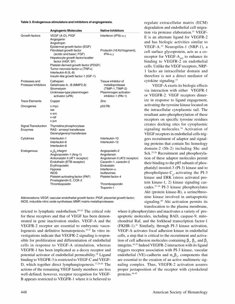
AngiogenesisBlood vessel development is characterized by two dis-tinct biologic processes, vasculogenesis, and angiogen-esis. Vasculogenesis, which is largely restricted to em-bryonic development, involves de novo endothelial celldifferentiation from mesodermal precursors as the pre-requisite for coordinated blood vessel generation.1 An-giogenesis, the process of new blood vessel formationfrom preexisting vessels, is responsible for the genera-tion of neovasculature in adult life, and occurs physi-ologically during wound healing and within female re-productive organs during the menstrual cycle, as well asin pathologic conditions such as proliferative retinopa-thy, arthritic synovium, and human malignancies.2 An-giogenesis is a multistep process that includes both acti-vation and resolution phases. The activation phase isresponsible for the sequential events of basement mem-brane degradation, endothelial cell proliferation andmigration, and capillary lumen formation. The resolu-tion phase is responsible for the maturation and stabili-zation of the newly formed microvasculature throughthe recruitment of pericytes, promotion of basementmembrane reconstitution, and subsequent extinction ofthe endothelial cell mitogenic response. A large numberof pro-angiogenic molecules and endogenous angiogen-
esis inhibitors that coordinate the angiogenic responsehave been delineated (Table 3).3 Vascular endothelialgrowth factor (VEGF), first identified in 1989 and laterisolated from the HL-60 myeloid leukemia cell line, is acritical regulator of vascular development that is respon-sible for activation of endothelial cell proliferation dur-ing vasculogenesis and the direction of capillary sprout-ing during angiogenesis.4 Indeed, gene inactivation stud-ies indicate that VEGF is essential to the neoplastic an-giogenic response.5
VEGF and Receptor Tyrosine KinasesThe VEGF-A molecule is a disulfide-linked homodimerrepresented by five different isoforms generated by al-ternate exon splicing of gene message.6 The correspond-ing VEGF monomers range in size from 121 to 206amino acids, with smaller molecules (i.e., 121, 145, and165 amino acids) representing the secreted and diffusableisoforms, whereas the larger proteins (189, 206 aminoacids) are sequestered by heparin sulfate residues presenton cell surfaces or within the extracellular matrix.7,8 Al-though all isoforms are biologically active, the VEGF
165
isoform predominates and is recognized as a more po-tent and bioavailable endothelial cell mitogen.9,10 Recentinvestigations indicate that the VEGF family is composedof five members in addition to the prototype, VEGF-A,including VEGF-B, VEGF-C, VEGF-D, VEGF-E andPIGF (placental growth factor).6 Within the arterial wall,VEGF is produced by smooth muscle cells in responseto oxidative stress and other stimuli.11 Autocrine pro-duction of VEGF and corresponding receptor upregu-lation is also demonstrable in endothelial cells in responseto hypoxia, nitric oxide, VEGF deprivation and othercellular stresses.12,13
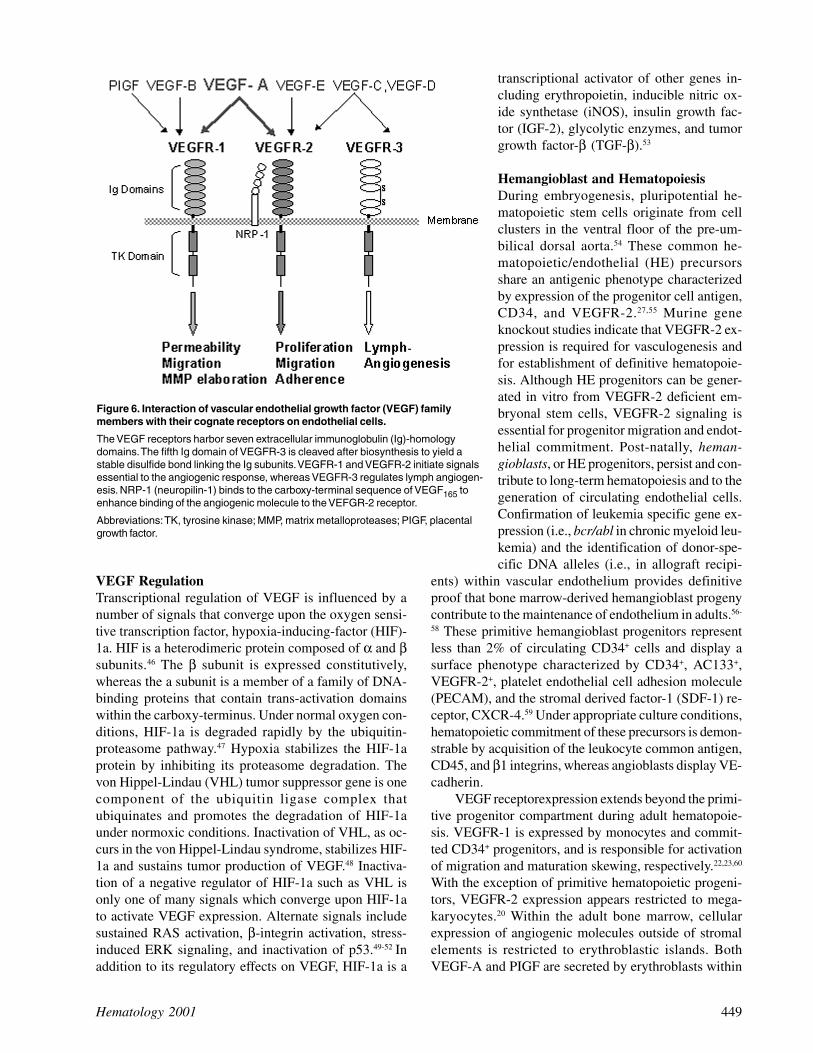
Trophic response to the VEGF family members isdirected by selective interaction with structurally ho-mologous type III receptor tyrosine kinases (RTKs), in-cluding VEGFR-1, originally termed fms-like tyrosinekinase (FLT-1),14 VEGFR-2 or kinase insert domain-con-taining receptor (KDR)/fetal liver kinase-1 (flk-1),15 andthe recently characterized VEGFR-3 or FLT-4 receptor(Figure 6).16 Each of these receptors contains seven ex-tracellular immunoglobulin homology domains that cre-ate the ligand-binding site, a single short transmembrane-spanning sequence, and a cytoplasmic tail that containsthe tyrosine kinase domain akin to that of the c-kit andPDGF-receptors.17,18 The external ligand-binding com-ponent of VEGFR-3 differs from the other VEGF re-ceptors in that the fifth immunoglobulin domain iscleaved during receptor processing to yield covalent,disulfide-linked subunits.19 In adults, VEGFR-1 andVEGFR-2 expression is limited to the vascular endothe-lium, monocytes (VEGFR-1), and primitive hematopoi-etic precursors (VEGFR-2), whereas VEGFR-3 is re-
* Arizona Cancer Center, 1515 N Campbell, Room 3945, POBox 245004, Tucson AZ 85774
448 American Society of Hematology
stricted to lymphatic endothelium.20-24 The critical rolefor these receptors and that of VEGF has been demon-strated in gene inactivation studies. VEGF-A and theVEGFR-2 receptor are essential to embryonic vascu-logenesis and definitive hematopoiesis.25-27 In vitro in-vestigations indicate that VEGFR-2 signaling is respon-sible for proliferation and differentiation of endothelialcells in response to VEGF-A stimulation, whereasVEGFR-1 has been implicated as a decoy receptor andpotential activator of endothelial permeability.28 Ligandbinding to VEGFR-3 is restricted to VEGF-C and VEGF-D, which together direct lymphangiogenesis.17,29,30 Theactions of the remaining VEGF family members are lesswell-defined; however, receptor recognition for VEGF-B appears restricted to VEGFR-1 where it is believed to
regulate extracellular matrix (ECM)degradation and endothelial cell migra-tion via protease elaboration.31 VEGF-E is an alternate ligand for VEGFR-2and has biologic activities similar toVEGF-A.32 Neuropilin-1 (NRP-1), acell surface glycoprotein, acts as a co-receptor for VEGF-A
165 to enhance its
binding to VEGFR-2 on endothelialcells. Unlike the VEGF receptors, NRP-1 lacks an intracellular domain andtherefore is not a direct mediator ofcytokine signaling.33
VEGF-A exerts its biologic effectsvia interaction with either VEGFR-1or VEGFR-2. VEGF receptors dimer-ize in response to ligand engagement,activating the tyrosine kinase located onthe intracellular cytoplasmic tail. Theresultant auto-phosphorylation of thesereceptors on specific tyrosine residuescreates docking sites for cytoplasmicsignaling molecules.34 Activation ofVEGF receptors in endothelial cells trig-gers recruitment of adaptor and signal-ing proteins that contain Src homologydomain-2 (Sh-2) including Shc andSck.35,36 Recruitment and phosphoryla-tion of these adaptor molecules permittheir binding to the p85 subunit of phos-phatidyl inositol-3 (PI-3) kinase and tophospholipase-C
g, activating the PI-3
kinase and ERK (stress activated pro-tein kinase-1, 2) kinase signaling cas-cades.37-39 PI-3 kinase phosphorylatesAkt (protein kinase-B), a serine/threo-nine kinase involved in antiapoptoticsignaling.40 Akt activation permits itstranslocation to the plasma membrane,
where it phosphorylates and inactivates a variety of pro-apoptotic molecules, including BAD, caspase-9, mito-chondrial Raf, and the forkhead transcription factor-1(FKHR-1).41 Similarly, through PI-3 kinase activation,VEGF-A activates focal adhesion kinase in endothelialcells, a step that is critical to the recruitment and activa-tion of cell adhesion molecules containing β
1, β
2, and β
3
integrins.42,43 Indeed VEGFR-2 interaction with its ligandtriggers receptor association with PI-3 kinase, vascularendothelial (VE)-cadherin and α
Vβ
3, components that
are essential to the creation of an active multimeric sig-naling complex. Thus, VEGFR-2 activation assuresproper juxtaposition of the receptor with cytoskeletalproteins.44,45
Table 3. Endogenous stimulators and inhibitors of angiogenesis.
VEGF RegulationTranscriptional regulation of VEGF is influenced by anumber of signals that converge upon the oxygen sensi-tive transcription factor, hypoxia-inducing-factor (HIF)-1a. HIF is a heterodimeric protein composed of α and βsubunits.46 The β subunit is expressed constitutively,whereas the a subunit is a member of a family of DNA-binding proteins that contain trans-activation domainswithin the carboxy-terminus. Under normal oxygen con-ditions, HIF-1a is degraded rapidly by the ubiquitin-proteasome pathway.47 Hypoxia stabilizes the HIF-1aprotein by inhibiting its proteasome degradation. Thevon Hippel-Lindau (VHL) tumor suppressor gene is onecomponent of the ubiquitin ligase complex thatubiquinates and promotes the degradation of HIF-1aunder normoxic conditions. Inactivation of VHL, as oc-curs in the von Hippel-Lindau syndrome, stabilizes HIF-1a and sustains tumor production of VEGF.48 Inactiva-tion of a negative regulator of HIF-1a such as VHL isonly one of many signals which converge upon HIF-1ato activate VEGF expression. Alternate signals includesustained RAS activation, β-integrin activation, stress-induced ERK signaling, and inactivation of p53.49-52 Inaddition to its regulatory effects on VEGF, HIF-1a is a
transcriptional activator of other genes in-cluding erythropoietin, inducible nitric ox-ide synthetase (iNOS), insulin growth fac-tor (IGF-2), glycolytic enzymes, and tumorgrowth factor-β (TGF-β).53
Hemangioblast and HematopoiesisDuring embryogenesis, pluripotential he-matopoietic stem cells originate from cellclusters in the ventral floor of the pre-um-bilical dorsal aorta.54 These common he-matopoietic/endothelial (HE) precursorsshare an antigenic phenotype characterizedby expression of the progenitor cell antigen,CD34, and VEGFR-2.27,55 Murine geneknockout studies indicate that VEGFR-2 ex-pression is required for vasculogenesis andfor establishment of definitive hematopoie-sis. Although HE progenitors can be gener-ated in vitro from VEGFR-2 deficient em-bryonal stem cells, VEGFR-2 signaling isessential for progenitor migration and endot-helial commitment. Post-natally, heman-gioblasts, or HE progenitors, persist and con-tribute to long-term hematopoiesis and to thegeneration of circulating endothelial cells.Confirmation of leukemia specific gene ex-pression (i.e., bcr/abl in chronic myeloid leu-kemia) and the identification of donor-spe-cific DNA alleles (i.e., in allograft recipi-
ents) within vascular endothelium provides definitiveproof that bone marrow-derived hemangioblast progenycontribute to the maintenance of endothelium in adults.56-
58 These primitive hemangioblast progenitors representless than 2% of circulating CD34+ cells and display asurface phenotype characterized by CD34+, AC133+,VEGFR-2+, platelet endothelial cell adhesion molecule(PECAM), and the stromal derived factor-1 (SDF-1) re-ceptor, CXCR-4.59 Under appropriate culture conditions,hematopoietic commitment of these precursors is demon-strable by acquisition of the leukocyte common antigen,CD45, and β1 integrins, whereas angioblasts display VE-cadherin.
VEGF receptorexpression extends beyond the primi-tive progenitor compartment during adult hematopoie-sis. VEGFR-1 is expressed by monocytes and commit-ted CD34+ progenitors, and is responsible for activationof migration and maturation skewing, respectively.22,23,60
With the exception of primitive hematopoietic progeni-tors, VEGFR-2 expression appears restricted to mega-karyocytes.20 Within the adult bone marrow, cellularexpression of angiogenic molecules outside of stromalelements is restricted to erythroblastic islands. BothVEGF-A and PIGF are secreted by erythroblasts within
Figure 6. Interaction of vascular endothelial growth factor (VEGF) familymembers with their cognate receptors on endothelial cells.
The VEGF receptors harbor seven extracellular immunoglobulin (Ig)-homologydomains. The fifth Ig domain of VEGFR-3 is cleaved after biosynthesis to yield astable disulfide bond linking the Ig subunits. VEGFR-1 and VEGFR-2 initiate signalsessential to the angiogenic response, whereas VEGFR-3 regulates lymph angiogen-esis. NRP-1 (neuropilin-1) binds to the carboxy-terminal sequence of VEGF165 toenhance binding of the angiogenic molecule to the VEFGR-2 receptor.
erythroid islands,61 suggesting that these cytokines mayserve to recruit nurse macrophages and promote β-integrin activation in a paracrine fashion.
Laboratory and animal studies employing recombi-nant human VEGF (rhu-VEGF) have shown that thecytokine exerts both growth-promoting and growth-sup-pressive effects on hematopoietic progenitors that arelineage- and maturation-dependent. Rhu-VEGF impairsmaturation of dendritic cells from CD34+ cells and pro-motes expansion of immature granulocyte-macrophageprogenitors in animal models60 while inhibiting the for-mation of primitive erythroid and multipotent progeni-tors.62 Similarly, VEGF promotes osteoclast differentia-tion and bone remodeling and, as a consequence, con-tributes to bone resorption in animal models.63,64 In re-ceptor-competent hematopoietic cells VEGF promotesresistance to radiation via induction of the antiapoptoticBCL-2 homologue, MCL-1, and activation of theKrüppel-type zinc finger transcription factor, ZK7.65-67
Hematologic MalignanciesTumor cells elaborate angiogenic factors as an essentialfeature of the malignant phenotype providing paracrinesignaling for tumor growth. Hematopoietic malignan-cies, as progeny of receptor-competent HE progenitors,are distinguished from other tumors by the potential forautocrine growth stimulation and thereby offer the pros-pect of clinical benefit with antiangiogenic therapy. Thus,in AML and MDS, for example, myeloblasts and leuke-mic monocytes secrete VEGF and display VEGF recep-tors.68,69 In leukemia cell lines, rhu-VEGF directly pro-motes colony-forming capacity and β-1 integrin activa-tion via the PI-3 kinase/Akt signal pathway, analogousto its effects in endothelial cells.69-71 Similarly, neutral-izing VEGF suppresses TNFα generation in MDS bonemarrow stroma and promotes the recovery of primitiveprogenitors.69 Table 4 summarizes the angiogenic pro-file of a number of hematologic malignancies and therelationship to clinical and biological features. Althoughbasic fibroblast growth factor (bFGF) production is de-monstrable in the majority of hematologic malignancies,expression on the malignant cells of functional bFGFreceptors appears limited to chronic myeloid leukemia(CML)72,73 and MM.74 The relevance of tumor-derivedangiogenic molecules to disease pathobiology is dem-onstrated by their linkage to prognosis and/or diseasebehavior. In NHL, for example, VEGF expression iscommon; however, co-expression of VEGF receptor(s)appears limited to intermediate- or high-grade lympho-mas and adversely impacts overall survival.75-77 Similarly,in AML, elevated cellular VEGF content is an indepen-dent prognostic variable associated with resistance toinduction chemotherapy and inferior overall and disease-free survival. Leukemia-derived VEGF appears essen-
tial for bone marrow engraftment of AML cells in SCIDmouse models.78,79 Indeed, rhu-VEGF promotes clono-genic growth of receptor competent AML cell lines andserves as an autocrine signal implicated in the centralcoalescence of immature myeloid precursors (i.e., ab-normal localized immature precursors [ALIP]) in ad-vanced MDS.69-71 The encouraging results of treatmentwith the antiangiogenic agent thalidomide in patientswith refractory myeloma suggests that angiogenesis rep-resents an appropriate therapeutic target in the hemato-logic malignancies.80
Antiangiogenic AgentsThe explosion in our understanding of neoplastic angio-genesis in the past decade has generated an ever-expand-ing catalogue of potential targets and therapeutics inclinical development. The angiogenesis antagonists canbe classified into five distinct types, including the pro-tease inhibitors that impact extracellular matrix remod-eling, inhibitors of activated endothelial cell prolifera-tion, inhibitors of survival signaling via vascular adhe-sion molecules, and agents that interfere with the gen-eration of angiogenic molecules or receptor activity(Table 5). Among protease inhibitors, the matrixmetalloprotease (MMP) inhibitors have undergone ex-tensive testing in patients with solid tumors and are nowbeing tested in hematologic malignancies. Most MMPsare soluble proenzymes that upon activation initiate pro-teolysis of basement membrane constituents such as col-lagen, laminin and proteoglycans.3 MMPs are classifiedaccording to their domain structure and substrate speci-ficity. A leading MMP candidate for antiangiogenictherapy in hematologic malignancies is the syntheticgelatinase inhibitor, AG3340 (Prinomastat),81 which,because of its selectivity, has limited clinical toxicity.The biological targets of gelatinases extend beyond base-ment membrane dissolution, and include the disruptionof integrin interactions and the generation of solubleTNFα and fas ligand via liberation from membranebound isoforms.82-84 A common structural feature ofMMP inhibitors is the presence of a metal binding group,in this case hydroxamate, that chelates the zinc ionpresent within the catalytic domain of the enzyme.AG3340 is currently completing phase II investigationsin patients with MDS.
The inhibitors of endothelial cell activation have un-dergone the most extensive testing in hematologic ma-lignancies. These agents, such as endostatin and syn-thetic pharmacophores, interfere with angiogenic fac-tor-induced endothelial cell migration and proliferationand may possess additional biological effects. Thalido-mide, a synthetic phamacophore, is a sedative and po-tent teratogen that has both antiinflammatory and anti-angiogenic properties. Its biological effects include in-
Hematology 2001 451
hibition of bFGF- and VEGF-induced angiogenesis, sup-pression of TNFα generation by potentiating mRNAdegradation, and the modulation of cell adhesion mol-ecule activation.85,86 Thalidomide and its analogs aug-ment natural killer cell cytotoxicity and exert direct cy-tostatic effects in myeloma cell lines unrelated to theirantiangiogenic properties.87,88 Indeed, in a study involv-ing 84 patients with refractory myeloma, durable antitu-mor responses were reported in up to 30% of patients.80
In MDS, approximately 30% of patients may achievered blood cell transfusion independence following treat-ment with thalidomide.89 Although phase II and phaseIII testing of thalidomide in both myeloma and MDSare underway, its use is limited by neurologic toxicity.The thalidomide analog, CC-5013 (Revimid), displays
greater in vitro potency, costimulates the Th 1 immuneresponse, and appears to be devoid of neurotoxicity. CC-5013 recently entered clinical trials in myeloma andMDS. Other angiogenic inhibitors that are completingtesting in hematologic malignancies include arsenic tri-oxide and the farnesyl transferase inhibitors (FTI),SCH66336 (Schering Pharmaceuticals) and R115777(Janssen). The FTIs inhibit ras protooncogene activa-tion, an upstream transcriptional regulator of VEGFessential for endothelial cell proliferation. Moreover,gene inactivation studies indicate that VEGF is neces-sary for ras-induced cell transformation, suggesting thattumorigenicity of the ras oncogene is VEGF-depen-dent.90 Preliminary results of phase I studies of R115777have shown single agent activity in AML.91
Table 4. Angiogenic profile of hematologic malignancies.
Agents that directly target angiogenic factors or theirreceptors offer the prospect for greater activity in recep-tor-competent hematologic malignancies by interrupt-ing autocrine receptor signaling. The humanized mono-clonal anti-VEGF antibody, Bevacizumab (Genentech)produces sustained neutralization of circulating VEGFand is now in phase II testing in MDS, lymphoma, AML,and solid tumors. The receptor tyrosine kinase inhibi-tors (RTKI) represent a particularly exciting class ofsynthetic, small molecule inhibitors of angiogenic re-ceptor signaling. The first receptor antagonist to enterclinical testing in hematologic malignancies is SU5416(Sugen), which impairs ligand-induced autophos-phorylation of the VEGFR-1 and VEGFR-2 receptorsand c-Kit. SU5416 inhibits VEGF-induced clonogenicresponse in leukemia cell lines and promotes apoptosisin myeloblasts from AML patients.70,92 Although it showspromising clinical activity in initial phase II testing,93
the utility of SU5416 is limited by its solubility and ne-cessity for intravenous administration. Other RTKIs are
entering phase II testing in AML and other receptor-competent hematologic malignancies, includingSU11248 (Sugen), PTK787/ZK222584 (Novartis), andAG13736 (Agouron). These orally bioavailable inhibi-tors demonstrate broad receptor specificity, inhibitingthe VEGF receptors, c-kit, PDGF, bFGF, and c-FMS.Although these agents may have inherent antineoplasticeffects in receptor-competent malignancies, they maybe more effective when combined with traditional DNA-interactive antineoplastics. As a group, this class ofagents presents enormous potential to impact survivalsignaling from cytokine receptors, selected constitutivelyactive TKs, integrin activation state, and angiogenic re-sponse. The angiogenic inhibitors are one of the mostexciting classes of therapeutics to enter clinical testingin hematologic malignancies. Their clinical potential,however, will not be realized for several years to come.
Hematology 2001 453
III. INHIBITING CELL CYCLE PROGRESSION AND
PROTEASOME DEGRADATION PATHWAYS:NEW AGENTS FOR LYMPHOMA AND MYELOMA
Bruce D. Cheson, MD*
During the past few decades, results of clinical trials inthe NHLs have failed to demonstrate a major impact onpatient survival. Research strategies in the indolent NHLlargely focused on comparisons of combinations andpermutations of regimens including alkylating agents ornucleosides with or without anthracyclines or interferon.After decades of comparing various combination regi-mens in the aggressive NHL, CHOP (cyclophosphamide,doxorubicin, vincristine and prednisone) remained thestandard. The recent availability of clinically activemonoclonal antibodies has the potential to alter our ap-proach to these patients. Nevertheless, there is consid-erable room for improvement in our current chemo-therapy. The recent recognition of distinct molecular tar-gets provides an opportunity to identify new drugs withunique mechanisms of action rather than relying onempiric combinations of nonspecific chemotherapyagents (Table 6). This section will present informationon two such novel compounds as examples for the fu-ture development of chemotherapy agents.
FlavopiridolProgression through the cell cycle is a tightly regulatedprocess controlled by the phosphorylation and proteoly-sis of regulatory proteins including cyclins, cyclin-de-pendent kinases (cdk), and their inhibitors (Figure 7;see color page 551). Positive regulatory function of thecell cycle is afforded by p53, the retinoblastoma protein(Rb), and the p16 (INK4A) family of cyclin-dependentkinase inhibitors. Protein kinases play a central role inthe function, proliferation, growth, transformation, anddeath of cells. The process of malignant transformationis associated with a progressive loss of cdk inhibitorsand overexpression of cyclins, leading to a growth andproliferation advantage for the malignant cell. An im-portant regulatory checkpoint occurs late in the G
1 phase
of the cell cycle where cyclin-dependent kinases are ac-tivated by cyclins to drive cells to the S phase.
The D-type cyclins, which associate with cdk4 andcdk6 to traverse G
1 and with Rb and p53 to regulate G
1/
S progression, are often mutated in malignancy. Thus,blocking cell cycle progression with cdk inhibitors maylead to growth arrest and apoptosis.
Mantle cell lymphoma (MCL) is a distinct clinical,genetic and molecular entity. Clinically, it exhibits theworst features of the aggressive and indolent NHL and
can behave in an aggressive manner. Like the indolentNHL, it is not a curable disease and has a median sur-vival of only 2.5-3 years.1 At the molecular level, MCLis characterized by overexpression of the G
1 cyclin,
cyclin D1, generally in association with the chromosomaltranslocation t(11;14)(q13;q32).2 This translocation jux-taposes the enhancer element of the immunoglobulinheavy chain region on chromosome 14 to the bcl-1 orPRAD-1 protooncogene encoding the cyclin D1 proteinon chromosome 11. As a consequence of the over-expression of cyclin D1, there is excess progression ofcells from G
1 through the S phase of the cell cycle. In
addition, p53 mutations in patients with MCL are asso-ciated with a poor outcome and also with blastic mor-phology.3 The overexpression of cyclin D1 makes MCLa suitable tumor against which to study agents that tar-get cyclin D1 (Figure 8; see color page 552).
Flavopiridol is a semisynthetic nonchlorinated fla-vone derivative that was first isolated from the plant al-kaloid rohitukine from the leaves and stems of Amoorarohituka, and later from Dysoxylum binectariferum. Bothplants are used in India as herbal medicines. It is thefirst active cyclin dependent kinase inhibitor to enterclinical trials in the US. Initial studies with a humanbreast cancer line showed that flavopiridol could inhibitin vitro growth with arrest of cells at G
1 or G
2 phases of
the cell cycle. In vitro activity against cycling as well asnoncycling cells has now also been demonstrated in anumber of tumor cell lines. Flavopiridol inhibits a vari-ety of protein kinases including cyclin D1, which is im-plicated in the pathogenesis of MCL, as well as cdk1,cdk23, and cdk4 through targeting of the ATP-bindingsite (Figure 9; see color page 552). It is active againstall cdks at concentrations of 100-300 nM, which may bea major factor in its in vitro antitumor activity. In addi-tion, it induces growth arrest, cytotoxic cell death andapoptotic changes in a variety of tumor types, includingleukemias and lymphomas. The drug exhibits compa-rable activity against resting and proliferating cells.
Table 6. New chemotherapy drugs for non-Hodgkin’slymphomas.
Preclinical studiesBased on in vitro observations that flavopiridol can killnoncycling cells, studies were conducted to test the hy-pothesis that it might be advantageous to combine thedrug with agents that inhibit cell cycle progression.Flavopiridol demonstrated sequence specific synergy invitro with cell cycle active agents including fludarabineand cytarabine, as well as paclitaxel, topotecan,gemcitabine, irinotecan, doxorubicin, etoposide,cisplatin, and 5-FU.4 The greatest activity is observedwhen chemotherapy agents are administered prior toflavopiridol. The sequence specificity has been thoughtto reflect the arrest of cells in G
1 and G
2 during and 24
hours following flavopiridol administration. Althoughschedules that approximate continuous infusion havebeen active in a number of cell lines, intermittent bolusschedules have shown activity against lymphoma andsolid tumor cell.
Flavopiridol induces apoptosis of B cell chronic lym-phocytic leukemia (B-CLL) and lymphoma cells (Table7).58 Flavopiridol is cytotoxic to CLL cells at concentra-tions of 0.1-0.3 µM with only a 24-hour drug exposure,with no additional benefit from more prolonged expo-sure. Whether apoptosis occurs in association with achange in expression of bcl-2 or bax is controversial.57
Whereas König et al5 noted an association with bcl-2expression, Byrd et al found apoptosis without bcl-2modulation, even in the setting of decreased p53 expres-sion, possibly related to cleavage of caspase 3.6,7 Themean attainable concentration in vivo was 2.4 times theconcentration that resulted in apoptosis of 50% of hu-man CLL cells in vitro.7 Unfortunately there was no ap-parent selectivity between the malignant lymphocytesand normal mononuclear cells. In the studies byAchenbach et al9 induction of apoptosis by flavopiridolappeared to be independent of bcl-2. In other studies,flavopiridol induced concentration dependent apoptosisof CLL cells with decreases in antiapoptotic proteinsMcl-1, X-linked inhibitor of apoptosis (XIAP), and BAG-1 in nearly all cases.10 Whether the disparities amongthese various studies regarding the role of bcl-2 reflectsdifferences in doses or other factors is not clear. Flavo-piridol may also have antiangiogenic properties.11
Arguello et al12 studied the toxicity and activity offlavopiridol in mice xenografts with either a leukemiaor lymphoma cell line. There was little effect on mostnormal tissues; however, the spleens were smaller withreduced white pulp and absent follicle centers. The sizeof the thymus was decreased and markedly depleted oflymphocytes. Peripheral lymph nodes were also reducedin size and without follicle centers. Following adminis-tration of flavopiridol by bolus i.v. or intraperitonealadministration, 11 of 12 human HL-60 xenografts un-derwent complete regressions, and the animals remained
disease-free for several months. Six of 8 animals with alymphoma cell line (SUDHL-4) underwent either a ma-jor (n = 2) or complete (n = 4) regression, and the ani-mals remained disease free for more than 60 days. Thiseffect was shown to be both dose and schedule depen-dent.
Clinical trialsFlavopiridol is the first cdk inhibitor to be tested in clini-cal trials. The first two phase I trials with this agent useda 72-hour infusion schedule given every 2 weeks. Thisschedule resulted in blood levels that were inhibitory invitro. The recommended phase II dose using this sched-ule was 78 mg/m2/d for 3 days. The dose limiting toxic-ity in the initial studies was secretory diarrhea. Whenantidiarrheal prophylaxis with cholestyramine andloperamide was used, hypotension became dose-limit-ing, along with a proinflammatory syndrome character-ized by fever, fatigue, tumor pain, and alterations in acutephase reactants. Responses were observed in one pa-tient each with renal cell carcinoma and colon cancer,and a minor response was reported in a patient with NHL.A number of patients also experience prolonged stabledisease, suggesting the possibility of a cytostatic effect.Other schedules of administration, including a 1-hourdaily bolus, have also been tested. The maximum toler-ated dose (MTD) on the 1-, 3-, and 5-day schedules were37.5, 50, and 62.5 mg/m2/d, respectively. Other trials areexploring schedules involving 1- and 3-hour infusionsor a bolus followed by a continuous infusion in CLLand other hematologic diseases and solid tumors.
Because of its effect on cyclin D1, flavopiridol wasconsidered early on as a potential agent for the treat-ment of patients with MCL. Two such trials have beenconducted: the Dana-Farber Institute, using a 72-hourinfusion, and in the National Cancer Institute (NCI)-Canada trial. In the latter flavopiridol was administeredat a dose of 50 mg/m2 over 1 hour daily for 3 days every 3weeks. Patients received from one to more than 6 cycles.Severe toxicities included diarrhea (n = 5, including 3 pa-tients who did not receive appropriate prophylaxis) andfatigue (78% overall, < 5% grade IV). The diarrhea cor-relates inversely with systemic glucuronidation of
Table 7. Effects of flavopiridol on CLL cells.
Induction of apoptosis
Independent of prior fludarabine
Activation of caspase 3
Cell cycle arrest
Decreased expression of p53 protein
Cytotoxicity independent of p53 status
Down-regulation of Mcl-1
Hematology 2001 455
flavopiridol.13 Hematologic toxicity has been mostly mildto moderate with a few cases of severe neutropenia. Anumber of patients also experienced thrombosis that wasthought to be drug related. In the Dana-Farber trial, ac-tivity was minimal (M. Shipp, personal communication).In the NCI-Canada trial there were 3 partial responsesand 11 patients with stable disease (E. Eisenhauer, J.Connors, personal communication). These disparateobservations suggest a possible schedule-dependent dif-ference in efficacy. Similar observations have been madein CLL trials (J. Byrd, personal communication). Thus,in phase II trials using a 1-hour schedule, activity hasbeen observed both in MCL and CLL where no activitywas noted using the longer infusion schedules.
Clearly, this agent has limited potential asmonotherapy in the lymphoid malignancies studied thusfar; however, it has potential in combination with a vari-ety of chemotherapy agents. Multiagent strategies arebeing planned, primarily with the shorter infusion sched-ule in a collaborative effort between Aventis Pharma-ceutical and the NCI.
Proteasome InhibitorsThe proteasome is a large, multicentric protease com-plex with a pivotal role in cellular protein regulation. Itis composed of a two copies of a 19S regulatory proteinthat recognizes substrates adorned with ubiquitin chains.The two copies form the 20S core particle that containsthe catalytic protease functions. The proteasome de-grades proteins that have been conjugated to ubiquitin(the ubiquitin-proteasome pathway)14-17 (Figure 10). Theubiquitin-proteasome pathway plays a critical role in thedegradation of intracellular proteins involved in cell cycle
control, transcription factor activation, apoptosis, celltrafficking, and tumor growth through an ATP-depen-dent process. Some of the proteins that undergo the deg-radation include the cyclins and cdk inhibitors. Manytumor cells depend on rapid cell cycling, which requiresexpression and degradation of numerous regulatory pro-teins. Cells accumulate in the G
2-M phase of the cell
cycle with a decrease of cells in G1. Unfortunately, the
indolent lymphoid malignancies are not rapidly cyclingand tend to be in G
0. Nevertheless, proteasome inhibi-
tors also induce apoptosis despite cellular accumulationof the cdk inhibitors p21 and p27 and irrespective ofp53 status.
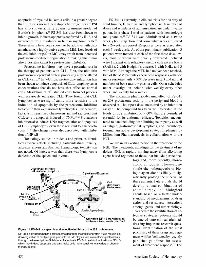
The proteasome is also required for activation ofthe nuclear transcription factor NF-κB, which plays arole in maintaining cell viability through the transcrip-tion of inhibitors of apoptosis in response to environ-mental stress or cytotoxic agents.18
Based on these observations, targeting the protea-some has become a novel new approach to cancertherapy. Several naturally occurring and syntheticproteasome inhibitors have been identified including theboronic acid peptides such as PS-341; the natural prod-uct lactacystin, a streptomyces metabolite; MG0132; anda synthetic peptide aldehyde. PS-341 is the compoundbeing most widely studied in the clinic. This dipeptidylboronic acid is a specific and selective inhibitor of the26S proteasome.17 PS-341 may also lead to an inductionof apoptosis (Figure 11). Moreover, enhanced tumor ef-ficacy in combination with other chemotherapy agentsand radiation has been suggested. In vitro studies havedemonstrated marked synergy between PS-341 and che-motherapy drugs such as the topoisomerase I inhibitor
irinotecan (CPT-11). CPT-11 can increaseNF-κB leading to greater transcription offactors that protect cells from apoptosis.19
PS-341 may block activation of NF-κB,thus potentiating the activity of the chemo-therapy drug. Since NF-kB can induce drugresistance, this agent may make cells morechemosensitive.20
PS-341 has demonstrated activityagainst many tumor types in vitro and is apoor substrate for the multi-drug resistancetransporter. It is also active even in the set-ting of overexpression of bcl-2 and hasshown a lack of acquired drug resistance.Using a prostate cancer cell line, Adams etal17 found that exposure of the cells to PS-341 results in accumulation in the G-Mphase of the cell cycle. Intravenous treat-ment of animals with this agent led to a60% decrease in tumor size. Anotherproteasome inhibitor, PSI, also induces
Figure 10. The ubiquitin-proteasome pathway.
The proteasome is a multicentric protease complex that plays a pivotal role in cellularprotein regulation. In order for a protein to be suitable for degradation, it must first beadorned with ubiquitin. The ubiquitin-proteasome pathway plays a critical role in thedegradation of intracellular proteins involved in cell cycle control and tumor growth.
456 American Society of Hematology
apoptosis of myeloid leukemia cells to a greater degreethan it affects normal hematopoietic progenitors.21 PSIhas also shown activity against a murine model ofBurkitt’s lymphoma.22 PS-341 has also been shown toinhibit growth, induces apoptosis conferred by IL-6, andovercomes drug resistance in human myeloma cells.23
These effects have been shown to be additive with dex-amethasone, a highly active agent in MM. Low levels ofthe cdk inhibitor p27 in MCL may result from increasedproteasome-mediated degradation,24 making this tumoralso a possible target for proteasome inhibitors.
Proteasome inhibitors may have a potential role inthe therapy of patients with CLL. First, the ubiquitinproteasome-dependent protein processing may be alteredin CLL cells.25 In addition, proteasome inhibition hasbeen shown to induce apoptosis of CLL lymphocytes atconcentrations that do not have that effect on normalcells. Masdehors et al25 studied cells from 50 patientswith previously untreated CLL. They found that CLLlymphocytes were significantly more sensitive to theinduction of apoptosis by the proteasome inhibitorlactacystin than were normal lymphocytes. Furthermore,lactacystin sensitized chemoresistant and radioresistantCLL cells to apoptosis induced by TNFα.26,27 Proteasomeinhibition also induces DNA fragmentation and apoptosisof CLL lymphocytes, even those resistant to glucocorti-coids.28,29 The changes were also associated with inhibi-tion of NF-κB.
Toxicology studies in rodents and primates identi-fied adverse effects including gastrointestinal toxicity,anorexia, emesis and diarrhea. Hematologic toxicity wasnot noted. Of interest was that there was lymphocytedepletion of the spleen and thymus.
PS-341 is currently in clinical trials for a variety ofsolid tumors, leukemias and lymphomas. A number ofdoses and schedules of administration are under investi-gation. In a phase I trial in patients with hematologicmalignancies30 PS-341 was administered as a twiceweekly bolus injection for 4 consecutive weeks followedby a 2-week rest period. Responses were assessed aftereach 6-week cycle. As of the preliminary publication, 3patients were treated at each of the first three dose lev-els, most of whom were heavily pretreated. Includedwere 1 patient with refractory anemia with excess blasts(RAEB), 2 with Hodgkin’s disease, 3 with NHL, and 3with MM. Although the MTD had not yet been reached,two of the MM patients experienced responses with onemajor response with > 50% decrease in IgG and normalnumbers of bone marrow plasma cells. Other schedulesunder investigation include twice weekly every otherweek, and weekly for 4 weeks.
The maximum pharmacodynamic effect of PS-341on 20S proteasome activity in the peripheral blood isobserved at 1-hour post dose, measured by an inhibitionassay.31 The compound has been well tolerated at thelevels of 20S inhibition of > 60% that are consideredessential for its antitumor efficacy. Toxicities encoun-tered to date including dose-limiting neuropathy as wellas fatigue, gastrointestinal symptoms, and thrombocy-topenia. An active development strategy is planned byMillennium Pharmaceuticals in collaboration with theNCI.
We are in an exciting period in the treatment of theNHL. The therapeutic paradigm for the treatment of in-dolent NHL is rapidly moving away from alkylatingagent-based regimens to those that include purine ana-
logs and, more recently, mono-clonal antibodies. However, nosingle chemotherapeutic or bio-logic agent alone is likely to sig-nificantly prolong the survival ofthese patients. Future trials shoulddevelop rational combinations ofchemotherapy and biologicalagents based on a better under-standing of mechanisms of drugaction and resistance, interactionsamong agents, and tumor biology.To expedite the identification of ef-fective strategies, patients shouldbe entered onto clinical trials ad-dressing important research ques-tions. Identification of the mostpromising of these drugs and regi-mens will be facilitated by recentlypublished guidelines for assess-ment of treatment response.32 The
Figure 11. PS-341 is a specific and selective inhibitor of the 26S proteasome.
NF-κB is activated when the proteasome degrades the inhibitor protein 1κBa resulting indownregulation of multiple gene products. NF-κB plays a role in maintaining cell viabilitythrough the transcription of inhibitors of apoptosis. PS-341 can block activation of NF-κB,which may induce apoptosis and also make cells more sensitive to a variety of chemo-therapy agents.
Hematology 2001 457
large number of new, unique therapeutic agents avail-able for clinical trials should provide optimism for im-proving the cure rate for patients with NHL.
REFERENCES
I. Antisense Oligonucleotides Directed at the Bcl-2Gene Message in Non-Hodgkin’s Lymphoma
1. Alizadeh AA, Eisen MB, Davis RE, et al. Distinct types ofdiffuse large B-cell lymphoma identified by gene expressionprofiling. Nature. 2000;403:503.
2. Ong ST, Le Beau MM. Chromosomal abnormalities andmolecular genetics of non-Hodgkin’s lymphoma. Semin Oncol.1998;25:447.
3. Gascoyne RD, Adomat SA, Krajewski S, et al. Prognosticsignificance of Bcl-2 protein expression and Bcl-2 generearrangement in diffuse aggressive non-Hodgkin’s lymphoma.Blood. 1997;90:244.
4. Reed JC. Bcl-2 family proteins: regulators of apoptosis andchemoresistance in hematologic malignancies. Semin Hematol.1997;34:9.
5. Yang E, Korsmeyer SJ. Molecular thanatopsis: a discourse onthe BCL2 family and cell death. Blood. 1996;88:386.
6. Reed JC. Double identity for proteins of the Bcl-2 family.Nature. 1997;387:773.
7. Zamecnik PC, Stephenson ML. Inhibition of Rous sarcomavirus replication and cell transformation by a specificoligodeoxynucleotide. Proc Natl Acad Sci U S A. 1978;75:280.
8. Stephenson ML, Zamecnik PC. Inhibition of Rous sarcomaviral RNA translation by a specific oligodeoxyribonucleotide.Proc Natl Acad Sci U S A. 1978;75:285.
9. Mizuno T, Chou MY, Inouye M. A unique mechanismregulating gene expression: translational inhibition by acomplementary RNA transcript (micRNA). Proc Natl Acad SciU S A. 1984;81:1966.
10. Izant JG, Weintraub H. Inhibition of thymidine kinase geneexpression by anti-sense RNA: a molecular approach to geneticanalysis. Cell. 1984;36:1007.
11. Wintersberger U. Ribonucleases H of retroviral and cellularorigin. Pharmacology & Therapeutics. 1990;48:259.
12. Sohail M, Hochegger H, Klotzbucher A, Guellec RL, Hunt T,Southern EM. Antisense oligonucleotides selected byhybridisation to scanning arrays are effective reagents in vivo.Nucleic Acids Res. 2001;29:2041.
13. Ho SP, Bao Y, Lesher T, et al. Mapping of RNA accessible sitesfor antisense experiments with oligonucleotide libraries. NatBiotechnol. 1998;16:59.
14. Ho SP, Britton DH, Stone BA, et al. Potent antisense oligo-nucleotides to the human multidrug resistance-1 mRNA arerationally selected by mapping RNA-accessible sites witholigonucleotide libraries. Nucleic Acids Res. 1996;24:1901.
15. Agrawal S, Temsamani J, Tang JY. Pharmacokinetics,biodistribution, and stability of oligodeoxynucleotidephosphorothioates in mice. Proc Natl Acad Sci U S A.1991;88:7595.
16. Raynaud FI, Orr RM, Goddard PM, et al. Pharmacokinetics ofG3139, a phosphorothioate oligodeoxynucleotide antisense tobcl-2, after intravenous administration or continuous subcuta-neous infusion to mice. J Pharmacol Exp Ther. 1997;281:420.
17. Jansen B, Schlagbauer-Wadl H, Brown BD, et al. bcl-2antisense therapy chemosensitizes human melanoma in Scidmice. Nat Med. 1998;4:232.
18. Monia BP, Johnston JF, Geiger T, Muller M, Fabbro D.Antitumor activity of a phosphorothioate antisenseoligodeoxynucleotide targeted against C-raf kinase [see
comments]. Nat Med. 1996;2:668.19. Weiner GJ, Liu HM, Wooldridge JE, Dahle CE, Krieg AM.
Immunostimulatory oligodeoxynucleotides containing the CpGmotif are effective as immune adjuvants in tumor antigenimmunization. Proc Natl Acad Sci U S A. 1997;94:10833.
20. Wooldridge JE, Ballas Z, Krieg AM, Weiner GJ.Immunostimulatory oligodeoxynucleotides containing CpGmotifs enhance the efficacy of monoclonal antibody therapy oflymphoma. Blood. 1997;89:2994.
21. Ballas ZK, Rasmussen WL, Krieg AM. Induction of NK activityin murine and human cells by CpG motifs inoligodeoxynucleotides and bacterial DNA. J Immunol.1996;157:1840.
22. Krieg AM, Yi AK, Matson S, et al. CpG motifs in bacterialDNA trigger direct B-cell activation. Nature. 1995;374:546.
23. Saijo Y, Uchiyama B, Abe T, Satoh K, Nukiwa T. Contiguousfour-guanosine sequence in c-myc antisense phosphorothioateoligonucleotides inhibits cell growth on human lung cancercells: possible involvement of cell adhesion inhibition. Jpn JCancer Res. 1997;88:26.
24. Buckheit RW Jr, Roberson JL, Lackman-Smith C, Wyatt JR,Vickers TA, Ecker DJ. Potent and specific inhibition of HIVenvelope-mediated cell fusion and virus binding by G quartet-forming oligonucleotide (ISIS 5320). AIDS Res HumRetroviruses. 1994;10:1497, 1994
25. Cotter FE, Johnson P, Hall P, et al. Antisense oligonucleotidessuppress B-cell lymphoma growth in a SCID-hu mouse model.Oncogene. 1994;9:3049.
26. Cotter FE. Antisense therapy for lymphomas. Hematol Oncol.1997;15:3.
27. Reed JC, Miyashita T, Takayama S, et al. BCL-2 familyproteins: regulators of cell death involved in the pathogenesisof cancer and resistance to therapy. J Cell Biochem.1996;60:23.
28. Reed JC. Bcl-2: prevention of apoptosis as a mechanism ofdrug resistance. Hematol Oncol. Clin North Am. 1995;9:451.
29. Miyashita T, Reed JC. bcl-2 gene transfer increases relativeresistance of S49.1 and Wehi7.2 lymphoid cells to cell deathand DNA fragmentation induced by glucocorticoids andmultiple chemotherapeutic drugs. Cancer Res. 1992;52:5407.
30. Miyashita T, Reed JC. Bcl-2 oncoprotein blocks chemotherapy-induced apoptosis in a human leukemia cell line. Blood.1993;81:151.
31. Kamesaki S, Kamesaki H, Jorgensen TJ, Tanizawa A, PommierY, Cossman J. bcl-2 protein inhibits etoposide-inducedapoptosis through its effects on events subsequent totopoisomerase II-induced DNA strand breaks and their repair[published erratum appears in Cancer Res 1994 Jun1;54(11):3074]. Cancer Res. 1993;53:4251.
32. Walton MI, Whysong D, PM OC, Hockenbery D, Korsmeyer SJ,Kohn KW. Constitutive expression of human Bcl-2 modulatesnitrogen mustard and camptothecin induced apoptosis. CancerRes. 1993;53:1853.
33. Reed JC, Kitada S, Takayama S, Miyashita T. Regulation ofchemoresistance by the bcl-2 oncoprotein in non-Hodgkin’slymphoma and lymphocytic leukemia cell lines. Ann Oncol.1994;5:61.
34. Keith FJ, Bradbury DA, Zhu YM, Russell NH. Inhibition of bcl-2 with antisense oligonucleotides induces apoptosis andincreases the sensitivity of AML blasts to Ara-C. Leukemia.1995;9:131.
35. Klasa RJ, Bally MB, Ng R, Goldie JH, Gascoyne RD, WongFM. Eradication of human non-Hodgkin’s lymphoma in SCIDmice by BCL-2 antisense oligonucleotides combined with low-dose cyclophosphamide. Clin Cancer Res. 2000;6:2492.
36. Dyer MJ, Lillington DM, Bastard C, et al. Concurrent activationof MYC and BCL2 in B cell non-Hodgkin lymphoma cell lines
458 American Society of Hematology
by translocation of both oncogenes to the same immunoglobu-lin heavy chain locus. Leukemia. 1996;10:1198.
37. Macpherson N, Lesack D, Klasa R, et al. Small noncleaved,non-Burkitt’s (Burkitt-Like) lymphoma: cytogenetics predictoutcome and reflect clinical presentation. J Clin Oncol.1999;17:1558.
38. Webb A, Cunningham D, Cotter F, et al. BCL-2 antisensetherapy in patients with non-Hodgkin lymphoma. Lancet.1997;349:1137.
39. Waters JS, Webb A, Cunningham D, et al. Phase I clinical andpharmacokinetic study of bcl-2 antisense oligonucleotidetherapy in patients with non-Hodgkin’s lymphoma. J ClinOncol. 2000;18:1812.
40. Jansen B, Wacheck V, Heere-Ress E, et al. Chemosensitisationof malignant melanoma by BCL2 antisense therapy. Lancet.2000;356:1728.
II. Targeting Angiogenesis in Hematologic Malig-nancies:
1. Hanahan D. Signaling vascular morphogenesis and mainte-nance. Science. 1997;277:48-50.
2. Risau W. Mechanisms of angiogenesis. Nature. 1997;386:671-674.
3. Liekens S, DeClercq E, Neyts J. Angiogenesis: regulators andclinical applications. Biochemical Pharmacology. 2001;61:253-270.
4. Leung DW, Cachianes G, Kuang W-J, Goeddel DV, Ferrara N.Vascular endothelial growth factor is a secreted angiogenicmitogen. Science. 1989;246:1306-1309.
5. Folkman J. Clinical applications of research on angiogenesis. NEngl J Med. 1995;333:1757-1763.
6. Tischer E, Mitchell R, Hartman T, et al. The human gene forvascular endothelial growth factor. Multiple protein forms areencoded through alternative exon splicing. J Biol Chem.1991:266:11947-11954.
7. Gitay-Goren H, Soker S, Vlodavsky I, Neufeld G. The bindingof vascular endothelial growth factor to its receptors isdependent on cell surface associated heparin-like molecules. JBiol Chem. 1992;267:6093-6098.
8. Roberts R, Gallagher J, Spooncer E, Allen TD, Bloomfield F,Dexter RM. Heparan-sulfate bound growth factors: a mecha-nism for stromal cell mediated haemopoiesis. Nature.1998;332:376-378.
9. Keyt BA, Berleau LT, Nguyen HV. The carboxyl-terminaldomain (111-165) of vascular endothelial growth factor iscritical for its mitogenic potency. J Biol Chem. 1996;271:7788-7795.
10. Soker S, Gollamudi-Payne S, Fidder H, Charmahelli H,Klagbrun M. Inhibition of vascular endothelial growth factor(VEGF)-induced endothelial cell proliferation by a peptidecorresponding to the exon 7-encoded domain of VEGF165. JBiol Chem. 1997;272:31582-31588.
11. Brogi E, Wu T, Namiki A, Isner JM. Indirect angiogeniccytokines upregulate VEGF and bFGF gene expression invascular smooth muscle cells, whereas hypoxia upregulatesVEGF expression only. Circulation. 1994;90:649-652.
12. Namiki A, Brogi E, Kearney M, et al. Hypoxia induces vascularendothelial growth factor in cultured human endothelial cells. JBiol Chem. 1995;270:31189-31195.
13. Waltenberger J, Mayr U, Pentz S, Hombach V. Functionalupregulation of the vascular endothelial growth factor receptorKDR by hypoxia. Circulation. 1996;94:1647-1654.
14. De Vries C, Escobeo JA, Ueno H, Houck K, Ferarra N,Williams LT. The fms-like tyrosine kinase receptor, a receptorfor vascular endothelial growth factor. Science. 1992;255:989-991.
15. Terman BL, Dougher-Vermazen M, Carrion ME, et al.Identification of the KDR tyrosine kinase as a receptor forvascular endothelial cell growth factor. Biochem Biophys ResCommun. 1992:187:1579-1586.
16. Hamada K, Oike Y, Nobuyuki T, et al. VEGF-C signalingpathways through VEGFR-2 and VEGFR-3 invasculoangiogenesis and hematopoiesis. Blood. 2000;96:3793-3800.
17. Joukov V, Pajusola K, Kaipainen A, et al. A novel vascularendothelial growth factor, VEGF-C, is a ligand for the Flt4(VEGFR-3) and KDR (VEGFR-2) receptor tyrosine kinases.EMBO J. 1996:15:290-298.
18. Waltenberger J, Claesson-Welsh L, Siegbahn A, Shibuya M,Heldin C-H. Different signal transduction properties of KDRand Flt1, two receptors for vascular endothelial growth factor. JBiol Chem. 1994;269:26988-26995.
19. Pajusola K, Aprelikova O, Pelicci G, Weich H, Claesson-WelshL, Alitalo K. Signaling properties of FLT4, a proteolyticallyprocessed receptor tyrosine kinase related to two VEGFreceptors. Oncogene. 1994:9:3545-3555.
20. Bellamy WT, Richter L, Frutiger Y, Grogan TM. Expression ofvascular endothelial growth factor (VEGF) and its receptors inhematopoietic malignancies. Cancer Res. 1999;59:728-733.
21. Ziegler BL, Valtieri M, Porada GA, et al. KDR receptor: a keymarker defining hematopoietic stem cells. Science.1999;285:1553-1558.
22. Barleon B, Sozzani S, Zhou D, Weich HA, Mantovani A,Marme D. Migration of human monocytes in response tovascular endothelial growth factor (VEGF) is mediated via theVEGF receptor flt-1. Blood. 1996;87:3336-3343.
23. Sawano A, Iwai S, Sakurai Y, Ito M, Shitara K, Nakahata T,Shibuya M. Flt-1, vascular endothelial growth factor receptor 1,is a novel cell surface marker for the lineage of monocyte-macrophages in humans. Blood. 2001:97:785-791.
24. Kaipainen A, Korhonen J. Mustonen T, et al. Expression of thefms-like tyrosine kinase 4 gene becomes restricted to lymphaticendothelium during development. Proc Natl Acad Sci U S A.1995;92:3566-3570.
25. Carmeliet P, Ferreira V, Breier G, et al. Abnormal blood vesseldevelopment and lethality in embryos lacking a single VEGFallele. Nature. 1996;380:435-439.
26. Ferrara N, Carver-Moore K, Chen H, et al. Heterozygousembryonic lethality induced by targeted inactivation of theVEGF gene. Nature. 1996;380:439-442.
27. Shalaby F, Rossant J, Yamaguchi TP, et al. Failure of bloodisland formation and vasculogenesis in Flk-1-deficient mice.Nature. 1995;376:62-66.
28. Hiratsuka S, Minowa O, Kuno J, Noda T, Shibuya M. Flt-1lacking the tyrosine kinase domain is sufficient for normaldevelopment and angiogenesis in mice. Proc Natl Acad Sci U SA. 1998;95:9349-9354.
29. Kukk E, Lymboussaki A, Taira S, et al. VEGF-C receptorbinding and pattern of expression with VEGFR-3 suggests arole in lymphatic vascular development. Development.1996;122:3829-3837.
30. Achen MG, Jeltsch M, Kukk E, et al. Vascular endothelialgrowth factor D (VEGF-D) is a ligand for the tyrosine kinasesVEGF receptor 2 (FLK1) and VEGF receptor 3 (Flt4). Proc NatlAcad Sci U S A. 1998;95:548-553.
31. Olofsson B, Korpelainen E, Pepper MS, et al. Vascularendothelial growth factor B (VEGF-B) binds to VEGF receptor-1 and regulates plasminogen activator activity in endothelialcells. Proc Natl Acad Sci U S A. 1998;95:11709-11714.
32. Meyer M, Clauss M, Lepple-Wienhues A, et al. A novelvascular endothelial growth factor encoded by Orf virus,VEGF-E, mediates angiogenesis via signaling through VEGFR-2 (KDR) but not VEGFR-1 (Flt-1) receptor tyrosine kinases.
Hematology 2001 459
EMBO J. 1999;18:363-374.33. Yamada Y, Takakura N, Yasue H, Ogawa H, Fujisawa H, Suda
T. Exogenous clustered neuropilin 1 enhances vasculogenesisand angiogenesis. Blood. 2001;97:1671-1678.
34. Veikkola T, Karkkainen M, Claesson-Welsh L, Alitalo K.Regulation of angiogenesis via vascular endothelial growthfactor receptors. Cancer Research. 2000;60:203-212.
35. Kroll J, Waltenberger J. The vascular endothelial growth factorreceptor KDR activates multiple signal transduction pathwaysin porcine aortic endothelial cells. J Biol Chem.1997;272:32521-32527.
36. Igarashi K, Shigeta K, Isohara T, Yaman T, Uno I. Sck interactswith KDR and Flt1 via its SH2 domain. Biochm Biophys ResCommun. 1998;251:77-82.
37. Takahashi T, Ueno H, Shibuya M. VEGF activates proteinkinase C-dependent, but Ras-independent Raf-independentRad-MEK-MAP kinase pathway for DNA synthesis in primaryendothelial cells. Oncogene. 1999;18;2221-2230.
38. Igarashi K, Isohara T, Kato T, Shigeta K, Yamano T, Uno I.Tryrosine 1213 of Flt1 is a major binding site of Nck and SHP-2. Biochem Biophys Res Commun. 1998;246:95-99.
39. Xia P, Aiello LP, Ishii H, et al. Characterization of vascularendothelial growth factor’s effect on the activation of proteinkinase C, its isoforms, and endothelial cell growth. J ClinInvestig. 1996;98:2018-2026.
40. Gerber H-P, McMurtrey A, Kowalski J, et al. Vascularendothelial growth factor regulates endothelial cell survivalthrough the phosphatidylinositol 3’-kinase/Akt signal transduc-tion pathway. J Biol Chem. 1998;273:30336-30343.
41. Coffer PJ, Jin J, Woodgett JR. Protein kinase B (c-Akt): amultifunctional mediator of phosphatidylinositol 3-kinaseactivation. Biochem J. 1998;335:1-13.
42. Liu ZY, Ganju RK, Wang JF, et al. Characterization of signaltransduction pathways in human bone marrow endothelial cells.Blood. 1997;90:2253-2259.
43. Byzova T, Goldman C, Pampori N, et al. A mechanism formodulation of cellular responses to VEGF: activation of theintegrins. Molecular Cell. 2000;6:851-860.
44. Soldi R, Mitola S, Strasly M, Defilippi P, Tarone G, BussolinoF. Role of αvβ3 in the activation of vascular endothelial growthfactor receptor-2. EMBO. 1999;18:882-892.
45. Carmeliet P, Lampugnani M-G, Moons L, et al. Targeteddeficiency or cytosolic truncation of the VE-cadherin gene inmice impairs VEGF-mediated endothelial survival andangiogenesis. Cell. 1999;98:147-157.
46. Semenza GL. Regulation of mammalian O2 homeostasis byhypoxia-inducible factor 1. Ann Rev Cell Dev Biol.1999;15:551-578.
47. Salceda S, Caro J. Hypoxia-inducible factor 1a (HIF-1a) proteinis rapidly degraded by the ubiquitin-proteasome system undernormoxic conditions. Its stabilization by hypoxia depends onredox-induced changes. J Biol Chem. 1997;272:22642-22647.
48. Yu F, White S, Zhao Q, Lee F. Dynamic, site-specific interac-tion of hypoxia-inducible factor-1a with the von Hippel-Lindautumor suppressor protein. Cancer Res. 2001;61:4136-4142.
49. Kerbel RS, Viloria-Petit A, Okada F, Rak J. Establishing a linkbetween oncogenes and tumor angiogenesis. Mol Med.1998;4:286-295.
50. Okada F, Rak JW, St Croix B, et al. Impact of oncogenes intumor angiogenesis: mutant K-ras up- regulation of vascularendothelial growth factor/vascular permeability factor isnecessary, but not sufficient for tumorigenicity of humancolorectal carcinoma cells. Proc Natl Acad Sci U S A.1998;95:3609-3614.
51. Arbiser JL, Moses MA, Fernandez CA, et al. Oncogenic H-rasstimulates tumor angiogenesis by two distinct pathways. ProcNatl Acad Sci U S A. 1997;94:861-866.
52. Ravi R, Mookerjee B, Bhujwalla ZM, et al. Regulation of tumorangiogenesis by p53-induced degradation of hypoxia-induciblefactor 1a. Genes Dev. 2000;14:34-44.
54. Marshall C. The embryonic origins of human haematopoiesis[review]. Br J Haematol. 2001;112:838-850.
55. Eichmann A, Corbel C, Nataf V, Vaigot P, Breant C, Le DouarinNM. Ligand-dependent development of the endothelial andhemapoietic lineages from embryonic mesodermal cellsexpressing vascular endothelial growth factor receptor 2. ProcNatl Acad Sci U S A. 1997;94:5141-5146.
56. Gunsilius E, Duba H-C, Petzer A, et al. Evidence fromleukaemia model for maintenance of vascular endothelium bybone-marrow-derived endothelial cells. Lancet. 2000;355:1688-1691.
57. Gao Z, McAlister V, Williams G. Repopulation of liverendothelium by bone-marrow-derived cells. Lancet.2001;357:932-933.
58. Ikpeazu C, Davidson M, Halteman D, Goodman S, Browning P,Brandt S. Donor origin of circulating endothelial progenitorsafter allogeneic bone marrow transplantation. Biology of Bloodand Marrow Transplantation. 2000;6:301-308.
59. Peichev M, Naiyer A, Pereira D, et al. Expression of VEGFR-2and AC133 by circulating human CD34+ cells identifies apopulation of functional endothelial precursors. Blood.2000;95:952-958.
60. Gabrilovich D, Ishida T, Oyama T, et al. Vascular endothelialgrowth factor inhibits the development of dendritic cells anddramatically affects the differentiation of multiple hematopoi-etic lineages in vivo. Blood. 1998;92:4150-4166.
61. Tordjman R, Delaire S, Plouet J, et al. Erythroblasts are asource of angiogenic factors. Blood. 2001;97:1968-1974.
62. Broxmeyer HE, Cooper S, Li ZH, et al. Myeloid progenitor cellregulatory effects of vascular endothelial cell growth factor. IntJ Hematol. 1995;62:203-215.
63. Gerber H-P, Vu TH, Ryan AM, Dowalski J, Werb Z, Ferrara N.VEGF couples hypertrophic cartilage remodeling, ossificationand angiogenesis during endochondral bone formation. NatMed. 1999;5:623-628.
64. Niida S, Kaku M, Amano H, et al. Vascular endothelial growthfactor can substitute for macrophage colony-stimulating factorin the support of osteoclastic bone resorption. J Exp Med.1999;190:293-298.
65. Kuramoto K, Uesaka T, Kimura A, Kobayashi M, Watanabe H,Katoh O. ZK7, a novel zinc finger gene, is induced by vascularendothelial growth factor and inhibits apoptotic death inhematopoietic cells. Cancer Research. 2000;60:425-430.
66. Katoh O, Takahashi T, Oguri T, et al. Vascular endothelialgrowth factor inhibits apoptotic death in hematopoietic cellsafter exposure to chemotherapeutic drugs by inducing MCL1acting as an anti-apoptotic factor. Cancer Research.1998;58:5565-5569.
67. Katoh O, Tauchi H, Kawaishi K, Kimura A, Satow Y. Expres-sion of the vascular endothelial growth factor (VEGF) receptorgene, KDR, in hematopoietic cells and inhibitory effect ofVEGF on apoptotic cell death caused by ionizing radiation.Cancer Research. 1995;55:5687-5692.
68. Fiedler W, Graeven U, Ergun S, et al. Vascular endothelialgrowth factor, a possible paracrine growth factor in humanacute myeloid leukemia. Blood. 1997;89:1870-1875.
69. Bellamy W, Richter L, Dirjani D, et al. Vascular endothelial cellgrowth factor is an autocrine promoter of abnormal localizedimmature myeloid precursors and leukemia progenitorformation in myelodysplastic syndromes. Blood. 2001;97:1427-1434.
70. List AF, Glinsmann-Gibson B, Stadheim C, Meuillet E, Bellamy
460 American Society of Hematology
W, Powis G. VEGFR-1 (Flt-1) and VEGFR-2 (KDR) stimulatethe proliferation of AML cells via the P13-kinase and Akt/protein kinase-b (PKB) signal pathway. Blood. 2000;96:301a.
71. Dias S, Hattori K, Zhu Z, et al. Autocrine stimulation ofVEGFR-2 activates human leukemic cell growth and migration.J Clin Invest 2000;106:511-521.
72. Aguayo A, Kantarjian H, Manshouri T, et al. Angiogenesis inacute and chronic leukemias and myelodysplastic syndromes.Blood. 2000;96:2240-2245.
73. Krejci P, Dvorakova D, Krahulcova E. et al. FGF-2 abnormali-ties in B cell chronic lymphocytic and chronic myeloidleukemias. Leukemia. 2001;15:228-237.
74. Plowright E, Li Z, Bergsagel L, et al. Ectopic expression offibroblast growth factor receptor 3 promotes myeloma cellproliferation and prevents apoptosis. Blood. 2000;95:992-998.
75. Salven P, Orpana A, Teerenhovi L, Joensuu H. Simultaneouselevation in the serum concentrations of the angiogenic growthfactors VEGF and bFGF is an independent predictor of poorprognosis in non-Hodgkin lymphoma: a single-institution studyof 200 patients. Blood. 2000;96:3712-3718.
76. Foss HD, Araujo A, Demel G, Klotzbach H, Hummel M, SteinH. Expression of vascular endothelial growth factor inlymphomas and Castleman’s disease. J Pathol. 1997:183:44-50.
77. Bellamy WT, Richter L, Frutiger Y, et al. Expression of vascularendothelial cell growth factor (VEGF) and its receptors in non-Hodgkin’s lymphoma (NHL). Proc Amer Assoc Cancer Res.1999;40:227.
78. Aguayo A, Estey E, Kantarjian H, et al. Cellular vascularendothelial growth factor is a predictor of outcome in patientswith acute myeloid leukemia. Blood. 1999;94:3717-3721.
79. Fusetti L, Pruneri G, Gobbi A, et al. Human myeloid andlymphoid malignancies in the non-obese diabetic/severecombined immunodeficiency mouse model: frequency ofapoptotic cells in solid tumors and efficiency and speed ofengraftment correlate with vascular endothelial growth factorproduction. Cancer Res. 2000;60:2527-2534.
80. Singhal S, Mehta J, Desikan R, et al. Antitumor activity ofthalidomide in refractory multiple myeloma. N Engl J Med.1999;341:1565-1571.
81. Price A, Shi Q, Morris D, et al. Marked inhibition of tumorgrowth in a malignant glioma tumor model by a novel syntheticmatrix metalloproteinase inhibitor AG3340. Clinical CancerResearch. 1999;5:845-854.
82. Gearing AJH, Beckett P, Christodoulou M, et al. Processing oftumor necrosis factor-α precursor by metalloproteinases.Nature. 1994;370:555-557.
83. Black RA, Rauch CT, Kozlosky CJ, et al. A metalloproteinasedisintegrin that releases tumor necrosis factor-alpha from cells.Nature. 1997;385:729-733.
84. Kayagaki N, Kawasaki A, Ebata T, et al. Metalloproteinase-mediated release of human Fas ligand. J Exp Med.1995;182:1777-1783.
85. D’Amato RJ, Loughman MS, Flynn E, Folkman J. Thalidomideis an inhibitor of angiogenesis. Proc Natl Acad Sci U S A.1994;91:4082-4085.
86. Corral LG, Haslett PAJ, Muller GW, et al. Differential cytokinemodulation and T cell activation by two distinct classes ofthalidomide analogs that are potent inhibitors of TNF-α. JImmunol. 1999;163:380-386.
87. Hideshima T, Chauhan D, Shima Y, et al. Thalidomide and itsanalogs overcome drug resistance of human multiple myelomacells to conventional therapy. Blood. 2000;96:2943-2950.
88. Davies FE, Raje N, Hideshima T, et al. Thalidomide andimmunomodulatory derivatives augment natural killercytotoxicity in multiple myeloma. Blood. 2001;98:210-216,
89. Raza A, Meyer P, Dutt T, et al. Thalidomide produces transfu-sion independence in patients with long-standing refractory
anemias and myelodysplastic syndromes. Blood 2001;9:958-965.
90. Shi YP, Ferrara N. Oncogenic ras fails to restore an in vivotumorigenic phenotype in embryonic stem cells lackingvascular endothelial growth factor (VEGF). Biochem BiophysRes Commun. 1999;254:480-483.
91. Karp JE, Lancet JE, Kaufmann SH, et al. Clinical and biologicactivity of the farnesyltransferase inhibitor R115777 in adultswith refractory and relapsed acute leukemias: a phase I clinical-laboratory correlative trial. Blood. 2001;97:3361-3369.
92. Smolich BD, Yuen HA, West KA, Giles FJ, Albitar M,Cherrington JM. The antiangiogenic protein kinase inhibitorsSU5416 and SU6668 inhibit the SCF receptor (c-kit) in ahuman myeloid leukemia cell line and in acute myeloidleukemia blasts. Blood. 2001;97:1413-1421.
93. Mesters RM, Padro T, Bieker R, et al. Stable remission afteradministration of the receptor tyrosine kinase inhibitor SU5416in a patient with refractory acute myeloid leukemia. Blood.2001;98:241-243.
94. Hussong JW, Rodgers GM, Shami PJ. Evidence of increasedangiogenesis in patients with acute myeloid leukemia. Blood.2000;95:309-313.
95. Padro T, Ruiz S, Bieker R, et al. Increased angiogenesis in thebone marrow of patients with acute myeloid leukemia. Blood.2000;95:2637-2644.
96. Janawska-Wieczorek A, Marquuez LA, Matsuzaki A, et al.Expression of matrix metalloproteinases (MMP-2 and –9) andtissue inhibitors of metalloproteinases (TIMP-1 and –2) inacute myelogenous leukaemia blasts: comparison with normalbone marrow cells. Br J Hematol. 1999;105:402-411.
98. Arimura K, Atima N, Ohtsubo H, et al. Matrixmetalloproteinase inhibitor inhibits apoptosis induction of MDSbone marrow cells. 1999;94:105a.
99. Mesa RA, Hanson CA, Rajkumar V, Schroeder G, Tefferi A.Evaluation and clinical correlations of bone marrow angiogen-esis in myelofibrosis with myeloid metaplasia. Blood.2000;96:3374-9980.
100. Rajkumar SV, Leong T, Roche PC, et al. Prognostic value ofbone marrow angiogenesis in multiple myeloma. ClinicalCancer Research. 2000;6:3111-3116.
101. Vacca A, Ribatti D, Presta M, et al. Bone marrowneovascularization, plasma cell angiogenic potential, andmatrix metalloproteinase-2 secretion parallel progression ofhuman multiple myeloma. Blood. 1999;93:3064-3073.
102. Dankbar B, Padro T, Leo R, et al. Vascular endothelial growthfactor and interleukin-6 in paracrine trumor-stromal cellinteractions in multiple myeloma. Blood. 2000;95:2630-2636.
103. Kuittinen O, Savolainen E-R, Koistinen P, Mottonen M,Turpeenniemi-Hujanen T. MMP-2 and MMP-9 expression inadult and childhood acute lymphatic leukemia (ALL).Leukemia Research. 2001;25:125-132.
104. Perez-Atayde AR, Sallan SE, Tedrow U, Connors S, Allred E,Folkman J. Spectrum of tumor angiogenesis in the bonemarrow of children with acute lymphoblastic leukemia. Am JPathol. 1997;150:815-821.
105. Bertolini F, Paolucci M, Peccatori F, et al. Angiogenic growthfactors and Endostatin in non-Hodgkin’s lymphoma. Br JHematol. 1999;106:504-509.
106. Vacca A, Ribatti D, Ruco L, et al. Angiogenesis extent andmacrophage density increase simultaneously with pathologicalprogression in B-cell non-Hodgkin’s lymphoma. Br J Cancer.1999;79:965-970.
107. Kini AR, Kay NE, Peterson LC. Increased bone marrowangiogenesis in B cell lymphocytic leukemia. Leukemia.2000;14:1414-1418.
Hematology 2001 461
108. Molica S, Vitelli G, Levato D, Gandolfo GM, Liso V.Increased serum levels of vascular endothelial growth factorpredict risk of progression in early B-cell chronic lymphocyticleukaemia. Br J Heamatol. 1999;107:605-610.
109. Chen H, Treweeke AT, West DC, et al. In vitro and in vivoproduction of vascular endothelial growth factor by chroniclymphocytic leukemia cells. Blood. 2000;96:3181-3187.
110. Kim KJ, Li B, Winer J, et al. Inhibition of vascular endothelialgrowth factor-induced angiogenesis suppresses tumor growthin vivo. Nature. 1999;362:841-844.
111. Gordon MS, Margolin K, Talpaz M, et al. Phase I safety andpharmacokinetic study of recombinant human ant-vascularendothelial growth factor in patients with advanced cancer. JClin Oncology. 2001;19:843-850.
112. Tulple A, Scadden DT, Espina BM, et al. Results of arandomized study of IM862 nasal solution in the treatment ofAIDS-related Kaposi’s sarcoma. J Clin Oncol. 2000;18:716-723.
113. Dinney CPN, Bielenberg DR, Perrotte P, et al. Inhibition ofbasic fibroblast growth factor expression, angiogenesis, andgrowth of human bladder carcinoma in mice by systemicinterferon-κ adminstration. Cancer Res. 1998;58:808-814.
114. Brekken RA, Overholser JP, Stastny VA, Waltenberger J,Minna JD, Thorpe PE. Selective inhibition of vascularendothelial growth factor (VEGF) receptor 2 (KDR/Flk-1)activity by a monoclonal anti-VEGF antibody blocks tumorgrowth in mice. Cancer Res. 2000;60:5117-5124.
115. Prewett M, Huber J, Li Y, et al. Antivascular endothelialgrowth factor receptor (fetal liver kinase 1) monoclonalantibody inhibits tumor angiogenesis and growth of severalmouse and human tumors. Cancer Res. 1999;59:5209-5218.
116. Sandberg JA, Parker VP, Blanchard KS, et al. Pharmacokinet-ics and tolerability of an antiangiogenic ribozyme(ANGIOZYME) in healthy volunteers. J Clin Pharm.2000;40:1462-1469.
117. Fong TA, Shawver LK, Sun L, et al. SU5416 is a potent andselective inhibitor of the vascular endothelial growth factorreceptor (Flk-1/KDR) that inhibits tyrosine kinase catalysis,tumor vascularization, and growth of multiple tumor types.Cancer Res. 1999;59:99-106.
118. Drevs J, Hofmann I, Hugenschmidt H, et al. Effects ofPTK787/ZK 222584, a specific inhibitor of vascular endothe-lial growth factor receptor tyrosine kinases, on primary tumor,metastasis, vessel density, and blood flow in a murine renalcell carcinoma model. Cancer Res. 2000;60:4819-4824.
119. Kumar CC, Malkowski M, Yin Z, et al. Inhibition of angiogen-esis and tumor growth by SCH221153, a dual αvβ3 and αvβ5
integrin receptor antagonist. Cancer Res. 2001;61:2232-2238.120. Kerr JS, Slee MA, Mousa SA. Small molecule av integrin
121. Kim Y-M, Jang J-W, Lee O-H, et al. Endostatin inhibitsendothelial and tumor cellular invasion by blocking theactivation and catalytic activity of matrix metalloproteinase 2.Cancer Res. 2000;60:5410-5413.
122. O’Reilly MS, Boehm T, Shing Y, et al. Endostatin: anendogenous inhibitor of angiogenesis and tumor growth. Cell.1997;88:277-285.
123. Pike SE, Yao L, Setsuda J, et al. Vasostatin, a calreticulinfragment, inhibits angiogenesis and suppresses tumor growth.J Exp Med. 1998;188:2349-2356.
124. Logothetis CJ, Wu KK, Finn LD, et al. Phase I trial of theangiogenesis inhibitor TNP-470 for progressive androgen-independent prostate cancer. Clinical Cancer Research.2001;7:1198-1203.
125. Fotsis T, Zhang Y, Pepper MS, et al. The endogenousoestrogen metabolite 2-methoxyoestradiol inhibits angiogen-
esis and suppresses tumour growth. Nature. 1994;368:237-239.
126. Lew YS, Brown SL, Griffin RJ, Song CW, Kim JH. Arsenictrioxide causes selective necrosis in solid murine tumors byvascular shutdown. Cancer Res. 1999;59:6033-6037.
127. Reichert A, Heisterkamp N, Daley GQ, Groffen J. Treatmentof Bcr/Abl-positive acute lymphoblastic leukemia in P190transgenic mice with the farnesyl transferase inhibitorSCH66336. Blood. 2001;97:1399-1403.
128. Peters DG, Hoover RR, Gerlach MJ, et al. Activity of thefarnesyl protein transferase inhibitor SCH66336 against BCR/Abl-induced murine leukemia and primary cells from patientswith chronic myeloid leukemia. Blood. 2001;97:1404-1412.
III. Inhibiting Cell Cycle Progression andProteasome Degradation Pathways: New Agentsfor Lymphoma and Myeloma
1. Argatoff LH, Connors JM, Klasa RJ, Horsman DE, GascoyneRD. Mantle cell lymphoma: a clinicopathologic study of 80cases. Blood. 1997;89:2067-2078.
2. Rimokh R, Berger F, Delsol G, et al. Rearrangement andoverexpression of the BCL1/PRAD1 gene in intermediatelymphocytic lymphomas and in t(11q13)bearing leukemias.Blood. 1993;81:3063-3067.
3. Greiner TC, Moynihan MJ, Chan WC, et al. p53 mutations inmantle cell lymphomas are associated with variant cytology andpredict a poor prognosis. Blood. 1996;87:4302-4310.
4. Bible KC, Kaurmann SH. Cytotoxic synergy betweenflavopiridol (NSC 649890, L868275) and various antineoplasticagents: the importance of sequence of administration. CancerRes. 1997;57:3375-3380.
5. König A, Schwartz GK, Mohammed RM, Al-Katib A,Gabrilove JL. The novel cyclin-dependent kinase inhibitorflavopiridol downregulates Bcl2 and induces growth arrest andapoptosis in chronic B-cell leukemia cell lines. Blood.1997;90:4307-4312.
6. Byrd JC, Shinn CA, Bedi A, et al. Flavopiridol has marked invitro activity against B-chronic lymphocytic leukemia (BCLL)and induces apoptosis independent of p53 status. Blood.1997;90(suppl 1):531a (abstr 2366).
7. Byrd JC, Shinn C, Waselenko JK, et al. Flavopiridol inducesapoptosis in chronic lymphocytic leukemia cells via activationof caspase3 without evidence of bcl2 modulation or depen-dence on functional p53. Blood. 1998;92:3804-3816.
8. Parker BW, Kaur G, Nieves-Niera W, et al. Early induction ofapoptosis in hematopoietic cell lines after exposure toflavopiridol. Blood. 1998;91:458-465.
10. Kitada S, Zapata JM, Andreeff M, Reed JC. Protein kinaseinhibitors flavopiridol and 7-hydroxy-staurosporinedownregulate antiapoptosis proteins in B cell chronic lympho-cytic leukemia. Blood. 2000;96:393-397.
11. Melillo G, Sausville EA, Cloud K, Lahusen T, Varesio L,Senderowicz AM. Flavopiridol, a protein kinase inhibitor,downregulates hypoxic induction of vascular endothelialgrowth factor expression in human monocytes. Cancer Res.1999;59:5433-5437.
12. Arguello F, Alexander M, Sterry JA, et al. Flavopiridol inducesapoptosis of normal lymphoid cells, causes immunosuppres-sion, and has potent antitumor activity in vivo against humanleukemia and lymphoma xenografts. Blood. 1998;91:2482-2490.
13. Innocenti F, Stadler W, Iyer L, Ramírez J, Vokes EE, Ratain MJ.Flavopiridol metabolism in cancer patients is associated with
462 American Society of Hematology
the occurrence of diarrhea. Clin Cancer Res. 2000;6:3400-3405.
14. Maki C, Huibregtse J, Howley P. In vivo ubiquination andproteasome-mediated degradation of p53. Cancer Res.1996;56:2649-2654.
15. Yoshida H, Kitamura K, Tanaka K, et al. Accelerated degrada-tion of PML-retinoic acid receptor a (PML RARα by all transretinoic acid in acute promyelocytic leukemia: possible role ofthe proteasome pathway. Cancer Res. 1996;56:2945-2948.
16. Adams J, Stein R. Novel inhibitors of the proteasome and theirtherapeutic use in inflammation. Ann Rep Med Chem.1996;31:279-288.
17. Adams J, Palombella VJ, Sausville EA, et al. Proteasomeinhibitors: a novel class of potent and effective antitumoragents. Cancer Res. 1999;59:2615-2622.
18. Beg AA, Baltimore D. An essential role for NF-κB in prevent-ing TNFa induced cell death. Science. 1996;274:782-784.
19. Wang CVY, Cusack JC, Liu R. Control of inducible chemoresis-tance: enhanced antitumor therapy through increased apoptosisby inhibition of NFκB. Nature. 1999;5:412-417.
20. Lin ZP, Boller YC, Amer SM, et al. Prevention of brefeldin-induced resistance to teniposide by the proteasome inhibitorMG132: involvement of NF-κB activation in drug resistance.Cancer Res. 1998;58:3059-3065.
21. Soglio D, Servida F, Delia D, et al. The apoptogenic response ofhuman myeloid leukaemia cells and of normal and malignanthaematopoietic progenitor cells to the proteasome inhibitor PSI.Br J Haematol. 2001;113:126-136.
22. Orlowski RZ, Eswara JR, Larond-Walker A, Grever MR,Orlowski M, Dang CV. Tumor growth inhibition induced in amurine model of human Burkitt’s lymphoma by a proteasomeinhibitor. Cancer Res. 1998;58:4342-4348,
23. Hideshima T, Richardson P, Chauhan D, et al. The proteasome6-inhibitor PS-341 inhibits growth, induces apoptosis, andovercomes drug resistance in human multiple myeloma cells.Cancer Res. 2001;61:3071-3076.
24. Chiarle R, Budel LM, Skolnik J, et al. Increased proteasomedegradation of cyclin-dependent kinase inhibitor p27 isassociated with a decreased overall survival in mantle celllymphoma. Blood. 2000;95:619-626.
25. Masdehors P, Omura S, MerleBeral H, et al. Increasedsensitivity of CLL-derived lymphocytes to apoptotic deathactivation by the proteasime-specific inhibitor lactacystin. Br JHaemtaol. 1999;105:752-757.
26. Delic J, Masdehors P, Omura S, et al. The proteasome inhibitorlactacystin induces apoptosis and sensitizes chemo andradioresistant human chronic lymphocytic leukaemia lympho-cytes to TNF α-initiated apoptosis. Br J Cancer. 1999;77:375-376.
27. Delic J, Masdehors P, Omura S, et al. The proteasome inhibitorlactacystin induces apoptosis and sensitizes chemo- andradioresistant human CLL lymphocytes to TNF α-inducedapoptosis. Br J Cancer. 1998;77:1103-1107.
28. Masdehors P, MerleVBeral H, Magdelenat H, Delic J. Ubiquitinproteasome system and increased susceptibility of BCLLlymphocytes to apoptotic death activation. Leuk Lymphoma.2000;38(56):499-504.
29. Chandra J, Niemer I, Gilbreath J, et al. Proteasome inhibitorsinduce apoptosis in glucocorticoid resistant chronic lympho-cytic leukemia lymphocytes. Blood. 1998;92:42204229.
30. Stinchcombe TE, Mitchell BS, Depcik V, et al. PS341 is activein multiple myeloma: preliminary report of a phase I trial of theproteasome inhibitor PS-341 in patients with hematologicmalignancies. Blood. 2000;96(suppl 1):516a (abstr 2219).
31. Nix D, Pien C, Newman R, et al. Clinical development of aproteasome inhibitor, PS341, for the treatment of cancer. ProcASCO. 2001;20:86 (abstr 339)
32. Cheson BD, Horning SJ, Coiffier B, et al. Report of anInternational Workshop to standardize response criteria fornon-Hodgkin’s lymphomas. Clin Oncol. 1999;17:1244-1253.