50
Basic Sciences of Renal Cell Carcinoma Dr. D. Ramu
| Date post: | 16-Jul-2015 |
| Category: |
Health & Medicine |
| Upload: | damuluri-ramu |
| View: | 144 times |
| Download: | 6 times |

Basic Sciences of Renal Cell Carcinoma
Dr. D. Ramu
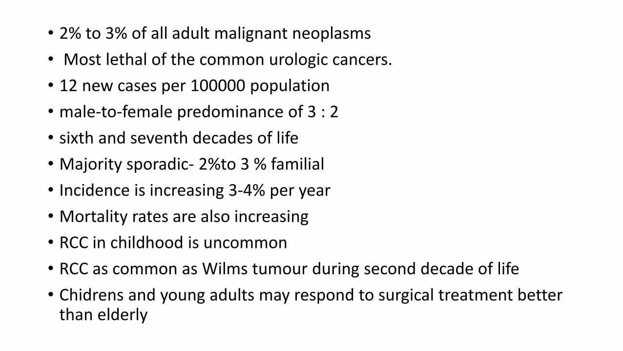
• 2% to 3% of all adult malignant neoplasms
• Most lethal of the common urologic cancers.
• 12 new cases per 100000 population
• male-to-female predominance of 3 : 2
• sixth and seventh decades of life
• Majority sporadic- 2%to 3 % familial
• Incidence is increasing 3-4% per year
• Mortality rates are also increasing
• RCC in childhood is uncommon
• RCC as common as Wilms tumour during second decade of life
• Chidrens and young adults may respond to surgical treatment better than elderly

India
• Incidence: 15-22/100,000/Yr, 2% of all malignant tumors,
• In India : Males: 1.2/100,000, Females : 0.5/ 100,000
• Indians living in western countries have higher incidence
• M:F = 2:1
• Age peak: 50 to 70 years
• India : mean age – 52 years
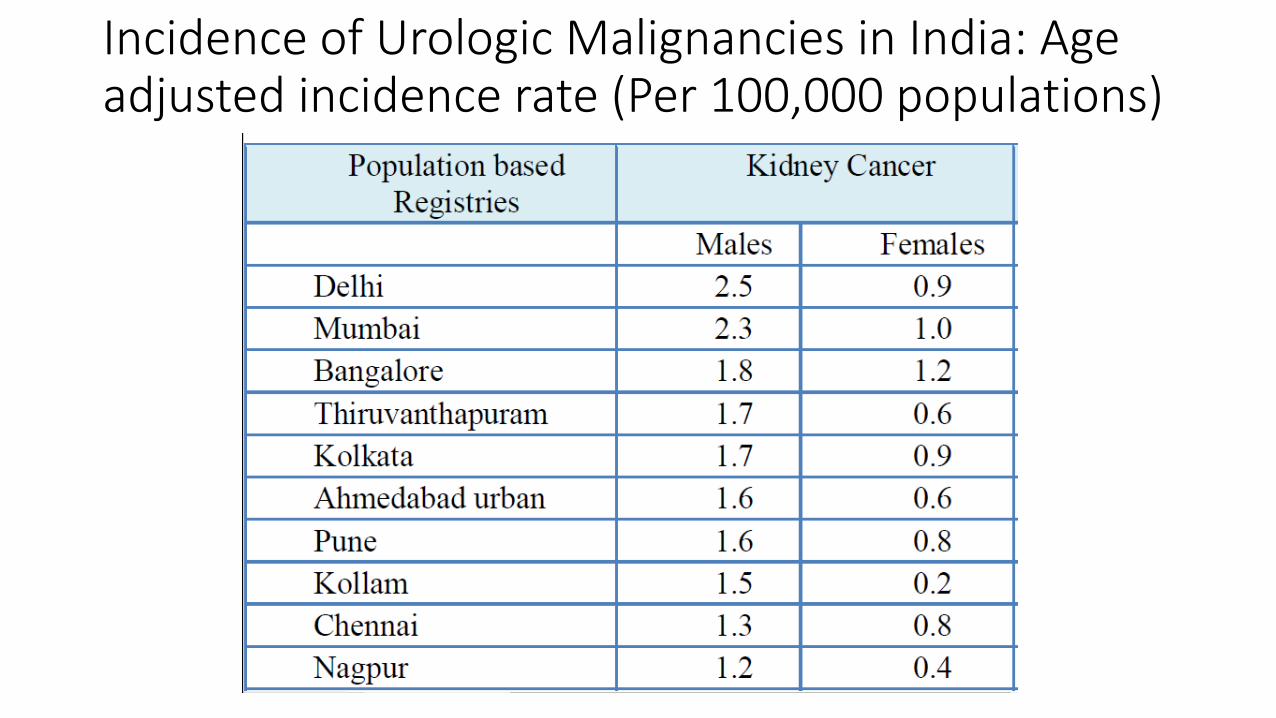
Incidence of Urologic Malignancies in India: Age adjusted incidence rate (Per 100,000 populations)
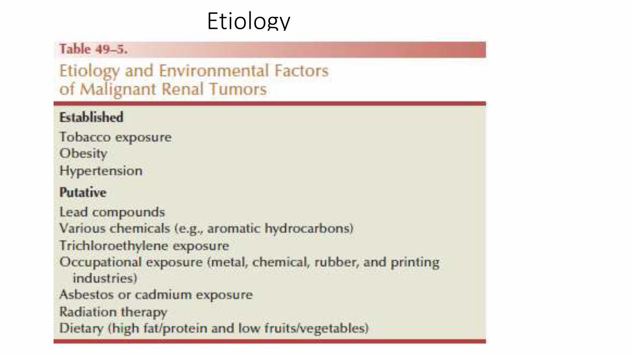
Etiology
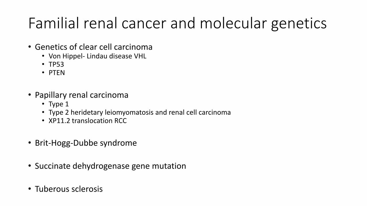
Familial renal cancer and molecular genetics
• Genetics of clear cell carcinoma• Von Hippel- Lindau disease VHL• TP53• PTEN
• Papillary renal carcinoma• Type 1• Type 2 heridetary leiomyomatosis and renal cell carcinoma• XP11.2 translocation RCC
• Brit-Hogg-Dubbe syndrome
• Succinate dehydrogenase gene mutation
• Tuberous sclerosis
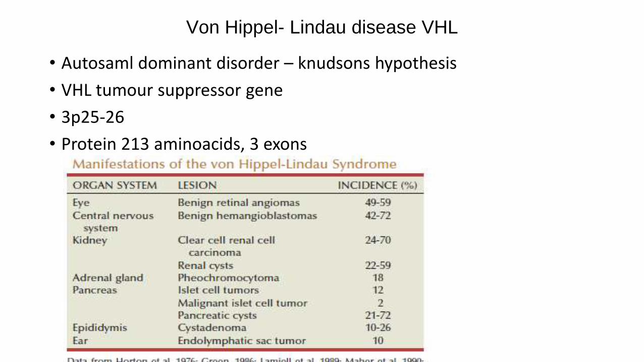
Von Hippel- Lindau disease VHL
• Autosaml dominant disorder – knudsons hypothesis
• VHL tumour suppressor gene
• 3p25-26
• Protein 213 aminoacids, 3 exons
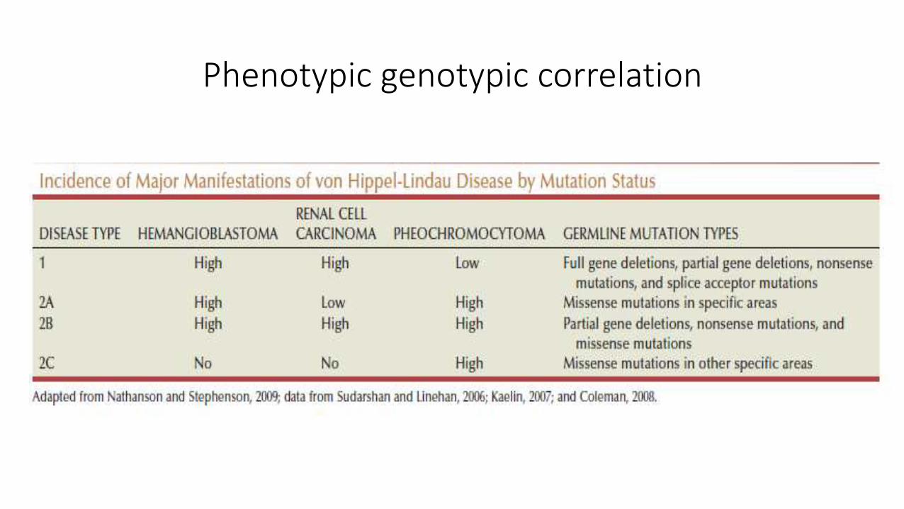
Phenotypic genotypic correlation
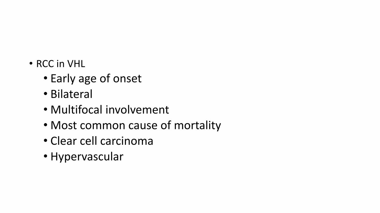
• RCC in VHL
• Early age of onset• Bilateral• Multifocal involvement• Most common cause of mortality• Clear cell carcinoma• Hypervascular
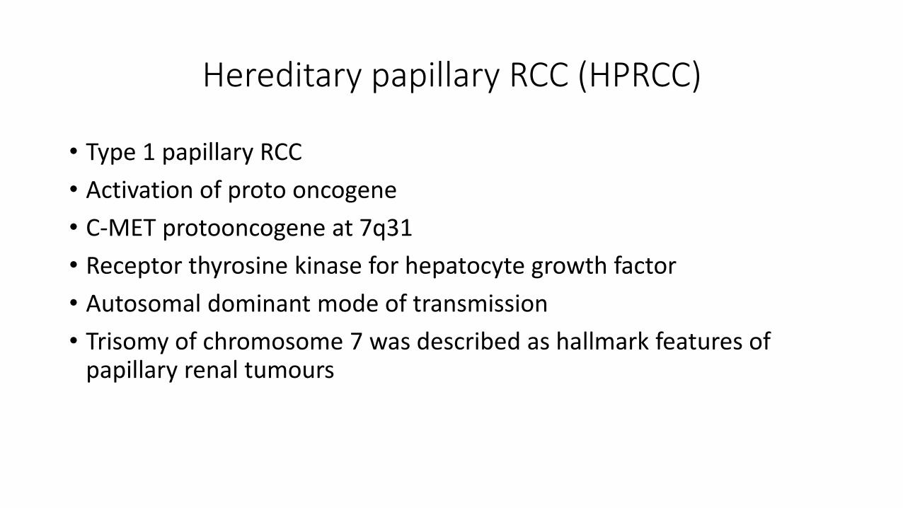
Hereditary papillary RCC (HPRCC)
• Type 1 papillary RCC
• Activation of proto oncogene
• C-MET protooncogene at 7q31
• Receptor thyrosine kinase for hepatocyte growth factor
• Autosomal dominant mode of transmission
• Trisomy of chromosome 7 was described as hallmark features of papillary renal tumours
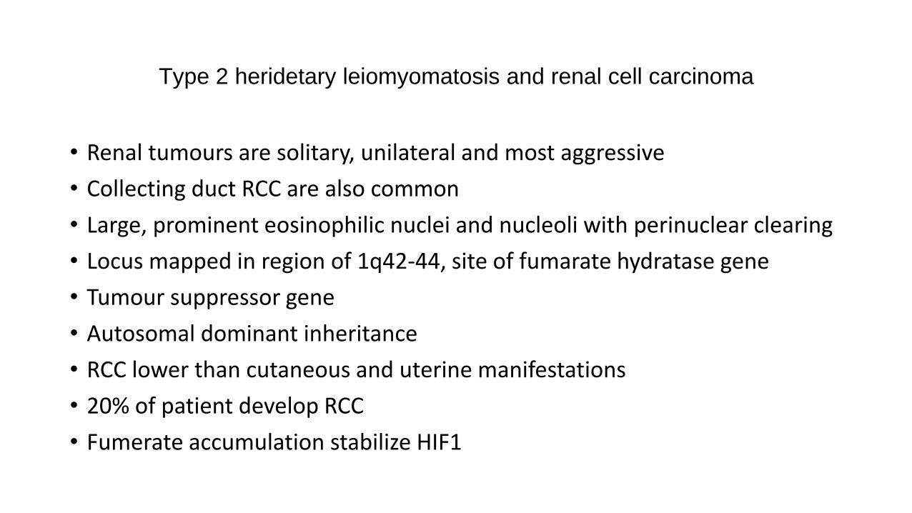
Type 2 heridetary leiomyomatosis and renal cell carcinoma
• Renal tumours are solitary, unilateral and most aggressive
• Collecting duct RCC are also common
• Large, prominent eosinophilic nuclei and nucleoli with perinuclear clearing
• Locus mapped in region of 1q42-44, site of fumarate hydratase gene
• Tumour suppressor gene
• Autosomal dominant inheritance
• RCC lower than cutaneous and uterine manifestations
• 20% of patient develop RCC
• Fumerate accumulation stabilize HIF1
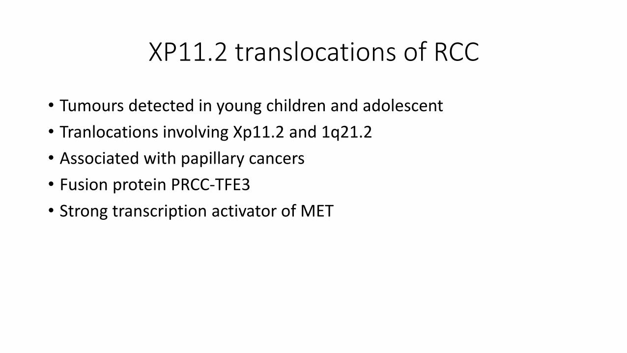
XP11.2 translocations of RCC
• Tumours detected in young children and adolescent
• Tranlocations involving Xp11.2 and 1q21.2
• Associated with papillary cancers
• Fusion protein PRCC-TFE3
• Strong transcription activator of MET
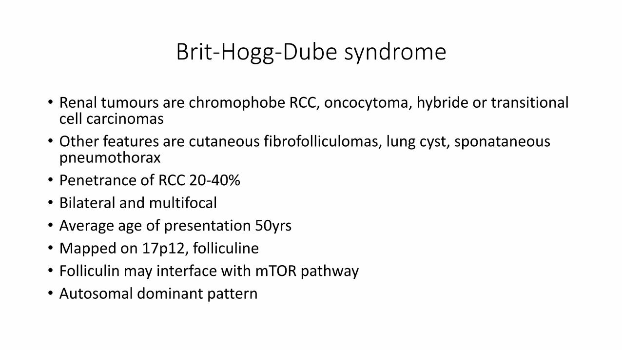
Brit-Hogg-Dube syndrome
• Renal tumours are chromophobe RCC, oncocytoma, hybride or transitional cell carcinomas
• Other features are cutaneous fibrofolliculomas, lung cyst, sponataneouspneumothorax
• Penetrance of RCC 20-40%
• Bilateral and multifocal
• Average age of presentation 50yrs
• Mapped on 17p12, folliculine
• Folliculin may interface with mTOR pathway
• Autosomal dominant pattern
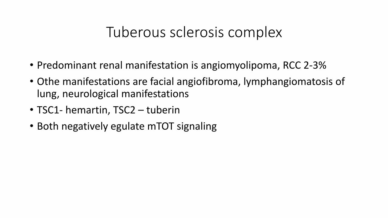
Tuberous sclerosis complex
• Predominant renal manifestation is angiomyolipoma, RCC 2-3%
• Othe manifestations are facial angiofibroma, lymphangiomatosis of lung, neurological manifestations
• TSC1- hemartin, TSC2 – tuberin
• Both negatively egulate mTOT signaling
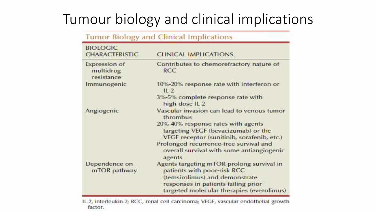
Tumour biology and clinical implications
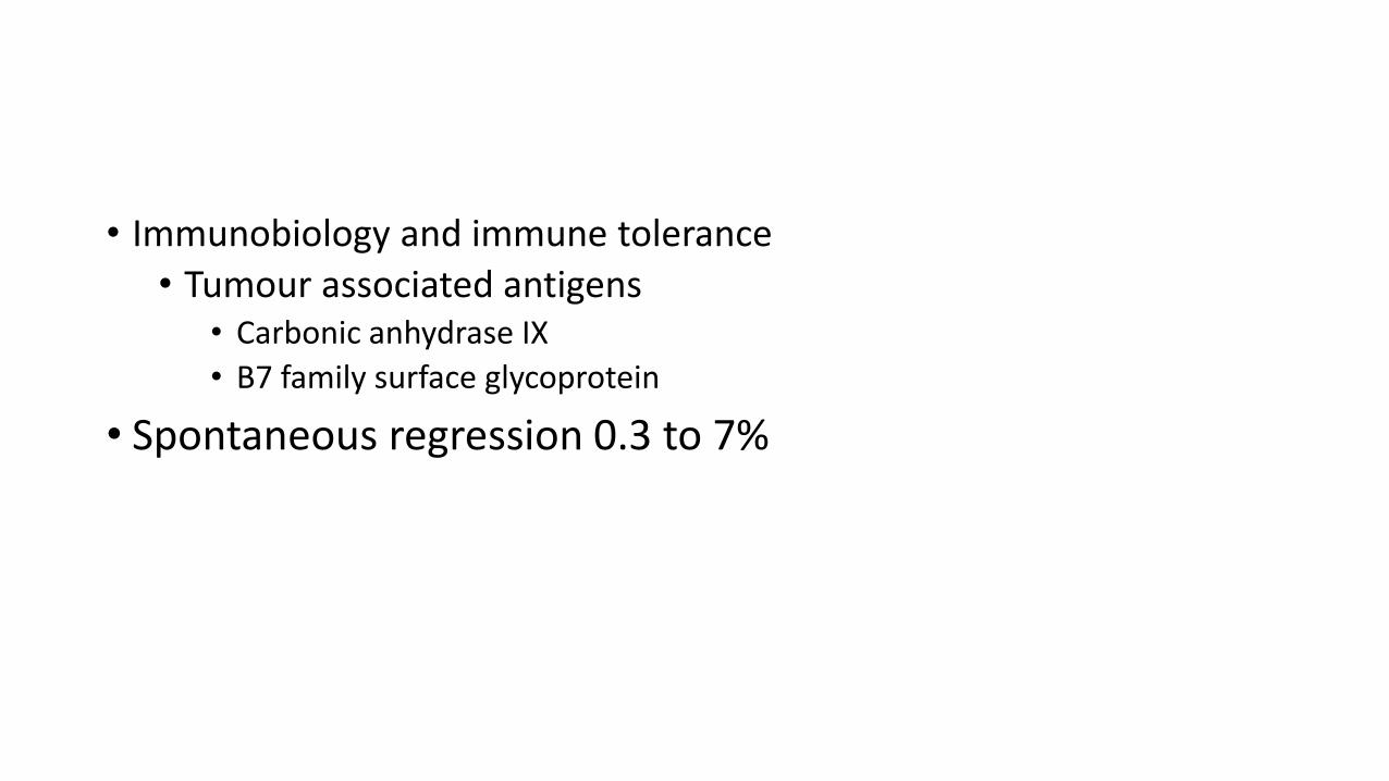
• Immunobiology and immune tolerance
• Tumour associated antigens• Carbonic anhydrase IX
• B7 family surface glycoprotein
• Spontaneous regression 0.3 to 7%
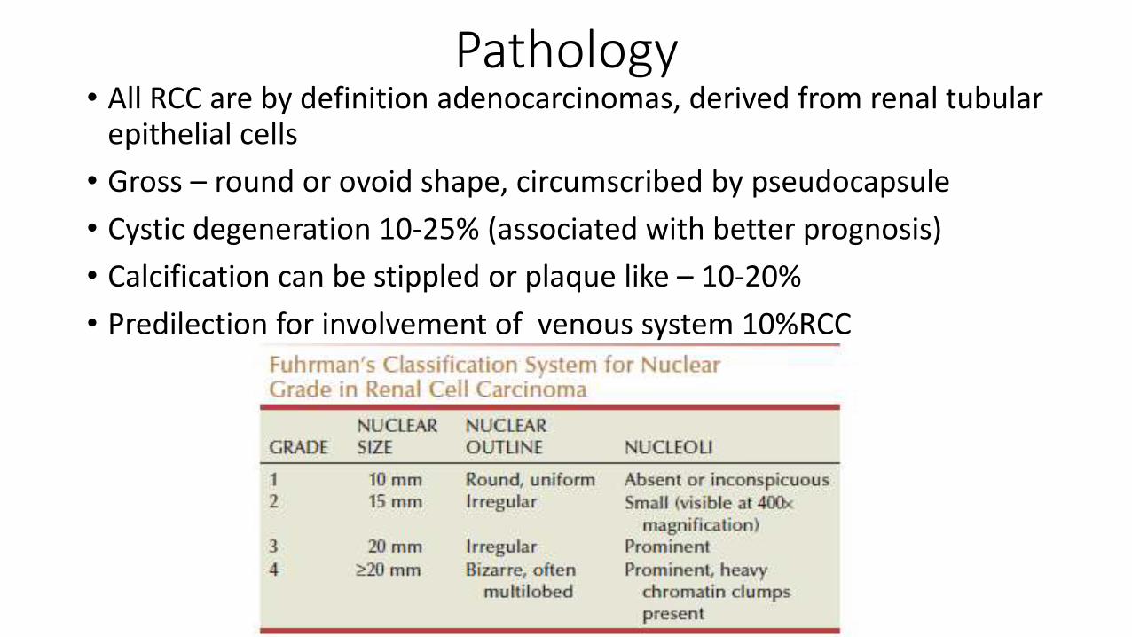
Pathology • All RCC are by definition adenocarcinomas, derived from renal tubular
epithelial cells
• Gross – round or ovoid shape, circumscribed by pseudocapsule
• Cystic degeneration 10-25% (associated with better prognosis)
• Calcification can be stippled or plaque like – 10-20%
• Predilection for involvement of venous system 10%RCC
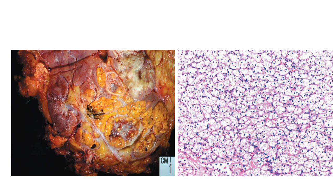
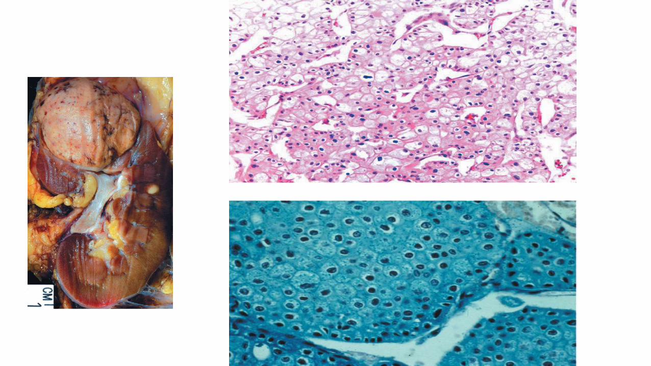
• Clear cell renal carcinoma• 70-80% of all RCC• Tumour typically yellow, highly vascular• Clear cell, granular cell, or mixed• Clear cell – round or polygonal with abundant cytoplasm containing
glycogen, cholesterol, cholesterol esters, and phospholipids, all of which are readily extracted by the solvents used in routine histologic preparations, contributing to the clear appearance of the tumor cells
• Granular cells -- eosinophilic cytoplasm and abundant mitochondria, can predominate.
• Two to 5 percent of clear cell RCC demonstrate sarcomatoid features• Clear cell RCC is more likely to exhibit venous tumor extension than any
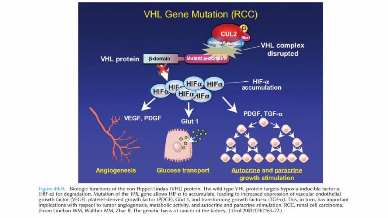
other subtype of RCC • Chromosome 3 alterations and VHL mutations are common in clear cell
RCC, and mutation or inactivation of this gene has been found in a majority of sporadic cases
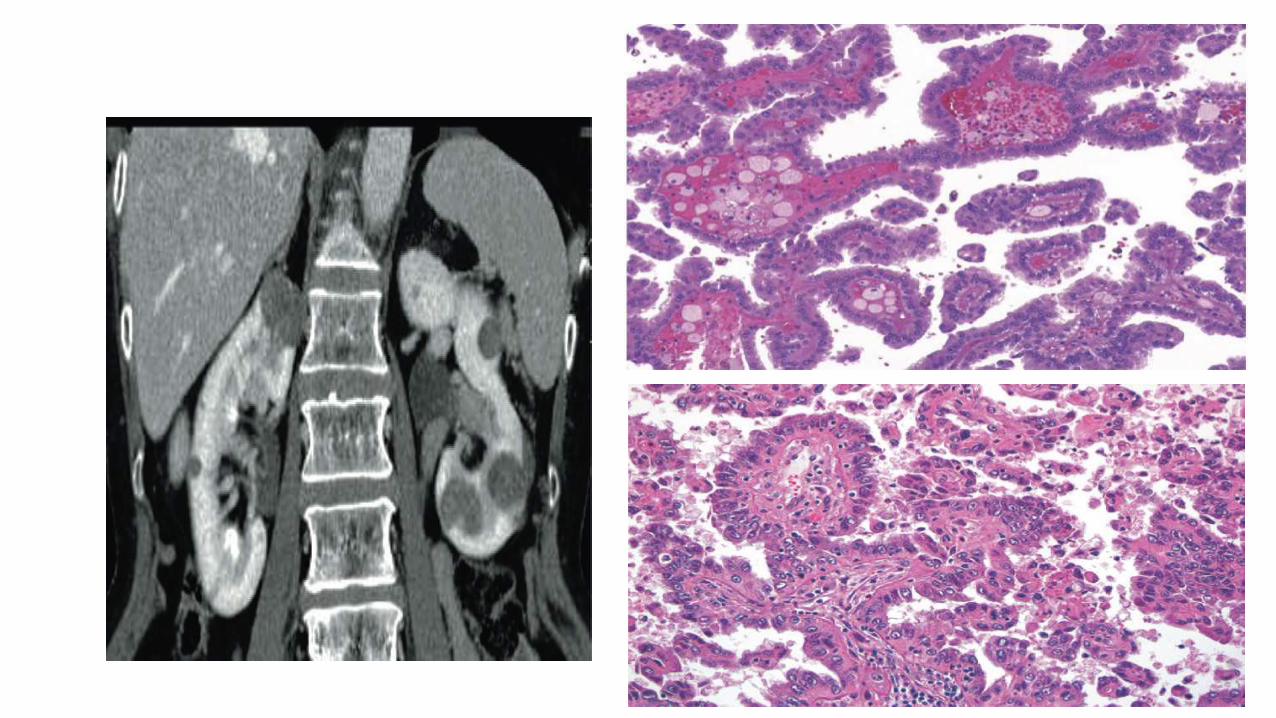
• Papillary Renal Cell Carcinoma• Second most common histologic subtype
• It represents 10% to 15% of all RCCs
• More commonly found in patients with end-stage renal failure and acquired renal cystic disease
• Basophilic or eosinophilic cells arranged in papillary or tubular configuration
• More than 50% or 75% of the tumor had to exhibit such architectural features to qualify as a papillary RCC
• Multicentricity, which approaches 40% in many series
• Type 1 papillary RCC, the more common form, consists of basophilic cells with scant cytoplasm
• Type 2 papillary RCC include potentially more aggressive variants with eosinophilic cells and abundant granular cytoplasm

• Chromophobe Renal Cell Carcinoma
• Derived from the cortical portion of the collecting duct
• It represents 3% to 5% of all RCCs
• The tumor cells typically exhibit a relatively transparent cytoplasm with a fine reticular pattern that has been described as a “plant cell” appearance
• Perinuclear clearing or “halo” is typically found and electron microscopic findings
• Microvesicles characteristically stain positive for Hale colloidal iron, indicating the presence of a mucopolysaccharide unique to chromophobe RCC
• Better prognosis for localized chromophobe RCC than for clear cell RCC but a poor outcome in the subset of patients with sarcomatoid features or metastatic disease
• Collecting Duct Carcinoma• Less than 1% of all RCCs • Occurred in younger patients, often in the third, fourth, or fifth
decades of life • up to 50% have metastatic disease at the time of detection • Ulex europaeus agglutinin 1 reactivity and positivity for E-cadherin
and c-KIT • On microscopic examination, these tumors consist of an admixture
of dilated tubules and papillary structures typically lined by a single layer of cuboidal cells, often creating a cobblestone appearance
• Renal Medullary Carcinoma
• Exclusively in association with the sickle cell trait
• Arise from the calyceal epithelium near the renal papillae
• Sarcomatoid Differentiation
• Sarcomatoid differentiation is characterized by spindle cell histology, positive staining for vimentin, infiltrative growth pattern, aggressive local and metastatic behavior, and poor prognosis
• Found in 1% to 5% of RCCs, most commonly in association with clear cell RCC or chromophobe RCC
• Unclassified Renal Cell Carcinoma• Most are poorly differentiated and are associated with a highly
aggressive biologic behavior and a particularly poor prognosis
Clinical Presentation• Kidney within the retroperitoneum, many renal masses remain
asymptomatic and nonpalpable until they are advanced.
• With the more pervasive use of noninvasive imaging for the evaluation of a variety of nonspecific symptom complexes, more than 50% of RCCs are now detected incidentally
• The classic triad of flank pain, gross hematuria, and palpable abdominal mass is now rarely found - too late triad 10%
• In advanced stage: hematuria 60%, flank pain-40%, palpable abdominal mass- 45%,
• Weight loss 35%, Anemia- 20%, varicocele, edema of leg (due to invasion of renal vein or IVC)
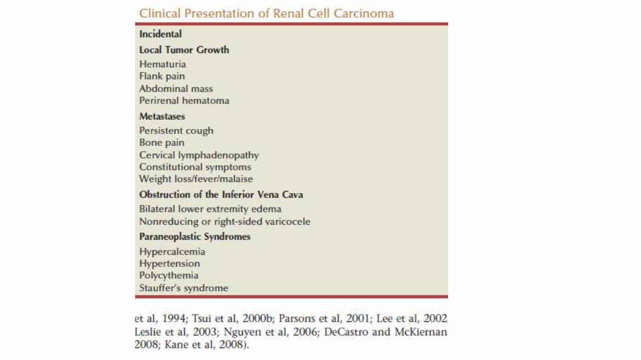


• METASTATIC SPREAD
• 30% of cases have distant metastasis at diagnosis
• Tumor < 3cm in diameter-usually without metastasis
• Hematogenous metastasis -Lung, liver, bone, CNS
• Lymphatic : to pelvic and para-aortic lymph nodes
• Local : to regional lymph nodes: para-aortic, paracaval, renal hilarlymph nodes
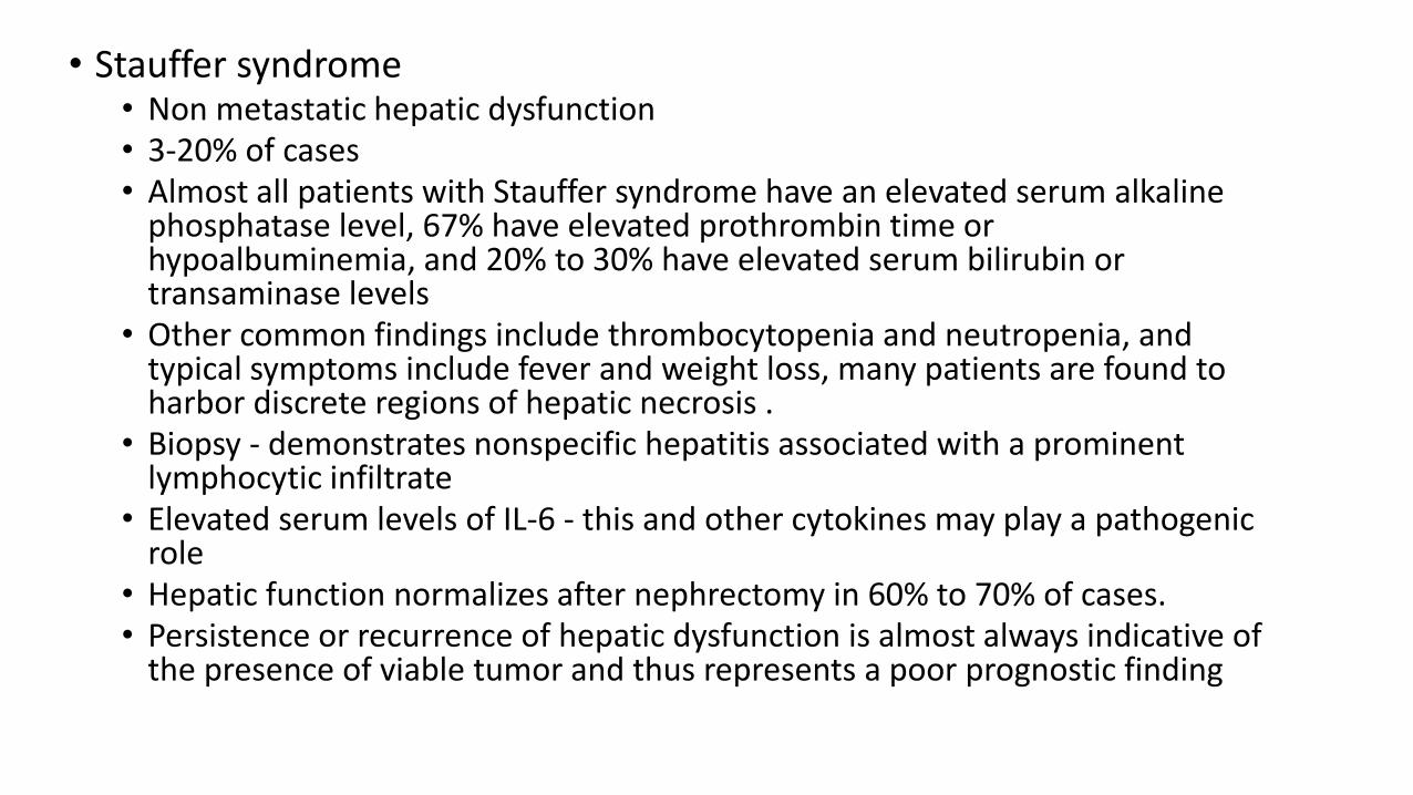
• Stauffer syndrome• Non metastatic hepatic dysfunction• 3-20% of cases• Almost all patients with Stauffer syndrome have an elevated serum alkaline
phosphatase level, 67% have elevated prothrombin time or hypoalbuminemia, and 20% to 30% have elevated serum bilirubin or transaminase levels
• Other common findings include thrombocytopenia and neutropenia, and typical symptoms include fever and weight loss, many patients are found to harbor discrete regions of hepatic necrosis .
• Biopsy - demonstrates nonspecific hepatitis associated with a prominent lymphocytic infiltrate
• Elevated serum levels of IL-6 - this and other cytokines may play a pathogenic role
• Hepatic function normalizes after nephrectomy in 60% to 70% of cases. • Persistence or recurrence of hepatic dysfunction is almost always indicative of
the presence of viable tumor and thus represents a poor prognostic finding

Screening and Clinical Associations
Investigations
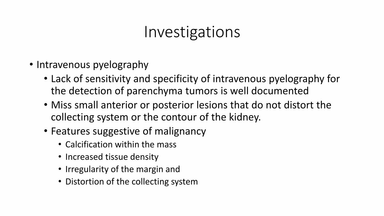
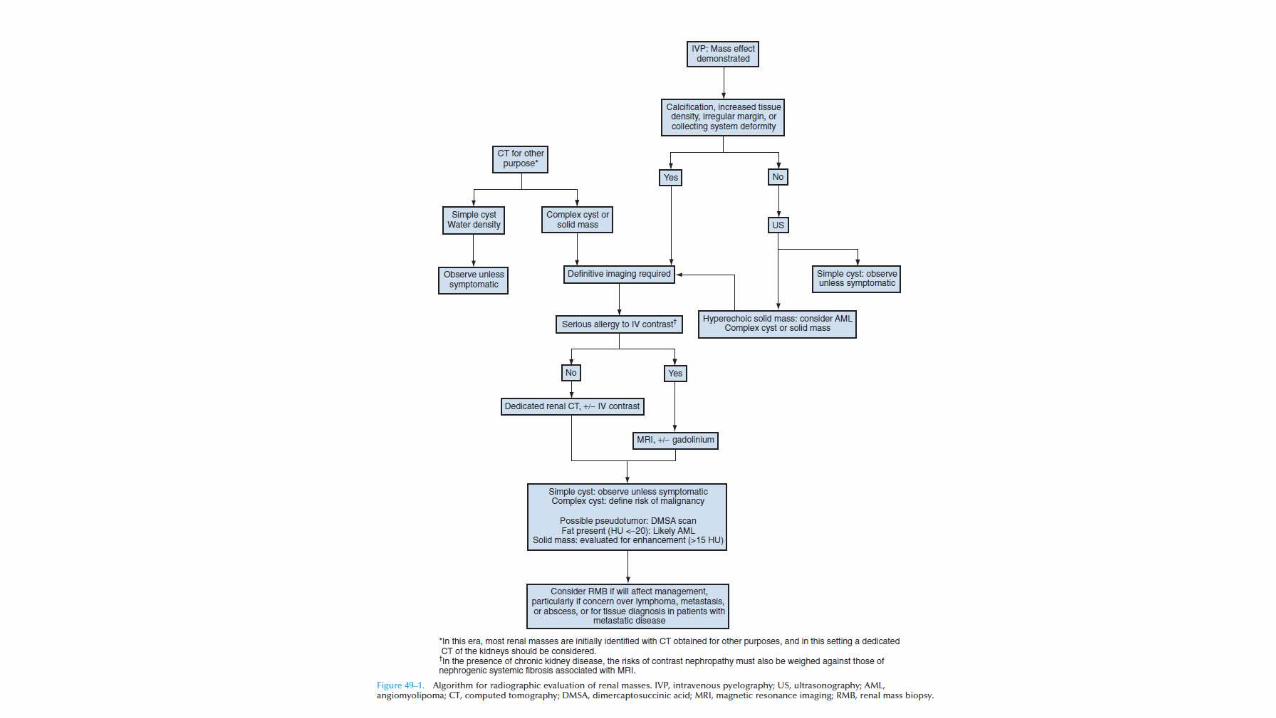
• Intravenous pyelography
• Lack of sensitivity and specificity of intravenous pyelography for the detection of parenchyma tumors is well documented
• Miss small anterior or posterior lesions that do not distort the collecting system or the contour of the kidney.
• Features suggestive of malignancy• Calcification within the mass
• Increased tissue density
• Irregularity of the margin and
• Distortion of the collecting system
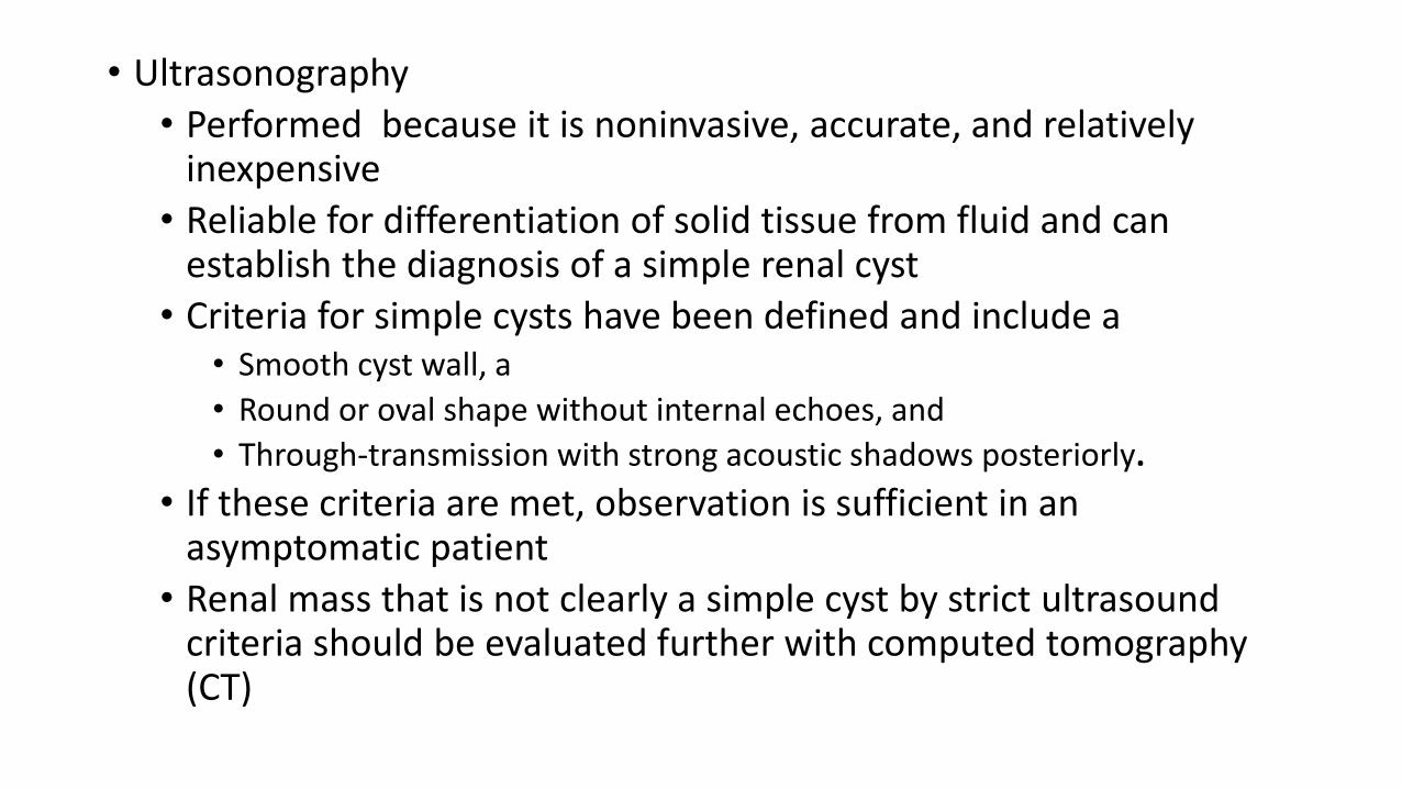
• Ultrasonography
• Performed because it is noninvasive, accurate, and relatively inexpensive
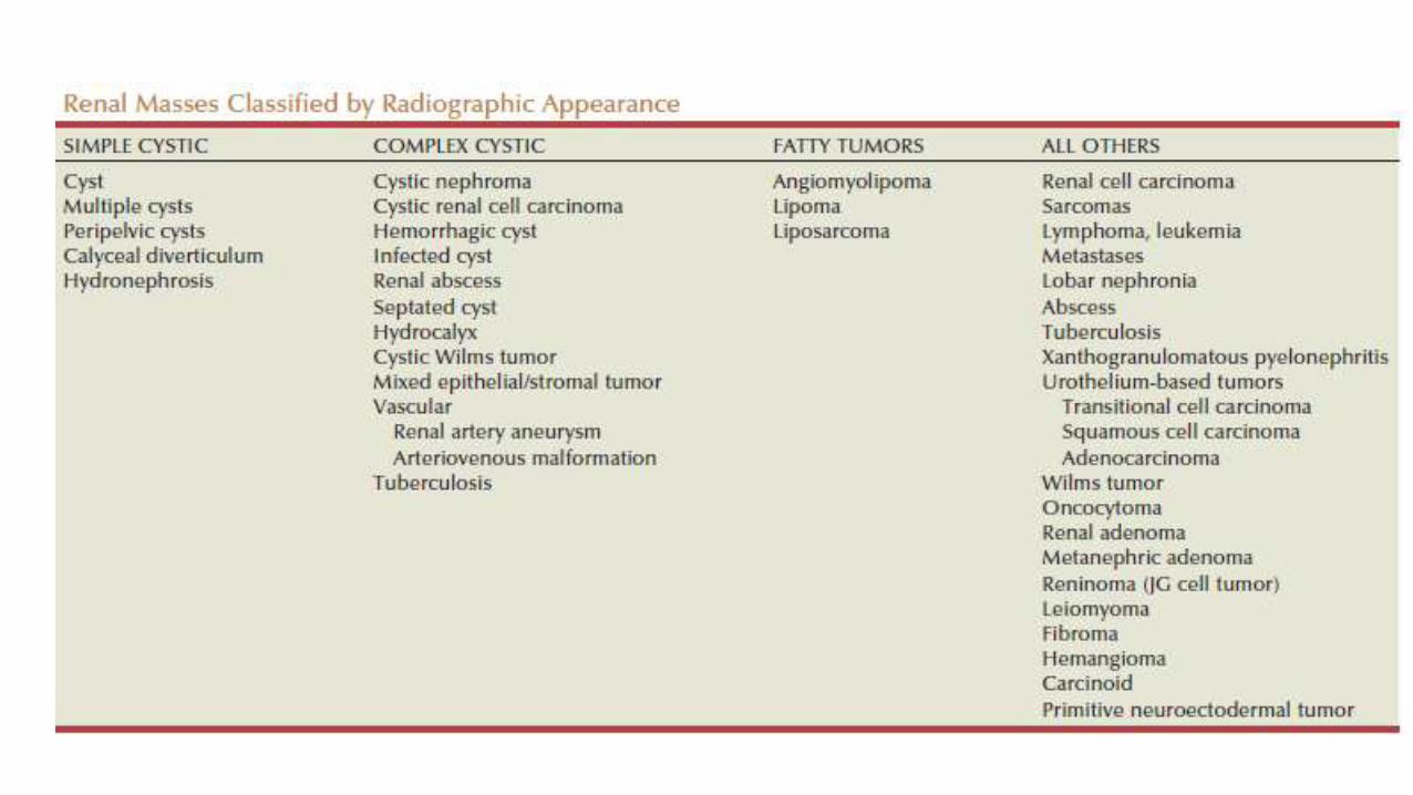
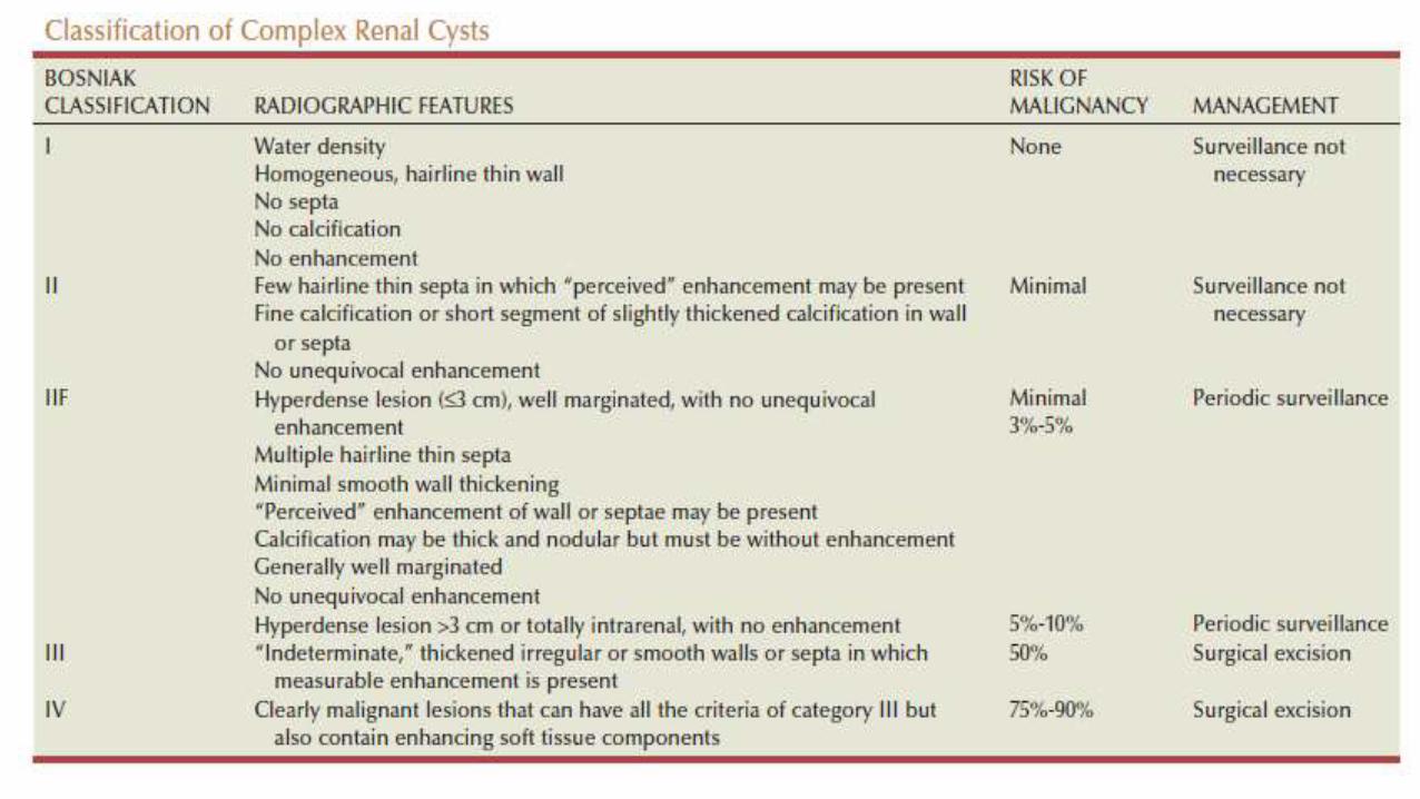
• Reliable for differentiation of solid tissue from fluid and can establish the diagnosis of a simple renal cyst
• Criteria for simple cysts have been defined and include a • Smooth cyst wall, a
• Round or oval shape without internal echoes, and
• Through-transmission with strong acoustic shadows posteriorly.
• If these criteria are met, observation is sufficient in an asymptomatic patient
• Renal mass that is not clearly a simple cyst by strict ultrasound criteria should be evaluated further with computed tomography (CT)
• CT scan• Any renal mass that enhances with intravenous administration of contrast
material on CT by more than 15 Hounsfield units (HU) should be considered an RCC until proved otherwise (Hartman et al, 2004).
• Solid masses that also have substantial areas of negative CT attenuation numbers (below −20 HU) indicative of fat are diagnostic of AMLs
• 10% to 20% of solid renal masses CT findings are indeterminate, and additional testing or surgical exploration is needed to establish a definitive diagnosis
• Magnetic resonance imaging (MRI)
• Renal arteriography has a limited role in the diagnostic evaluation of renal masses and is primarily reserved for patients with concomitant renal artery disease
• Renal mass biopsy is now being revisited for the evaluation of renal masses• False-negative rate of renal mass biopsy was thought to be 18%
• Overall accuracy is greater than 80%.
• Assessment of tumor grade and histologic type, which reflects tumoraggressiveness, is also accurate in the majority of cases
• The risks of clinically significant perinephric bleeding and pneumothorax also appear to be low (<1%), and needle tract seeding is exceedingly rare when centrally located, infiltrative renal masses are excluded.
• Renal mass biopsy is now being considered more frequently, particularly in
• Younger, healthy patients who are unwilling to accept the uncertainty associated with renal mass biopsy are still typically managed primarily based on radiographic and clinical considerations.
• Suspicion of renal abscess
• When RCC must be differentiated from metastatic malignant disease
• Renal lymphoma

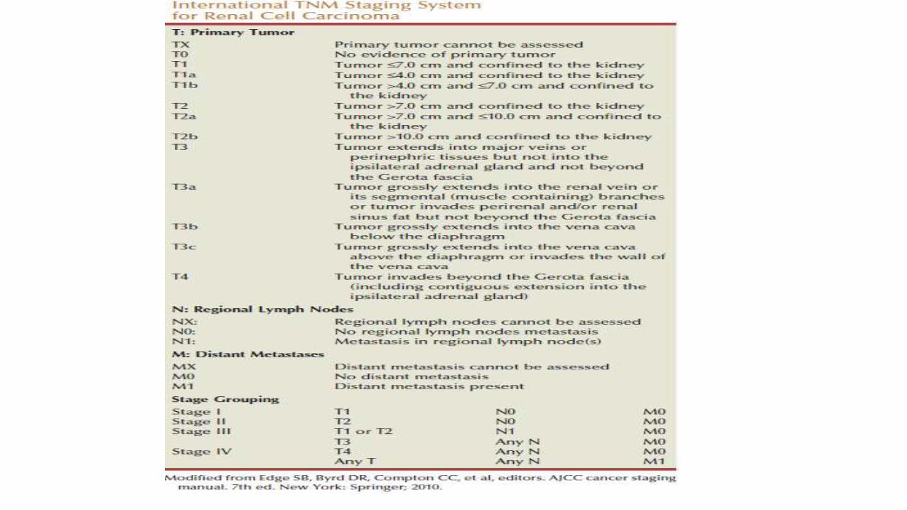
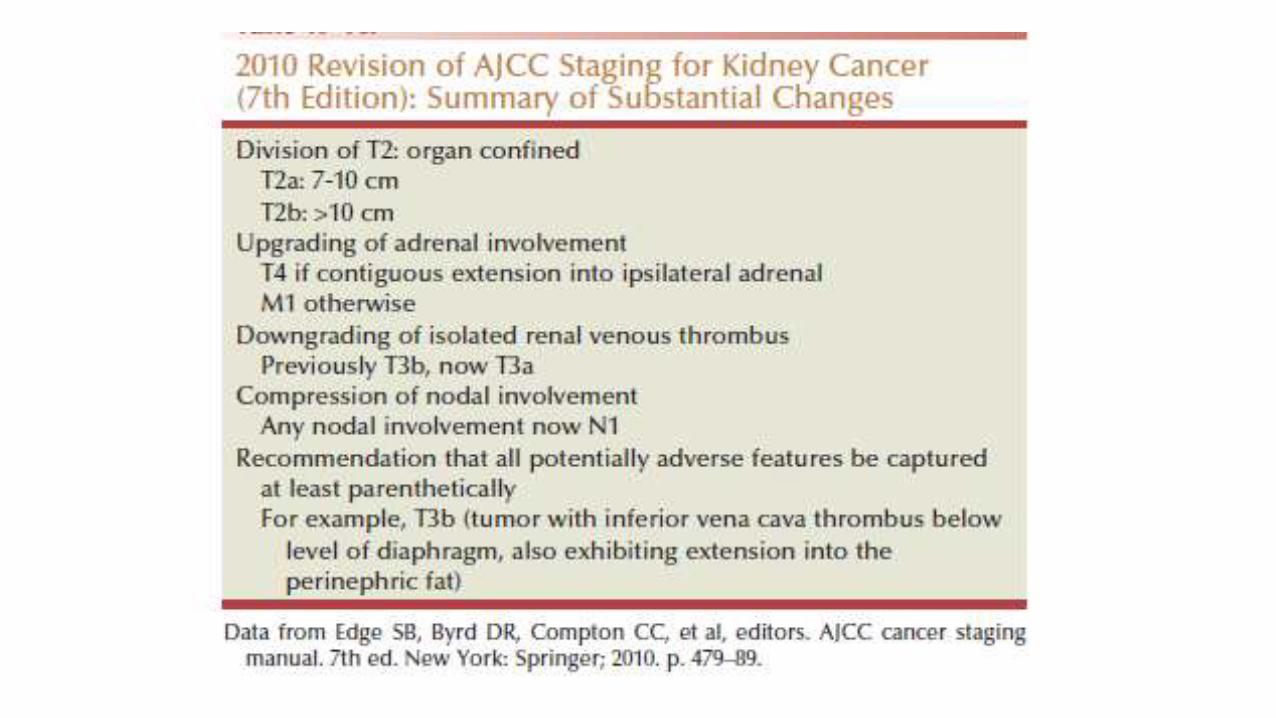
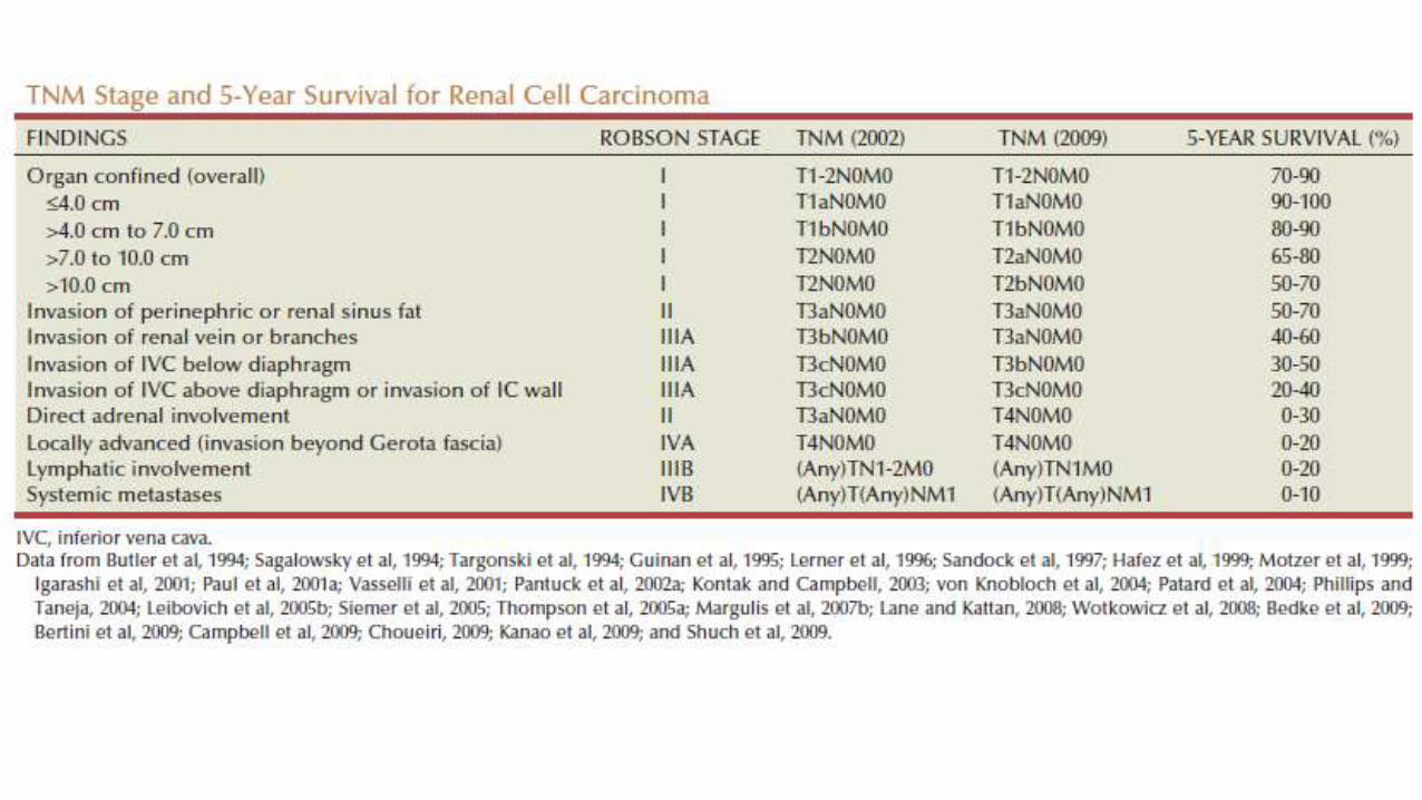
staging


• Prognostic Factors – MSKCC Scoring System
• Poor-prognosis patients are defined as those having ≥ 3 more factors: these are predictors of short survival:
• Serum LDH > 1.5 times the upper limit of normal
• Hemoglobin (g/dl) : < lower limit of normal
• Corrected serum Calcium > 10 mg/dL (2.5 mmol/L)
• Interval less than a year from original diagnosis to start of systemic therapy
• Karnofsky Performance Score ≤ 70
• ≥ 2 sites of organ metastasis