25
Reaching the Information Limit in Cryo-EM of Biological Macromolecules: Experimental Aspects -Robert M. Glaeser and Richard J. Hall (2011)
| Date post: | 13-Dec-2015 |
| Category: |
Documents |
| Upload: | emmeline-franklin |
| View: | 219 times |
| Download: | 0 times |

Reaching the Information Limit in Cryo-EM of Biological Macromolecules:Experimental Aspects
-Robert M. Glaeser and Richard J. Hall (2011)
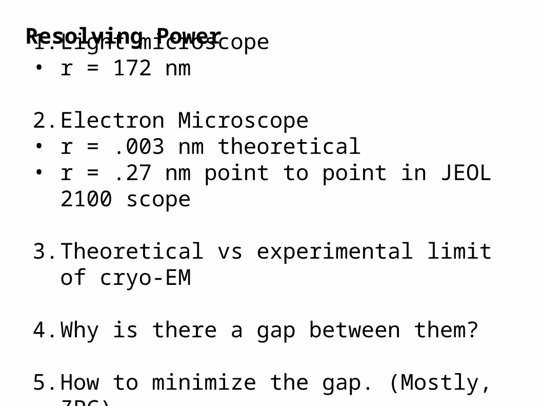
1. Light microscope• r = 172 nm
2. Electron Microscope• r = .003 nm theoretical• r = .27 nm point to point in JEOL 2100 scope
3. Theoretical vs experimental limit of cryo-EM
4. Why is there a gap between them?
5. How to minimize the gap. (Mostly, ZPC)
Resolving Power
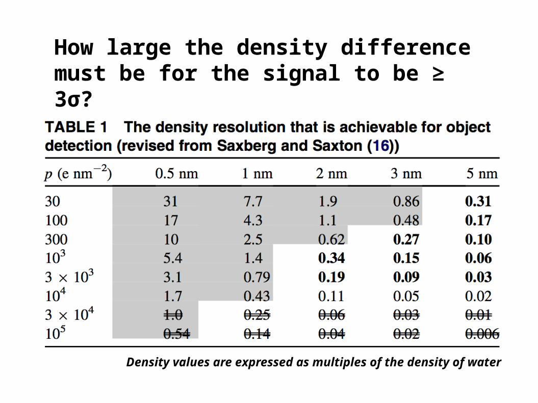
How large the density difference must be for the signal to be ≥ 3σ?
Density values are expressed as multiples of the density of water

Atomic resolution(3Å) information by Henderson (1995)
•Theoretically ideal condition :1. Homogenous (identical) objects2. perfect contrast transfer 2. noise free detector
•Specimen size to align properly : 40 kDa1. Depends on the contrast (signal) &
exposure
• Particle numbers for atomic res. : 12,000
1. e- exposure required for the image 2. e- exposure that damages the molecule
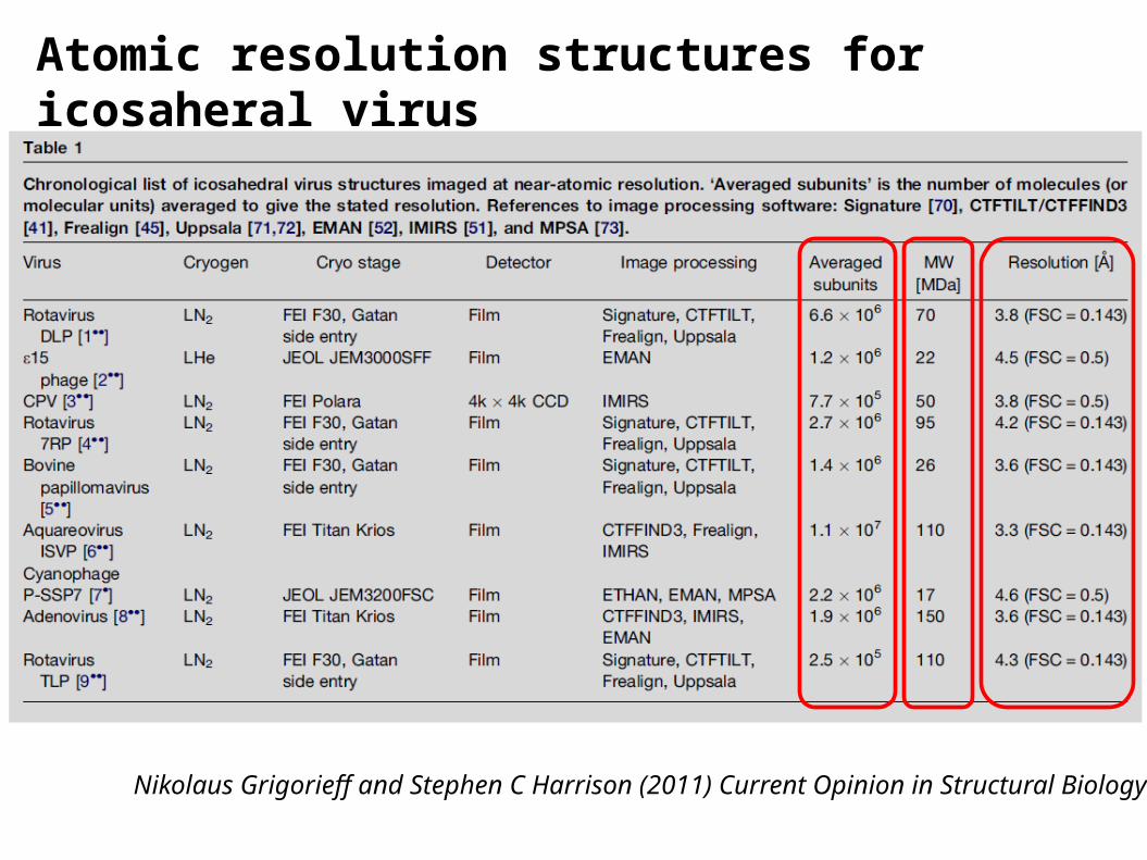
Atomic resolution structures for icosaheral virus
Nikolaus Grigorieff and Stephen C Harrison (2011) Current Opinion in Structural Biology
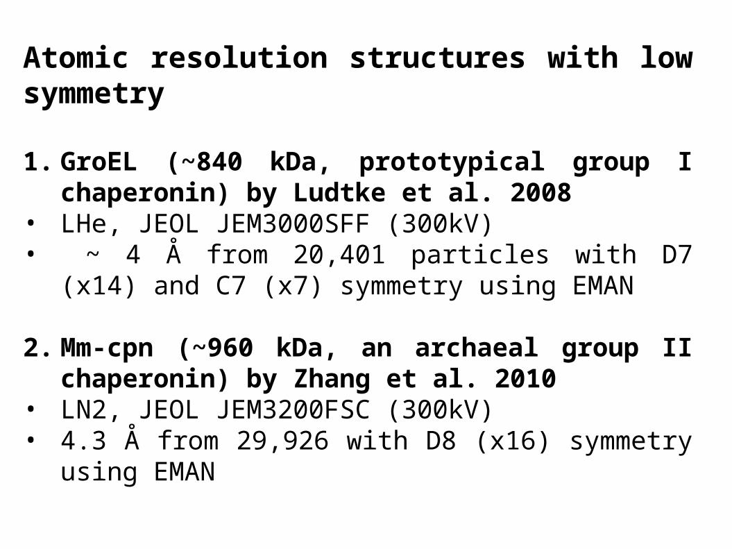
Atomic resolution structures with low symmetry
1. GroEL (~840 kDa, prototypical group I chaperonin) by Ludtke et al. 2008
• LHe, JEOL JEM3000SFF (300kV)• ~ 4 Å from 20,401 particles with D7 (x14) and C7 (x7)
symmetry using EMAN
2. Mm-cpn (~960 kDa, an archaeal group II chaperonin) by Zhang et al. 2010
• LN2, JEOL JEM3200FSC (300kV)• 4.3 Å from 29,926 with D8 (x16) symmetry using EMAN
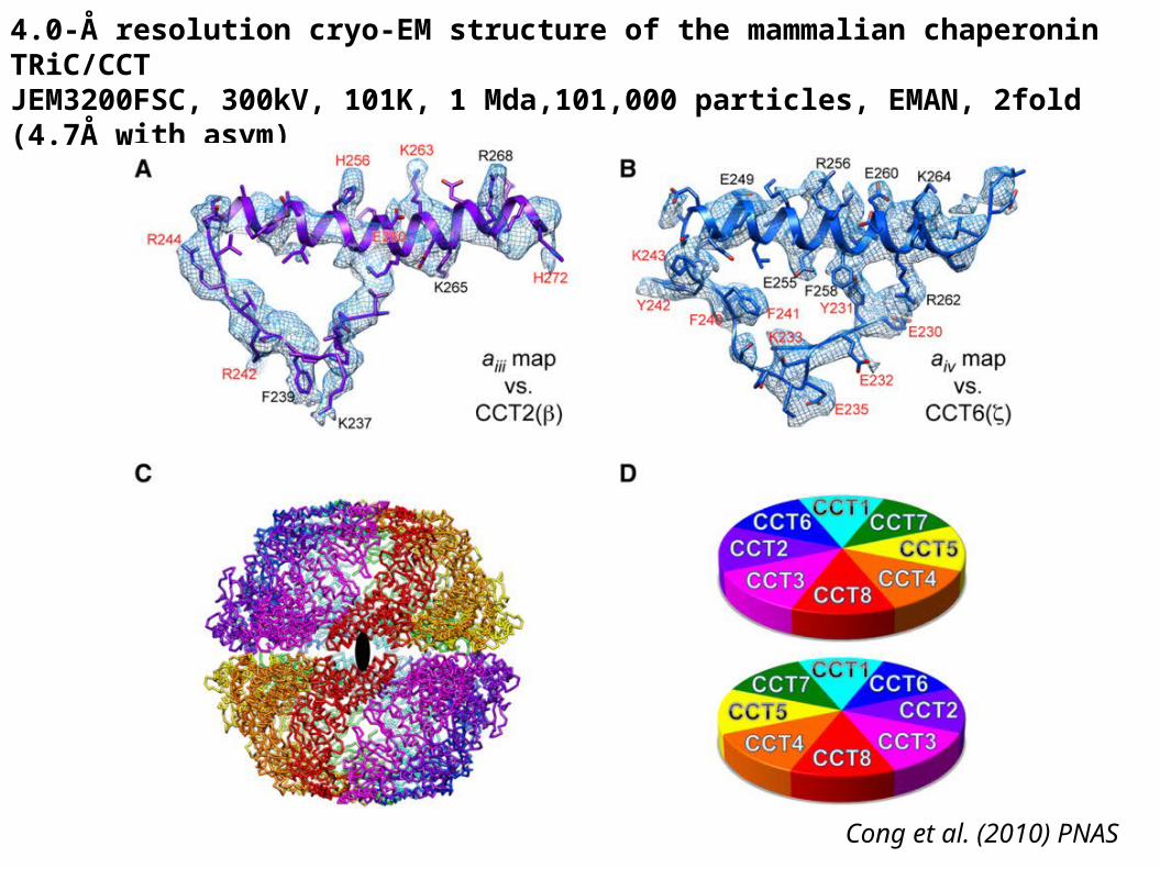
4.0-Å resolution cryo-EM structure of the mammalian chaperonin TRiC/CCTJEM3200FSC, 300kV, 101K, 1 Mda,101,000 particles, EMAN, 2fold (4.7Å with asym)
Cong et al. (2010) PNAS
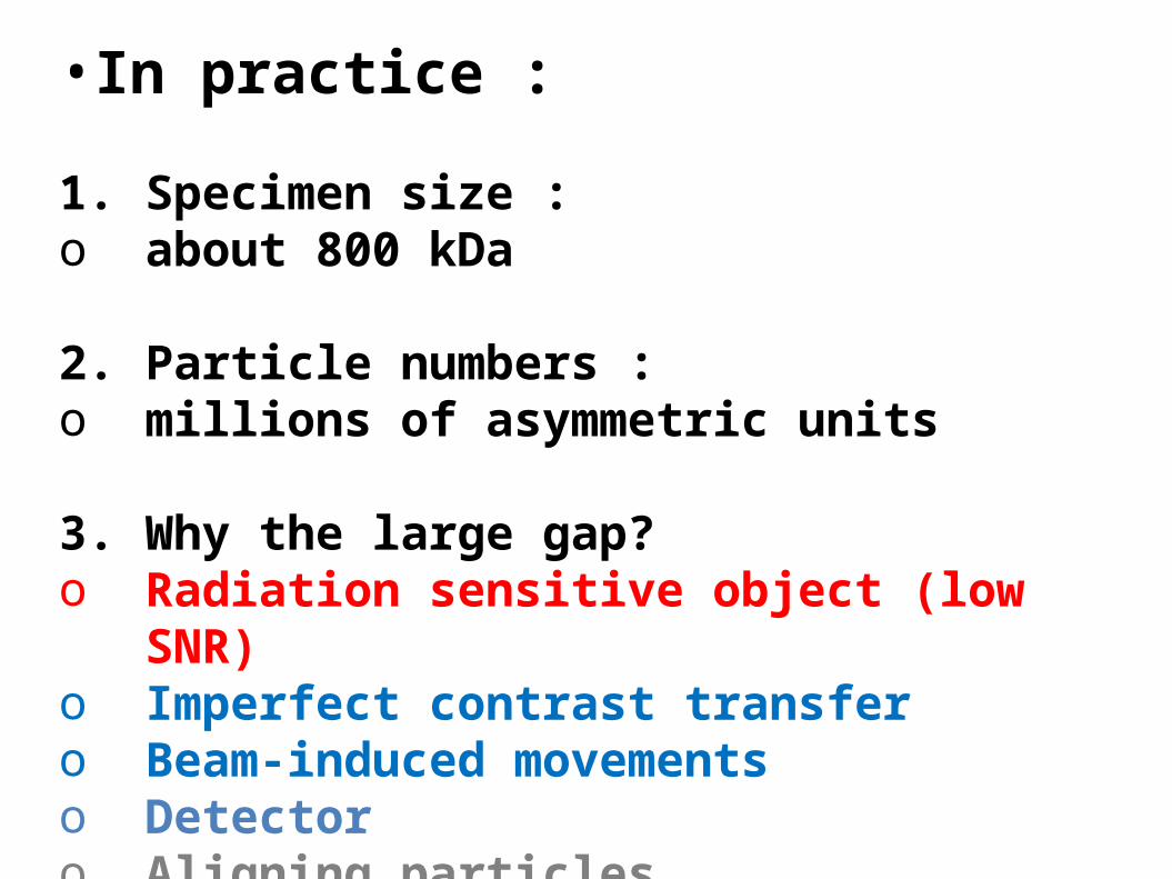
•In practice :
1. Specimen size : o about 800 kDa
2. Particle numbers : o millions of asymmetric units
3. Why the large gap?o Radiation sensitive object (low SNR)o Imperfect contrast transfer o Beam-induced movements o Detectoro Aligning particles (reconstruction)
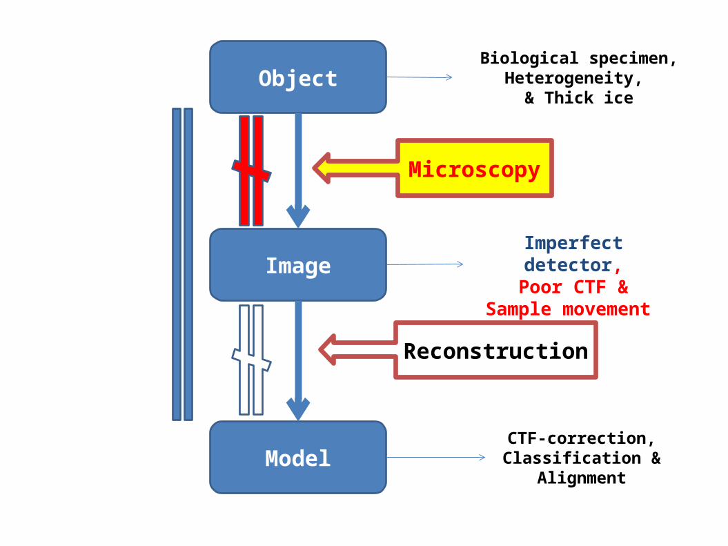
Object
Image
Model
Microscopy
Reconstruction
Biological specimen, Heterogeneity,
& Thick ice
Imperfect detector,Poor CTF &
Sample movement
CTF-correction, Classification &
Alignment
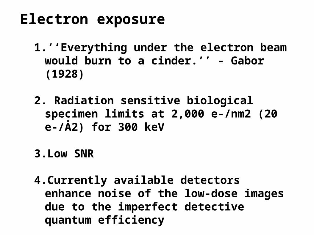
Electron exposure
1. ‘‘Everything under the electron beam would burn to a cinder.’’ - Gabor (1928)
2. Radiation sensitive biological specimen limits at 2,000 e-/nm2 (20 e-/Å2) for 300 keV
3.Low SNR
4.Currently available detectors enhance noise of the low-dose images due to the imperfect detective quantum efficiency
Ideal contrast transfer
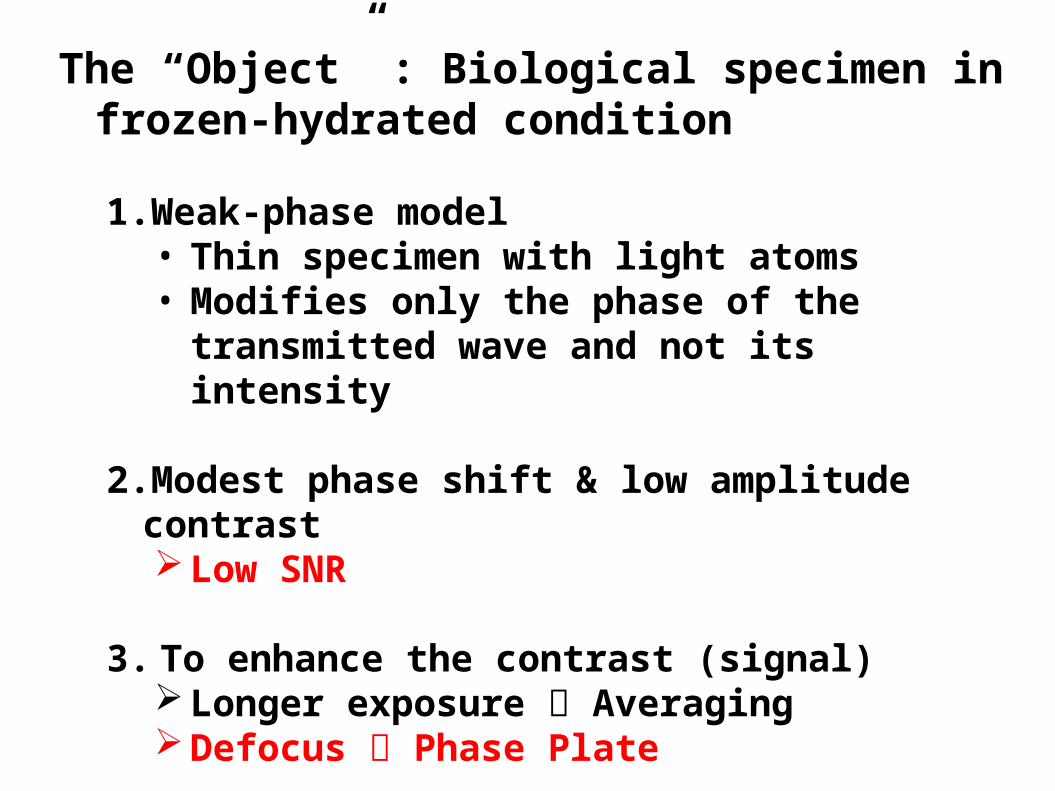
The “Object” : Biological specimen in frozen-hydrated condition
1.Weak-phase model • Thin specimen with light atoms• Modifies only the phase of the transmitted
wave and not its intensity
2.Modest phase shift & low amplitude contrast Low SNR
3. To enhance the contrast (signal) Longer exposure Averaging Defocus Phase Plate

Defocus (under focus)
1.Enhance the contrast
2.Poor signal transfer
1 um √3 um
coherent
incoherent
Frank (2006)

A quarter-wave plate to apply a 90 phase shift to the scattered wave relative to the unscattered wave (Zernike, 1955)
* Phase contrast is stronger at in-focus than defocus.
Taylor series and assuming Φ(r) << 1,
Phase shift by an object C
Phase-shifted wave function
2007, Frank

Phase plate (Zernike phase contrast cryo-electron microscopy)

Chang et al. (2010) Structure : Simulated pol II images
•In-Focus
•Enhanced contrast
Higher SNR
Especially at low resolution
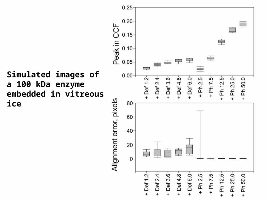
Simulated images of a 100 kDa enzymeembedded in vitreous ice
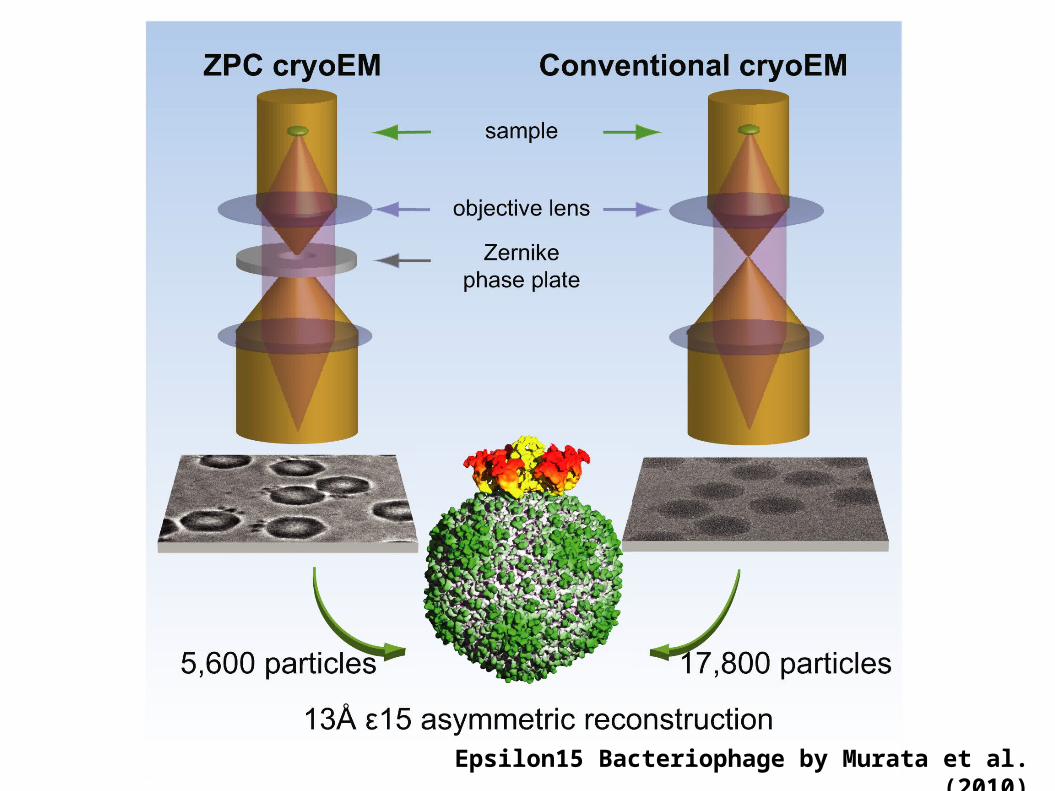
Epsilon15 Bacteriophage by Murata et al. (2010)

5600 1500 500 100

Minimizing beam-induced movement

Minimizing beam-induced movement
Bacteriorhodopsin 2D crystal
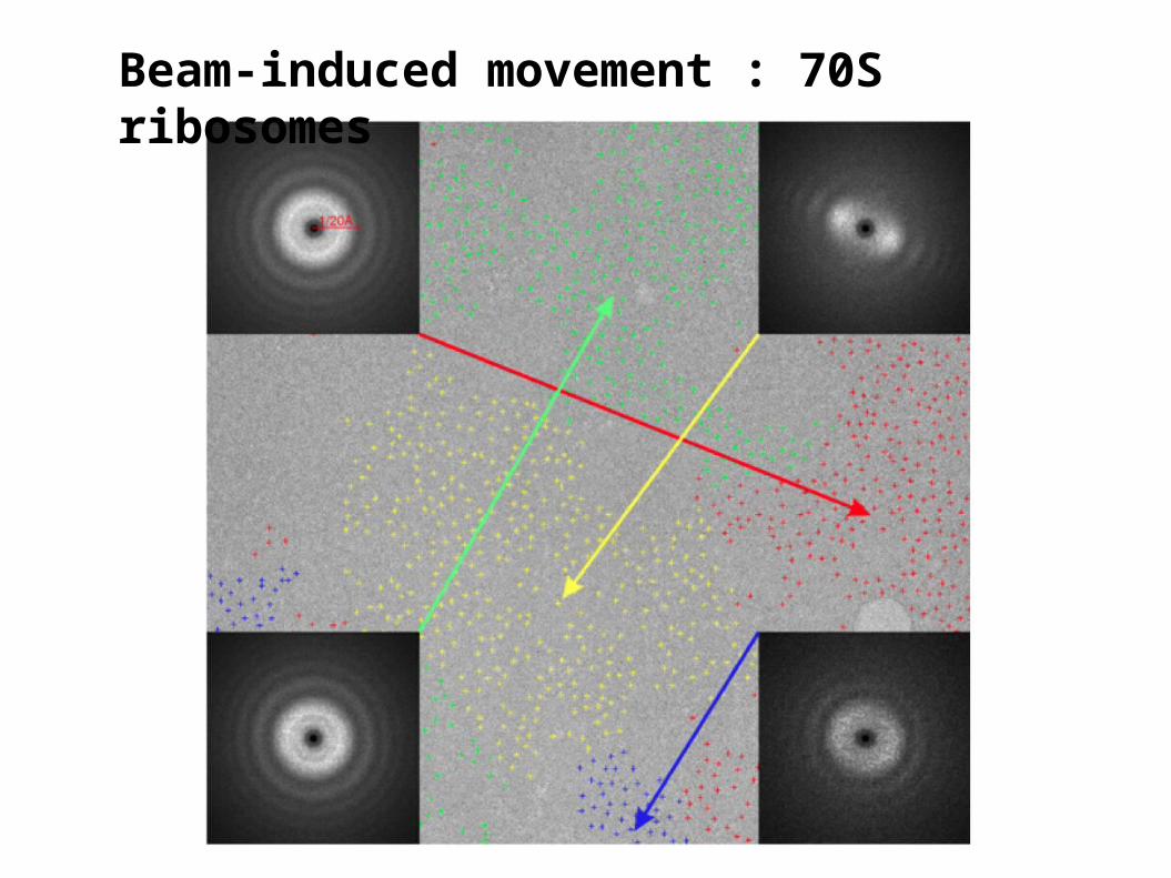
Beam-induced movement : 70S ribosomes
Efforts to minimize the movement
1.Limiting the size of illuminated area.
2. Improving the electrical conductivity of the
support film.
3.However, only partial reduction has been
achieved.
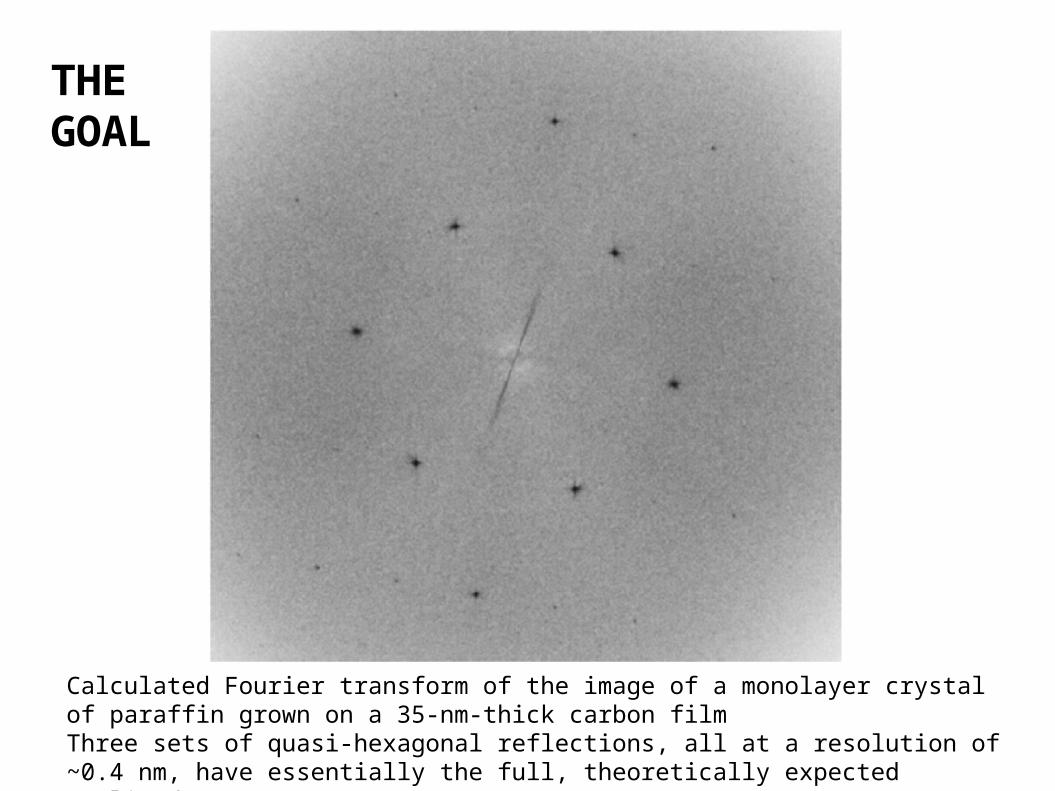
Calculated Fourier transform of the image of a monolayer crystal of paraffin grown on a 35-nm-thick carbon filmThree sets of quasi-hexagonal reflections, all at a resolution of ~0.4 nm, have essentially the full, theoretically expected amplitude
THE GOAL

lBarriers for the information limit and how to reach it
l1. Imperfect DQE of the detector Noise-free detector l Pixilated electron counter
l2. Poor CTF Ideal phase-contrast transfer functionl Charging-free quarter-wave plate
l3. Beam-induced movement ??!!
l4. Reconstruction • CTF-correction
l Reliable classfication (different conformational states)l Perfect alignment


