RecA Protein from the Extremely Radioresistant BacteriumDeinococcus radiodurans: Expression, Purification,
and CharacterizationJong-Il Kim,1† Ajay K. Sharma,2‡ Stephen N. Abbott,1 Elizabeth A. Wood,1 David W. Dwyer,1
Aaron Jambura,1 Kenneth W. Minton,2 Ross B. Inman,1,3 Michael J. Daly,2 and Michael M. Cox1*Department of Biochemistry1 and Department of Molecular Virology,3 University of Wisconsin—Madison, Madison, Wisconsin
53706, and Department of Pathology, Uniformed Services University of the Health Sciences, Bethesda, Maryland 208142
Received 18 July 2001/Accepted 2 November 2001
The RecA protein of Deinococcus radiodurans (RecADr) is essential for the extreme radiation resistance of thisorganism. The RecADr protein has been cloned and expressed in Escherichia coli and purified from this host.In some respects, the RecADr protein and the E. coli RecA (RecAEc) proteins are close functional homologues.RecADr forms filaments on single-stranded DNA (ssDNA) that are similar to those formed by the RecAEc. TheRecADr protein hydrolyzes ATP and dATP and promotes DNA strand exchange reactions. DNA strandexchange is greatly facilitated by the E. coli SSB protein. As is the case with the E. coli RecA protein, the useof dATP as a cofactor permits more facile displacement of bound SSB protein from ssDNA. However, there areimportant differences as well. The RecADr protein promotes ATP- and dATP-dependent reactions with dis-tinctly different pH profiles. Although dATP is hydrolyzed at approximately the same rate at pHs 7.5 and 8.1,dATP supports an efficient DNA strand exchange only at pH 8.1. At both pHs, ATP supports efficient DNAstrand exchange through heterologous insertions but dATP does not. Thus, dATP enhances the binding ofRecADr protein to ssDNA and the displacement of ssDNA binding protein, but the hydrolysis of dATP is poorlycoupled to DNA strand exchange. The RecADr protein thus may offer new insights into the role of ATPhydrolysis in the DNA strand exchange reactions promoted by the bacterial RecA proteins. In addition, theRecADr protein binds much better to duplex DNA than the RecAEc protein, binding preferentially to double-stranded DNA (dsDNA) even when ssDNA is present in the solutions. This may be of significance in thepathways for dsDNA break repair in Deinococcus.
Bacteria belonging to the family Deinococcaceae are themost radiation-resistant organisms known (2, 19, 40). Despiteubiquitous distribution and ancient derivation, only seven spe-cies of this family have been described (35). Of these species,Deinococcus radiodurans is the only one for which systems ofgenetic manipulation have been developed, and these havebeen used in the molecular analysis of its DNA repair pathways(15, 16) and in its development for bioremediation of radio-active waste environments (5, 14, 27). Adding to the growingresource of genetic technologies available for D. radiodurans isthe recent whole-scale sequencing, annotation, and analysis ofits genome (28, 35, 36, 56). D. radiodurans is the first repre-sentative with a completely sequenced genome from a bacterialbranch of extremophiles—the Thermus/Deinococcus group.Phylogenetic tree analysis, combined with the identification ofseveral synapomorphies (shared derived characters) betweenThermus and Deinococcus, support that it is a very ancientbranch localized in the vicinity of the bacterial tree root (35).
D. radiodurans is a polyextremophile (48), showing remark-able resistance to a range of severe damage caused by ionizing
radiation, desiccation, UV radiation, oxidizing agents, or elec-trophilic mutagens (40). This bacterium is most famous for itsresistance to ionizing radiation. It survives acute exposures togamma radiation that exceed 1,700,000 rads without lethalityor induced mutation (18) and is capable of vigorous growth inthe presence of chronic irradiation (6,000 rads per h) (27, 55).D. radiodurans can survive 100 to 200 irradiation-inducedDNA double-stranded breaks (DSBs) per haploid genome,yielding �1,000 DSB fragments in a typical polyploid cell (18,28). Although its myriad resistance phenotypes stem from ef-ficient DNA repair processes, the mechanisms underlying thisrepair remain poorly understood (35). Its extreme DNA dam-age resistance phenotype appears to be very complex, deter-mined collectively by features revealed by analysis of its ge-nome (35, 56) as well as by many more subtle structuralpeculiarities of proteins and DNA that are not readily inferredfrom the sequences. Remarkably, the number of DNA repairenzymes identified in D. radiodurans by genomic annotation(35, 56) is less than reported for Escherichia coli. Further, arecent analysis of predicted expression levels [E(g)] (based oncodon usage) of the main repair proteins of D. radiodurans andE. coli showed that neither set of repair genes is likely to beexpressed at high levels (22). There is one exception. RecAprotein is predicted to be highly expressed in both organisms;the level of E. coli RecA (RecAEc) expression was predicted tobe high [E(g) � 1.48], and D. radiodurans RecA (RecADr)expression was predicted to be dramatically high [E(g) � 2.04].Given these general similarities in expression of repair func-
* Corresponding author. Mailing address: Department of Biochem-istry, University of Wisconsin—Madison, 433 Babcock Dr., Madison,WI 53706-1544. Phone: (608) 262-1181. Fax: (608) 265-2603. E-mail:[email protected].
† Present address: Department of Applied Microbiology, SeoulWomen’s University, Nowon-Gu, Seoul, Korea.
tions, another explanation for the radioresistance of D. radio-durans has been sought. For example, unlike E. coli, the repairproteins of D. radiodurans may operate in the context of highlyorganized clusters of aligned multiple identical chromosomes(17, 35). This chromosomal structure would simplify the searchfor homology during repair and facilitate the function of whatappears to be a conventional set of repair enzymes in D. ra-diodurans.
RecA or RecA-like proteins have been found throughoutthe phylogenetic tree, including archaea, bacteria, yeast,plants, and mammals; they play a critical role in biologicalprocesses that require homologous DNA pairing and recom-bination (4, 10, 49). D. radiodurans is no exception, and its recAgene appears very similar to those found in Thermus spp. andother gram-positive bacteria. For example, the RecAs of D.radiodurans and Thermus aquaticus have 69% amino acid se-quence identity. In comparison, the RecAs of E. coli and D.radiodurans exhibit a 53% sequence identity. A functionalRecADr protein is central to the expression of the resistancephenotypes of D. radiodurans (41). For example, disruption ofrecADr greatly diminishes the bacterium’s ability to recoverfrom acute DNA damage (6, 18, 20, 41) and prevents its growthin the presence of chronic radiation (55). A direct comparisonof the D. radiodurans and E. coli RecA proteins could proveenlightening. The biological function of the RecADr proteinhas been tightly linked to efficient repair of DSBs. The primaryfunction of the RecAEc protein appears to lie in the repair ofa broad range of stalled replication fork structures (9, 11, 12,25).
RecADr is normally expressed constitutively at low levels inD. radiodurans (M. Lipton, Pacific Northwest National Labo-ratory, Richmond, Wash., personal communication) and isonly transiently expressed at high levels following extremeDNA damage (6). Thus, it has been difficult to purify signifi-cant quantities of this recombinase directly from D. radio-durans, and several previous attempts to purify cloned RecADr
in E. coli were unsuccessful (6, 20, 40).These difficulties have been overcome, and we report here
the cloning and expression of RecADr protein in two differentE. coli expression systems. One involves the construction of afusion protein to aid in purification, and the other is a directexpression of the native protein. Both proteins have been pu-rified to near-homogeneity, and their in vitro activities havebeen examined at some length. In general, the RecADr proteinforms filaments on DNA and possesses activities that resemblethose of the RecAEc protein. However, certain molecular func-tions, particularly binding to duplex DNA, the hydrolysis ofnucleoside triphosphate (NTP) cofactors, and the response ofDNA strand exchange to NTP hydrolysis, provide a source ofsome sharp contrasts between the E. coli and D. radioduransRecA proteins.
MATERIALS AND METHODS
Enzymes and reagents. Restriction endonucleases, T4 DNA ligase, theIMPACT-T7 cloning system, chitin beads, and E. coli RecA protein were pur-chased from New England Biolabs. E. coli single-stranded DNA (ssDNA) bind-ing protein (SSB) was purified as described previously (31) and stored frozen at�70°C in a buffer containing 20 mM Tris-HCl (40% cation, pH 8.4), 0.15 MNaCl, 1 mM EDTA, 1 mM �-mercaptoethanol, and 50% glycerol. The concen-tration of SSB was determined by absorbance at 280 nm using an extinctioncoefficient of ε280 � 1.5 A280 mg�1 ml�1 (32). Plasmid DNA purification and gel
extraction kits were purchased from Qiagen. [�-S]ATP, Tris buffer, creatinephosphokinase, and phosphocreatine were purchased from Boehringer Mann-heim. Restriction endonucleases were purchased from New England Biolabs.Ceramic hydroxyapatite type II was purchased from Bio-Rad. Phosphoenolpyru-vate, pyruvate kinase, lactic dehydrogenase, NADH, ATP, and proteinase Kwere purchased from Sigma.
DNA. Circular duplex and single-stranded DNA (ssDNA) from M13mp8 andits derivatives (M13mp8.52) were prepared using methods described previously(51). Circular ssDNA, supercoiled double-stranded DNA (dsDNA), and nickedcircular �X174 DNA were purchased from New England Biolabs. The concen-trations of ssDNA and dsDNA were determined by absorbance, using A260 � 1as equivalent to 36 and 50 �g ml�1, respectively. Linear duplex �X174 DNA wasprepared by digestion of supercoiled DNA with PstI endonuclease. LinearssDNA was prepared by annealing a synthetic oligonucleotide to the circularsingle-stranded �X174 DNA at a site encompassing the unique PstI recognitionsequence, with digestion with PstI endonuclease. M13mp8 dsDNA was linearizedby digestion with EcoRI. M13mp8.52 dsDNA was linearized by cleavage with therestriction endonuclease MscI. The linear ssDNA was purified using the Qia-quick gel extraction kit (Qiagen).
Cloning of D. radiodurans recA gene. The recA gene from D. radiodurans usedin this study was cloned in two different ways.
The first utilized the IMPACT-T7 expression system as described previously(20) and subcloned as pJDC101 (6). To clone the recA gene into theIMPACT-T7 vector, two PCR forward and reverse primers were designed withan NdeI site (underlined) flanking at the N-terminal side (5 TTTTTTCATATGAGCAAGGACGCCACCAAAG 3) and a SmaI site (underlined) at the C-terminal side (5 TTTTTTCCCGGGCGCTTCGGCGGCTTCGGGC 3). DNAwas amplified by PCR using an Advantage-GC Genomic PCR kit (Clontech) asdescribed by the manufacturer. The PCR product was agarose gel purified, andthe DNA was digested with the NdeI and SmaI restriction enzymes. The cleavedDNA was ligated in frame to the pTYB2 plasmid vector of the IMPACT-T7system, which was also restricted with NdeI/SmaI. The result is a cloned gene inwhich the recADr gene is fused to an intein- and chitin-binding domain. Therecombinant plasmid DNA was transformed into competent cells of the E. colistrain BLR(DE3) (Novagen) and plated onto Luria-Bertani (LB) agar platescontaining ampicillin (100 �g/ml). The colonies were picked and grown in LBliquid media, and plasmid DNA was prepared from different bacterial colonies.Plasmids were checked for the correct insert DNA by restriction analysis andDNA sequencing.
The second method involved direct cloning of the recADr gene into thepET21A vector (Novagen). The recADr gene was PCR amplified from genomicDNA extracted from D. radiodurans bacterial cells (ATCC 13939), using thefollowing primers: 5CCAATAGCATATGAGCAAGGACGCCA and 5CGGGATCCCAAGAGGAGGTTTAC. The 1,123-bp PCR product was gel purifiedand then digested overnight with 30 U each of NdeI and BamHI. It was ligatedto pET21A that had been digested with the same enzymes and transformed intothe nonexpression host DH5. Several clones were sequenced. The first threehad point mutations, but the fourth had the complete wild-type recADr gene andwas selected as pEAW158. The pEAW158 construct was transformed into thehost STL2669/pT7pol26 (a gift from Susan Lovett, Brandeis University) forexpression. This strain lacks the recAEc gene and carries a plasmid expressing thebacteriophage T7 RNA polymerase under lac control.
Purification of RecADr protein. The recombinant plasmid containing D. radio-durans recA gene fused to the intein-, chitin-binding domain (pAS18) in E. coliwas inoculated into LB medium containing 100 �g of ampicillin/ml at 37°C.When the culture reached an optical density at 600 nm of 0.6, it was induced with0.3 mM isopropyl-�-D-thiogalactopyranoside (IPTG) (final concentration) for4 h. The cells were harvested, resuspended in buffer A (20 mM Tris Cl [pH 7.5]–1mM EDTA–1.5 M KCl–0.1% Tween 20) containing pepstatin and leupeptinprotease inhibitors (final concentration, 1 �g/ml each), and lysed by passing thecells three times through a French press cell (American Instrument Co.) at 900lb/in2. The lysate was incubated at 4°C for 1 h with slow shaking and centrifugedat 27,000 � g for 20 min, and the clarified crude cell lysate was loaded onto achitin agarose column. The chitin column was then rinsed with 10 volumes ofbuffer A and then quickly flushed with buffer B (20 mM Tris Cl [pH 7.5]–0.5 MNaCl–1 mM EDTA) containing 50 mM dithiothreitol (DTT). The column wasthen left at 4°C for about 16 h. This step produces efficient self-cleavage of theRecA protein from the intein portion of the vector. The latter portion remainsattached to the chitin agarose column. Then the protein was eluted from thecolumn with 5 volumes of buffer B. The protein was further concentrated usingan ultrafiltration cell concentrator (Amicon) and dialyzed against buffer C (20mM phosphate buffer [pH 7.5]–0.1 mM EDTA). This protein was loaded on ahydroxyapatite column and rinsed with 10 volumes of buffer C. The protein was
then eluted with a phosphate buffer step gradient (50 to 400 mM). The individualprotein fractions were checked for nuclease contamination by incubating themwith �X174 RFI DNA, linear dsDNA, and circular ssDNA. In each case, theamount of RecADr used was twice that used in typical experiments, the incuba-tions were carried out under standard reaction conditions for 2 h, and thereactions were analyzed by gel electrophoresis. The fractions without any de-tectable nuclease contamination were pooled and dialyzed extensively againstbuffer D (20 mM Tris Cl [pH 7.5]–0.1 mM EDTA–1 mM DTT–10% glycerol). Asdescribed in Results, the resulting protein has one extra glycine residue attachedto the C terminus, and we refer to this protein as RecA*Dr.
The native RecADr protein was purified using a different procedure. TheSTL2669/pT7pol26 cells containing pEAW158 were grown in LB broth contain-ing 100 �g of ampicillin/ml and 40 �g of kanamycin/ml at 37°C to an opticaldensity at 600 nm of just greater than 0.5. They were then induced with 0.4 mMIPTG and grown for three more hours at 37°C before harvesting. The cells (50g) from a 6-liter culture were resuspended in a buffer (200 ml) containing 25%sucrose and 250 mM Tris-HCl (80% cation, pH 7.5) and lysed by adding ly-sozyme (final concentration, 1.4 mg/ml). After 1 h of stirring at 4°C, EDTA (10mM final concentration) was added and the cells were sonicated. The insolublematerial was removed by centrifugation at 31,000 � g for 1 h at 4°C. The DNAand proteins were precipitated by adding 5% Polymin P (final concentration,0.5% vol/vol) slowly to the supernatant. The lysate was stirred for 30 min at 4°Cand centrifuged at 9,000 rpm for 15 min. The pellet was washed twice with Rbuffer (20 mM Tris-HCl, 80% cation [pH 7.5], 10% glycerol, 1 mM DTT) plus 50mM ammonium sulfate, by resuspending it with stirring for 15 min at 4°Cfollowed by centrifugation at 9,000 rpm for 20 min each time. Using the sameprocedure, the final pellet from the above wash steps was washed with R bufferplus 200 mM ammonium sulfate to elute the RecADr protein, followed bycentrifugation at 14,000 rpm for 30 min. Ammonium sulfate (0.2002 g/ml) wasadded to the supernatant, and the precipitated proteins were removed by cen-trifugation. The pellet was discarded. Additional ammonium sulfate (0.145 g/mlof supernatant) was then added to precipitate the RecADr protein, followed bycentrifuged at 14,000 rpm for 30 min. The pellet was washed twice with R bufferplus 0.377 g of ammonium sulfate/ml to remove as much Polymin P as possibleand then dissolved in R buffer plus 200 mM ammonium sulfate and dialyzedagainst R buffer containing 50 mM KCl. The dialyzed solution was loaded at aflow rate of 150 ml/h onto a 60-ml DEAE-Sepharose column, equilibrated withR buffer plus 50 mM KCl. The column was washed with R buffer plus 50 mMKCl. Fractions containing D. radiodurans RecA protein were combined anddialyzed against P (350 mM) buffer containing 350 mM potassium phosphate(pH 7.5), 10% glycerol, 0.1 mM EDTA, and 1 mM DTT, and then loaded ontoa 70-ml Bio-Gel hydroxyapatite column, equilibrated with the same P (350 mM)buffer. The RecA protein was eluted from the column with 2 volumes of P (500mM phosphate) buffer. The individual protein fractions were checked for nucle-ase contamination by incubating fractions with �X174 RFI DNA, linear dsDNA,and circular ssDNA as described above. Fractions containing purified RecAprotein were pooled and dialyzed against storage buffer (20 mM Tris-HCl, 80%cation [pH 7.5], 10% glycerol, 1 mM DTT), aliquoted, and stored at �80°C.Typical yields of the native protein were 0.3 to 0.5 mg per g of cells.
The extinction coefficient for RecADr protein was determined by a publishedprocedure (30), modified as described previously (37). The results of threedeterminations were averaged to give an extinction coefficient of RecADr proteinas ε280 � 0.372 A280 mg ml�1. The RecADr protein contains no tryptophanresidues. N-terminal sequence analysis was carried out at the Protein and NucleicAcid Chemistry Laboratories, Department of Molecular Biology and Pharma-cology, Washington University School of Medicine, St. Louis, Mo., and wascarried out with protein samples that were not identified to the sequencinglaboratory personnel. Matrix-assisted laser desorption ionization (MALDI) massspectrometry was carried out in the Mass Spectrometry Bioanalytical Facility atthe University of Wisconsin Biotechnology Center.
DNA strand exchange reactions. RecA-dependent DNA strand exchange re-actions were carried out as described previously (3, 13), between circular ssDNAand the linear duplex DNA (derived from either �X174 or M13mp8). Unlessotherwise stated, all reactions were carried out at 37°C in solutions containing 25mM Tris-acetate (80% cation, pH 7.5), 1 mM DTT, 5% glycerol, 3 mM potas-sium glutamate, 10 mM (or the indicated concentration) magnesium acetate, andan ATP-regenerating system (10 U/ml of pyruvate kinase, 3.3 mM phosphoenol-pyruvate, or 10 U of creatine kinase/ml, 12 mM phosphocreatine). DNA, SSB,ATP (or dATP), and RecA protein concentrations are indicated for each exper-iment. A preincubation of ssDNA with RecADr protein at 37°C for 5 min wasfollowed by addition of ATP and SSB. After an additional 5-min incubation,linear duplex DNA was added to start the DNA strand exchange reactions. Theorder of addition is described in figure legends for each experiment in which it
was changed. Aliquots (20 �l unless otherwise indicated) of strand exchangereactions described above were removed at each time point, and the reactionswere stopped by addition of 5 �l of gel loading buffer (0.125% bromophenolblue, 25 mM EDTA, 25% glycerol, 5% sodium dodecyl sulfate [SDS]). Thesealiquots were stored on ice until after the last time point was taken. Samples wereelectrophoresed in an 0.8% agarose gel with TAE buffer (40 mM Tris-acetate[pH 8.0], 1 mM EDTA), stained with GelStar nucleic acid gel stain (FMCBioProducts) or ethidium bromide, and photographed with UV light using a geldocument camera. The DNA bands were quantified with ImageQuant software(version 4.2). In order to correct for variability in sample loading onto theagarose gel, the band corresponding to full-length circular hybrid duplex productwas quantified as the fraction of the total fluorescing DNA in a given gel lane,excluding only the band corresponding to the ssDNA.
ATPase assay. ATP hydrolysis was monitored as previously described (3, 21,42). All reactions were done at 37°C. ATP (or dATP) hydrolysis was measuredby a coupled spectrophotometric assay. Absorbance measurements were ob-tained on a Cary 300 spectrophotometer equipped with two six-position, ther-mojacketed cuvette holders attached to a constant-temperature water circulator.Reaction mixtures contained 25 mM Tris-acetate (80% cation, pH 7.5), 1 mMDTT, 5% glycerol, 3 mM potassium glutamate, and 10 mM magnesium acetate.Concentrations of RecADr protein, DNA, SSB, and ATP (or dATP) are indi-cated in figure legends. Where the pH was varied, the buffer was a 25 mMconcentration of either Tris-acetate or morpholineethanesulfonic acid-NaOH.An ATP regenerating system (3 mM phosphoenolpyruvate, 10 U of pyruvatekinase ml�1, and 3 mM potassium glutamate) and a coupling system (2 mMNADH and 10 U of lactate dehydrogenase ml�1) were also included. WhendATP was used, the ATP regenerating and coupling enzymes were increased to25 U of pyruvate kinase ml�1 and 60 U of lactate dehydrogenase ml�1. Reactionswere initiated by the addition of ATP (or a mixture of ATP and SSB) after allother components were incubated at 37°C for 10 min. Changes in the order ofaddition are indicated in figure legends. Regeneration of ATP from ADP andphosphoenolpyruvate is coupled to the conversion of NADH to NAD� (in thereaction catalyzed by lactate dehydrogenase), which can be monitored by adecrease in absorbance at 380 nm. Although the absorbance maximum forNADH occurs at 340 nm, absorbances were measured at 380 nm to remainwithin the linear absorbance range of the spectrophotometer for an extendedlength of time as required by these experiments. Rates of ATP hydrolysis (�M/min) were calculated from �A380 min�1 obtained at steady state, using anextinction coefficient of 1,210 M�1 cm�1 at 380 nm for NADH. In most cases,absorbances were continuously measured over a period of 2 h.
Electron-microscopic studies. The formation of RecA filament onto circularssDNA, linear ssDNA, and dsDNA was visualized by electron microscopy. Car-bon films mounted on electron microscopy grids were activated with denaturedbovine serum albumin (BSA) in the following way. A solution containing 0.1%BSA and 0.5% N-lauroyl sarcosine (sodium salt) was placed in a boiling waterbath for 10 min. After cooling to 25°C, carbon grids were floated on drops of thedenatured BSA for 5 min and then on drops of water followed by immersion inwater (three times for 10 s). Grids were finally dried under a heat lamp andstored in a desiccator.
Reactions between RecADr and circular ssDNA or linear dsDNA (�X174)consisted of RecA*Dr (2 uM), HEPES (25 mM, pH 7.5), ssDNA or dsDNA (6uM), creatine phosphokinase (10 U/ml), phosphocreatine (12 mM). After incu-bation for 10 min at 37°C, SSB and dATP were added (2 �M and 3.3 mM,respectively) followed by a further incubation for 1 min at 37°C. This was thenimmediately followed by the addition of �-S-ATP (final concentration, 1 mM).
The above-described samples were immediately diluted 10-fold with 200 mMammonium acetate plus 10% glycerol, adsorbed to denatured BSA-activatedcarbon grids for 3 min, washed with 200 mM ammonium acetate plus 10%glycerol, and stained with uranyl acetate (5%) plus 10% glycerol. Grids were thenimmersed in water for 10 s and dried. Grids were finally rotary shadowed withplatinum at an angle of about 7 degrees.
RESULTS
Experimental rationale. The goal of these experiments wasstraightforward. We wished to characterize the RecADr pro-tein as part of a larger effort to discover the molecular basis ofthe extreme radioresistance of the bacterium D. radiodurans.These initial efforts focused on protein purification and anexamination of classical RecA activities.
VOL. 184, 2002 DEINOCOCCUS RADIODURANS RecA PROTEIN 1651
RecA gene cloning strategy. Because very low-level expres-sion of the RecADr gene in the absence of inducer (e.g., IPTG)was reported to be lethal to E. coli cells (20), we tried severaldifferent expression systems. We were eventually able to suc-cessfully express the RecADr protein in two ways, as a fusionprotein with an intein at the C-terminal region (using theIMPACT-T7 system in E. coli) and as the native protein (in thepET21A vector). The pTYB2 plasmid vector is designed tocreate a fusion between the gene of interest (in this case D.radiodurans RecA), 55-kDa intein, and the DNA encoding asmall 5-kDa chitin binding domain at the C terminus of theintein for efficient column affinity purification of the three-partfusion protein. The N terminus of intein undergoes a self-cleavage at low temperatures in the presence of thiols, such asDTT, �-mercaptoethanol, or cysteine (7, 8). The recADr geneflanking NdeI at the N terminus and SmaI at the C terminuswas cloned into pTYB2 vector as described in Materials andMethods. In this case, the removal of the intein leaves oneextra glycine residue at the C terminus of the RecADr protein.Although this protein promoted many RecA activities, as de-scribed below, the one extra glycine residue did appear to havemodest effects on the function of the RecADr protein, as de-scribed in Results, and we refer to this protein as the RecA*Dr
protein.In cloning the native protein, we found an unusual number
of point mutations among the isolated clones. These mutationsincluded F229L, K245E, and A348V. Based on the previousreports of RecADr toxicity, it is possible that these changesinactivated the protein, but we did not follow up on them. Wedid isolate one clone that lacked sequence alterations and usedthis one for expression and purification (plasmid pEAW158).We do not know if a mutation elsewhere in the plasmid or inthe host strain genome permitted the survival of this clone.However, it has remained stable (subsequent sequencing—twice over a period of more than a year—has revealed no newsequence changes) and expresses the RecADr protein at highlevels after induction.
Purification of recombinant RecADr protein from E. coli. Inorder to ascertain the biochemical properties of the D. radio-durans RecA protein, it was necessary to purify this protein tohomogeneity. After expression and purification on a chitincolumn, the RecA-intein fusion was cleaved by DTT underconditions described by New England Biolabs. However, re-sidual nucleases were still present in the preparation afterthese procedures were completed. Therefore, several addi-tional purification steps were explored, with success achievedusing a hydroxyapatite column, which efficiently removed thenuclease contamination. The native protein was purified with acombination of Polymin P precipitation and chromatographyon DEAE-Sepharose and hydroxyapatite, a general strategythat we have employed successfully for a number of bacterialRecA proteins and RecA mutants.
The purity of D. radiodurans RecA protein was checked bySDS-polyacrylamide gel electrophoresis after Coomassie bluestaining (Fig. 1). D. radiodurans RecA* migrates somewhatmore slowly than native RecADr, even though the two differ byonly one glycine residue. Interestingly, the native RecADr pro-tein migrated slightly faster than the E. coli RecA protein.Although RecADr is nine amino acids longer (20), the calcu-lated Mr for native RecADr protein is 38,013 (361 amino acid
residues excluding the initiating methionine), only slightlyhigher than the 37,842 calculated for the RecAEc protein (352residues without the initiating Met). Direct N-terminal se-quencing of the native RecADr protein purified from E. coliconfirmed that the N-terminal Met residue was absent in theprotein purified from E. coli, but otherwise it perfectly con-formed to the expected RecADr sequence (SKDATKE). Inaddition, a sample of the native RecADr protein was subjectedto MALDI mass spectrometry. This yielded a measured massof 37,987 and 37,970 Da in two trials, 26 and 43 less than thecalculated mass, respectively. These values are well within theerror of the method (0.2%). For comparison, a sample of pureRecAEc protein was subjected to MALDI mass spectrometryin the same series of trials, yielding a mass of 37,802 Da (40 lessthan the calculated mass). The RecA*Dr protein was subjectedto Western blotting along with the RecAEc protein, and bothproteins reacted strongly with antibodies to the RecAEc pro-tein (data not shown). We estimate that the RecADr proteinpreparations used in this study were more than 95% homoge-neous, and all were free of detectable endo- and exonucleases.
Electron microscopy. RecA*Dr protein was bound to circu-lar ssDNA or linear dsDNA in the presence of �-S-ATP, andthe mixtures were spread and examined. The RecA*Dr proteinformed filaments on both DNAs that were essentially indistin-guishable from the filaments formed by the RecAEc protein(Fig. 2). The filaments had the characteristic striated appear-ance of normal RecA filaments, and the DNA was extended.When RecADr was incubated with linear dsDNA, as describedin Materials and Methods, essentially all of the DNA wascompletely filamented by RecA*Dr (Fig. 2A). These experi-ments also suggested to us that the RecADr protein bound todsDNA more readily than the RecAEc protein. Similar exper-iments with RecA*Dr protein bound to circular ssDNA areshown in Fig. 2B. A large proportion of the DNA moleculeswere completely filamented in these trials. A minor proportionof the filamented DNA had variable regions that are boundwith SSB. Additionally, there was a minor proportion ofssDNA circles that were not filamented but rather bound withSSB (examples of the low-contrast and highly condensed, SSB-
FIG. 1. SDS-polyacrylamide gel electrophoresis of purified RecAproteins. Lane 1 contains molecular size markers, as indicated. TheRecADr and RecA*Dr proteins are compared, with the RecAEc proteinincluded as a reference.
bound DNA circles can also be seen in Fig. 2B). There ap-peared to be a subtle increase in the clarity of the striations inthe dsDNA complexes, relative to those on ssDNA, which wasreproduced in several experiments.
ATPase activity. ATP hydrolysis was monitored with a cou-pled spectrophotometric assay (29, 42). As seen in Fig. 3, theRecA*Dr protein will catalyze the hydrolysis of either ATP ordATP. Using linear ssDNA as a cofactor, the dATP hydrolysisexhibits an apparent pH optimum between pHs 6.3 and 7.2.The decline above pH 7.2 is more gradual than would beexpected if it reflected the simple ionization of an amino acidresidue on the protein, and we therefore suspected that thedecline might reflect a complex effect of pH on binding ofRecADr to the linear DNA. The assembly and disassembly ofthe RecAEc protein is unidirectional (10), and there are mul-tiple steps that could be affected by pH. While not exploring allof these parameters in detail, we carried out a few experimentswith circular ssDNA. Here, nucleation of filament formationshould result in a completely coated DNA (assuming the nu-cleation and extension of filament formation are similar tothose of the RecAEc protein). At high pH, we observed thatthe rate of dATP hydrolysis increased when the circular DNA
was used, suggesting that the inherent rate of dATPase wasconstant or nearly so over a broader range of high pHs.
In contrast, very little hydrolysis of ATP was observed underthe conditions normally used to monitor this activity with theRecAEc protein (pH 7.5). This result was obtained consistentlyin four trials. This property was reminiscent of those observedwith the RecA protein from Bacillus subtilis, which also pro-moted the hydrolysis of dATP but not ATP (33). More recentlyit has been shown that modest adjustments of the reactionconditions would unmask ATPase activity in the B. subtilisRecA protein (52), and we decided to further investigate theuse of ATP by the RecADr protein. A pH-rate profile showedthat this RecA protein does hydrolyze ATP, but only at pHsbelow 7.5 (Fig. 3A).
A more extended set of experiments was carried out with thenative RecADr protein (Fig. 3). The results with circularssDNA and ATP were quite similar to those obtained withRecA*Dr, although there was somewhat more ATP hydrolysisdetected at the higher pHs (Fig. 3B). For dsDNA, a pH opti-mum for ATP hydrolysis was observed between pHs 6.5 and7.5, with a severe drop-off in rates above pH 7.5. UnlikeRecAEc protein, the RecADr protein hydrolyzes ATP more
FIG. 2. Electron microscopy of the RecA*Dr protein bound to DNA derived from bacteriophage �X174. (A) RecADr filamented linear dsDNA.(B) RecADr filamented circular ssDNA. This view was chosen because it includes in the background, at lower contrast, several collapsed DNAcircles bound with SSB.
VOL. 184, 2002 DEINOCOCCUS RADIODURANS RecA PROTEIN 1653
rapidly on dsDNA than on ssDNA at pH 7.5. This providedanother clue that the RecADr protein possessed an enhancedcapacity to bind dsDNA relative to the RecAEc protein. FordATP hydrolysis, rates were generally a little faster, with a
broader optimum between pHs 7 and 9 (Fig. 3B). The rateswith ssDNA were obtained in the presence of the E. coli SSBprotein. We also examined the pH rate profile of hydrolysisusing poly(dT) as the DNA and without SSB. These rates weresubstantially higher for both ATP and dATP and exhibited abroader pH optimum (Fig. 3C). This could reflect some specialproperty of poly(dT) as a cofactor, or an inhibitory effect of theSSB protein, and is explored further below.
The time course of NTP hydrolysis with a ssDNA cofactorwas generally linear with time under these conditions (withexcess RecADr protein), beginning a minute or so after thereaction was initiated (data not shown). With dsDNA as acofactor, a lag was observed before the establishment of asteady state under most conditions (data not shown). For ATP,steady-state hydrolysis was observed with a lag of less than 3min at pH 6.5. The lag increased to just over 13 min at pH 7.5.At pH 8.5 the lag disappeared but the rate slowed markedly.For dATP, the reaction at pH 6.5 was relatively slow butexhibited no evident lag. A slight lag and a higher rate ap-peared at pH 7.5, and a longer lag and a higher ultimate ratewas seen at pH 8.5. In general, the RecADr protein appearedto bind to dsDNA faster than the E. coli RecA protein (45, 46)under many conditions, especially at intermediate pHs forATP and higher pHs for dATP.
To learn more about the competition between E. coli SSBand RecADr protein, we carried out RecADr protein titrationsusing 6 �M circular ssDNA (Fig. 4), employing the nativeRecADr protein and the ATPase assay as an indirect measureof binding of RecADr protein to DNA. When the standardorder of addition was observed [RecADr protein and DNAincubated together first, with SSB and (d)ATP added to startthe reaction], the rates of hydrolysis for both ATP and dATPleveled off abruptly at an approximately 2 �M concentration ofRecADr protein, suggesting a binding stoichiometry of oneRecADr monomer per three nucleotides of ssDNA (Fig. 4A).This binding stoichiometry is the same as that observed for theRecAEc protein, and the transition in the binding curve is quitesharp. This suggests that the higher rates observed on poly(dT)(Fig. 3) reflect some special stimulatory property of thepoly(dT) and do not indicate that the rate of hydrolysis onother DNA substrates is limited by competition with SSB.
The competition with SSB follows patterns reminiscent ofthe results reported for the RecAEc protein (39). When ATP isused as a cofactor, the addition of E. coli SSB prior to theRecADr protein has a strong inhibitory effect on the apparentbinding of the RecADr protein to the ssDNA (Fig. 4B). This issimilar to the effects seen with the RecAEc protein, in whichSSB addition prior to RecA produces an inhibition of filamentnucleation. However, if RecA is bound first, SSB facilitatescomplete RecA filament formation by eliminating the second-ary structure before being displaced by RecA filament exten-sion. We note that even when RecADr was added first, ratesof ATP hydrolysis declined somewhat over time when theRecADr protein was present at subsaturating concentrations,suggesting that SSB could gradually displace the RecADr pro-tein, as described below. The rates of ATP hydrolysis reportedin Fig. 3B and 4 reflect the initial rate observed during the first10 min of the reaction.
With dATP as a cofactor, the order of addition makes littledifference, and both the rates and the transition point in the
FIG. 3. Effects of pH on the hydrolysis of ATP and dATP by theRecADr protein. Circular single-stranded (css or SS), linear single-stranded (lss), and linear double-stranded DNA from �X174 linear-ized with PstI restriction enzyme (DS) were used as DNA cofactors.(A) (d)ATP hydrolysis reactions of the RecA*Dr protein. Closed andopen symbols represent dATP and ATP hydrolysis, respectively. Cir-cles and squares represent reactions with circular and linear ssDNA,respectively. Reactions were carried out as described in Materials andMethods, and reaction mixtures contained 5 �M ssDNA, 3 �MRecA*Dr protein, 0.5 �M E. coli SSB protein, and a 3 mM concentra-tion of either ATP or dATP. (B) Effect of pH on the DNA-dependent(d)ATP hydrolytic activities of the native RecADr protein. Reactionswere carried out as described in Materials and Methods, and reactionmixtures contained 2 mM ATP (or dATP), 2 �M RecADr, 0.5 �M E.coli SSB protein, and 5 �M ssDNA (or 10 �M dsDNA). The E. coliSSB protein was omitted in the dsDNA-dependent reactions. (C) Re-actions were the same as the ssDNA reactions in panel B, but poly(dT)was substituted for the ssDNA.
titration are similar (Fig. 4C). This suggests that the RecADr
protein competes very well for ssDNA binding with E. coli SSBin the presence of dATP.
In similar titrations with the RecA*Dr protein in the pres-ence of dATP, we did note some apparent inhibition of ssDNAbinding by E. coli SSB when the two proteins were added at thesame time. This was reflected in a titration curve that did not
level off until twice as much RecA*Dr was present than shouldbe needed to saturate the DNA (data not shown). Thus, theextra Gly residue in RecA*Dr may compromise somewhat thecapacity of this mutant protein to compete with E. coli SSB.
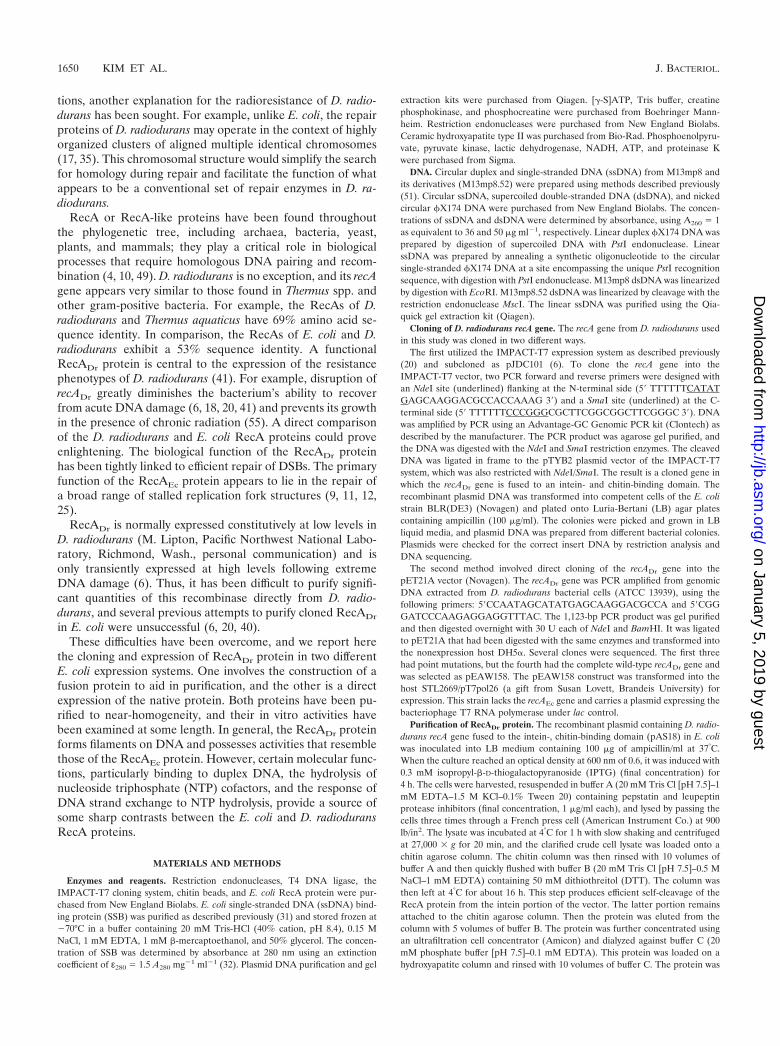
Titrating with the nucleotide cofactor allowed an estimationof the S0.5 for ATP and dATP at pH 7.5 (Fig. 5). The S0.5
(point for half saturation) for ATP appears to be quite high(Fig. 5A). When RecADr protein is bound to ssDNA, the S0.5
is approximately 1.2 mM. On dsDNA, ATP is not only hydro-lyzed faster, it exhibits a lower S0.5 of approximately 600 �M.The observed S0.5 values for dATP were lower, about 250 �Mfor both ssDNA and dsDNA. The S0.5 observed for the hydro-lysis of dATP by RecA*Dr bound to ssDNA was nearly 1 mM,again suggesting that this form of the protein is somewhatcompromised in some functions (data not shown).
DNA strand exchange. The RecADr protein will promoteDNA strand exchange with either ATP or dATP (Fig. 6).However, the two nucleotide cofactors have different effects onthe reactions. At pH 7.5, dATP is hydrolyzed two to threetimes faster than ATP (Fig. 3 and 4). Nevertheless, the DNAstrand exchange promoted in the presence of ATP is muchmore efficient, reaching completion in a quarter or less of thetime needed for the dATP-dependent reaction. Direct mea-surements of the ATP and dATPase activities during DNAstrand exchange indicated that the slow reaction in the pres-ence of dATP was not caused by a large decline in dATPhydrolysis (Table 1). For ATP, rates of hydrolysis actuallyincreased somewhat during DNA strand exchange relative tothe ssDNA-dependent ATPase rates. Essentially identicalDNA strand exchange reactions were observed under theseconditions for the RecA*Dr protein (data not shown). Thus,the apparently weaker competition of this RecADr variant withE. coli SSB and the higher S0.5 for dATP do not appear toaffect the DNA strand exchange activities of that protein.
Further characterization focused entirely on the nativeRecADr protein. The effects of pH, magnesium ion concentra-tion, and (d)ATP concentration are also explored in Fig. 6. Theeffects of pH on strand exchange do not perfectly parallel theeffects of pH on DNA-dependent ATP or dATP hydrolysis.With ATP, a good strand exchange reaction is seen betweenpHs 7 and 8.5. Even though ATP hydrolysis on ssDNA anddsDNA declines above pH 7.5 (Fig. 3), the rates during strandexchange are higher (Table 1). The hydrolysis of dATP isoptimal or nearly so between pHs 7 and 9 (Fig. 3 and Table 1),but efficient DNA strand exchange is seen only at the higherpHs (8 and above). The optimal concentration of Mg2� ionsfor the ATP-dependent DNA strand exchange reaction at pH7.5 is between 5 and 15 mM (Fig. 6D). ATP supports DNAstrand exchange at concentrations above 700 �M, while dATPis effective at concentrations above 200 �M. The (d)ATP ti-tration of the DNA strand exchange reaction was done at pH8.1, where both ATP and dATP promote a reaction. The re-quirements for these nucleotides are consistent with the higherS0.5 value observed for ATP in Fig. 5. At pH 7.5, where theATP-mediated strand exchange reaction is much faster thanthe dATP reaction (Fig. 6A), the use of a mixture of ATP anddATP (1 mM each) slowed the strand exchange reaction by afactor of 2 relative to a reaction with 2 mM ATP (data notshown).
The effects of the E. coli SSB were explored at pH 8.1 (data
FIG. 4. Effect of native RecADr protein concentration on the rateof (d)ATP hydrolysis. (A) Reactions were carried out as described inMaterials and Methods, and reaction mixtures contained 6 �M circularsingle-stranded �X174 DNA, 0.6 �M E. coli SSB, 2 mM ATP (ordATP), and the indicated concentration of native RecADr protein.Reactions carried out with the two different nucleotide cofactors areidentified in the figure. (B) Order of addition effects in the presence ofATP. Reactions were carried out as for panel A, with 2 mM ATP.RecA protein was added to ssDNA, which was followed by addition ofATP and SSB (RecADr3SSB reactions), or SSB protein was added tossDNA, which was followed by addition of ATP and RecA protein(SSB3RecADr reactions). (C) Reactions were carried out as for panelB, but with dATP replacing ATP.
VOL. 184, 2002 DEINOCOCCUS RADIODURANS RecA PROTEIN 1655
not shown). With either nucleotide cofactor, SSB was ex-tremely important to the yield of a strand exchange reaction.With ATP, essentially no reaction occurred with ATP unlessSSB was also present. With dATP, some reaction was seenwithout SSB, but it was minimal. As with RecAEc protein, theorder of addition was important. Addition of SSB before theRecADr protein nearly abolished the strand exchange reactionwith ATP and slowed the reaction with dATP.
We also explored the capacity of the RecADr protein topromote DNA strand exchange through a heterologous inser-tion of 52 bp in the duplex DNA substrate. In these reactions,the �X174 DNAs used in most of the experiments were re-placed by M13mp8 circular ssDNA and M13mp8.52 linearduplex DNA. The latter DNA contains a 52-bp heterologousinsert in M13mp8 (23, 51) and is linearized so as to place theinsertion near the center of the linear duplex DNA substrate.In the presence of ATP, the insertion slowed the strand ex-change slightly, but an efficient reaction was observed never-theless at both pHs 7.5 and 8.1 (data not shown). In contrast,
dATP supported the formation of paired joint molecules atboth pHs, but very little complete strand exchange product wasseen. This suggests that the 52-bp insertion is a much moresubstantial barrier to reaction when dATP is used as the nu-cleotide cofactor.
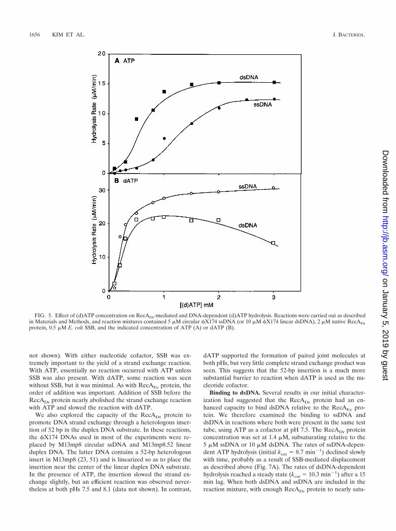
Binding to dsDNA. Several results in our initial character-ization had suggested that the RecADr protein had an en-hanced capacity to bind dsDNA relative to the RecAEc pro-tein. We therefore examined the binding to ssDNA anddsDNA in reactions where both were present in the same testtube, using ATP as a cofactor at pH 7.5. The RecADr proteinconcentration was set at 1.4 �M, subsaturating relative to the5 �M ssDNA or 10 �M dsDNA. The rates of ssDNA-depen-dent ATP hydrolysis (initial kcat � 8.7 min�1) declined slowlywith time, probably as a result of SSB-mediated displacementas described above (Fig. 7A). The rates of dsDNA-dependenthydrolysis reached a steady state (kcat � 10.3 min�1) after a 15min lag. When both dsDNA and ssDNA are included in thereaction mixture, with enough RecADr protein to nearly satu-
FIG. 5. Effect of (d)ATP concentration on RecADr-mediated and DNA-dependent (d)ATP hydrolysis. Reactions were carried out as describedin Materials and Methods, and reaction mixtures contained 5 �M circular �X174 ssDNA (or 10 �M �X174 linear dsDNA), 2 �M native RecADrprotein, 0.5 �M E. coli SSB, and the indicated concentration of ATP (A) or dATP (B).
rate one but not both, the rates of ATP hydrolysis were thosecharacteristic of binding to dsDNA (Fig. 7A). This was truewhether the dsDNA was homologous or heterologous. A lag inATP hydrolysis was not observed when both DNAs werepresent.
To ensure that transfer to dsDNA was not compelled by thepresence of the E. coli SSB protein, we carried out additionalexperiments without SSB and by substituting poly(dT) (whichhas no secondary structure) for the ssDNA (Fig. 7B). We also
used dATP in this experiment. The poly(dT)-dependent dATPhydrolysis was substantially greater than that observed withdsDNA (see Fig. 3). Again, when the DNAs were mixed, therates of hydrolysis were those characteristic of binding todsDNA. The experiments of Fig. 7B were carried out at pH8.1, where there is no evident lag in the direct binding ofRecADr protein to dsDNA, as described above. The timecourse of dATP hydrolysis in the reaction including bothDNAs appeared to start out at a higher rate before slowing tothe dsDNA-dependent rate after 10 min, suggesting thatssDNA may be bound first and may prime the binding todsDNA in some way.
DISCUSSION
We report here the purification and initial characterizationof the RecA protein from the radioresistant bacterium D.radiodurans. In many respects, the protein has the propertiesone would expect of a close homologue of the E. coli RecAprotein. It forms the striated filaments on DNA that are char-acteristic of RecA protein and hydrolyzes both ATP anddATP. It also promotes an efficient DNA strand exchangereaction. The contrasts come in the details, and several seemquite significant. The presence of dATP allows the RecADr
protein to compete better with E. coli SSB for binding sites onssDNA, and the S0.5 for dATP hydrolysis is much lower thanthe S0.5 for ATP hydrolysis. Nevertheless, dATP hydrolysissupports RecADr-mediated DNA strand exchange poorly un-der many conditions and only minimally supports DNA strandexchange through heterologous insertions in the duplex DNAsubstrate. ATP, which is hydrolyzed more slowly than dATPunder most conditions, supports an efficient DNA strand ex-change that readily bypasses heterologous insertions in theDNA substrates. Structural and mechanistic comparisons ofthese proteins may eventually yield clues to the molecular basisfor the coupling between ATP hydrolysis and DNA strandexchange in this system. In addition, the RecADr protein ap-pears to bind dsDNA much faster than does the RecAEc pro-tein. More significantly, the RecADr protein binds preferen-tially to dsDNA in reactions that also contain ssDNA, under atleast one set of reaction conditions. This is true even when thetwo DNAs are heterologous and cannot undergo DNA strandexchange.
The reactions of bacterial RecA proteins and their eukary-otic homologues are almost always greatly stimulated by
FIG. 6. DNA strand exchange promoted by the RecADr protein.Reactions were carried out as described in Materials and Methods, andreaction mixtures contained 6 �M �X174 circular ssDNA, 12 �M�X174 linear dsDNA, 2 �M native RecADr protein, and 0.6 �M E. coliSSB. Reactions also contained 2 mM ATP (or dATP) unless otherwiseindicated. Substrate DNA (�X174 linear dsDNA), product (nickedcircular dsDNA), and reaction intermediates (joint molecules) aredenoted as S, P, and JM, respectively. (A) Comparison of ATP anddATP reactions at pH 7.5. (B) Effects of ATP concentration. Reac-tions were carried out at pH 8.1. (C) Effects of dATP concentration.Reactions were carried out at pH 8.1. (D) Effect of Mg2� concentra-tion on DNA strand exchange. Reactions were carried out at pH 7.5.The standard reaction mixture described above and in Materials andMethods was altered by including only the indicated concentration ofMg(acetate)2. (E) Effects of pH on the ATP-dependent DNA strandexchange reaction. The standard reaction was altered by includingbuffers of the indicated pH. (F) Effects of pH on the dATP-dependentDNA strand exchange reaction. The standard reaction was altered byincluding buffers of the indicated pHs.
TABLE 1. Rates of ATP (or dATP) hydrolysisa
Activity and pHs
Rate of hydrolysis (�M/min)
dsDNA-dependent
During strandexchange
ssDNA-dependent
ATP hydrolysispH 7.5 15.5 15.7 11.5pH 8.1 3.6 10.2 8.3
ssDNA binding proteins. In many cases, there may be littlespecies-specific protein-protein interaction. The SSBs of manyorganisms are often interchangeable, with the eukaryotic rep-lication protein A (RPA) stimulating the strand exchange re-actions of bacterial RecA proteins and bacterial SSB substitut-ing well for RPA in stimulating the reactions of yeast Rad51protein (1, 43). For our work on RecADr protein, we do not yethave the cognate D. radiodurans SSB protein and have reliedon the E. coli SSB protein. There is a very large stimulation ofRecADr-mediated DNA strand exchange by the E. coli SSB.However, we cannot eliminate the possibility that the SSB mayalter some of these results, perhaps allowing for better strandexchange under some conditions where it was not observedhere. This remains a goal of future research.
The work reported here provides a comparison of two dif-ferent RecADr proteins, generated by different cloning proce-dures. The variant we have designated RecA*Dr has an extraglycine at the C terminus. In its capacity to form filaments onDNA and to promote DNA strand exchange, this protein ap-pears to be very similar in its properties to the native RecADr
protein isolated from E. coli. The only differences noted in theset of experiments done with this protein were an increasedS0.5 for dATP hydrolysis (on ssDNA) and a reduced capacity ofthis protein to compete with E. coli SSB for ssDNA bindingsites in the presence of dATP. The C-terminal domain of RecAprotein protrudes from the RecA filament, and there is noevidence that it plays a role in monomer-monomer interactionsin the filament (49, 53). However, the last 25 amino acid
FIG. 7. The RecADr protein binds to dsDNA in preference to ssDNA. (A) Reactions were carried out at pH 7.5 and contained 1.4 �M RecADrprotein, DNA as indicated (5 �M css �X174 DNA (ssDNA), 10 �M lds �X174 (Hm dsDNA or dsDNA), and/or 10 �M lds M13mp8 DNA (HtdsDNA), 0.4 �M E. coli SSB, 2 mM ATP, and an ATP regenerating system. (B) Reactions were carried out at pH 8.1, and reaction mixturescontained 1.0 �M RecADr protein, DNA as indicated [4 �M poly(dT) and/or 8 �M lds M13mp8 DNA], no E. coli SSB, 2 mM dATP, and a dATPregenerating system.
residues at the C terminus are not resolved in the publishedstructure (53), and this protein segment could in principle foldback to interact with many parts of the RecA protein surface.The effects of this extra glycine at the C terminus may suggestan interaction or function of the C terminus that affects bind-ing of RecA to both DNA and nucleotide cofactors.
Stole and Bryant (52a) have shown that combinations ofwild-type or mutant RecAEc proteins and nucleotide cofactorswith an S0.5 value of less than about 200 �M are generallyunable to establish the active conformation needed for RecA-mediated DNA strand exchange. This does not seem to be thecase for the RecADr protein. The S0.5 value for ATP hydrolysisat pH 7.5, when the RecADr protein is bound to an ssDNA, ismore than 1 mM. However, a facile DNA strand exchangereaction is seen under these same conditions as long as theATP concentration is high enough. Interestingly, the S0.5 valuefor ATP hydrolysis in the presence of dsDNA (about 600 �M)appears to correlate better with the ATP requirements seen inDNA strand exchange (Fig. 5A) than does the S0.5 value forATP hydrolysis in the presence of ssDNA (about 1.2 mM).
The significance of some of the RecADr observations will beunclear until work is done to examine intracellular conditionsin D. radiodurans. The few measurements that have been re-ported indicate that the normal concentration of dNTPs in anE. coli cell is generally less than 200 �M (44). The size of thenucleotide pools in D. radiodurans is not known. If they aresimilar in size to those in E. coli (and if the intracellular pH isabove 7), then the RecADr protein may have a limited capacityto hydrolyze dATP under normal in vivo conditions. In view ofthe generally negative effects that dATP hydrolysis has on theDNA strand exchange reactions of the RecADr protein, onemight expect that in vivo conditions would favor the hydrolysisof ATP. The relatively low levels of RecADr-mediated ATPhydrolysis at pH 7.5 is similar to the lack of ATP hydrolysisreported for the B. subtilis RecA protein (33), although morerecent data indicates that ATP is hydrolyzed by the B. subtilisprotein if the conditions are altered somewhat (52).
The RecADr protein promotes complete DNA strand ex-change with full-sized DNA substrates derived from �X174 orM13mp8 with good efficiency. We presume that ATP bindingand/or hydrolysis play a role in this reaction, since DNA strandexchange does not occur in the absence of NTP or dNTPcofactor. It has been established that the RecA protein andRecA homologues, such as the Rad51 protein, promote DNApairing and strand exchange with little or no need for thehydrolysis of NTP cofactors (23, 24, 26, 38, 47, 51, 54). In thecase of RecA protein, NTP hydrolysis may contribute a motorfunction that augments the inherent DNA strand exchangereaction, permitting it to move past DNA structural barriers,rendering it unidirectional, and allowing 4-strand exchangereactions to occur (3, 23, 24, 34, 50, 51). Such activities mightbe useful in a chromosomal repair capacity. This work providesa jumping-off point for a more complete analysis of the recom-binational DNA repair processes in D. radiodurans and forfurther investigations into the role of ATP hydrolysis in RecA-mediated DNA strand exchange.
ACKNOWLEDGMENTS
We thank Nirmala D. Sharma for technical assistance in the cloning,expression, and purification of recombinant RecA*Dr protein; Michael
N. Flora of the USUHS Biomedical Instrumentation Center for oli-gonucleotide synthesis and DNA sequencing; and Maria Schnös andDavid Inman for assistance in electron microscopy.
This work was supported by grants GM32335-18 (from the NationalInstitutes of Health; to M.M.C.), DE-FG02-98ER6283 (from the Mi-crobial Genome Program, U.S. Department of Energy; to M.J.D.),FG02-97ER62492 and FG07-97ER20293 (from the DOE; to M.J.D.),GM39933-09 (from the National Institutes of Health; to M.J.D.), andGM14711-33 (from the National Institutes of Health; to R.B.I.).
Jong-Il Kim and Ajay K. Sharma contributed equally to this work.
REFERENCES
1. Alani, E., R. Thresher, J. D. Griffith, and R. D. Kolodner. 1992. Character-ization of DNA-binding and strand-exchange stimulation properties of y-RPA, a yeast single-strand-DNA-binding protein. J. Mol. Biol. 227:54–71.
2. Battista, J. R., A. M. Earl, and M. J. Park. 1999. Why is Deinococcusradiodurans so resistant to ionizing radiation? Trends Microbiol. 7:362–365.
3. Bedale, W. A., and M. Cox. 1996. Evidence for the coupling of ATP hydro-lysis to the final (extension) phase of RecA protein-mediated DNA strandexchange. J. Biol. Chem. 271:5725–5732.
4. Bianco, P. R., R. B. Tracy, and S. C. Kowalczykowski. 1998. DNA strandexchange proteins: a biochemical and physical comparison. Front. Biosci.3:560–603. [Online.] http//www.bioscience.org/.
5. Brim, H., S. C. McFarlan, J. K. Fredrickson, K. W. Minton, M. Zhai, L. P.Wackett, and M. J. Daly. 2000. Engineering Deinococcus radiodurans formetal remediation in radioactive mixed waste environments. Nat. Biotech-nol. 18:85–90.
6. Carroll, J. D., M. J. Daly, and K. W. Minton. 1996. Expression of recA inDeinococcus radiodurans. J. Bacteriol. 178:130–135.
7. Chong, S., F. B. Mersha, D. G. Comb, M. E. Scott, D. Landry, L. M. Vence,F. B. Perler, J. Benner, R. B. Kucera, C. A. Hirvonen, J. J. Pelletier, H.Paulus, and M. Q. Xu. 1997. Single-column purification of free recombinantproteins using a self-cleavable affinity tag derived from a protein splicingelement. Gene 192:271–281.
8. Chong, S., Y. Shao, H. Paulus, J. Benner, F. B. Perler, and M. Q. Xu. 1996.Protein splicing involving the Saccharomyces cerevisiae VMA intein. Thesteps in the splicing pathway, side reactions leading to protein cleavage, andestablishment of an in vitro splicing system. J. Biol. Chem. 271:22159–22168.
9. Cox, M. M. 2001. Historical overview: searching for replication help in all ofthe rec places. Proc. Natl. Acad. Sci. USA. 98:8173–8180.
10. Cox, M. M. 1999. Recombinational DNA repair in bacteria and the RecAprotein. Prog. Nucleic Acids Res. Mol. Biol. 63:310–366.
11. Cox, M. M. 2001. Recombinational DNA repair of damaged replicationforks in Escherichia coli: questions. Annu. Rev. Genet. 35:53–82.
12. Cox, M. M., M. F. Goodman, K. N. Kreuzer, D. J. Sherratt, S. J. Sandler,and K. J. Marians. 2000. The importance of repairing stalled replicationforks. Nature 404:37–41.
13. Cox, M. M., and I. R. Lehman. 1981. RecA protein of Escherichia colipromotes branch migration, a kinetically distinct phase of DNA strand ex-change. Proc. Natl. Acad. Sci. USA 78:3433–3437.
14. Daly, M. J. 2000. Engineering radiation-resistant bacteria for environmentalbiotechnology. Curr. Opin. Biotechnol. 11:280–285.
15. Daly, M. J., and K. W. Minton. 1996. An alternative pathway of recombi-nation of chromosomal fragments precedes recA-dependent recombinationin the radioresistant bacterium Deinococcus radiodurans. J. Bacteriol. 178:4461–4471.
16. Daly, M. J., and K. W. Minton. 1995. Interchromosomal recombination inthe extremely radioresistant bacterium Deinococcus radiodurans. J. Bacte-riol. 177:5495–5505.
17. Daly, M. J., and K. W. Minton. 1995. Resistance to radiation. Science270:1318.
18. Daly, M. J., L. Ouyang, P. Fuchs, and K. W. Minton. 1994. In vivo damageand recA-dependent repair of plasmid and chromosomal DNA in the radi-ation-resistant bacterium Deinococcus radiodurans. J. Bacteriol. 176:3508–3517.
19. Ferreira, A. C., M. F. Nobre, F. A. Rainey, M. T. Silva, R. Wait, J. Burghardt,A. P. Chung, and M. S. da Costa. 1997. Deinococcus geothermalis sp. nov.and Deinococcus murrayi sp. nov., two extremely radiation-resistant andslightly thermophilic species from hot springs. Int. J. Syst. Bacteriol. 47:939–947.
20. Gutman, P. D., J. D. Carroll, C. I. Masters, and K. W. Minton. 1994.Sequencing, targeted mutagenesis and expression of a recA gene requiredfor the extreme radioresistance of Deinococcus radiodurans. Gene 141:31–37.
21. Iype, L. E., E. A. Wood, R. B. Inman, and M. M. Cox. 1994. RuvA and RuvBproteins facilitate the bypass of heterologous DNA insertions during RecAprotein-mediated DNA strand exchange. J. Biol. Chem. 269:24967–24978.
22. Karlin, S., and J. Mrazek. 2001. Predicted highly expressed and putativealien genes of Deinococcus radiodurans and implications for resistance toionizing radiation damage. Proc. Natl. Acad. Sci. USA 98:5240–5245.
23. Kim, J. I., M. M. Cox, and R. B. Inman. 1992. On the role of ATP hydrolysis
VOL. 184, 2002 DEINOCOCCUS RADIODURANS RecA PROTEIN 1659
in RecA protein-mediated DNA strand exchange. I. Bypassing a short het-erologous insert in one DNA substrate. J. Biol. Chem. 267:16438–16443.
24. Kim, J. I., M. M. Cox, and R. B. Inman. 1992. On the role of ATP hydrolysisin RecA protein-mediated DNA strand exchange. II. Four-strand exchangesJ. Biol. Chem. 267:16444–16449.
25. Kowalczykowski, S. C. 2000. Initiation of genetic recombination and recom-bination-dependent replication. Trends Biochem. Sci. 25:156–165.
26. Kowalczykowski, S. C., and R. A. Krupp. 1995. DNA-strand exchange pro-moted by RecA protein in the absence of ATP: implications for the mech-anism of energy transduction in protein-promoted nucleic acid transactions.Proc. Natl. Acad. Sci. USA 92:3478–3482.
27. Lange, C. C., L. P. Wackett, K. W. Minton, and M. J. Daly. 1998. Engineer-ing a recombinant Deinococcus radiodurans for organopollutant degrada-tion in radioactive mixed waste environments. Nat. Biotechnol. 16:929–933.
28. Lin, J. Y., R. Qi, C. Aston, J. P. Jing, T. S. Anantharaman, B. Mishra, O.White, M. J. Daly, K. W. Minton, J. C. Venter, and D. C. Schwartz. 1999.Whole-genome shotgun optical mapping of Deinococcus radiodurans. Sci-ence 285:1558–1562.
29. Lindsley, J. E., and M. M. Cox. 1990. Assembly and disassembly of RecAprotein filaments occurs at opposite filament ends: relationship to DNAstrand exchange. J. Biol. Chem. 265:9043–9054.
30. Lohman, T. M., K. Chao, J. M. Green, S. Sage, and G. T. Runyon. 1989.Large-scale purification and characterization of the Escherichia coli rep geneproduct. J. Biol. Chem. 264:10139–10147.
31. Lohman, T. M., J. M. Green, and R. S. Beyer. 1986. Large-scale overpro-duction and rapid purification of the Escherichia coli ssb gene product.Expression of the ssb gene under PL control. Biochemistry 25:21–25.
32. Lohman, T. M., and L. B. Overman. 1985. Two binding modes in Escherichiacoli single strand binding protein-single stranded DNA complexes. Modula-tion by NaCl concentration. J. Biol. Chem. 260:3594–3603.
33. Lovett, C. M. J., and J. W. Roberts. 1985. Purification of a RecA proteinanalogue from Bacillus subtilis. J. Biol. Chem. 260:3305–3313.
34. MacFarland, K. J., Q. Shan, R. B. Inman, and M. M. Cox. 1997. RecA as amotor protein. Testing models for the role of ATP hydrolysis in DNA strandexchange. J. Biol. Chem. 272:17675–17685.
35. Makarova, K. S., L. Aravind, Y. I. Wolf, R. L. Tatusov, K. W. Minton, E. V.Koonin, and M. J. Daly. 2001. Genome of the extremely radiation-resistantbacterium Deinococcus radiodurans viewed from the perspective of compar-ative genomics. Microbiol. Mol. Biol. Rev. 65:44–79.
36. Makarova, K. S., Y. I. Wolf, O. White, K. Minton, and M. J. Daly. 1999.Short repeats and IS elements in the extremely radiation-resistant bacteriumDeinococcus radiodurans and comparison to other bacterial species. Res.Microbiol. 150:711–724.
37. Marrione, P. E., and M. M. Cox. 1995. RuvB protein-mediated ATP hydro-lysis: functional asymmetry in the RuvB hexamer. Biochemistry 34:9809–9818.
38. Menetski, J. P., D. G. Bear, and S. C. Kowalczykowski. 1990. Stable DNAheteroduplex formation catalyzed by the Escherichia coli RecA protein in theabsence of ATP hydrolysis. Proc. Natl. Acad. Sci. USA 87:21–25.
39. Menetski, J. P., and S. C. Kowalczykowski. 1989. Enhancement of Esche-richia coli RecA protein enzymatic function by dATP. Biochemistry 28:5871–5881.
40. Minton, K. W. 1994. DNA repair in the extremely radioresistant bacteriumDeinococcus radiodurans. Mol. Microbiol. 13:9–15.
41. Minton, K. W. 1996. Repair of ionizing-radiation damage in the radiationresistant bacterium Deinococcus radiodurans. Mutat. Res. 363:1–7.
42. Morrical, S. W., J. Lee, and M. M. Cox. 1986. Continuous association ofEscherichia coli single-stranded DNA binding protein with stable complexesof RecA protein and single-stranded DNA. Biochemistry 25:1482–1494.
43. Namsaraev, E. A., and P. Berg. 2000. Rad51 uses one mechanism to driveDNA strand exchange in both directions. J. Biol. Chem. 275:3970–3976.
44. Neuhard, J., and P. Nygaard. 1987. Purines and pyrimidines, p. 445–473. InF. C. Neidhardt, J. L. Ingraham, K. B. Low, B. Magasanik, M. Schaechter,and H. E. Umbarger (ed.), Escherichia coli and Salmonella typhimurium:cellular and molecular biology, vol. 1. American Society for Microbiology,Washington, D.C.
45. Pugh, B. F., and M. M. Cox. 1988. General mechanism for RecA proteinbinding to duplex DNA. J. Mol. Biol. 203:479–493.
46. Pugh, B. F., and M. M. Cox. 1987. Stable binding of RecA protein to duplexDNA. Unraveling a paradox. J. Biol. Chem. 262:1326–1336.
47. Rehrauer, W. M., and S. C. Kowalczykowski. 1993. Alteration of the nucle-oside triphosphate (NTP) catalytic domain within Escherichia coli recAprotein attenuates NTP hydrolysis but not joint molecule formation. J. Biol.Chem. 268:1292–1297.
48. Richmond, R. C., R. Sridhar, and M. J. Daly. 1999. Physicochemical survivalpattern for the radiophile Deinococcus radiodurans: a polyextremophilemodel for life on Mars. SPIE 3755:210–222.
49. Roca, A. I., and M. M. Cox. 1997. RecA protein: structure, function, and rolein recombinational DNA repair. Prog. Nucleic Acid Res. Mol. Biol. 56:129–223.
50. Shan, Q., and M. M. Cox. 1998. On the mechanism of RecA-mediated repairof double-strand breaks: no role for four-strand DNA pairing intermediates.Mol. Cell 1:309–317.
51. Shan, Q., M. M. Cox, and R. B. Inman. 1996. DNA strand exchange pro-moted by RecA K72R. Two reaction phases with different Mg2� require-ments. J. Biol. Chem. 271:5712–5724.
52. Steffen, S. E., and F. R. Bryant. 1999. Reevaluation of the nucleotide cofac-tor specificity of the RecA protein from Bacillus subtilis. J. Biol. Chem.274:25990–25994.
52a.Stole, E., and F. R. Bryant. 1995. Spectroscopic demonstration of a linkagebetween the kinetics of NTP hydrolysis and the conformational state of theRecA-single-stranded DNA complex. J. Biol. Chem. 270:20322–20328.
53. Story, R. M., I. T. Weber, and T. A. Steitz. 1992. The structure of the E. coliRecA protein monomer and polymer. Nature 355:318–325.
54. Sung, P., and S. A. Stratton. 1996. Yeast Rad51 recombinase mediates polarDNA strand exchange in the absence of ATP hydrolysis. J. Biol. Chem.271:27983–27986.
55. Venkateswaran, A., S. C. McFarlan, D. Ghosal, K. W. Minton, A. Vasilenko,K. Makarova, L. P. Wackett, and M. J. Daly. 2000. Physiologic determinantsof radiation resistance in Deinococcus radiodurans. Appl. Environ. Micro-biol. 66:2620–2626.
56. White, O., J. A. Eisen, J. F. Heidelberg, E. K. Hickey, J. D. Peterson, R. J.Dodson, D. H. Haft, M. L. Gwinn, W. C. Nelson, D. L. Richardson, K. S.Moffat, H. Y. Qin, L. X. Jiang, W. Pamphile, M. Crosby, M. Shen, J. J.Vamathevan, P. Lam, L. McDonald, T. Utterback, C. Zalewski, K. S.Makarova, L. Aravind, M. J. Daly, K. W. Minton, C. M. Fraser, et al. 1999.Genome sequence of the radioresistant bacterium Deinococcus radioduransR1. Science 286:1571–1577.