j our na l ho me page: www.elsev ier .com/ locate /vacc ine
ecent advances in dengue pathogenesis and clinical management
ameron P. Simmonsa,b,c,∗, Kirsty McPhersonc, Nguyen Van Vinh Chaud, D.T. Hoai Tame,aul Youngf, Jason Mackenziec, Bridget Willsa,b
Oxford University Clinical Research Unit, Hospital for Tropical Diseases, 764 Vo Van Kiet street, District 5, Ho Chi Minh City, Viet NamCentre for Tropical Medicine, Nuffield Department of Medicine, University of Oxford, Oxford, United KingdomDepartment of Microbiology and Immunology, University of Melbourne, Parkville, Victoria 3010, AustraliaHospital for Tropical Diseases, 764 Vo Van Kiet, District 5, Ho Chi Minh City, Viet NamUniversity of Medicine and Pharmacy, 217 Hong Bang, District 5, Ho Chi Minh City, Viet NamSchool of Chemistry and Molecular Biosciences, University of Queensland, Australia
r t i c l e i n f o
rticle history:vailable online 14 October 2015
a b s t r a c t
This review describes and commentates on recent advances in the understanding of dengue pathogenesisand immunity, plus clinical research on vaccines and therapeutics. We expand specifically on the roleof the dermis in dengue virus infection, the contribution of cellular and humoral immune responses to
eywords:engueaccineathogenesismmunologyirology
pathogenesis and immunity, NS1 and mechanisms of virus immune evasion. Additionally we review aseries of therapeutic intervention trials for dengue, as well as recent clinical research aimed at improvingclinical diagnosis, risk prediction and disease classification.
The underlying mechanistic causes of the dominant clinicaleatures of severe dengue, i.e. a transient increase in vascularermeability and a hemorrhagic diathesis, remain enigmatic. Inrinciple, acquiring deeper insights into the mechanistic drivers ofhe clinically important features of dengue should enable improvedreatment strategies and uncover novel drug targets. Yet neatlyissecting the pathogenesis of any infectious disease syndrome isever straightforward and dengue is no exception. Host and virusariables shape the clinical outcome of any given dengue virusDENV) infection. For the host, there is undoubtedly a physiologicalnd immunological component (humoral and cellular) that influ-nces whether infection (or re-infection) has a benign outcomer results in disease that manifests across a gradient of severity.here must also be a virological aspect, such that some virusesre simply better equipped to replicate and reach high titers in
human (or mosquito) host. Neither of these processes work insolation. Rather, the outcome of exposure to an infectious Aedes
osquito will always depend on a constellation of “positive” and
∗ Corresponding author at: Oxford University Clinical Research Unit, Hospital forropical Diseases, 764 Vo Van Kiet street, District 5, Ho Chi Minh City, Viet Nam.el.: +84 904434839.
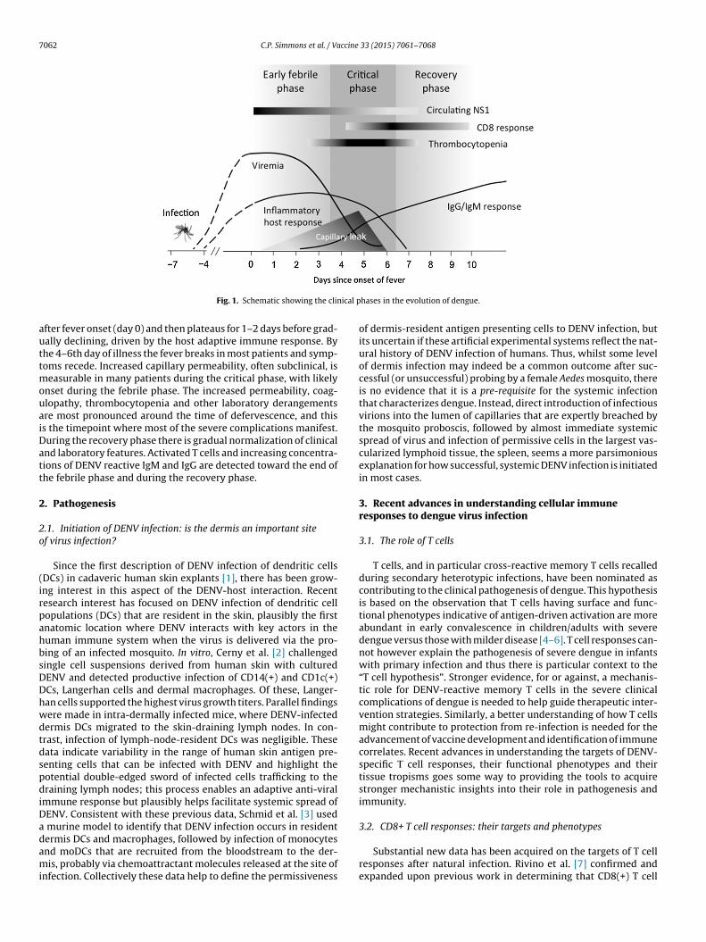
“negative” host and viral factors that each influences the over-all clinical evolution of the infection. Layered upon this are thebenefits that careful clinical management can have in preventingor treating the life-threatening complications that may occur; inmany endemic countries improvements in clinical managementmean that case fatality rates amongst hospitalized cases have beenreduced from 20% to less than 0.5%. Yet there is still room to dobetter. This might be via development of more effective, evidence-based fluid resuscitation strategies for cases with severe shock, orthe arrival of specific anti-viral therapeutics or disease modifiersable to reduce the duration and/or severity of generalized symp-toms in the tens of millions of uncomplicated dengue cases thatoccur globally. Central to all considerations of patient-centeredresearch findings in dengue is an understanding of the temporal“windows” that exist in the evolution of disease. These “windows”are represented schematically in Fig. 1. Beyond all else, they under-score the dynamic nature of disease evolution and that clinicalresearch studies must always consider timing of interventions,observations and sampling in their design and reporting.
Following the bite of an infected mosquito the virus dissem-inates and infects multiple lymphoid and non-lymphoid tissues.A viremia ensues that is presumed to be a proxy for the under-
lying severity of tissue infection. The viral burden accumulatesto the point that generalized clinical symptoms (fever, headache,myalgia) develop, presumably secondary to a host antiviral statein which interferon expression is abundant. Viremia peaks shortly
nder the CC BY license (http://creativecommons.org/licenses/by/4.0/).
7062 C.P. Simmons et al. / Vaccine 33 (2015) 7061–7068
ical p
auttmouaiDatt
2
2o
(irpahbsDDhwdtdspdiDadami
Fig. 1. Schematic showing the clin
fter fever onset (day 0) and then plateaus for 1–2 days before grad-ally declining, driven by the host adaptive immune response. Byhe 4–6th day of illness the fever breaks in most patients and symp-oms recede. Increased capillary permeability, often subclinical, is
easurable in many patients during the critical phase, with likelynset during the febrile phase. The increased permeability, coag-lopathy, thrombocytopenia and other laboratory derangementsre most pronounced around the time of defervescence, and thiss the timepoint where most of the severe complications manifest.uring the recovery phase there is gradual normalization of clinicalnd laboratory features. Activated T cells and increasing concentra-ions of DENV reactive IgM and IgG are detected toward the end ofhe febrile phase and during the recovery phase.
. Pathogenesis
.1. Initiation of DENV infection: is the dermis an important sitef virus infection?
Since the first description of DENV infection of dendritic cellsDCs) in cadaveric human skin explants [1], there has been grow-ng interest in this aspect of the DENV-host interaction. Recentesearch interest has focused on DENV infection of dendritic cellopulations (DCs) that are resident in the skin, plausibly the firstnatomic location where DENV interacts with key actors in theuman immune system when the virus is delivered via the pro-ing of an infected mosquito. In vitro, Cerny et al. [2] challengedingle cell suspensions derived from human skin with culturedENV and detected productive infection of CD14(+) and CD1c(+)Cs, Langerhan cells and dermal macrophages. Of these, Langer-an cells supported the highest virus growth titers. Parallel findingsere made in intra-dermally infected mice, where DENV-infectedermis DCs migrated to the skin-draining lymph nodes. In con-rast, infection of lymph-node-resident DCs was negligible. Theseata indicate variability in the range of human skin antigen pre-enting cells that can be infected with DENV and highlight theotential double-edged sword of infected cells trafficking to theraining lymph nodes; this process enables an adaptive anti-viral
mmune response but plausibly helps facilitate systemic spread ofENV. Consistent with these previous data, Schmid et al. [3] used
murine model to identify that DENV infection occurs in resident
ermis DCs and macrophages, followed by infection of monocytesnd moDCs that are recruited from the bloodstream to the der-is, probably via chemoattractant molecules released at the site of
nfection. Collectively these data help to define the permissiveness
hases in the evolution of dengue.
of dermis-resident antigen presenting cells to DENV infection, butits uncertain if these artificial experimental systems reflect the nat-ural history of DENV infection of humans. Thus, whilst some levelof dermis infection may indeed be a common outcome after suc-cessful (or unsuccessful) probing by a female Aedes mosquito, thereis no evidence that it is a pre-requisite for the systemic infectionthat characterizes dengue. Instead, direct introduction of infectiousvirions into the lumen of capillaries that are expertly breached bythe mosquito proboscis, followed by almost immediate systemicspread of virus and infection of permissive cells in the largest vas-cularized lymphoid tissue, the spleen, seems a more parsimoniousexplanation for how successful, systemic DENV infection is initiatedin most cases.
3. Recent advances in understanding cellular immuneresponses to dengue virus infection
3.1. The role of T cells
T cells, and in particular cross-reactive memory T cells recalledduring secondary heterotypic infections, have been nominated ascontributing to the clinical pathogenesis of dengue. This hypothesisis based on the observation that T cells having surface and func-tional phenotypes indicative of antigen-driven activation are moreabundant in early convalescence in children/adults with severedengue versus those with milder disease [4–6]. T cell responses can-not however explain the pathogenesis of severe dengue in infantswith primary infection and thus there is particular context to the“T cell hypothesis”. Stronger evidence, for or against, a mechanis-tic role for DENV-reactive memory T cells in the severe clinicalcomplications of dengue is needed to help guide therapeutic inter-vention strategies. Similarly, a better understanding of how T cellsmight contribute to protection from re-infection is needed for theadvancement of vaccine development and identification of immunecorrelates. Recent advances in understanding the targets of DENV-specific T cell responses, their functional phenotypes and theirtissue tropisms goes some way to providing the tools to acquirestronger mechanistic insights into their role in pathogenesis andimmunity.
3.2. CD8+ T cell responses: their targets and phenotypes
Substantial new data has been acquired on the targets of T cellresponses after natural infection. Rivino et al. [7] confirmed andexpanded upon previous work in determining that CD8(+) T cell
ccine
eNCtNlctdaaim
arCeCYidmtddopntmhito
nTttcclt
3
cdpaCatbfDirhpmlpt
C.P. Simmons et al. / Va
pitopes are predominantly located in the nonstructural proteinsS3 and NS5. Similarly Weiskopf et al. [8] identified DENV-reactiveD8+ T cell responses in Sri Lankan blood donors; 408 immunoreac-ive peptides were identified, two thirds of which were located inS3, NS5 or NS4b. Finally, following tetravalent vaccination with
ive attenuated DENV, Weiskopf et al. [9] demonstrated CD8+ Tell responses were universally (99.8%) against non-structural pro-eins, with 97% directed toward NS3 and NS5. Collectively, theseata underscore the importance of DENV non-structural proteinss CD8+ T cell immunogens during natural infection; this remainsn important consideration for vaccine strategies that, along withnduction of neutralizing antibody, also seek to induce CD8+ T cell
emory.Whether DENV-reactive CD8+ T cell responses contribute mech-
nistically to vascular leakage, the hallmark of severe dengue,emains unresolved. Based on observations of HLA-A*11-restrictedD8+ T cell responses to the NS3133–142 epitope, Mongkolsapayat al. [10] have suggested original antigenic sin occurs amongstD8+ T cell populations during secondary heterotypic infection.et these conclusions have been drawn from observations made
n early convalescence, when CD8+ T cell responses peak, and noturing the febrile and critical phase, when vascular leakage com-ences and is most prominent. Indeed, Dung et al. [11] reported
hat NS3133–142-specific T cells were undetectable until after theevelopment of plasma leakage among infected Vietnamese chil-ren. In contrast, Freiberg et al. [12] detected very low frequenciesf activated NS3133–142-specific CD8+ T cells during the febrilehase in both primary and secondary dengue cases, but foundo evidence that these responses correlated with immune sta-us exposure or current disease severity. Although traditionally
easured in the blood, Rivino and colleagues [13] also identifiedighly activated and proliferating NS3133–142-specific CD8+ T cells
n experimentally induced skin blisters on dengue cases. Whetherhese dermis-infiltrating CD8+ T cells are relevant to pathogenesisf the clinical and laboratory features of dengue is uncertain.
The next phase of research into CD8+ T cells and pathogenesiseeds to take an expansive approach to measuring epitope-specific
cells (e.g. using panels of Class I tetramer reagents, targeting morehan one specificity) and investigate responses longitudinally in thehree phases of disease; early febrile, critical phase and convales-ence, with sample sizes large enough to capture the spectrum oflinical outcomes, from the very mild to the severe. These are chal-enging studies to perform but are necessary if the field is to advancehe understanding of CD8+ T cells in pathogenesis and immunity.
.3. CD4+ T cells responses: their targets and phenotypes
In contrast to CD8+ T cell responses, DENV-specific CD4+ Tells have been less well characterized. In patients with secondaryengue, Rivino et al. [7] demonstrated that CD4+ T cell epitopes areredominantly located in structural proteins e.g. envelope, capsid,nd NS1, which themselves are major targets of the B cell response.irculating CD4+ T cells during early convalescence had the surfacend functional phenotype of follicular helper T cells, suggesting thathey are interacting with B cells in vivo, presumably to assist anti-ody production. Whether the size of the acute expansion of theollicular helper T population in blood is predictive of the anti-ENV neutralizing Ab titer deserves investigation as a possible
mmune correlate of vaccine or infection-driven humoral immuneesponses. Mangada et al. [14] examined CD4+ cells in 6 donors whoad received monovalent live attenuated DENV vaccine 12 monthsrior. Stimulation with heterologous serotype peptide resulted in
ore TNF�-producing cells than IFN� producers relative to stimu-
ation with homologous peptides, suggesting differential functionalhenotypes amongst these cell populations that are dependent onhe type of antigenic stimulation. This repeats a theme also evident
33 (2015) 7061–7068 7063
in studies of CD8+ T cells, that partial peptide agonists elicit adiverse range of functional phenotypes in cross-reactive T cells [15].In a mouse model, Yauch et al. [16] identified that DENV2-specificCD4+ T cells were of a Th1 phenotype and could mediate in vivocytotoxicity and that immunization with dominant CD4+ T cell epi-topes led to enhance viral clearance. Whether results in murinemodels are informative for understanding human immunization ordisease remains uncertain, but these results underscore the needfor further investigations of CD4+ T cells in dengue pathogenesis.
3.4. Mast cells
Mast cells (MCs), whilst traditionally associated with hyper-sensitivity responses, express a wide range of Fc receptors andhence are candidates for involvement in dengue pathogenesis. StJohn et al. demonstrated in a mouse model that MCs are activatedin vivo during experimental DENV infection and MC-deficient micehad greatly reduced vascular leakage compared to MC-sufficientcontrols. Treatment of experimentally infected animals with MC-stabilizing drugs also ameliorated vascular leakage [17]. In humans,MC-derived vasoactive products such as chymase, a serine pro-tease, are elevated in the peripheral blood of primary and secondarydengue cases [17,18]. As attractive as MCs might be as “actors” inthe pathogenesis of severe dengue, there remain critical questions.For example, to explain the clinical observation that secondaryheterotypic DENV infection is associated with greater risk of severedisease, the “MC hypothesis” requires some level of Ab-Ag, (e.g.viral particles or NS1) to trigger Fc receptor aggregation on MCsand their activation/degranulation; this has yet to be shown in aconvincing fashion. Moreover, the timing of MC activation wouldneed to occur in a manner temporally consistent with the evolutionof vascular leakage, i.e. most pronounced leading up to the time ofdefervescence in severe cases [19]. Thus, whilst MCs are intrigu-ing, further clinical studies, possibly even drug probe studies, areneeded to better understand their contribution to human disease.
3.5. Humoral immunity
Antibodies are believed to be critical mediators of resolution ofinfection, immunity to reinfection and in some circumstances, arebelieved to be risk factors for severe disease, i.e. antibody depend-ent enhancement (ADE) of infection. Recent results of phase IIband III clinical trials of Sanofi Pasteur’s recombinant live atten-uated tetravalent dengue vaccine (CYD-TDV) have questionedthe decades-old assumption that plaque reduction neutralizationassays (PRNT50) are good predictors of immune status. Specifi-cally, CYD-TDV elicited tetravalent virus neutralizing antibodiesafter three doses yet offered very low levels of efficacy againstDENV-2 and modest efficacy against DENV-1 [20,21]. That the rela-tionship between PRNT50 titer and immunity to DENV-2 is complexwere reinforced by Buddhari et al. [22] who utilized data from geo-graphic cluster studies in Thailand. They found an overall significantpositive association between baseline PRNT50 titers to DENV-1, -2and DENV-4 and immunity from homotypic symptomatic infectionduring the follow-up period. However the threshold for definingimmunity to DENV-2 was unclear and might be higher than forother serotypes. Thus, traditionally accepted PRNT50 values (i.e.titer >10) regarded as denoting “immunity” to DENV may notbe accurate, especially so for DENV-2. This has provided motiva-tion to the field to explore alternative correlative assays. This isbeing enabled in part by productive basic research that has iden-tified new, highly potent virus-neutralizing human mAbs elicited
by natural infection. Mapping the epitope targets of these mAbshas revealed many target quaternary epitopes on the virus sur-face [23–25]. Some are serotype specific (e.g. 14C10 targetingDENV-1 [26] and 5J7 that targets DENV-3 [24]) whilst others are
7 ccine
cBipbddteeftsveots
3
urtocrEc
peaaAii[hutotr[atidrgptra
3o
dciom
064 C.P. Simmons et al. / Va
ross-reactive (e.g. the so-called Envelope Dimer Epitope mAbs7 and C10) [27]. The existence of rare but potently neutraliz-
ng, cross-reactive human mAbs elicited by natural infection isrovocative. Potentially, such Abs, and their epitope targets, coulde deployed for pan-serotype therapeutic indications or vaccineevelopment efforts. Additionally, these mAbs could be used forevelopment and/or validation of 2nd generation virus neutraliza-ion assays that replace or augment the PRNT50 measurement. Forxample, 2nd generation assays might compete individual or pan-ls of highly potent virus neutralizing mAbs with polyclonal serarom vaccinees for binding to viral particles or recombinant pro-eins and thus determine a titer of antibodies in polyclonal serapecific for the critical epitope regions on the virus. Finally, foraccine development, it is likely that strategies employing DENVnvelope protein subunits as immunogens will fail to elicit somef these quaternary epitope binding immune responses; whetherhis significantly reduces the probability of success of this vaccinetrategy is unknown.
.6. Immune subversion
A recent expert review has reported the latest mechanisticnderstanding of how DENV manipulates intracellular antiviralesponses and directly inhibits cellular signaling cascades in ordero favor its own replication [28]. Here, we will focus on the rolef lipids, and induction of the autophagy and endoplasmic reti-ulum (ER) stress pathways during DENV infection. Intracellulareplication of DENV occurs on the cytoplasmic side of remodeledR membranes and requires sufficient lipid resources from the hostell to enable efficient encapsidation and assembly of virions.
Recent evidence suggests lipid droplets and the autophagyathway positively contribute to the pool of lipid resources thatnables DENV genome encapsidation and assembly [29,30]. Henceutophagy, sometimes regarded as a biochemical arm of the innatenti-viral immune response, actually benefits DENV replication.dditionally, cholesterol has been shown to play a pivotal role
n the replication cycle of DENV and other flaviviruses, contribut-ng not only to efficient replication but also to immune evasion31–33]. Although, the mechanism by which dengue regulates lipidomeostasis and recruitment for these purposes is not currentlynderstood, the membrane remodeling protein NS4A appearso be a likely candidate [34,35]. In addition to autophagy it isbserved that all flaviviruses modulate the ER stress/unfolded pro-ein response. DENV, JEV and WNV all activate Xbp-1, ATF6 and IRE1egulatory factors to promote cell survival and immune evasion36–41]. Although the exact underlying molecular and functionalspects of this induction are not currently understood it is pertinento highlight that Xbp-1 can promote lipid biosynthesis [42], IRE1s involved in regulated RNA decay pathway promoting anti-viralefense [43] and ATF6 can regulate activation of innate immuneesponses and cell death [44]. Thus there appears to be an inte-rated involvement of autophagy and ER stress/UPR modulation toromote lipid balance, intracellular replication and survival. Givenhe seemingly central role for these cellular responses in DENVeplication key enzymes or by-products within these pathways arettractive candidates for antiviral therapeutics.
.7. Recent advances in understanding virological determinantsf DENV transmission
Whilst humans are dead-end hosts for many Flaviviruses, theynamics of human-to-mosquito transmission of DENV has been
entral to its successful emergence. Accumulated data from empir-cal infection studies on human subjects conducted in the first halff the twentieth century showed that humans can be infectious toosquitoes from 1.5 days prior to the onset of symptoms to around
33 (2015) 7061–7068
5 days after the commencement of symptoms [45–49]. Recent stud-ies have quantified the factors shaping transmission to either Aedesaegypti or Ae. albopictus. In infected humans, the concentration ofvirus circulating in the blood, and the duration that it circulates,influences the likelihood of a permissive Aedes mosquito becom-ing infected after imbibing a blood meal [50,51]. Nguyet et al. [50],and separately Whitehorn et al. [51] experimentally measured theplasma viremia characteristics in Vietnamese adult dengue casesthat led to DENV infection of directly blood-fed Aedes aegypti or Ae.albopictus. For Ae. aegypti, the plasma viremia required to infect 50%of mosquitoes differed between serotypes and was ∼10-fold lowerfor DENV-1 and DENV-2 (6.29 or 6.51 log10 RNA copies/ml) thanfor DENV-3 and DENV-4 (7.49 or 7.52 log10 RNA copies/ml). ForAe. albopictus the 50% mosquito infectious dose was highly similarto that in parallel-fed Ae. aegypti, suggesting equal permissivenessbetween these species for initial infection of midgut tissues. Inaddition, Nguyet et al. [50] demonstrated that patients with a highearly viremia have a longer window of infectiousness to Ae. aegypti.Collectively, these findings define the viremia level that interven-tions such as vaccines and antivirals must target for prevention oramelioration to reduce DENV transmission.
3.8. The evolving story of NS1
More than 40 years ago, a series of papers described a sol-uble complement fixing (SCF) antigen in dengue virus infectedmice and cell culture [52–54]. This SCF antigen was identified asa secreted non-structural viral protein [55] that was later desig-nated NS1, following the sequencing of the first flavivirus genome[56]. It was immediately seen as a potential player in the pathogen-esis of severe disease primarily because of the reported associationbetween high levels of complement consumption and dengueshock syndrome (DSS) [57]. These studies lead to an expansion ofinterest in the role of complement pathway engagement by dengueviruses in infected patients and in particular, the role of immunecomplexes in potentiating the severity of disease [53,58–61]. How-ever, the underlying in vivo mechanism of complement activationand the role of secreted NS1 has remained a matter of conjec-ture ever since. The development of NS1 capture assays [62,63]and the discovery that high levels of circulating NS1 in patientsera early during the course of infection correlate with progres-sion to severe disease [64,65] has provided further impetus toresearch in NS1 as a mediator of disease. These studies have leadto a greater understanding of the structure and trafficking of thisprotein within and from infected cells, its proposed role in viralreplication, potential as a vaccine candidate, value in diagnos-tic applications and its role in pathogenesis in vivo through itsinteraction with an ever increasing number of host cell targets(reviewed in Muller and Young [66]). Not surprisingly, these hostcell binding partners have been shown to comprise a number ofdifferent complement pathway components in addition to otherhost cell regulatory proteins. These include the complement regu-lation protein factor H (fH), complement inhibitory factor clusterin,complement proteins C4 and proC1s/C1s, hnRNP C1/C2, STAT3�,thrombin/prothrombin and has been shown to trigger the gener-ation of C5b-9 and SC5b-9 complexes [66]. The recent publicationof the crystal structure of both dengue and WNV NS1 has providedsome clues into the structural basis of NS1-host cell protein engage-ment [67]. Both dimer and hexamer forms of NS1 reveal threedistinct structural domains, a hydrophobic �-roll (residues 1–29),an �/�-wing (residues 38–151; comprising a RIG-I like fold) and acentral �-ladder (residues 181–352). These are all connected via a
3-stranded �-sheet (residues 30–37 and 152–180). The hydropho-bic N-terminal �-roll, along with a hydrophobic loop extensionfrom the connector (residues 159–162) provide a hydrophobic faceto the dimeric form, thereby explaining membrane association of
ccine
tblaiohssfnottfbdbp
3
roaatdewemcuwc
3
waepmlpinedoau[ddp
titcphw
C.P. Simmons et al. / Va
his otherwise hydrophilic protein as well as its ability to assem-le as a hexameric lipoparticle that carries a lipid cargo. The RIG-I
ike wing domain is intriguing as it suggests that NS1 may act asn RNA sensor, interacting with dsRNA recognition systems of thennate immune response. However RIG-I is located in the cytoplasmf cells, on the opposite side of the ER membrane to NS1 and soow it could operate in this way is unknown. Akey et al. [68] havepeculated that the wing domain may interfere with RLR-dsRNAignaling. Perhaps the most revealing structural insight has comerom comparisons with crystal structures of complement compo-ents bound to other pathogen proteins [68]. A common featuref these structures is the association of anti-parallel �-sheets fromhe pathogen proteins with the conserved complement control pro-ein (CCP) domain of their complement partners. The CCP domain isound in factor H, C1s, C4 and C4 binding proteins, all demonstratedinding partners of NS1. The �-roll and �-ladder are clearly can-idates for the NS1 domains responsible for complement proteininding. Mutagenesis and binding studies should quickly reveal theotential in vivo role for these interactions.
.9. Clinical management
Until such time as safe and effective vaccines are available andoutinely deployed in endemic countries, ensuring good clinicalutcomes for individuals with symptomatic dengue must rely on
combination of early case detection and prompt and appropri-te case management, allied with close follow-up to identify andreat severe manifestations as soon as they occur. However, sinceengue shares clinical features with a wide variety of illnessesspecially during the early febrile phase, large numbers of patientsith possible dengue are often admitted to healthcare facilities in
ndemic areas, primarily for observation, and seasonal epidemicsay overwhelm health service capacity. Improvements in clini-
al diagnosis of dengue and risk prediction for severe disease arergently needed, especially in settings with a high case burdenhere appropriate allocation of limited resources is crucial to out-
ome.
.9.1. Improving clinical diagnosis and risk predictionAs noted earlier the clinical presentation of dengue varies
idely. While the majority of symptomatic patients recover after short illness, a small proportion progress to more severe dis-ase, typically manifesting as a vasculopathy characterized bylasma leakage and a hemorrhagic diathesis. Plasma leakageay be profound, particularly in children, sometimes resulting in
ife-threatening dengue shock syndrome (DSS). Other severe com-lications do occur, particularly severe liver and/or neurological
nvolvement, but are less frequent. The 2009 World Health Orga-ization (WHO) revised classification system defines two majorntities, dengue and severe dengue, in place of the more complexengue fever/dengue hemorrhagic fever categories previously rec-mmended [69] Although the revised classification is more easilypplicable for clinical and epidemiological purposes, debate contin-es regarding the utility of both systems for pathogenesis research70–72]; however efforts are on-going within the internationalengue research community to develop agreed standards for theetailed discrimination of dengue clinical phenotypes for use inathogenesis studies and/or therapeutic intervention trials.
Given the protean manifestations of clinical dengue, it is clearhat observational studies need to be very large to allow meaningfulnterpretation of the relative importance of different clinical fea-ures. In one recent study that enrolled more than 5000 Vietnamese
hildren presenting within 72 h of fever onset with clinically sus-ected dengue [73] of whom 1692 were subsequently confirmed toave dengue, a diagnostic algorithm using the patient’s age, totalhite cell count and platelet count at presentation, resulted in
33 (2015) 7061–7068 7065
sensitivity and specificity of around 75% for diagnosis of dengue.Inclusion of additional clinical and routine laboratory data did notimprove significantly on the performance characteristics of theEarly Dengue Classifier, but use in conjunction with an NS1 rapidtest improved the sensitivity to over 90%. However it is notablethat this classifier would only be relevant in similar epidemiologi-cal settings with a high burden of pediatric dengue cases. A secondmajor prospective dataset describing the clinical features and man-agement for over 1700 Vietnamese children with DSS admittedto a single hospital [19], has also been published recently; clini-cal signs and symptoms were generally consistent with empiricaldescriptions of DSS, although at presentation 9% of cases were stillfebrile and almost one-third had no bleeding during the entire ill-ness episode. Only 8 patients died, confirming that with prompt,assiduous clinical care by experienced staff the outcome of thispotentially fatal condition can be excellent. These data were alsoused to develop a prognostic model to identify patients at risk ofdeveloping profound or recurrent shock [74]. Earlier presentationwith DSS, and more severe hemodynamic compromise at presen-tation, were also identified as important risk factors for recurrentshock in the only other published study to examine this issue [75],which used data collected from 444 children managed at two otherhealthcare facilities in southern Vietnam. The other risk factorsidentified differed between the two studies, highlighting the factthat elements of study design including study definitions, timingof observations in a disease with a rapidly evolving natural history,sample size, etc., can affect the comparability and generalizabilityof research findings in the wider context.
The ability to identify, during the febrile phase, patients athigh risk of progression, i.e. those likely to benefit from admis-sion for close observation and early intervention with supportivetherapy, has become the focus of intense research effort in recentyears. The 2009 classification encompasses a set of ‘warning signs’,derived in part from a dataset describing almost 2000 patients withdengue recruited across Asia and Latin America [76]. However, amajor limitation of this and all other related research publishedto date has been the limited number of patients who have devel-oped severe disease while under detailed observation. Recognizingthis limitation, and the importance of providing a clear evidence-base for any future refinement of the WHO guidelines, a majorclinical study is presently underway, coordinated by one of thethree large EU funded consortia that are currently working ondengue research themes [77]. The study aims to recruit 8–10,000outpatients of all ages, presenting with possible dengue at sitesacross 9 endemic countries within the first 3 days of fever (Clin-icalTrials.govID: NCT01550016. www.idams.eu). Participants arefollowed daily to identify readily available clinical and laboratoryparameters, and/or viral and immunological markers, that differ-entiate between dengue and other common febrile illness, andsecondly to identify any features that predict likely progression toa more severe disease course. Almost 6000 participants have beenrecruited to date, and the study is expected to report toward theend of 2016. It is to be hoped that with this very large patient cohort,clinical and/or laboratory warning signs with high predictive powercan be identified. In addition, while previous efforts have alwaysused data collected at a single time-point to assess risk, this datasetmakes it possible to evaluate the evolution of particular symptomsor laboratory values over time for their utility in risk prediction.
3.9.2. Therapeutic interventionsCurrent management strategies continue to focus on support-
ive care [78]. Individual case-management relies largely on careful
monitoring to recognize vascular leakage and provide judiciousfluid replacement, combined with prompt volume resuscitationfor patients who do develop DSS. Regrettably no formal researchto establish an evidence-base to support these fluid resuscitation
ecommendations has taken place for 10 years [79], likely reflectinghe practical difficulties inherent in such trials, as well as the veryarge sample sizes that would be required and the financial cost.owever, given the on-going controversy surrounding fluid resus-itation in critical care generally [80,81], there is a clear need toddress these difficulties [82]. On the other hand, in the face of thever-increasing burden of dengue globally [83], the developmentf specific therapeutic interventions directed toward symptomaticengue, rather than DSS specifically, has become a major focus ofurrent research efforts.
First, there is considerable interest in the potential utility ofntiviral agents. An effective and safe antiviral therapy, if givenrally to outpatients with dengue, might be expected not only toecrease the burden on healthcare services in endemic areas, butotentially might also reduce on-going transmission of the virus toosquitoes and new human hosts. Compounds that target a num-
er of possible virus and host proteins are in development, but only few agents have so far been assessed in formal clinical trials inengue-infected humans [84]. These include chloroquine, balapi-avir (a polymerase inhibitor developed for treatment of the relatedirus, hepatitis C) and celgosivir (a cellular glucosidase inhibitor)85–87]. Unfortunately, although the safety assessments were gen-rally satisfactory, there was no evidence in any of these trials of
benefit in reducing plasma viremia or in preventing the devel-pment of complications. Subsequent investigation of the lack ofesponse to balapiravir has indicated that dengue virus infectionikely limits conversion of the prodrug to the active moiety [88]. Its also noteworthy that all three trials were conducted in southeastsian adults, a population at relatively low risk for complications;
o power any subsequent trials in similar populations to a robustlinical efficacy endpoint would likely require enrolment of sev-ral thousand patients, so the development of reliable algorithmso identify high-risk patients could greatly facilitate the conduct ofuch trials in the future.
An alternative intervention strategy involves suppression of theost immune response. A number of small clinical trials have exam-
ned the efficacy of corticosteroids in patients with DSS, but withnconsistent results. However most of the work was carried outver 25 years ago, and the studies were underpowered and lackedtringent randomization or allocation concealment [89]. Notablylso, the steroids were administered after onset of shock, whichs likely to be too late in the disease evolution to exert a ben-ficial effect. An alternative strategy involves early interventionuring the febrile phase in an attempt to prevent or attenuateevere disease. One randomized, blinded, placebo-controlled clin-cal trial of early prednisolone was recently conducted in Vietnamn 225 children with confirmed dengue, with the primary goal ofssessing safety during the phase of active viral replication [90].ther than a trend to hyperglycaemia in high-dose steroid recipi-nts the trial showed no evidence of harm with early prednisolonese compared to placebo, but there was also no evidence of effi-acy in preventing DSS. Prednisolone conferred only a small changen the whole blood gene expression profile, with only 81 trans-ripts from 64 genes differentially abundant between high-doserednisolone and placebo recipients [91]. Secondly, no attenuationf early-convalescent T cell responses or plasma cytokine levelsas observed. Overall the influence of prednisolone on immune
esponse parameters in dengue patients was minimal, in line withhe trial evidence showing lack of impact on clinical laboratory end-oints or the clinical phenotype. One possible explanation coulde that even commencing prednisolone therapy within the first2 h is too late to attenuate the infection-driven processes that
ead to the development of complications. Use of higher doseherapy might be considered as an option, but given the trendo hyperglycemia among participants receiving the 2 mg/kg pred-isolone dose, it is unlikely that clinicians would consider this an
33 (2015) 7061–7068
acceptable risk to take with a treatment likely to be administered tolarge numbers of patients in the community. Interestingly, the fail-ure of prednisolone to show a benefit in either clinical or laboratoryoutcomes for dengue patients leaves the door open for the hypoth-esis that mast cell mediators play a role in dengue pathogenesis ascorticosteroids have no effect on mast cell degranulation [92].
Another agent that is under active investigation as a ther-apeutic for dengue is lovastatin, one of the statin group oflipid-lowering drugs. Additional to their well-established role incardiovascular risk modification [93], statins are recognized to haveanti-inflammatory and endothelial-stabilising properties. Retro-spective studies have suggested potential benefit from adjunctivestatin therapy in severe sepsis [94], although in more recent for-mal randomized controlled trials statin use did not improve clinicaloutcomes in patients with sepsis-associated Acute Respiratory Dis-tress Syndrome or reduce exacerbations of chronic obstructivepulmonary disease [95–97]. Given that endothelial dysfunction isone of the defining features of severe dengue, and that, in additionto its immunomodulatory properties, lovastatin has been shownto inhibit dengue virus replication in vitro [98], a randomizedplacebo-controlled trial to evaluate the safety and tolerability ofearly lovastatin therapy in 300 Vietnamese adults with dengue wasinitiated in 2011 (ISRCTN03147572) [99]. The trial is expected toreport shortly.
Finally, the results of the Adult Dengue Platelet Study (ADEPT,ClinicalTrials.gov: NCT01030211), a prospective randomized open-label trial to examine the safety and efficacy of prophylacticplatelet transfusion in Singaporean adults with severe dengue-related thrombocytopenia (platelet count below 20,000/�l) but nobleeding, are also likely to be published shortly. Thrombocytopeniais frequently observed in patients with dengue, but is rarely accom-panied by clinically relevant bleeding. Prophylactic transfusion ofplatelets is common however [100–102], despite a lack of evidenceof benefit, significant cost, and the acknowledged risks of fluid over-load, allergic reactions and transmission of blood–borne pathogens.A recent publication from Pakistan describing a randomized open-label study of 87 patients with dengue and a platelet count below30,000/�l [103], concluded that administration of platelets did notprevent development of severe bleeding or shorten the time to ces-sation of bleeding, but was associated with significant harm. If theresults of the ADEPT trial, with a larger sample size and a more strin-gent platelet cutoff, prove to be similar, it is to be hoped that thisevidence will be rapidly incorporated into international and localmanagement guidelines, so as to inform rational decision-makingon this important aspect of supportive care for dengue patients.
4. Conclusions
Unquestionably, the phase III vaccine clinical trial results ofCYD-TDV have reminded the research community that there isstill much to learn with regards to understanding induction andexpression of immunity to DENV. Further, there remain knowledgegaps in clinical pathogenesis that preclude a wider, evidence-basedspectrum of therapeutic strategies for improved case management.Focused research efforts on these critical bottlenecks will hopefullylead to improvements in disease prevention and management inthe next decade.
References
[1] Wu SJ, Grouard-Vogel G, Sun W, Mascola JR, Brachtel E, Putvatana R, et al.
Human skin Langerhans cells are targets of dengue virus infection. Nat Med2000;6:816–20.
[2] Cerny D, Haniffa M, Shin A, Bigliardi P, Tan BK, Lee B, et al. Selective suscep-tibility of human skin antigen presenting cells to productive dengue virusinfection. PLoS Pathog 2014;10:e1004548.
[3] Schmid MA, Harris E. Monocyte recruitment to the dermis and differentia-tion to dendritic cells increases the targets for dengue virus replication. PLoSPathog 2014;10:e1004541.
[4] Duangchinda T, Dejnirattisai W, Vasanawathana S, Limpitikul W, Tangtha-wornchaikul N, Malasit P, et al. Immunodominant T-cell responses to denguevirus NS3 are associated with DHF. Proc Natl Acad Sci USA 2010;107:16922–7.
[5] Mongkolsapaya J, Duangchinda T, Dejnirattisai W, Vasanawathana S, Avirut-nan P, Jairungsri A, et al. T cell responses in dengue hemorrhagic fever: arecross-reactive T cells suboptimal. J Immunol 2006;176:3821–9.
[6] Zivna I, Green S, Vaughn DW, Kalayanarooj S, Stephens HA, Chandanayingy-ong D, et al. T cell responses to an HLA-B*07-restricted epitope on the dengueNS3 protein correlate with disease severity. J Immunol 2002;168:5959–65.
[7] Rivino L, Kumaran EA, Jovanovic V, Nadua K, Teo EW, Pang SW, et al. Differ-ential targeting of viral components by CD4+ versus CD8+ T lymphocytes indengue virus infection. J Virol 2013;87:2693–706.
[8] Weiskopf D, Angelo MA, de Azeredo EL, Sidney J, Greenbaum JA, FernandoAN, et al. Comprehensive analysis of dengue virus-specific responses sup-ports an HLA-linked protective role for CD8+ T cells. Proc Natl Acad Sci USA2013;110:E2046–53.
[9] Weiskopf D, Angelo MA, Bangs DJ, Sidney J, Paul S, Peters B, et al. The humanCD8+ T cell responses induced by a live attenuated tetravalent dengue vaccineare directed against highly conserved epitopes. J Virol 2015;89:120–8.
[10] Mongkolsapaya J, Dejnirattisai W, Xu XN, Vasanawathana S, Tangthaworn-chaikul N, Chairunsri A, et al. Original antigenic sin and apoptosis in thepathogenesis of dengue hemorrhagic fever. Nat Med 2003;9:921–7.
[11] Dung NT, Duyen HT, Thuy NT, Ngoc TV, Chau NV, Hien TT, et al. Timing ofCD8+ T cell responses in relation to commencement of capillary leakage inchildren with dengue. J Immunol 2010;184:7281–7.
[12] Friberg H, Bashyam H, Toyosaki-Maeda T, Potts JA, Greenough T, KalayanaroojS, et al. Cross-reactivity and expansion of dengue-specific T cells during acuteprimary and secondary infections in humans. Sci Rep 2011;1:51.
[13] Rivino L, Kumaran EA, Thein TL, Too CT, Gan VC, Hanson BJ, et al. Virus-specificT lymphocytes home to the skin during natural dengue infection. Sci TranslMed 2015;7:278ra35.
[14] Mangada MM, Rothman AL. Altered cytokine responses of dengue-specificCD4+ T cells to heterologous serotypes. J Immunol 2005;175:2676–83.
[15] Bashyam HS, Green S, Rothman AL. Dengue virus-reactive CD8+ T cells displayquantitative and qualitative differences in their response to variant epitopesof heterologous viral serotypes. J Immunol 2006;176:2817–24.
[16] Yauch LE, Prestwood TR, May MM, Morar MM, Zellweger RM, Peters B, et al.CD4+ T cells are not required for the induction of dengue virus-specific CD8+T cell or antibody responses but contribute to protection after vaccination. JImmunol 2010;185:5405–16.
[17] St John AL, Rathore AP, Raghavan B, Ng ML, Abraham SN. Contributions of mastcells and vasoactive products, leukotrienes and chymase, to dengue virus-induced vascular leakage. Elife 2013;2:e00481.
[18] Furuta T, Murao LA, Lan NT, Huy NT, Huong VT, Thuy TT, et al. Association ofmast cell-derived VEGF and proteases in Dengue shock syndrome. PLoS NeglTrop Dis 2012;6:e1505.
[19] Lam PK, Tam DT, Diet TV, Tam CT, Tien NT, Kieu NT, et al. Clinical characteris-tics of Dengue shock syndrome in Vietnamese children: a 10-year prospectivestudy in a single hospital. Clin Infect Dis 2013;57:1577–86.
[20] Sabchareon A, Wallace D, Sirivichayakul C, Limkittikul K, Chanthavanich P,Suvannadabba S, et al. Protective efficacy of the recombinant, live-attenuated,CYD tetravalent dengue vaccine in Thai schoolchildren: a randomised, con-trolled phase 2b trial. Lancet 2012;380:1559–67.
[21] Capeding MR, Tran NH, Hadinegoro SRS, Ismail HIHJM, ChotpitayasunondhT, Chua MN, et al. Clinical efficacy and safety of a novel tetravalent denguevaccine in healthy children in Asia: a phase 3, randomised, observer-masked,placebo-controlled trial. Lancet 2014;384:1358–65.
[22] Buddhari D, Aldstadt J, Endy TP, Srikiatkhachorn A, Thaisomboonsuk B,Klungthong C, et al. Dengue virus neutralizing antibody levels associatedwith protection from infection in thai cluster studies. PLoS Negl Trop Dis2014;8:e3230.
[23] Smith SA, de Alwis AR, Kose N, Jadi RS, de Silva AM, Crowe Jr JE. Isolation ofdengue virus-specific memory B cells with live virus antigen from humansubjects following natural infection reveals the presence of diverse novelfunctional groups of antibody clones. J Virol 2014;88:12233–41.
[24] Fibriansah G, Tan JL, Smith SA, de Alwis R, Ng TS, Kostyuchenko VA, et al. Ahighly potent human antibody neutralizes dengue virus serotype 3 by bindingacross three surface proteins. Nat Commun 2015;6:6341.
[25] Dejnirattisai W, Wongwiwat W, Supasa S, Zhang X, Dai X, Rouvinsky A, et al.A new class of highly potent, broadly neutralizing antibodies isolated fromviremic patients infected with dengue virus. Nat Immunol 2015;16:170–7.
[26] Teoh EP, Kukkaro P, Teo EW, Lim AP, Tan TT, Yip A, et al. The structural basisfor serotype-specific neutralization of dengue virus by a human antibody. SciTransl Med 2012;4:139ra83.
[27] Rouvinski A, Guardado-Calvo P, Barba-Spaeth G, Duquerroy S, Vaney MC,Kikuti CM, et al. Recognition determinants of broadly neutralizing humanantibodies against dengue viruses. Nature 2015;520:109–13.
[28] Green AM, Beatty PR, Hadjilaou A, Harris E. Innate immunity to dengue virus
infection and subversion of antiviral responses. J Mol Biol 2014;426:1148–60.
[29] Samsa MM, Mondotte JA, Iglesias NG, Assunc ão-Miranda I, Barbosa-Lima G,Da Poian AT, et al. Dengue virus capsid protein usurps lipid droplets for viralparticle formation. PLoS Pathog 2009;5:e1000632.
33 (2015) 7061–7068 7067
[30] Mateo R, Nagamine CM, Spagnolo J, Méndez E, Rahe M, Gale M, et al. Inhi-bition of cellular autophagy deranges dengue virion maturation. J Virol2013;87:1312–21.
[31] Mackenzie JM, Khromykh AA, Parton RG. Cholesterol manipulation byWest Nile virus perturbs the cellular immune response. Cell Host Microbe2007;2:229–39.
[32] Soto-Acosta R, Mosso C, Cervantes-Salazar M, Puerta-Guardo H, Medina F,Favari L, et al. The increase in cholesterol levels at early stages after denguevirus infection correlates with an augment in LDL particle uptake and HMG-CoA reductase activity. Virology 2013;442:132–47.
[33] Rothwell C, Lebreton A, Young Ng C, Lim JY, Liu W, Vasudevan S, et al. Choles-terol biosynthesis modulation regulates dengue viral replication. Virology2009;389:8–19.
[34] Miller S, Kastner S, Krijnse-Locker J, Buhler S, Bartenschlager R. Non-structuralprotein 4A of Dengue virus is an integral membrane protein inducing mem-brane alterations in a 2K-regulated manner. J Biol Chem 2007;282:8873–82.
[35] Roosendaal J, Westaway EG, Khromykh A, Mackenzie JM. Regulated cleavagesat the West Nile virus NS4A-2K-NS4B junctions play a major role in rearrang-ing cytoplasmic membranes and Golgi trafficking of the NS4A protein. J Virol2006;80:4623–32.
[36] Pena J, Harris E. Early dengue virus protein synthesis induces extensiverearrangement of the endoplasmic reticulum independent of the UPR andSREBP-2 pathway. PLoS ONE 2012;7:e38202.
[37] Ambrose RL, Mackenzie JM. ATF6 signaling is required for efficient West Nilevirus replication by promoting cell survival and inhibition of innate immuneresponses. J Virol 2013;87:2206–14.
[38] Ambrose RL, Mackenzie JM. West Nile virus differentially modulates theunfolded protein response to facilitate replication and immune evasion. J Virol2011;85:2723–32.
[39] Yu CY, Hsu YW, Liao CL, Lin YL. Flavivirus infection activates the XBP1 pathwayof the unfolded protein response to cope with endoplasmic reticulum stress.J Virol 2006;80:11868–80.
[40] Yu C, Achazi K, Niedrig M. Tick-borne encephalitis virus triggers inositol-requiring enzyme 1 (IRE1) and transcription factor 6 (ATF6) pathways ofunfolded protein response. Virus Res 2013;178:471–7.
[41] Medigeshi GR, Lancaster AM, Hirsch AJ, Briese T, Lipkin WI, Defilippis V, et al.West Nile virus infection activates the unfolded protein response, leading toCHOP induction and apoptosis. J Virol 2007;81:10849–60.
[42] Sriburi R, Jackowski S, Mori K, Brewer JW. XBP1: a link between the unfoldedprotein response, lipid biosynthesis, and biogenesis of the endoplasmic reti-culum. J Cell Biol 2004;167:35–41.
[43] Cho Jin A, Lee A-H, Platzer B, Cross Benedict CS, Gardner Brooke M, De LucaH, et al. The unfolded protein response element IRE1� senses bacterial pro-teins invading the ER to activate RIG-I and innate immune signaling. Cell HostMicrobe 2013;13:558–69.
[44] Wu J, Rutkowski DT, Dubois M, Swathirajan J, Saunders T, Wang J, et al.ATF6alpha optimizes long-term endoplasmic reticulum function to protectcells from chronic stress. Dev Cell 2007;13:351–64.
[45] Cleland JB, Bradley B. Dengue fever in Australia: its history and clinical course,its experimental transmission by Stegomyia fasciata, and the results of Inoc-ulation and other Experiments. J Hyg (London) 1918;16:317–418.
[46] Cleland JB, Bradley B, Macdonald W. Further experiments in the etiology ofdengue fever. J Hyg (London) 1919;18:217–54.
[47] Chandler ACR L. Observations on the etiology of dengue fever. Am J Trop Med1923;3:233–62.
[48] Siler JFH, Hitchens MWAP. Results obtained in the transmission of denguefever. J Am Med Assoc 1925;84:1163–72.
[49] Siler JF, Hall MW, Hitchens AP. Dengue: its history, epidemiology, mechanismof transmission, etiology, clinical manifestations, immunity and prevention.Philipp. J. Sci 1926.
[50] Nguyet MN, Duong TH, Trung VT, Nguyen TH, Tran CN, Long VT, et al.Host and viral features of human dengue cases shape the population ofinfected and infectious Aedes aegypti mosquitoes. Proc Natl Acad Sci USA2013;110:9072–7.
[51] Whitehorn J, Kien DT, Nguyen NM, Nguyen HL, Kyrylos PP, Carrington LB, et al.Comparative susceptibility of Aedes albopictus and Aedes aegypti to denguevirus infection after feeding on blood of viremic humans: implications forpublic health. J Infect Dis 2015.
[52] Brandt WE, Chiewslip D, Harris DL, Russell PK. Partial purification and char-acterization of a dengue virus soluble complement-fixing antigen. J Immunol1970;105:1565–8.
[53] Russell PK, Chiewsilp D, Brandt WE. Immunoprecipitation analysis of sol-uble complement-fixing antigens of dengue viruses. J Immunol 1970;105:838–45.
[54] Smith TJ, Brandt WE, Swanson JL, McCown JM, Buescher EL. Physical andbiological properties of dengue-2 virus and associated antigens. J Virol1970;5:524–32.
[55] Cardiff RD, Brandt WE, McCloud TG, Shapiro D, Russell PK. Immunological andbiophysical separation of dengue-2 antigens. J Virol 1971;7:15–23.
[56] Rice CM, Lenches EM, Eddy SR, Shin SJ, Sheets RL, Strauss JH. Nucleotidesequence of yellow fever virus: implications for flavivirus gene expression
and evolution. Science 1985;229:726–33.
[57] Russell PK, Intavivat A, Kanchanapilant S. Anti-dengue immunoglobulins andserum beta 1 c-a globulin levels in dengue shock syndrome. J Immunol1969;102:412–20.
[58] Bokisch VA, Top Jr FH, Russell PK, Dixon FJ, Muller-Eberhard HJ. The potentialpathogenic role of complement in dengue hemorrhagic shock syndrome. NEngl J Med 1973;289:996–1000.
[59] Ruangjirachuporn W, Boonpucknavig S, Nimmanitya S. Circulating immunecomplexes in serum from patients with dengue haemorrhagic fever. Clin ExpImmunol 1979;36:46–53.
[60] Sobel AT, Bokisch VA, Muller-Eberhard HJ. C1q deviation test for the detectionof immune complexes, aggregates of IgG, and bacterial products in humanserum. J Exp Med 1975;142:139–50.
[61] Theofilopoulos AN, Wilson CB, Dixon FJ. The Raji cell radioimmune assay fordetecting immune complexes in human sera. J Clin Invest 1976;57:169–82.
[62] Young PR, Hilditch PA, Bletchly C, Halloran W. An antigen capture enzyme-linked immunosorbent assay reveals high levels of the dengue virus proteinNS1 in the sera of infected patients. J Clin Microbiol 2000;38:1053–7.
[63] Alcon S, Talarmin A, Debruyne M, Falconar A, Deubel V, Flamand M. Enzyme-linked immunosorbent assay specific to Dengue virus type 1 nonstructuralprotein NS1 reveals circulation of the antigen in the blood during the acutephase of disease in patients experiencing primary or secondary infections. JClin Microbiol 2002;40:376–81.
[64] Libraty DH, Young PR, Pickering D, Endy TP, Kalayanarooj S, Green S, et al.High circulating levels of the dengue virus nonstructural protein NS1 early indengue illness correlate with the development of dengue hemorrhagic fever.J Infect Dis 2002;186:1165–8.
[65] Avirutnan P, Punyadee N, Noisakran S, Komoltri C, Thiemmeca S, Auethavor-nanan K, et al. Vascular leakage in severe dengue virus infections: a potentialrole for the nonstructural viral protein NS1 and complement. J Infect Dis2006;193:1078–88.
[66] Muller DA, Young PR. The flavivirus NS1 protein: molecular and structuralbiology, immunology, role in pathogenesis and application as a diagnosticbiomarker. Antiviral Res 2013;98:192–208.
[67] Akey DL, Brown WC, Dutta S, Konwerski J, Jose J, Jurkiw TJ, et al. Flavivirus NS1structures reveal surfaces for associations with membranes and the immunesystem. Science 2014;343:881–5.
[68] Akey DL, Brown WC, Jose J, Kuhn RJ, Smith JL. Structure-guided insights onthe role of NS1 in flavivirus infection. Bioessays 2015;37:489–94.
[69] Organization WH. Dengue: guidelines for diagnosis, treatment, preventionand control – New edition. Geneva: World Health Organization; 2009.
[70] Horstick O, Jaenisch T, Martinez E, Kroeger A, See LL, Farrar J, et al. Compar-ing the usefulness of the 1997 and 2009 WHO dengue case classification: asystematic literature review. Am J Trop Med Hyg 2014;91:621–34.
[71] Farrar JJ, Hien TT, Horstick O, Hung NT, Jaenisch T, Junghanns T, et al. Dogmain classifying dengue disease. Am J Trop Med Hyg 2013;89:198–201.
[72] Halstead SB. Dengue: the syndromic basis to pathogenesis research Inutilityof the 2009 WHO case definition. Am J Trop Med Hyg 2013;88:212–5.
[73] Tuan NM, Nhan HT, Chau NV, Hung NT, Tuan HM, Tram TV, et al. Sensitivityand specificity of a novel classifier for the early diagnosis of dengue. PLoS NeglTrop Dis 2015;9:e0003638.
[74] Lam PK, Hoai Tam DT, Dung NM, Hanh Tien NT, Thanh Kieu NT, Simmons C,et al. A Prognostic model for development of profound shock among childrenpresenting with dengue shock syndrome. PLOS ONE 2015;10:e0126134.
[75] Huy NT, Thao NT, Ha TT, Lan NT, Nga PT, Thuy TT, et al. Development of clin-ical decision rules to predict recurrent shock in dengue. Crit Care 2013;17:R280.
[76] Alexander N, Balmaseda A, Coelho IC, Dimaano E, Hien TT, Hung NT, et al. Mul-ticentre prospective study on dengue classification in four South-east Asianand three Latin American countries. Trop Med Int Health 2011;16:936–48.
[77] Jaenisch T, Sakuntabhai A, Wilder-Smith A. Dengue research funded bythe European Commission-scientific strategies of three European dengueresearch consortia. PLoS Negl Trop Dis 2013;7:e2320.
[78] Organization WH. Handbook for clinical management of dengue; 2012.[79] Wills BA, Nguyen MD, Ha TL, Dong TH, Tran TN, Le TT, et al. Comparison of
three fluid solutions for resuscitation in dengue shock syndrome. N Engl JMed 2005;353:877–89.
[80] Myburgh JA, Finfer S, Bellomo R, Billot L, Cass A, Gattas D, et al. Hydrox-yethyl starch or saline for fluid resuscitation in intensive care. N Engl J Med2012;367:1901–11.
[83] Bhatt S, Gething PW, Brady OJ, Messina JP, Farlow AW, Moyes CL, et al. Theglobal distribution and burden of dengue. Nature 2013;496:504–7.
[84] Whitehorn J, Yacoub S, Anders KL, Macareo LR, Cassetti MC, Nguyen Van VC,et al. Dengue therapeutics, chemoprophylaxis, and allied tools: state of theart and future directions. PLoS Negl Trop Dis 2014;8:e3025.
[85] Tricou V, Minh NN, Van TP, Lee SJ, Farrar J, Wills B, et al. A randomized con-trolled trial of chloroquine for the treatment of dengue in Vietnamese adults.PLoS Negl Trop Dis 2010;4:e785.
[86] Low JG, Sung C, Wijaya L, Wei Y, Rathore AP, Watanabe S, et al. Efficacy andsafety of celgosivir in patients with dengue fever (CELADEN): a phase 1b,randomised, double-blind, placebo-controlled, proof-of-concept trial. LancetInfect Dis 2014;14:706–15.
[87] Nguyen NM, Tran CN, Phung LK, Duong KT, Huynh Hle A, Farrar J, et al. A ran-domized, double-blind placebo controlled trial of balapiravir, a polymeraseinhibitor, in adult dengue patients. J Infect Dis 2013;207:1442–50.
[88] Chen YL, Abdul Ghafar N, Karuna R, Fu Y, Lim SP, Schul W, et al. Activation ofperipheral blood mononuclear cells by dengue virus infection depotentiatesbalapiravir. J Virol 2014;88:1740–7.
[89] Panpanich R, Sornchai P, Kanjanaratanakorn K. Corticosteroids for treatingdengue shock syndrome. Cochrane Database Syst Rev 2006:CD003488.
[90] Tam DT, Ngoc TV, Tien NT, Kieu NT, Thuy TT, Thanh LT, et al. Effects ofshort-course oral corticosteroid therapy in early dengue infection in Viet-namese patients: a randomized, placebo-controlled trial. Clin Infect Dis2012;55:1216–24.
[91] Nguyen TH, Nguyen TH, Vu TT, Farrar J, Hoang TL, Dong TH, et al. Corticoste-roids for dengue – why don’t they work? PLoS Negl Trop Dis 2013;7:e2592.
[92] Cohan VL, Undem BJ, Fox CC, Adkinson Jr NF, Lichtenstein LM, Schleimer RP.Dexamethasone does not inhibit the release of mediators from human mastcells residing in airway, intestine, or skin. Am Rev Respir Dis 1989;140:951–4.
[93] Taylor F, Huffman MD, Macedo AF, Moore TH, Burke M, Davey Smith G,et al. Statins for the primary prevention of cardiovascular disease. CochraneDatabase Syst Rev 2013;1:CD004816.
[94] Bruyere R, Vigneron C, Prin S, Pechinot A, Quenot JP, Aho S, et al. Impact ofprior statin therapy on the outcome of patients with suspected ventilator-associated pneumonia: an observational study. Crit Care 2014;18:R83.
[95] Papazian L, Roch A, Charles PE, Penot-Ragon C, Perrin G, Roulier P, et al.Effect of statin therapy on mortality in patients with ventilator-associatedpneumonia: a randomized clinical trial. JAMA 2013;310:1692–700.
[96] Truwit JD, Bernard GR, Steingrub J, Matthay MA, Liu KD, Albertson TE, et al.Rosuvastatin for sepsis-associated acute respiratory distress syndrome. NEngl J Med 2014;370:2191–200.
[97] Criner GJ, Connett JE, Aaron SD, Albert RK, Bailey WC, Casaburi R, et al. Sim-vastatin for the prevention of exacerbations in moderate-to-severe COPD. NEngl J Med 2014;370:2201–10.
[98] Martinez-Gutierrez M, Castellanos JE, Gallego-Gomez JC. Statins reducedengue virus production via decreased virion assembly. Intervirology2011;54:202–16.
[99] Whitehorn J, Van Vinh Chau N, Truong NT, Tai LT, Van Hao N, Hien TT, et al.Lovastatin for adult patients with dengue: protocol for a randomised con-trolled trial. Trials 2012;13:203.
[100] Whitehorn J, Rodriguez Roche R, Guzman MG, Martinez E, Gomez WV, Naing-golan L, et al. Prophylactic platelets in dengue: survey responses highlight lackof an evidence base. PLoS Negl Trop Dis 2012;6:e1716.
[101] Lye DC, Lee VJ, Sun Y, Leo YS. Lack of efficacy of prophylactic platelet transfu-sion for severe thrombocytopenia in adults with acute uncomplicated dengueinfection. Clin Infect Dis 2009;48:1262–5.
[102] Tomashek KM, Biggerstaff BJ, Ramos MM, Perez-Guerra CL, Garcia Rivera EJ,
Sun W. Physician survey to determine how dengue is diagnosed, treated andreported in puerto rico. PLoS Negl Trop Dis 2014;8:e3192.
[103] Khan Assir MZ, Kamran U, Ahmad HI, Bashir S, Mansoor H, Anees SB, et al.Effectiveness of platelet transfusion in dengue fever: a randomized controlledtrial. Transfus Med Hemother 2013;40:362–8.


![2011 [Advances in Virus Research] Volume 81 __ Coronavirus Pathogenesis](https://static.documents.pub/doc/80x56/613ca6749cc893456e1e82e2/2011-advances-in-virus-research-volume-81-coronavirus-pathogenesis.jpg)


![The Histopathological Features of Muscular Dystrophies€¦ · the pathogenesis of the muscular dystrophies was totally mysterious [2]. With advances of molecular genetics, the pathogenesis](https://static.documents.pub/doc/80x56/6037b6dc870f1e2dfa2bc62b/the-histopathological-features-of-muscular-dystrophies-the-pathogenesis-of-the-muscular.jpg)


