Reconstruction and restoration of neglected ruptured patellar tendon usingsemitendinosus and gracilis tendons with preserved distalinsertions: Two case reports
Bin Chen ⁎,1, Runguang Li 1, Sheng ZhangDepartment of Orthopaedics and Traumatology, Nanfang Hospital, Southern Medical University, Guangzhou, Guangdong 510515, PR China
Neglected rupture of the patellar tendon is rare but becomes more difficult to repair the longer it is leftuntreated. The most common rupture sites are the inferior pole of the patella and distal insertion. Proximalretraction of the patella and extensor mechanism adhesions makes the treatment more difficult than acutetendon rupture. We report two patients with neglected patellar tendon rupture treated by reconstruction andrestoration using semitendinosus–gracilis (STG) tendons with preserved distal insertions. Preserved distalinsertion provided sufficient blood supply to accelerate healing, while combined fixation with tension-reducing wire, offered the initial stability of the closed-loop sutured tendon. Both patients reacquired nearnormal strength and stability of the patellar tendon and restoration of function after operation andrehabilitation.
Management of neglected patellar tendon ruptures remains adifficult therapeutic endeavor, and surgical repair is often lessfavorable because of contracture, adhesion, atrophy of the quadricepsmuscle, and proximal patellar migration [1–3]. Treatment goalsinclude restoration of the extensor mechanism, both structurally andfunctionally, to allow active knee extension [4]. Simple reapproxima-tion of the torn tendon ends is often difficult when repair has beendelayed more than six weeks. A cadaveric study led by Mihalko et al[5]. carried out the stability comparison of suturation with non-absorbable sutures using three vertical bone tunnels in the patella andhamstrings autograft augmentation for patellar tendon ruptures anddemonstrated a statistical trend toward decreased gap formationwithan augmented patella tendon at the repair site under simulateddynamic knee motion. Several methods of treatment for this difficultproblem have been described, including autogenous tissue-graftingusing the semitendinosus and/or gracilis tendon [1,2], contralateralbone–patellar tendon–bone [6], turndown of the quadriceps tendon[7], lateral gastrocnemius muscle belly and part of an Achilles tendon[8]; extensor-mechanism allograft using bone–patellar tendon–boneallograft [9] and Achilles tendon [10,11]; or some artificiallysynthesized material [12] have been reported. Methods to relocatethe patella to its anatomic location or provide additional support to the
reconstructed tendon include preoperative traction, intraoperativetraction, external fixation, and quadricepsplasty. Thus, variousoperative techniques and rehabilitation programs have been de-scribed, but there is no “gold standard” treatment protocol. In thisarticle, we describe an improved surgical technique for reconstructionof the patellar tendon using semitendinosus–gracilis (STG) tendongrafts with preserved distal insertions in addition to fixation withtension-reducing wire. To our knowledge, the method that couldprovide blood supply to closed-loop suture points to facilitate healingof the sutured closed-loop tendon and offer the initial stability toreconstruction of patellar tendon was rarely reported.
2. Surgical technique
Preoperative patellar traction was not used in our patients. Thepatient is placed under spinal anesthesia and intravenous antibioticprophylaxis is administered. He is positioned on the operating table ina supine position. The procedure starts with range of motion andligamentous examination of both knees. After exsanguinating thelimb and applying tourniquet ischemia, the knee is prepped anddraped in the usual sterile fashion. A modification of the techniquedescribed by Cadambi et al [1] and Timo Jarvela et al [4] was used toreconstruct the patellar tendon. An anterior midline skin incision wasmade from the proximal pole of the patella to approximately 6 cmdistal to the tibial tuberosity. In both patients, we found that most ofthe patellar tendon was atrophic, and residual tissues were thin andfragile, thus precluding direct suture. Scar tissue in the remnants ofthe patellar tendon was excised. Adhesions were lysed to mobilize thepatella distally. Another 4 cm incision was made over the surface of
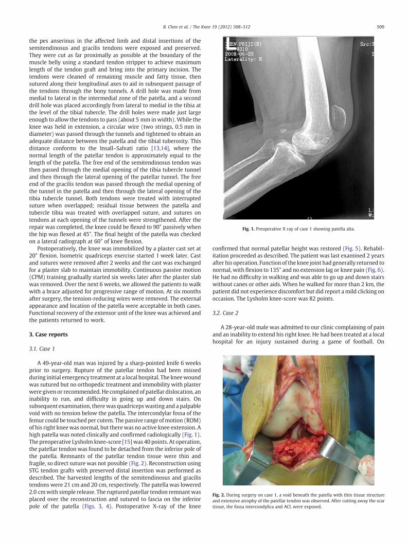
Fig. 1. Preoperative X ray of case 1 showing patella alta.
Fig. 2. During surgery on case 1, a void beneath the patella with thin tissue structureand extensive atrophy of the patellar tendon was observed. After cutting away the scartissue, the fossa intercondylica and ACL were exposed.
509B. Chen et al. / The Knee 19 (2012) 508–512
the pes anserinus in the affected limb and distal insertions of thesemitendinosus and gracilis tendons were exposed and preserved.They were cut as far proximally as possible at the boundary of themuscle belly using a standard tendon stripper to achieve maximumlength of the tendon graft and bring into the primary incision. Thetendons were cleaned of remaining muscle and fatty tissue, thensutured along their longitudinal axes to aid in subsequent passage ofthe tendons through the bony tunnels. A drill hole was made frommedial to lateral in the intermedial zone of the patella, and a seconddrill hole was placed accordingly from lateral to medial in the tibia atthe level of the tibial tubercle. The drill holes were made just largeenough to allow the tendons to pass (about 5 mm inwidth).While theknee was held in extension, a circular wire (two strings, 0.5 mm indiameter) was passed through the tunnels and tightened to obtain anadequate distance between the patella and the tibial tuberosity. Thisdistance conforms to the Insall–Salvati ratio [13,14], where thenormal length of the patellar tendon is approximately equal to thelength of the patella. The free end of the semitendinosus tendon wasthen passed through the medial opening of the tibia tubercle tunneland then through the lateral opening of the patellar tunnel. The freeend of the gracilis tendon was passed through the medial opening ofthe tunnel in the patella and then through the lateral opening of thetibia tubercle tunnel. Both tendons were treated with interruptedsuture when overlapped; residual tissue between the patella andtubercle tibia was treated with overlapped suture, and sutures ontendons at each opening of the tunnels were strengthened. After therepair was completed, the knee could be flexed to 90° passively whenthe hip was flexed at 45°. The final height of the patella was checkedon a lateral radiograph at 60° of knee flexion.
Postoperatively, the knee was immobilized by a plaster cast set at20° flexion. Isometric quadriceps exercise started 1 week later. Castand sutures were removed after 2 weeks and the cast was exchangedfor a plaster slab to maintain immobility. Continuous passive motion(CPM) training gradually started six weeks later after the plaster slabwas removed. Over the next 6 weeks, we allowed the patients to walkwith a brace adjusted for progressive range of motion. At six monthsafter surgery, the tension-reducing wires were removed. The externalappearance and location of the patella were acceptable in both cases.Functional recovery of the extensor unit of the knee was achieved andthe patients returned to work.
3. Case reports
3.1. Case 1
A 49-year-old man was injured by a sharp-pointed knife 6 weeksprior to surgery. Rupture of the patellar tendon had been missedduring initial emergency treatment at a local hospital. The kneewoundwas sutured but no orthopedic treatment and immobility with plasterwere given or recommended. He complained of patellar dislocation, aninability to run, and difficulty in going up and down stairs. Onsubsequent examination, therewas quadricepswasting and a palpablevoid with no tension below the patella. The intercondylar fossa of thefemur could be touchedper cutem. The passive range ofmotion (ROM)of his right kneewas normal, but therewas no active knee extension. Ahigh patella was noted clinically and confirmed radiologically (Fig. 1).The preoperative Lysholmknee-score [15]was 40 points. At operation,the patellar tendon was found to be detached from the inferior pole ofthe patella. Remnants of the patellar tendon tissue were thin andfragile, so direct suture was not possible (Fig. 2). Reconstruction usingSTG tendon grafts with preserved distal insertion was performed asdescribed. The harvested lengths of the semitendinosus and gracilistendons were 21 cm and 20 cm, respectively. The patella was lowered2.0 cmwith simple release. The ruptured patellar tendon remnantwasplaced over the reconstruction and sutured to fascia on the inferiorpole of the patella (Figs. 3, 4). Postoperative X-ray of the knee
confirmed that normal patellar height was restored (Fig. 5). Rehabil-itation proceeded as described. The patient was last examined 2 yearsafter his operation. Function of the knee joint had generally returned tonormal, with flexion to 135° and no extension lag or knee pain (Fig. 6).He had no difficulty in walking and was able to go up and down stairswithout canes or other aids. When he walked for more than 2 km, thepatient did not experience discomfort but did report amild clicking onoccasion. The Lysholm knee-score was 82 points.
3.2. Case 2
A 28-year-old male was admitted to our clinic complaining of painand an inability to extend his right knee. He had been treated at a localhospital for an injury sustained during a game of football. On
Fig. 3. During surgery on case 1, distal insertions of semitendinosus and gracilis wereexposed and the tendons separated.
510 B. Chen et al. / The Knee 19 (2012) 508–512
examination, the patella hadmigrated proximally. The range of flexionwas 0–130° and the extensor lag was 10° in his knee. Radiographsrevealed a high location of the patella. The preoperative Lysholmknee-score was 49 points. A rupture of the patellar ligament from theinferior pole of the patella was identified during the operation. Scartissue was excised and the semitendinosus and gracilis tendons wereused to reconstruct the patellar ligament as described. In this patient,24 cm of the semitendinosus tendon and 22 cm of the gracilis tendonwere harvested and his patella was lowered by 2.5 cm. The rupturedpatellar tendon remnant was sutured to strengthen the reconstruc-tion. Postoperative X-ray confirmed that normal patellar height wasrestored. Rehabilitation proceeded as described. The patient was lastexamined 1.5 years after his operation. On examination, there was noextensor lag and flexion was 0–135°. His knee functions had generallyreturned to normal with no pain reported. He could participate inrelatively rigorous exercise at 18 months after surgery. The Lysholmknee-score was 87 points.
Semitendinosustendon
Gracilis
tendon
Insertions
Fig. 4. The free end of the semitendinosus tendon was passed through the tunnel in the tibmedial. The free end of the gracilis tendon went through the patellar tunnel from medial totissue between patella and tubercle tibia was treated with overlapped suture, and sutures oshowed the key site in interrupted suture nearer to STG insertions.
4. Discussion
Patellar tendon disruptions are relatively uncommon knee injuriescompared to fractures, ligament sprains, or meniscal tears [16]. Thesetears may be traumatic or they may occur spontaneously in patientswith other underlying diseases, such as rheumatosis [17], metabolicabnormalities [9], or hormonal disorders [18]. In addition, patellartendon disruption has been observed in patients receiving local steroidinjection therapy [19], after total knee arthroplasty [1,4,10], andfollowing anterior cruciate ligament reconstruction with a bone–patellar tendon–bone autograft [20]. Even if the proper physicalexamination is performed, the acute tear is accompanied by substantialswelling and hematoma formation, which may make palpation of thetendon defect difficult, leading to neglected patellar tendon rupture[16]. Additional information fromX-ray examination can show a patellaalta. In addition, knee MRI can also be a useful, non-invasive andaccurate tool for providing additional information suchas the location ofthe rupture, the condition of tendon, and the appearance of thesurrounding tissues [21]. We reported two cases of neglected patellardisruption; case 1was stabbed, and case 2 was injured during sport, andboth injuries were missed on initial emergency treatment.
Neglected rupture of the patellar tendon is a rare occurrence, so alarge patient series on the subject is not available. Several techniqueshave been used to relocate the patella to its anatomic position andrepair the patellar tendon, but there is no widely accepted method.Timo Jarvela et al. [4] also used a STG graft for reconstruction of thepatellar tendonwith an interference screw and staple fixation, but theSTG was free. They did not keep STG insertions and not make full useof the role provided by insertions. We adapted our improved methodfrom Cadambi et al [1]. In their technique, the insertions weremaintained when treating rupture of the patellar tendon with STG,but they did not drill a hole in the tibial. Instead, they sutured the freeend of the semitendinosus tendon to its insertion or to the proximaltibial periosteum after it passed through the tunnel in the patella. Thetendons used were attached more medially, however, so that forcewas not well distributed, which may alter the tracking of the patella.
In contrast, we performed reconstruction and restoration of aruptured patellar tendon using STG tendons with preserved distalinsertions in combined with tension-reducing wire. We suggest thatthis modified technique has several advantages.
Gracilis
tendon
Semitendinosustendon
ia tubercle medial to lateral, and then through the tunnel in the patella from lateral tolateral. Both tendons were treated with interrupted suture when overlapped. Residualn tendons at each tunnel opening were strengthened. The white arrow in right figure
Fig. 5. Postoperative X ray in case 1 revealed that the patella had returned to normal height.
511B. Chen et al. / The Knee 19 (2012) 508–512
First, semitendinosus and gracilis tendons are rich in tendon fibers,which are stronger than those of the distal iliotibial tract, fascia lata, orquadriceps-patellar retinaculum. Indeed, STG are used extensively foranterior cruciate ligament reconstruction [22]. In addition, thisresection results in only modest damage [23].
Second, by preserving distal insertion of the tendon and pullingtendons through tunnels in the bone, the integration between tendonand bone is strengthened and stability is maximized after construc-tion. It was reported that tendon-bone healing requires three monthsor more [24,25]. We retained the tibial insertion of the STG, whichcould provide additional stability during the early stages of tendon-bone healing. In addition, the medial point of the tibial tunnel, whichwas the cross and suture point of the two tendons, was close to theoriginal insertion of the STG. We also surmised that it is important toretain blood supply at this point from the original insertion of the STGto promote healing of the tendon. Healing at the crossing point iscrucial, particularly for a closed-loop closure, and could provide foradditional stability of the loop. Using an animal model, Papachristouet al. [26] compared ACL reconstruction with semitendinosus tendonautograft with or without maintaining the tibial insertion. They found
Fig. 6. Photo-documentation of knee function at two years postoperative follow-up in patie135°. The track of patella was normal. On front and lateral views, STG tendons can be obsefemoris was contracted. The site of the original patellar ligament was empty, suggesting succrelied on healing of the sutured closed tendon loop, particularly on the recovery of the suturbone.
that necrosis of the graft was observed 3 weeks after surgery andprogressive revascularization and maturation required 6 to 12 weeksin the control group (without maintaining the tibial insertion).Retaining the tibial insertion of the semitendinosus autograftappeared to preserve its viability and avoid avascular necrosis andrevascularization that occurs with the use of a free tendon autograft.They concluded that harvesting the semitendinosus tendon withoutdetachment of the tibial attachment could preserve a sufficientamount of blood supply to keep it viable. Sung-Jae Kim [27] suggestedthat a more viable graft is obtained and more firm distal fixation isachieved by preservation of the tibial insertion of hamstring tendonsduring ACL reconstruction.
Third, when the tendons are passed in opposite directions throughthe tunnels, force is distributed on both sides of original patellartendon, which allows for even transmission of tension and facilitatesrecovery of the correct patellar locus for maximal range of motion ofthe knee joint. Fourth, tension-reducing wire was used for furtherstabilization. The wire could transmit tension load from the patelladirectly to the tubercle tibia; when the knees bend, the patellae ispulled downward by the wire, rendering the patellar tendon
nt 1. The injured knee achieved a complete ROM, with active motion ranging from 0 torved distending from the two sides of the patella (black arrow) when the quadricepsessful replacement of the ligament by the STG tendons. The success of this replacemented crossed tendons adjacent to the tibial tubercle and healing between the tendon and
tensionless and greatly aiding natural recovery [20,28]. In addition,the wires helped maintain proper patellar tracking. In our two clinicalcases, the patellar locus was recovered after operation.
Generally, the semitendinosus and gracilis tendons are shorter inChinese people than in other races. In case 1, we obtained asemitendinosus tendon 21 cm in length, and a gracilis tendon 20 cmin length. In case 2, the resected semitendinosus tendon was 24 cm inlength and gracilis tendon was 22 cm in length. Williams [28] wasable to obtain 32 cm and 30 cm lengths from amale Caucasian patient.The longest possible tendonmay be related to patient height and race.Thus, use of both the semitendinosus and gracilis tendons may benecessary for patients of shorter stature.
It is obviously necessary tomaintain the track, rotation, andheight ofthe patella. It is especially important to lower the patella to its properposition (where the inferior pole is on top of intercondlar fossa of thefemur at 45° of knee flexion). The knee should allow for at least 90°flexion, while the patellar tendon should be relaxed when the knee isextended passively. In our patients, thepatellawas lowered by 2 cmand2.5 cm, respectively. We also found that the length of the reconstructedpatellar tendon was approximately equaled to the length of patella asreported previously [13,14]. For patients in which it is difficult to lowerthe patella, the medial and lateral sulcus of the suprapatellar bursashould first be released and subperiosteal dissection of vastusintermedius performed in the front of the femur. In addition, the lateralretinaculum or medial retinaculum should also be released. This mayincrease the chance of patellar ischemia and necrosis, however [29]. Ifthe patella still cannot be lowered to a normal position, Z-lengthening ofthe rectus femoris may be considered, or patellar traction may be usedpreoperatively to restore the height of patella. The Ilizarov method hasalso been used to relocate the patella to its original anatomic position[30]. In the two patients we treated, normal height was achieved duringsurgery simply by release without preoperative patellar traction. Thisgreatly reduced the duration of immobility while still achievingsatisfactory postoperative recovery.
The first 6 months after surgery are critical for the restoration of kneejoint function. Proper functional training can significantly improvefunction. After one year following surgery, however, additional functionalrestoration will be rather limited [31], so therapy must be started as soonas possible. In our report, we allowed the patients to remove the plasterslab and begin CPM training and walking with a brace 6 weeks afteroperation. We judged that the reconstructed patellar tendon was strongand stable enough for rehabilitation at this time. Acceptable functionalrecovery of the knee joint was achieved in both patients, although not tothe level of the unaffected knee or to degree achieved followingrestoration of a newly ruptured patellar tendon [32]. Compared tofunction before surgery, both patients were satisfied with their postop-erative recovery and both returned to working and leisure activities.
In conclusion, combined use of STG tendons with preserved distalinsertions and tension-reducing fixation facilitates good functionalrecovery in patients with neglected patellar tendon rupture. Thissuccess was due to the initial strength and achieved stability and to thepreservation of blood supply that mitigated ischemic necrosis. Thesurgery allowedbothpatients to beginCPMtrainingwithin 6 weeks andsatisfactory function of the knee joint was restored within 1.5–2 years.The technique is relatively simple and feasible, and warrants furtherinvestigation.
5. Conflict of interest
We have no conflict of interest in the article.
References
[1] Cadambi A, Engh GA. Use of a semitendinosus tendon autogenous graft for ruptureof the patellar ligament after total knee arthroplasty. A report of seven cases.J Bone Joint Surg Am 1992;74:974–9.
[2] BekD,DemiralpB,KomurcuM, SehirliogluA.Neglectedpatellar tendon rupture: a caseof reconstruction without quadriceps lengthening. J Orthop Traumatol 2008;9:39–42.
[3] Mandelbaum BR, Bartolozzi A, Carney B. A systematic approach to reconstructionof neglected tears of the patellar tendon. A case report. Clin Orthop Relat Res 1988:268–71.
[4] Jarvela T, Halonen P, Jarvela K, Moilanen T. Reconstruction of ruptured patellartendon after total knee arthroplasty: a case report and a description of analternative fixation method. Knee 2005;12:139–43.
[5] Mihalko WM, Vance M, Fineberg MJ. Patellar tendon repair with hamstringautograft: a cadaveric analysis. Clin Biomech (Bristol, Avon) 2010;25:348–51.
[6] Milankov MZ, Miljkovic N, Stankovic M. Reconstruction of chronic patellar tendonrupture with contralateral BTB autograft: a case report. Knee Surg SportsTraumatol Arthrosc 2007;15:1445–8.
[7] Scuderi C. Ruptures of the quadriceps tendon; study of twenty tendon ruptures.Am J Surg 1958;95:626–34.
[8] Chiou HM, Chang MC, Lo WH. One-stage reconstruction of skin defect and patellartendon rupture after total knee arthroplasty. A new technique. J Arthroplasty1997;12:575–9.
[9] ElGuindy A, Lustig S, Servien E, Fary C, Weppe F, Demey G, et al. Treatment ofchronic disruption of the patellar tendon in osteogenesis imperfecta with allograftreconstruction. Knee 2010;18:121–4.
[10] Crossett LS, Sinha RK, Sechriest VF, Rubash HE. Reconstruction of a rupturedpatellar tendon with achilles tendon allograft following total knee arthroplasty.J Bone Joint Surg Am 2002;84-A:1354–61.
[11] Lewis PB, Rue JP, Bach Jr BR. Chronic patellar tendon rupture: surgical reconstructiontechnique using 2 Achilles tendon allografts. J Knee Surg 2008;21:130–5.
[12] Fukuta S, Kuge A, Nakamura M. Use of the Leeds–Keio prosthetic ligament for repairof patellar tendon rupture after total knee arthroplasty. Knee 2003;10:127–30.
[13] Insall J, Salvati E. Patella position in the normal knee joint. Radiology 1971;101:101–4.
[14] Ahmed AD. Radiological assessment of the patella position in the normal kneejoint of adult Nigerians. West Afr J Med 1992;11:29–33.
[15] Bengtsson J, Mollborg J,Werner S. A study for testing the sensitivity and reliability ofthe Lysholm knee scoring scale. Knee Surg Sports Traumatol Arthrosc 1996;4:27–31.
[16] McGrory JE. Disruption of the extensor mechanism of the knee. J Emerg Med2003;24:163–8.
[17] Prasad S, Lee A, Clarnette R, Faull R. Spontaneous, bilateral patellar tendon rupturein a woman with previous Achilles tendon rupture and systemic lupuserythematosus. Rheumatology (Oxford) 2003;42:905–6.
[18] Chen CH, Niu CC, Yang WE, Chen WJ, Shih CH. Spontaneous bilateral patellartendon rupture in primary hyperparathyroidism. Orthopedics 1999;22:1177–9.
[19] Clark SC, Jones MW, Choudhury RR, Smith E. Bilateral patellar tendon rupturesecondary to repeated local steroid injections. J Accid Emerg Med 1995;12:300–1.
[20] Milankov Ziva M, Semnic R, Miljkovic N, Harhaji V. Reconstruction of patellartendon rupture after anterior cruciate ligament reconstruction: a case report.Knee 2008;15:419–22.
[21] Sutherland F, Dawson JS, Moran CG. MRI detection of partial rupture of the patellartendon in association with multiple ligament injuries of the knee its surgicalimportance. Injury Extra 2005;36:6–8.
[22] Chen L, Cooley V, Rosenberg T. ACL reconstruction with hamstring tendon. OrthopClin North Am 2003;34:9–18.
[23] Tashiro T, Kurosawa H, Kawakami A, Hikita A, Fukui N. Influence of medialhamstring tendon harvest on knee flexor strength after anterior cruciate ligamentreconstruction. A detailed evaluation with comparison of single- and double-tendon harvest. Am J Sports Med 2003;31:522–9.
[24] Weiler A, Hoffmann RF, Bail HJ, Rehm O, Sudkamp NP. Tendon healing in a bonetunnel. Part II: histologic analysis after biodegradable interference fit fixation in amodel of anterior cruciate ligament reconstruction in sheep. Arthroscopy2002;18:124–35.
[25] Nebelung W, Becker R, Urbach D, Ropke M, Roessner A. Histological findings oftendon-bone healing following anterior cruciate ligament reconstruction withhamstring grafts. Arch Orthop Trauma Surg 2003;123:158–63.
[26] Papachristou G, Nikolaou V, Efstathopoulos N, Sourlas J, Lazarettos J, Frangia K,et al. ACL reconstruction with semitendinosus tendon autograft withoutdetachment of its tibial insertion: a histologic study in a rabbit model. KneeSurg Sports Traumatol Arthrosc 2007;15:1175–80.
[27] Kim SJ, Kim HK, Lee YT. Arthroscopic anterior cruciate ligament reconstructionusing autogenous hamstring tendon graft without detachment of the tibialinsertion. Arthroscopy 1997;13:656–60.
[28] Williams S, Ireland J, Zebdeh MYE. Late reconstruction of the patellar tendon: twocase reports. Knee 1997;4:113–5.
[29] Chow FY, Wun YC, Chow YY. Simultaneous rupture of the patellar tendon and theanterior cruciate ligament: a case report and literature review. Knee Surg SportsTraumatol Arthrosc 2006;14:1017–20.