106


RECORDS OF THE
ZOOLOGICAL SURVEY OF INDIA
OCCA.SIONAL PAPER NO. IS7
Caryophyllidean Cestode Fauna of India
by
M. HAFEEZULLAH
Zoological Survey of India, Calcutta:.
Edited by the Director, Zoological Survey of India, Calcutta
1993

© Copyright: Government of India,1993
Published: November, 1993
Price: Inland: Rs. 120·00 Foreign: £ 6·00 $ 8'00
Printed in India by A. K. Chatterjee at Jnanodaya Press, SSB, Kabi Sukanta Sarani, Calcutta 700 085 and published
by the Director, Zoological Survey of India, Calcutta

RECORDS
OFi'THE
ZOOLOGICAL SURVEY OF INDIA
Occasional Paper
No. 157 1993
CONTENTS
Systemetic Index Introduction Material and Method Generalised Morphology and Anatomy Outline of Development and Life Cycle Systematic Account Species of Uncertain Systematic Position Discussion Host-Parasite List Parasite-Host List Summary Acknowledgements References
••• . . . . , .
... • • •
...
...
...
...
Pages 1-102
(i) 1 4 S
18 22 89 90 92 94 96 96 96

SYSTEMATIC INDEX
Page
Pamily I. CARYOPHYLLAEIDAE Leuckart 25 Genus 1. Paracaryophyllaeos Kulakovskaya 25
1. Paracaryophyllaeus lepidocephali (Kundu) 26 2. Paracaryophyllaew ostiobramensis
(Gupta and Sinha) 29 Genus 2. PseuodocaryopbylJaeos Gupta 31
3. Pseudocaryophyllaeus indica Gupta ... 31 Family II. CAPINGENTIDAE Hunter 37
Genus 3. Adenoscolex Fotedar 38 4. Adenoscolex oreini Fotedar 38
Genus 4. Breviscolex Kulakovskaya 40 s. Breviscolex aurangabadensis (Shinde) 41 6. Breviscolex naldurgensis (Shinde et al.) 45
Family III. L YTOCESTIDAE HUDter 47 Genus S. Lytocestos Cohn 48
7. Lytocestus birmanicus Lynsdale 49 8. Lytocestus filiformis (Woodland) 52 9. Lytocestus fossilisi (Gupta) 54
10. Lytocestus indicu,$ (Moghe) 60 11. Lytocestus longicollis Rama Devi 65
Genus 6. Djombangia Bovien 67 12. Djombanria Penetrans Bovien 68
Genus 7. Bovienia Fuhrmann 71 13. Bovlenia bilocula (Murhar) 72
14. Bovienia ilishai Zaidi and Khan 79 15. Bovienia serialis (Bovien) 81
Genus 8. Lytocestoides Baylis ... . .. 86
16. LyttJcestoides paithanenlfs Shinde and Deshmukh . .. 87

INTRODUCTION
As a matter of fact, till recently, little attention was paid to the co11ection and study of this group of cestodes in India. It was Moghe (1925) who initiated the study in India on this group by describing Caryophyllaeus indicus from the common walking cat-fish, C/mill' batrachus (L.), from Nagpur. Woodland (1926) indicated that the species belongs to the senus Lytocestus Cohn, 1908. Acting on this clue, Moghe (1931) redescribed it as L1tDC"IUS l"dlcus. Moghe was joined by Mehra who (1930) presented a paper in the 17th Science Congress Association on Caryophy/laeus kashmirensis from the fresh water hill stream fl.h SchlzDthorax micropogon Heckel from near Srinagar, Kashmir. The paper remained in abstract form and its full detail along with iJ1ustrations was never published. While discussing its systematic position, Mehra (loc. cit.) observed that "this species apparently connects the new two genera CaryophyUaeus and Lytocestus." Fotedar (1958) described .4d'''olcDlex oreini n. gen., D. sp. from the fresh water fish Oreinus sinuatus (Heckel) from Kashmir in the family Capingentidae Hunter, 1927, but did not compare this species with Car)1Dphyllaeus kashmirensis Mehra, 1930. Mackiewicz (1972) considered C. kashmirensis as 'p,eles Inquirenda but later on he (1981a) opined that fresh material of this species should be collected from the same host and locality, studied and compared with Adenoscolex oreini Fotedar, 1958. Agarwal (1985) considered it as a synonym of A oreini. In the present work this species has been considered as species of uncertain status till its fresh material is collected, studied and compared with A oreini. Gupta (1961), Gupta and Sinha (1980, 1984), Verma (1971), Gupta and Parmar (1984, 1986~ 1990), Gupta and Singh (1984), Agrawal and Singh (1985), Pandey (1973) and Pandey and Tiwari (1989) from Uttar Pradesh; Murhar (1963), Mackiewicz and Murhar (1972), Shinde (1970), Shinde and Deshmukh (1975, 1980) and Sinde et ale (1987) from Maharashtra; Satpute and Agarwal (1974, 1980 a & b), Niyogi et ale (1982), Mackiewicz and Murhar (1972) from Madhya Pradesh; Rama Devi (1973) from Andhra Pradesh; Sahay and Sahay (1977) and Kanth et al. (1984) from Bihar; Kundu (1985) and Kundu et ale (1985) from West Bengal; and Gupta (1961) and Chakravarty and Tandon (1989) from Assam have significantly contributed to the Indian fauna of Caryophyllidea. Lynsdale (1956), Singh (1975) and Zaidi and Khan (1976) described one species each from Burma, Nepal and Pakistan. Hafeezullah (1986) reviewed the status of various Indian species of the genus Djombangia Bovien, 1926. Agarwal (J 985) reviewed the caryophyllid fauna of India, and Mackiewicz (1981a) discussed the status of species of India, Nepal and Pakistan. Nama (1979) reported the occurrence of Crescentovitus hi/oculus (1979) in Rajasthan.
CF 1

REe. ZOOL. SURV. INDIA, Occ. PAPER No. 151
The caryophyllideans form a small group of single-segmented cestode parasites with about 145 described species under 50 genera. These figures include 40 species under 14 genera from Indian region (inclusive of Pakistan, Nepal and Burma). Bovenla ilishai Zaidi and Khan, 1976 is the only species known from Pakistan; so is the case with Lytocestus fossilis Singh, 1975 from Nepal; and Lytocestus birmanicus Lynsdale, 1956 has been reported from Burma and India both. Bovenia nagpurensis n. sp. was reported by Murhar (1977) from the fish Clarias batrachus from Nagpur in the proceedings of the 65th Indian Science Congress held at Bhubaneswar. in 1977, but it remained in abstract form; its full detail was never published. Similarly, accounts of Morvekia chotanagpurensis n. gen., n. sp. and Neolytocestus vitellodiscontinuatus n. gen., n. sp. from Clarias batrachus from Chotonagpur was presented by Sahay and Sahay (1977) in tbe proceedings of the First National Conventions of Indian Helminthologists held at Bhubaneswar in 1977. These taxa also remained in abstract forms; they were never published in detail. Therefore, these two new genera and new species are not considered in the present work. Johri (1959) described Hunteroides mystei as a new caryophyllid genus and species from Mystus seenghala (Sykes) in Delhi State. According to Schmidt (1986) this is actually a true cestodarian adding a new genus and new species to the subclass Cestodaria Monticelli, 1891, but Mackiewicz (1981a) and Dubinina (1982) considered it to be Gephrolina paragonopora (Woodland, 1923) earlier described from the same host from India. Recently, Chakravarty and Tandon (1989), while giving accounts of caryophyllids from north·easterm region of India, reported Lytocestus jili./ormis (Woodland, 1923) from the fish Clarias batrachus from Guwahati (Assanl, India), which was originally described from the fish Mormyrus caschive from the river Nile (Sudan, Africa). In the same paper they also reported Lytocestus birmanicus Lynsdale, 1956 which was originally described from Rangoon, Burma.
HOW CAR YOPHYLLIDEA DIFFERS FROM CESTODARIA
Caryophyllids are held apart fronl cestodarians, another set of monozoic cestodes like (Amphilina and Gyrocotylt:) since the latters have no scolex at al1, the positions of male and female gonopores are entirely different, the shelled larva is 10-hooked, and they parasitise turtles also in addition to fishes including marine fishes.

RA,aZULLAH: Caryophyllidean Cestode Fauna of India
HOW CARYOPHYLLIDEA DIFFERS FROM ORDERS WITH SEGMENTED STROBILA
3
The polyzoic cestodes strikingly differ from monozoic caryophllideans in being strobililte and segmented usually with one set (or two) of reproductive systems in each segment. They parasitise all groups of vertebrates and intermediate hosts are usuaUy arthropods. Caryophyllids require fresh water oligochaete annelids (usually tubificids) only as intermediate hosts and never need copepods or amphipods to complete their development to the procercoid stage. Polyploidy and parthenogenesis although recorded so far to occur only in some species of caryophyllids but these twin phenomenons are not known in polyzoic cestodes. Further, the caryophyllid genus Archigetes completes its life cycle and matures progeneticeally in the invertebrate host (tubificids) itself. This phenomenon also is not known in polyzoic cestodes.
Nature of Caryophyllidea: It is an established fact that, like polyzoic cestodes, the caryophyllid monozoic cestodes do not have germinative cells behind the scolex i.e. in the region of neck which serves as the zone of proliferation and consequent strobilization ~ut it is not satisfactorily known that this monozoic condition of caryophyllids is original and primary or secondarily achieved by dropping segmented strobilate stage. It goes to mean whether they never had a strobilate stage or a strobil ate stage existed in them at one time. This dispute gives birth to another question whether the oaryophyllid cestodes are progenetic or neotenic. The twin questions are rather easy to answer with respect to Archigetes which has a cercomor-bearing stage and in the oligocbaete-dwelling stage the gonopore is not functional because of an integumental oovering, and in Archigetes iowensis these two larval characteristics are lost when ingested by fish. So, the precocious sexual maturity of the procercoid of A. iowensis is indeed progenetic. The real difficulty lies with the Caryophyllaeus-like caryophyUids where scientific evidences are rather obscure regarding primary or secondary nature of unseg ..
mented strobila and consequent progenesis or neoteny.
Many theories have been propounded by various investigators to solve this difficulty but each theory has its own criticisms also. However, the pseudophyllidean theory e~joys wide acceptance. It postulates that caryopbyllids are members of the Order Pseudophyllidea, since the life cycles of the caryophyUideans represent the abbreviated life cycles of pseudopbyllideans with the ad.ditional occurrence of progenesis (neoteny) at the plerocercoid larval stage in which strobilate stages no longer develop. They are merely precocious progenetic larvae that have dropped the strobilate stage. However, MaQkiewic2; (1972) sUIQmarises Ule whole problem as follows; "All of these theories are

4 REC. ZOOL. SURV. INDIA, OCC. PAPER No. IS7
subject to criticism but the fact that caryophyllids have: a nonciliated, non-free-swimming larva; an annelid intermediate host; a single set of reproductive organs in a non-segmented body, which zoological opinion generally accepts as a condition preceding strobilization; a large number of species which have scoleces generaJ]y unlike any found in strobilate tapeworms, with the possible exception of EublJthrium exhibited extensive radiation of morphological types; a worldwide distribution; and occur predominantly in primitive, teleost freshwater fishes, argues forcefully in my opinion, for their being regarded as nOD
neotenic cestodes, distinct from but closely related to the Pseudophyllidea. If the above analysis is correct then it would appear proper to regard the cercomere
bearing stage of Archigetes as a neo tenic or progenetic caryophyllid and all others (Plerocercoid-like) as genuine non-progenetic adult stages."
MATERIAL AND METHOD
For correct study of worms, the elucidation of the various internal organ systems of their fresh specimens is essential. This is achieved by adopting their correct processing technique. In this connection the recent manual of Pritchard and Kruse (1982) can be successfully used. However, a brief technique for processing caryophyllid material is given below.
As far as possible, live fresh water fish hosts (specially belonging to the Orders Siluriformes and Cyprinifornles) should be examined in order to recover live specimens of caryophyllid cestodes. Dead or iced fish hos ts may not give specimens in good condition. Care should be taken that the scolex of the specimen is not lost. The live specimens so obtained should be studied first and shape and structure of the scolex and their variations, if any, noted in the field book. Attempt should also be made to study the position and number of male and female genital pores as well as presence or absence of genital atrium. This is important because these structures may not be studied correctly in the whole mounts.
Fixation: Fixative sbould not be used on the specimens unless and until contraction has completely stopped, or relaxation and fixation can be done simultaneously depending upon the experience in order to achieve better results. A fixative is a chemical reagent which preserves the specimen in life-like condition without brittleness. However" a perfect fixation is yet to be known. These live specimens are then taken in a small bottle containing cold or hot fixative FA (7% solution of formalin with 3% to 7% glacial acetic acid, the amount of acetic acid being not critical) or AFA (5 prts glacial ~cetic acid, 10 parts formalin and 85 parts alcohol) or s% fo~malin sol~tion wl1ichev~r

HAPEEZULLAH: Caryophyllidean Cestode Fauna of India s
Is benificial known througb experience. The bottle is then shaken vigorously for about one minute so that the contraction of the specimens stops and they get relaxed and fixed. This fixed material can now be stored in air-tight vials containing 70% alcohol with a little glycerine, and label with collection data inserted therein.
Staining: This material is stained for whole mounts. Before staining, the material may be flattened under coverglass pressure with gentle pressing using the tip of a needle. Overfllattening between two slides should be avoided. In order to get good results from whole mounts, staining of the material should be done in borax cormine, acetic carmine, Semichon's carmine or even haematoxylin. It has been found that retrogressive staining yields better results than progressive one. So the material should be overstained with the chosen stain and then gradually de-stained to the desired differenciation. To de-stain overstained specimens, 5% acidulated alcohol in the case of carmine or SOlo aquous HCL in the case of haemotoxylin is used. The stained material should be debydrated in alcohol grades, cleared in clove oil and xylol, mounted in Canada balsam and dried on a regulated hot plate or drying oven at about 56°0. Two
labels having details of. the host on one and identification of the worm on the other should be affixed on the slide.
Sectioning: For serial sectioning, a part of the material should be separately fixed with FA or 4% formalin. AFA should not be used for this purpose. The unpressed or unftattened material should be used for serial cross-soctioning througb testicular region and serial sagittal-sectioning through the posterior quarter of the body. The cross-sections will help determine the position and· arrangement of vitellaria with respect to the inner longitudinal muscles and the sagittal sections will tell about the details of the male and female genital ducts as well as the number of genital pores and absen~e or presence of genital atrium. By following this technique correct and complete study of worm can be made.
All Indian investigators should deposit the holotypes and para types of worms with the National Helminthological Collections of the Zoological Survey of India, Calcutta.
GENERALISED MORPHOLOGY AND ANATOMY
Mackiewicz (1972) in his very informative and useful review of world caryopbyUids has given their various body shapes (his figs. 1-22 and 41-63) and different structures of ~olt!ces (his fiss. 27-40). He (1982) has further furnishe4 additional scolex types (his figs.

6 REG. ZOOL. SURV. INDIA, Oce. PAPER No, 1S7
loculus -----+.~~
neck reljon---~+
viteltarium----+-4A. (preovarianl
~ •• de'eren.
eaternal seminal vesicle (ej.cul.tory duct)
_jtcul.tory duct
female lonopor.
&I'erine ,lands
"a,ina
.emin.I receptlcle
ootype
viteUlrium (po.tovl,ian)
.xcr.tory pore
~--scole.
~.---....... .... -"---I---ellcretory
a
(osmoregulatory) canals
outer longitudinal muscles
~4--- vitellarium
~.ef-~-- testis
Xl-..f---- inner loncitudin,1 mu,cl,s
cirrus s,c
cirrus
m.'e lonopore uterovalinllduct
uterus 3
uterus 2
ovary
vitelline res,rvolr
vitellin. duct
uterus 1
eacretory bladder
opuculum
hook
boss
b
Fig. lA: Caryophyllidean morphology and anatomy, (After Mackiewicz, 1972). a. Hypothetical species illustrating principal features. b. Eg~ of Archigeles $p. showio$ on~bQspbero.

IlAiB!zULLAH: Caryophyllidean Cestode Fauna o/India
c.
b
e
Fig. IB: Caryophyllidean specimens of a single population. a. b, c. Variations in scolex shape.
a
d
d. Follicles of vitellaria and ovary meeting on one side. no t meeting OD the other side; ovarian follicles r<aching posterior end of body.
e. Ovarian follicles not reacbioa posterior end of body.
1

8 REe. tOOL. SURV. tNDIA, OCC. PAPER No. 157
ptod pgd
Fig. 2: Hypothetical caryopbyllidean indicating various morphological and anatomical regions (After Mackiewicz, 1982).
pvd-previtellaria distance, ptd-pre-testes distance, povd-pre-ovarlan vitellaria field, tl-testes field, pgd-post-gonopore distance, wg-width at gonopore, ogd-ovary .. gonopore distance, plod-post-ovarian distance,
ptovd-post·ovarian vitellaria distance, o-ovary, I-testes and v-vitellaria.

HAF!EZULLAH: Caryophyllidean Cestode Fauna oj India
18-11) and devised some terminology to describe their shapes and structures. In the same paper, he has attempted to describe various morphological and anatomical regions with the help of suitable terminologies. However, a generalised morphology and anatomy of caryopbyllids is briefly furnished here .
a
o • • • •• It
d
g
••• • •• • t • - •••
b
e
h
c
f
I
FiS- 3A ~ Terminology for some scolex types of caryophyllidean genera (After Mackiewicz, 1982).
a. Tholate, b. Cuneiform, c. Cuneicrispitate or PJabellate, d. Cuneifimbriate, e. Cuneiloculate, f. Biacetabulate, g. Monobothriate, h. Bulbate, i. Choano
companuJate.
The bodies of caryophyllidean cestodes are usually long and narrow in shape. They may be broad also (Djombangia Bovien, N oto!ytocestus Johnston and Muirhead and
CF 2

10 REC. tOOL. SURV. INDIA, Oce. PAPER No. 157
a c.
d e f
9
PiS. 3D: Some scolex types of Indian caryophyllideaas.

RAFJ!EZULLAH: Caryophyllidean Cestode Fauna of India 11
Salaftotaenia Johnston). The body is usually distinguishable into scolex, neck and strobila or main body (Fig. 2). In some genera e.g. Balanotaenia Lvtocestoides Khawia N Dlo-, 'J ,
I,toceslus, ThallDphyllaeus, Paracaryophyllaeus, Hun trella, Pllovitellaria, AdenoscDlex, EdllntDnla and Breviscolex. the neck is absent or is not distinct from the main body. The neck in caryophyllid monozoic cestodes is different from that in poly zoic cestodes. In the latter, the neck or if it is not present the posterior part of the scolex has germinal cells and is known as the region of proliferation which produces a chain of segments each of which contains one or two sets of reproductive organs. Thus, the strobila or main body is segmented and chain-like. Contrarily, in caryophyllid cestodes the neck on the posterior part of the scolex does not act as the region of proliferation. So the strobila is not chainlike and remains unsegmented with only one set of reproductive organs. The neck in this case is short, constriction-like or very long and narrow.
The scoleces in caryophyllids are variously shaped (Figs. 3A, 3B) and are different
in structure and specialisation. In its simplest for~ it may be very short, smooth and indistinguishable from the main body as is found in Breviscolex, Adenoscolex and Paracaryophyllaeus, or it may be "globular or ovate, smooth, unspecialised and borne on a long and narrow neck as occurs in Capingentoides Gupta, 1961, PseudDcaryophyllaeus Gupta, 1961 and Lytocestus IDnglcollis Ramadevi, 1973, or it may be globular or ovate with a terminal introvert and concentration of gland cells be10w the apex as happens in Djombangia Bovien, 1926, or it may be bell-shaped with a prominent collar around the base and an apical funnel as in Caryoaustralus Mackiewicz and Blair, 1980, or it may be with several furrows, constricted off from body or not as in Wenyonia Woodland, 192 J, or its anterior margin may be folded or frilled as in Caryophyllaeus Muller, 1787 and Khawia Hsu, 1935 respectively. Some of the genera have acetabula, sucker, locula, bothria or some combination of them. Thus, the specialisation of scolex is varied, but rostellum and hooks have not yet been reported on any caryophyllid s~olex as is found in some polyzoic cestodes. The shape of scolex may also vary within one and the same species (Figs. 29 a, b ;.40 h, c, d; 46 b; IB a, b, c).
Some caryopbyllids like Archiqetes lowensis Calentine, 1962 attain precocious sexual maturity in the coelome of its freshwater oligochaete annelid retaining the 'larval cercomer with the larval hooks at the posterior end. So, of all the cestodes, only caryophyUids have the examples which mature in an invertebrate.
Generally the male and female gonopores (Figs. lA, 4) are situated in the posterior part of the body midventrally, say in the last I/Sth to 1/4th of body length, but in WenYDnia Woodland, 1923 and Caryoaustralus Mackiewicz and Blair, 1980 they are in anterior half of the worm, in latter the gonopore being single. In M arkevitschia Kulakovskaya and Achmerov, 1965 and Pliovitellaria Fischthal, 1951, the gonopores occur a bit posterior to equatorial plane~ the latter ~en\ls havin~ only one ~onopore. In Djombangia

12 REC. ZooL. SURV. INDIA,.()CC. PAPER No. 157
the genital pores are located immediately in front of ovarian commissure while in Balanotaenia they are behind it. In genera like Caryophyllaeus, the female gonopore is present just behind the male pore on the flat ventral surface, there being no common genital atrium (Fig. 4a). In genera like Atractolytocestus Anthony, 1958, Lytocestus Cohn, 1908 and Djombangia Bovien, 1926, the male and female gonopores are separate as in the previous case but both of them are situa ted in a large shallow depression of body surface called common genital atrium (Fig, 4b). There is a third condition also which
a b c
Fig. 4. Oonopore -types as shown in midsagittal sections (diagrammatic) (After Mackiewicz, 1972).
Abbreviations: AT-atrium; C-cirrus; Ejd-ejaculatory duct; FO-female gonoporo; HD-hermaphroditic duct: MG-male gonoporo; OB-Ovarian bridse or commissure: U-uterus; UVD-uterovasinal duct, VA-vagina.
occurs in genera like CaryoauJ/raluI, PlIovltellarla, Blacetabulum, Caryophylleldes and lsoglarldaerla. In this case, the terminal male and female ducts first join to form a short hermaphroditic duct which ultimately opens as a single gonopore on the fiat ventral surface of the body, there being no common genital atrium as in the second case (Fig. 4c).
REPRODUCTIVE SYSTEMS
CaryopbyUids are single-segmented cestodes since there is no trace Df external or Internal segmentation in their strobila. The single segment is provided with one set each

~LAH; Caryop~idean Cestode Fauna of India 13
of male and female reproductive organs (Fig. la). Sexes are not separate i. e. ga"ochoristcism does not occur. They are hermaphroditic or monoecious. Generally the male reproductivo organs mature first and produce sperms which remain stored till the ovary matures and produces eggs. The maturation of testes earlier than ovary is known as protandry or androgyny. The reverse of this phenomenon i. e. gynandry or protogyn} is not known in caryophyllids.
Male ReprDductlve System :
The main male organs are the testes which are many and produce sperms and the associated ducts and their terminal modificatiBtts drain the sperms towards male pore.
The testes lie in the medullary zone, extending from anterior to coil of vas deferens upto slightly behind neck (if present) or scolex, except in the family Balanotaeniidae in which they occur in the cortical zone along with vitellaria (Fig. 8d). From each testis arises a fine duct called vas efferens. All the vasa efferentia unite to form a common coiled vas deferens which lies anterior to cirrus sac in the central medulla. Before entering the cirrus sac the vas deferens may dialate and become muscular which part is known as external seminal vesicle, e. g. in Archigetes, Biacetabulum, M onobothrium, etc., or it may be simple. Within the cirrus sac the vas deferens may form a convoluted ejaculatory duct or swell into internal seminal vesicle as happens in Caryophyl/aeus laticeps. In other cases the vas deferens immediately enters into a muscular swollen sac called cirrus sac and leads into the ejaculatory duct. This is surrounded by a bulbus muscular structure called cirrus which is the male copulatory organ and is usually eversible and unspined. The cirrus opens to the exterior ventrally in the three different ways as discussed earlier and shown in Figs. 4a, b, c.
Ftmale Reproductive System :
The main female organ is a single ovary which produces ova," the associated gtands provide nutrition, membrane and eggsheli to the zygote, and the ducts lead the egg to the female pDre.
U sualIy the ovary is follicular and in many genera it is ·H .. sbaped situated' near the posterior end of body except in Markevitschia, pliovitellaria and Wenyonla in which genera it is situated in the middle-third of the body. It may also· be butterfty .. shaped, dumbbell-shaped, u- or v-shaped or shaped like an inverted' A' in different genera. It may be compact also as in the genus Caryophyllaeides. The two arms of the ovary are connected by a transverse band called ovarian commissure. The vitellaria or vitelline glands are situated in the cortical field (in the family Lytocestidae, Fig. 8e, c'), in the medullary zone (in the family Caryophyllaeida.e, Fig. 8a1 a'), partly cortical

14 REC. ZOOL. SURV. INDIA, OCC. PAPER No. lS7
and partly medullary (in the family Capingentidae, Fig. 8b, b' ), or cortical along with testes (in the family Balanotaenidae) (Figs. 8d, lib, 12b). So, the occurrence of vitellaria in one field or the other, alone or together with testes is of familial importance .
a
• ~ • ••
b
Fig. S: a. Pos,ovarian vitellaria present (diagrammatic; after Schmidt, 1970, 1986). b. Postovarian vitellaria absent (diagrammatic; after Schmidt~ 1970, 1986).
In certain genera e. g. A den os colex , Edlinton/o, B re vis eolex etc. a group of postovarian vitelline follicles may also be present (Figs. lA, S). The vitellaria may extend from behind the neck up to the level of cirrus sac or eveh beyond posteriorly. In some species the pre- and post-vitelline follicles may be laterally continuous. The viteJlarine follicles may be continuous or discontinuous with the follicles of the anterior horns of the ovary in the same species (Figs. IB e, d). In some specimens the ovarian follicles may not extend up to the posterior end of the body while in others of the same species they intermingle near the posterior end giving the false impression of the presence of post ovarian vitellaria (Figs. IB d, e). The vitelline glands provide material for the egg-shell formation and nutrition for the developing embryo.
Th~ mature ova leave the ovary through a small duct called ,oviduct which arises from the posterior margin of the ovarian commissure. The oviduct has a controlling sphincter called the ovicapt. It receives a duct from vitelline reservoir and a spermioduct from the vagina or seminal receptacle. Beyond that the proximal part of the oviduct functions as a chamber where sperms fertilize the ova and thus zygote is formed. As the oviduct passes further posterior it dialates to form ootype. The ootype is s"Urro\lnded b~ two types of

HAFEIZULLAH: CarYDphyllidean Cestode Fauna 0/ India 1S
unicellular glands, the serous gland cells which are fewer in number and the mucous gland c,lI, which are much more in number. The two types of cells are together called Mehles' ,land. How eggshell formation takes place in Caryopbyllid cestodes is not known with certainty. Probably it takes place as it happens in polyzoic cestodes. The Mehles' glands
• • •• ·4 ..
.. , ;' ~" . .. .~ ~o· , __ ._. 0 .'
.. , ........ -- .,.. ....
a
...
b
e • . ,
Fig. 6 : 8. Ovarian lobes entirely medullary (diagrammatic ; after Schmidt, 1970, 1986), b. Ovarian lobes party cortical (diagrammatic ,. after Schmidt, 1970, 1986).
secrete a very thin membrane around the zygote and the accompanying vitelline cells. The accompanying vitelline cells then form the sclerotin of the eggsh~ll from within. Thus the eggshell formation is completed. The egg then leaves the ootype and passes into a tubular coiled structure known as uterus. It then ascends and in the pre-commissural field it gets thrown into lateral coils. The middle part of the uterus is usually provided wi th gland cells. The proximal and the distal parts are devoid of such cells. The vagina is a long tube which posteriorly drains into the oviduct but anteriorly it does not open independently on the ventral surface of the body. Usually it joins with the terminal part of the uterus to form a short utero-vaginal duct which communicates to the ventral surface of the body as
female pore. The manner of opening of the male and female pores have already been discussed earlier.
16 REe. ZOOL. -SURV. tN.DIA, OCC. PAPER-·No. 157
OSMOREGULATORY SYSTEM
It is the water balancing system in the body of the worm. Excess of fluid is removed from the body parenchyma and some metabolic waste products are also excreted. This is accomplished by a protonephridial type of paired and interconnected descending and ascending longitudinal canal systems in which the organ of osmoregulation is flame cell. Ultimately the unwanted extra water is excreted out through the excretory vesicle and the excretory pore at the posterior end. The details of this system are avoided since it is not much of taxonomic importance in Caryopbyllidea.
NERVOUS SYSTEM
This system also is not of taxonomic value in caryophyllid cestodes. Moreover, knowledge on this system is very scanty and meagre. There are at least two main lateral nerve cords. Other minute details are not known.
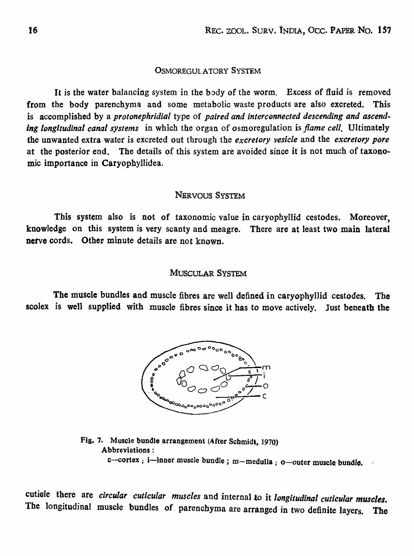
MUSCULAR SYSTEM
The muscle bundles and muscle fibres are well defined in caryophyllid cestodes. The scolex is well supplied with muscle fibres since it has to move actively. Just beneath the
Fig. 7. Muscle bundle arrangement (After Schmidt, 1970) Abbreviations:
c-cortex; i-inner muscle bundle; m-medulla; o-outer musclQ bundle. \
cutiole there are circular cuticular muscles and internal ~o it longitudinal cuticular muscles. The longitudinal muscle bundles of parenchyma are arranged in two definite layers. The

ItAPIEZULLAH: Caryophyllidean Cestode Fauna of India 17
,,.11" longitudinal muscle bundles are arranged in a definite ring and divide the cross-section of the body into inner medulla and outer cortex (Fig. 7). The outer longitudinal muscle bundles are also arranged in a definite layer and surround the cortical region. The arrangement of
c
1:»' c' a'
v
• 1
t
cL
Fig. 8.: Cross-sections illustrating inner longitudinal muscle arrangement for various families (dialrammatic).
Caryophyllaeidae: Capingentidae ~ a. Vitellaria anDular a" Vitellaria lateral
Lytocestidae : c. Vitellaria annular c'" Vitellaria lateral
Abbreviations :
b. Vitel1aria annular b' Vitellaria lateral
Balanotaeniidae : d. Vitellafia and testes cortical.
i-inner longitudinal muscle bundles, t - testes; v - vitellaria.
inner longitudinal muscles in relation to the distribution of vitellaria is of great taxonoroic importance, since family classification of caryophyllid cestode depends on this character. (Fig. 8).
CF 3

18 REC. ZOOL. SURV. INDIA, OCC. PAPER No. ls1
OUTLINE OF DEVELOPMENT AND LIFE CYCLE
Caryopbyllideans are characteristically small, long and narrow cestodes having no true holdfast structure (scolex) and with only one set of reproductive organs. Their body plan is monozoic. They parasitise the intestine of bottom feeding fresh water teleost fishes primarily belonging to the Orders Cypriniformes and Siluriformes, their intermediate hosts invariably being freshwater oligochaete annelids usually belonging to the family Tubificidae. The genus Archigetes is the sole exception whose species progenitically attain sexual maturity in the invertebrate hosts without involving vertebrate hosts in the life cycle.
The eggs (Fig. 1 b) are thin-shelled and operculate and have a single ovum and 3-5 vitelline cells. The operculum is very minute and is difficult to see in utero with the aid of a microscope. In most species the eggs are discharged in water unembryonated where their development takes place through embryonation and hexacanth embryos or onchospheres are formed, but in Archigetes, Djombangio, Wenyonia, Huntrella and Biacetabulum the embryonation starts and is completed in utero, and when the eggs are expelled out in water the oncbospheres have already been formed. A caryopbyllid onchosphere has been defined to be an embryo which develops in the egg with a nonci1iated membrance and three pairs of hooks, the middle one being the longest. The eggs with onchospheres are ingested by the tubificid intermediate hosts in whose body-cavity further development takes place. The hatching of eggs takes place due to the mechanical pressure applied by the strong stretching and contracting mo~~ments of the onchosphere on the operculum of the egg. The onchosphere develops into a procercoid larva which is characterised to be soft· bodied with a hold. fast structure (scolex), primordia of gonads and a cercomer having the six onchospheric hooks. Wardle and Mcleod (1952) hold that "The procercoid may be described as a solidbodied larva in which the onchospberic hooks are retained and in which the future holdfast has not yet differentiated-. Mackiewicz (1972) seems to disagree with this definition in so far as the differentiation of the holdfast (scolex) is concerned and gives his definition like this: "The infective procercoid is characterised by a cercomer containing the six hooks of the onchosphere, a scolex that does not invaginate and shows a high degree of differentiation that is characteristic of the particular genus and, except for an increase in size, is retained through to the sexually mature stage, and the rudiments of a single set of reproductive organs". This definition of procercoid larva seems to be very compr~hensive, complete and meaningful.
As mentioned earlier, a species of Archigetes can complete its life cycle progenitically in the coelome of an 0 Iigochaete annelid intermediate host itself but it has also been shown beyond any shadow of doubt that a benthic feeding fish may occasional1y be

IlAPE!zULLAH: Caryoph)'lIidean Ce$tode Fauna of India
ANNEUOUTEN
Sy YERTEBRATE HOST
Cyprinul carpio L
PROGENETIC DEVELOPMENT'
ProcerOlid dIYelopment
INVERTEBRATE HOST
Limn_Hul
fIof(meilterf
CI.pared,
£UI ma, embryonate in utero. Annelid and I,avid Archlletel die. T&!p8worm elilliberated in mud.
lntectwe OIKOIpher.
A
c
pott in,.IUon
Fi,. 9: Life cycle of Archigetes iowenS/, Calootioe, 1962 (Simpilfied after 'Mackiewic1;. 1972).
19

REC. ZOOL. SURV. INDIA, Oce. PAPER No. ··IS7
&y DEfiNITIVE HOST
Catostomus commerlonl Lac.
t G 62 day,
development ,..,1IiI .... iIle'I complete
cercomere
embqonic hooks
~:+--- vttellarlum
~--t"tls
Procercoid development
uterus
lonopor8
ovary
• ,~lum-....,cII~
[!GGEATlNI
IY
14 CIa,. Infective oncos"..,. non <fila'"
INTERMEDIA T£ HOlTS. Tubi',. templ.tonISouth""
T. tubit,. MUtI" JANNELIDA : OLiGOCHAlMJ
Fig. 10: Life cycle of Biacelobulum macrocephalum McCrae, 1962 (Simplified after Mackiewicz, 1972).
vlt ..... ee. nucteul

HAFEEZULLAH: Caryophyllidean Cestode Fauna of India 21
involved in the life cycle if the oligochaete annelid infected with procercoid larva is ingested before the development of the functional reproductive organs in the procercoid. Thus, in literature it has been indicated that invertebrate and vertebrate cycles of Archigetes iQwensis represent two physiological strains. Szidat (1937) believed that Archigetes is the annelid stage whereas Biacetahulum is the vertebrate stage.
It is generally accepted and there is no evidence to the contrary that the caryophyUid life cycles involve only the freshwater oligochaete annelids and no other invertebrates as the intermediate hosts.
Mackiewicz (1972) has suggested that at least three types of caryophyUid life cycles can be recognised. They are :
Progenetic type: The life cycle does not involve a vertebrate (fish) host and the procorcoid larva with its differentiated scolex and a cercomer having the six oncho .. spheric hooks develops functional gonads and thus becomes sexually mature in the body-cavity of the invertebrate fresh water oligochaete annelid itself. .Arehigetes siebold;, A. limnDdrili and A. iowensis are best examples of this type of life cycle. However, A. siebold; and A. iowensis can mature in vertebrate (fish) hosts also if the invertebrate hosts harbouring procercoid larvae are eaten by them before becoming progenetic.
Interrupted type: In this type of life cycle, the procercoid larva not only grows to a large size in the intermediate in vertebrate host, it is morphologically well developed
also but the functional sexual maturity remains checked and interrupted. To break this interruption and to bring it to functional sexual maturity a vertebrate (fish) is required, This happens in Caryophyllaeus spp.
Complete type: In this type, the procercoid grows to a large size in the oligochaete annelid with some primordia of germ cells. Further development will not take place in this invertebrate host. At this stage, the annelid harbouring the infective larva or the young procercoid is ingested by a fish host. In the intestine of this vertebrate, the Jtalted gr.owth gets going ahead and eventualIy sexual maturity is attained. This type of lif~ cycle is exemplified by Khawia sinensis and Caryophyllaeus latieeps.

22 REC. ZOQL. SURV. INDIA, OCC. PAPER No. 157
SYSTEMATIC ACCOUNT
Class CESTOIDEA Rudoiphi, 1808
Subclass CESTODA Carus, 1863
Order CAR YOPHYLLIDEA Beneden "in Carus, 1863.
Order Caryopbyllidea Beneden in Carus
1863. Caryopbyllidea Benedeo in Carus, Handb. Zoo/., 2: 422-600,
Small group of single-segmented cestodes without any trace of external or internal segmentation, with a single set of reproductive organs and having poorly or well defined scolex. Male and female pores separate, occasionally common, lying midventrally at varying positions; if separate, very close to each other. Body plan monozoic, zone of proliferation behind scolex being absent (unlike polyzoic cestodes), thus chains of progttids or segments not formed. Shelled embryo nonciliated and six-hooked.
Complete diagnosis of the Order is detailed below:
Diagnosis: Body unsegmented, elongate, foliate or oval. Scolex marked off froPt
rest of body or not; smooth or specialised with folds, shallow grooves, frills, crenulations, shallow bothria or loculi, acetabular suckers, apical disc or introvert etc. Neck formed or not. Male and female gonopores usually separate, rarely common, mid ventral at varying levels, usually in posterior part of body (in anterior half of body in Wen,,,nia Woodland, 1923 and posterior to ovarian commissure in Balanotaenia Joh'nsoo', 1924). Common genital atrium present or not. Testes preuterine, usually medullary (Cortical in Balanotaeniidae). Cirrus sac present immediately behind testes. Ovary; bilobed
(H-shaped, bow tie- or dumbbell-shaped, inverted A-shaped or U-sbaped); lobes 'folIicular~ connected by a transverse bridge called isthmus or commissure. Uterus much coiled" in median field, joining vagina terminally before opening outside as female pore just behind male pore, or male and female pores opening outside together as common gonopore. Vitellaria follicular, lateral or surrounding medulla; cortic'al, medullary or both. Eggs operculate. Embryo hexcanth. Freshwater oligochaeta annelids serving as intermediate hosts. Definitive hosts freshwater benthic feeding fishes; only genus Archigetes sexually maturing in coelome of a tubificid oligochaete (cercomer present); adults of this genus also occur in intestine of freshwater teleost fishes (cercomer- flPsent), specially cyprinids, "

HAl'I!ZULLAii: Caryophyliidean Cestode Fauna of India
Type family: Caryophyllaeidae Leuckart, 1878.
Other families: Lytocestidae Hunter, 1927
Capingentidae Hunter, 1930
Bolanotaeniidae Mackiewiez and Blair, 1978.
Key to families of Caryophyllidea [ After Mackiewicz and Blair (1978) ]
23
1. Testes and vitellaria in cortical parenchyma ; neither testes nor vitellaria internal to inner longitudinal muscles Balanotaeniidae Mackiewicz and Blair, 1978.
Testes and vitellaria in separate planes; testes, vitellaria, or both internal to inner longitudinal muscles •.. 2.
2. Vitellaria completely in cortical parenchyma ; inner longitudinal muscles separate medullary testes from corticaJ vitellaria Lytocestidae Hunter, 1927.
Vitellaria either completely in medullary parenchyma or partially in medullary and cortical parenchyma 3.
3. Vitellaria and testes in_ medullary parenchyma; inner longitudinal muscles external to vitellaria ..• Caryopbyllaeidae Leuckart, 1878.
Vitellaria partially in medullary and cortical parenchyma; inner longitudinal muscles between adjacent vitelline fallicles Capingentidae Hunter, 1930.
a
b
Fig. 11: Balanotaenia bancrofti Johnston, 1924 a. Entire Worm, b. Cross-section.

Roc. ZOOL. SURV. INDIA, Oce. PAPER No. 1 .. 51'
The family Balanotaeniidae (Figs. 11; 12) has not been reported so far from India~ region. It has been reported only from Australia and Papua New Guinea. The family has been characterised by cortical distribution of vitellaria and testes being external to inner longitudinal muscles, medullary ovary and gonopore slightly anterior, over or posterior to
a
1lHS~ .:A;lir-f-'- V T C
b
Fig. 12: Balallolaenia newguillsis Mackiewicz and Blair· 1978 ~
a. Entire worm, b. Cross-section.
ILM
v
ovarian commissure. following two species.
It includes only one genus Balanotaenia Johnston 1924 and the ,
B. bancrofti Johnston, 1924, in Tandanus tandanZif Mitchel Mithel (Siluriformes: Plotosidae), from Murray Bridge, Murray River, South Australia (Fig. 11).
B. newguinensis Mackiewicz and Blair, 1978, in Tandanu$ brevidorsalis (Gunther), from near Port Moresby, Brown River, Papua New Guinea (Fig. 12).

RAPEEZULLAH: Caryophyllidean Cestode Fauna of India rzs
Family I. CARYOPHYLLAEIDAE Leuckart, 1878
1878. Caryopbyllaeidae Leuckart, Z. wiss. Zool. Suppl., 30 : 593·606. 1922. Caryopbyllaeinae i'NybeIio, Goteburgs Kugl. Veteusk. Vitterh.-Samh. Handb., Fjardo
Puljdeo, 26: 1-228. 1927. Caryapbyllaeinae Hunter, J. Parasit., 14 ~ 16-26. 1927. Wenyoninae Hunter, J. Parasit., 14 : 16·26. 1952. Wenyonidae: Wardle and Meleod, The Zoology of Tapeworms, University of Minnesota
Press, Minneapolis, 780 pp.
Diagnosis: Vitellaria entirely medullary being always internal to and surrounded by inner longitudinal muscle layer, typically annularly arranged, occasionally lateral; gonopore(s) and ovary situated in last fourth of body except Wenyonia Woodland, 1923 in which gonopores are in anterior half of body and uterus with a coat of uterine glands. Ovary is in posterior half of body.
rrype genus: Caryopbyllaeus Mueller, 1787.
Key to Indian genera of Caryphyllaeidae
Scolex broad, poorly defined, un specialised, not marked off from rest of body; neck absent; testes few, arranged in two parallel longitudinal rows; uterine coils extending anterior to cirrus sac; postovarian vitelline follicles present
Paracaryophyllaeus Kulakovskaya, 1961.
Scolex oval, well defined, unspecialised, distinctly marked off from body; neck present, long narrow; testes numerous, strewn in medulla anterior to cirrus sac; uterine coils not extending anterior to cirrus sac; postovarian vitelline follicles absent ... Pseudocaryophyllaeus Gupta, 1961.
Genus 1. Paracaryophyllaeus Kulakovskaya
1961. Paracaryophyllaeus Kulakovskaya, Parazit. Sh., 20: 339-355.
Diagnosis: Scolex broad, smooth, unspecialised, not marked off from rest of body; neck absent; testes arranged in two longitudinal rows on each side in meduJIa restricted much posterior to anterior level of vitellaria; cirrus sac comparatively small; ovary H-shaped, medullary; uterine coils extending anterior to cirrus sac; genital pores separate; seminal receptacle present or absent; vitellaria external and anterior to medullary testes; postovarian set of viteUaria present. Parasites of cyprinoid fishes. Russia, India.
CF 4

26 REC. ZooL. SURV. INDIA, OCC. PAPER No. 1S1
Type species: P dubininae Kulakovskaya, 1961;. in Misgurnus angusticautiatus ; Russia.
Key to Indian species of Paracaryophyllaeur
Scolex slightly rounded and widened as compared to rest of body; testes 16-20 in number; postovarian region comparatively less extensive
P lepidocephali (Kundu, 1985)
Scolex truncated, not wider than re,st of body; testes 16-41 in number; postovarian region comparatively much extensive p. ostiobramensis
(Gupta and Parmer, 1984).
1. Paracaryophyllaeus lepidocephali (Kundu) n. comb. (Figs. 13, 14)
1985. Lytocestoides iepidocephali Kundu, Bull. zool. Surv. India, 7 (2-3) : 285.
Material examined: Host-Lepidocepha/us gun tea (Hamilton). Loacb, (Cypriniformes: Cobitidae) ; location-intestine; localities-Garapota and Canning Town (West Bengal) ; no. of specimens-7 + 1, on 5 + 1 slides; ZSI Regd. Nos. W. 7548/1 to W 7552/1.
Description: Body elongated, 4·77 long, 0·72 wide at ovarian region, slightly narrowing towards both ends. Scolex simple, smooth, unspecialised, slightly widened. Neck absent.
Testes globular, 16-20 in number, 0·14-0·19 in diameter, medullary, arranged in two longitudinal rows in front of cirrus sac (alignment may get disturbed during flattening), anterior extent remaining restricted to a level much posterior to anterior level of vitellaria. Vas deferens in loose coils in front of cirrus sac, surrounded by posteriormost testes. External seminal vericle absent. Cirrus sac comparatively small, globular or slightly oval, situated in anterior part of last third of body, enclosing much coiled ejaculatory duct, opening independently on midventral surface of body.
Ovary H-shaped, medullary; wings follicular, 0·38-0-45 long, connected by a narrow strip of isthmus. Uterus in lateral coils, extending almost up to or anterior to cirrus sac (in additional material from Canning Town). Seminal "receptacle absent. Vagina joining uterus near its distal end forming uterovaginal canal opening as female gonopore just behind maJe gonopore. Genital atrium undetectable. Shell gland complex behind ovarian isthmus. Vitel1ine follicles smaller than testes, extending from a little behind scolex to anterior horns of ovarian wings. Postovarian set of vitelline follicles present. Preovarian and postovarian vitellaria are connected by small and narrow follicles lateral to ovary_

IJAIrDzULLAH: Caryophyllidean Cestode Fauna of India
Bggs ova), 40-S0x 20 .. 30 llm.
27
Remarks: Kundu (1985) described the present species under the genus Lytocestoides Baylis, 1928. A re-examination of the material of this species and the study of additional
a b
Fig. 13: Paracaryophyllaeus /epidocepha/i (Kundu, 1985) a. Anterior half of body (After Kundu, 1985), b. Posterior half of body (Redrawn from holotype).
material (a broken specimen) from the type host Lepidocephalus guntea examined at Canning Town, West Bengal, reveal that the uterine coils extend almost up to level of anterior limit of cirrus sac in Kundu's material whereas they extend anterior to cirrus sac in the additional . broken material. The species cannot belong to the genus Lytocestoides because its scolex is slightly widened, far less number (16-20) of testes characteristically arranged in two longitudinal rows, remaining restricted much behind anterior level of vitellaria, and the uterus extending anterior to cirrus sac. Thus, the species fits more appropriately in the genus faracary,phyilaeul Kula~ovs~a)'a? 1961 rather tba.n in L)'tocestoide$,

28 REe. ZOOL. SURV. INDIA, Oce. PAPER NO. IS7
The type species Paracaryophyllaeus dubininae Kulaskovskaya, 1961 was also described from a cobitid fish from Russia, P. lepidocephali (Kundu, 1985) closely resembles the
a b
Pig. 14: Paracaryophyllaeus /epidocephali (Kundu, 1985) from additional material from Canning Town
a. Anterior half of body, b. Middle-third of body ..
type species. Apparently, there seem to be no pronounced morphological differences between t hem, except that in P. lepidocephali some vitel1ine follicles lateral to ovarian wings COQQect

HAPEIZULLAH: CarYDphyllidean Cestode Fauna of India 29
the preovorian and postovarian sets of vitellaria and the absence of seminal receptacle. The literature on P. dubi"inae could not be made available for adequate comparision.
The discovery of this species from India extends the distribution of the genus Paracaryophyllaeus from Palaearctic to Oriental region also.
DlstrlbutitJII: West Bengal: Garapota (District: North 24-Parganas), Canning Town (District : South 24 .. parganas).
2. ParacaryopbyDaeos ostiobramensis (Gupta and Sinha) n. comb.
(Fig. 15)
1984. Pllovitellarla ostiobramensis Gupta and Sinha, Indian J_ Helminth., 36 (1) : 73.
Material examined: Nil.
Host-OstiDbrama cotio (Hamilton), (Cypriniformes : Cyprinidae); location-intestine; locality-Lucknow (river Gomti); no. of specimens-9 mature + 6 immature, no transverse sections_
DescriptiDn: Body elongated, 6'87-9'66 long, 0-60-1-05 wide. Scolex undifferentiated from rest of body, poorly defined, truncated, unspecialised'(without bothria). Neck
absent.
Testes 16-41, in two longitudinal row (Alignment shown disturbed probably
during processing). ovoid, 0'06-0'19 in diameter, medullary, extending from much
behind (about 2'00-3'00) anterior level of vitellaria to cirrus sac. External seminal vesicle absent. Vas deferens a small straight duct in front of cirrus sac. Cirrus sac 0-01-0-46 x 0·35-0-4fi, ovoid, situated at 1,93-3-86 from posterior end of body, enclosing much convoluted and bell-shaped ejaculatory duct, opening as male gonopore on mid ventral surface_
Ovary H-sbaped, much anteriorly removed (1-22-2t 46) from posterior end of body; ovarian wings 0'35-0'75 long, connected by an isthmus_ Ootype and shell gland complex posterior to ovarian isthmus. Uterus coiled, first descending posteriorly to some distance, then ascending anteriorly up to almost anterior level of cirtus sac (as is evident from Fig. t Sb) and then passing posteriorly up to posterior end of cirrus sac joining vagina to form a short uterovaginal canal. Uterovaginal canal opening as female gODopore immediately behind male pore. Seminal receptacle not knowu. Vitenarium
fol1icular, follicles smaller than testes, entirely medullary surrounding testes, extending from some distance behind scolex to cirrus sac. Postovarian set of vitelline follioles present. Preovarian and postovarian vitel1aria connected laterally by a row of vitelline follicles,

30 REC. ZOOL. SURV. INDIA, OCC. PAPER No. IS7
Eggs oval, 30-49 X 35-48 lim.
Excretory vesicle Y -shaped; pore terminal.
Remarks: The genus Pliovitellarium Fischthal, 19S1 is characterised by having one dorsal and one ventral deep acetabulum-like bothria on the scolex, one common genital
-pore, presence of external seminal vesicle and uterus remaining restricted to posterior level of
a
b
Pig. 15: Paracaryophyl/eus .osteobramellsis (After Gupta and Sinha. 1984), a. Anterior part of body. b. Posterior part of body.
vitellaria. Judging from Figs. 1 Sa, b, male and female pores appear to be separate, external seminal vesicle is absent, uterus extends almost to anterior level of cirrus sac ~n~ tl1e testes ~ommenc~ from a level much behind anterior level of vitellaria.

HAPEEiULLAH: CarYophyllldean Cestode Fauna of India 31'
Moreover, there does not seem to be present a single bothrium on the scolex. In all probabilities it appears to be an artefact of fixation rather than a real acetabulum-like bothrium. Thus, the present species does not appear to be referrable to the genus Pllovltellarium Fiscthal, 1951. It fits more appropriately in the genus Paracaryophyllaeus Kulaskovskaya, 1961 which has an uDspecialised scolex not marked off from rest of body. two longitudinal rows of testes which commence from a level much behind anterior level of vitellaria, uterus extending almost up to anterior level of cirrus sac or even beyond and postovarian set of vitelline fol1icles.
Thus, there are two species of Paraceryophyllaeus from Ind ia: P. lepidocephali (Kundu, 1985) and p. ostiobramensis (Gupta and Parmar, 1984) both from cypriniformes fishes. The latter differs from the former in having truncated and not widened apex of scolex, more number (16-41) of testes and comparatively longer postovarian region of body.
Genus 2. Pseodocaryopbyllueos Gupta
1961. Pseudocaryophyl/aeus Gupta, Pr~c. helminth. Soc. Washington, 28 (1) 2 43. 1961. Capingentoides Gupta. Proc. helminth. Soc. Washington, 28 (1) : 46.
Diagnosis: Scolex oval or conical, truncated at apex or not, unspeciaJised, distinctly marked off from neck behind. Neck present, long, narrow. Testes in most of medullary parenchyma auterior to cirrus sac. External seminal vesicle absent. Ovary H-sbaped. Uterus not extending anterior to cirrus sac. Two separate genital pores. Preovarian vitellaria surrounding testes, internal to inner longitudinal muscles. Seminal receptacle absent. Postovarian vitelline follicles absent. Parasites of siluroid fishes, India.
Type species: Pseudocaryophyllaeus indica Gupta, 196J, India; in Clarias batrachus (L.) ; India.
3. Pseodocaryophyllaeus indica Gupta
(Figs. 16-20)
1961. Psendocaryophy/laens indica Gupta, Proc. helminth, Soc. Wash .• 28 (1) : 43. 1961. Capingenioides batrachii Gupta, Proc. helminth, Soc. Wash., 28 (I) : 46. 1984. Pseudocaryophyllaeus mackiewiczi Gupta, V. and Parmar, Indian J. Helminth., (1982). 34
(2): 136. 1984. Pseudocaryophyllaeus ritai Oupta~ V. and Singh. Indian J. Helminth., (1983),35 (1): 11. 1984. Pseudocaryophyllaeus lucknowensis Gupta and Sinha, Indian J. Helminth., 36 (1) : 73.

32 REe. ZOOL. SURV. INDIA, dct. PAPER No. is?
a
c b
Fig. 16: Pseudocaryophyllaeus indica (Aftc)r Gupta, 1961) a. Scolex and part of neck, b. Main body and part of neck, c. Cross-section of body.

"AJB!ZULLAH : CarYDphyliidean Cestode Fauna Df India 33
a
b
Fig. 17: Cap ingen to ides hatrachii (After Gupta. 1961) a. Scolex. b. Posterior balf of body, c. Cross-sec:tioD of body.
CF S

REC. ZOOL. SURV. INDIA, Oce. PAPER NO. IS7
Material examined: Nil_
Host-Clarias batrachus (L.), Walking cat-fish, (Siluriformes: Clariidae), Hetero .. pneustes fossilis (Hamilton), Stinging cat-fish, (Siluriformes: Heteropneustidae) and Rita rita (Hamilton), Rita (SiIuriformes: Bagridae); location-intestine; localitiesGuwahati (river Brahmaputra) ; Lucknow (river Gomti) and Gorakhpur (river Rapti) ; no.
of specimens-nutnerous_
Description: Body elongated, about 13,0-25 0 0 long, 0'65-1·35 in maximum width. Scolex oval or cone-shaped, truncated anteriorly, distinctly marked off from neck behind. Neck slender, long, 5-04·8-74 long, 0'175-0·365 wide_ Trunk or main body cylindrical,
7'48-14-96 long, posterior end rounded.
a b
Fig. 18: Pseudocaryophyllaeus mackiewiczi (After Gupta and Parmar. 1984) a. Scolex and part of neck, b. Posterior half of body.
Testes numerous, spherical to oval, 0·14-0'28 X 0'06-0-11, medullary, extending from a level behind anteriormost vitelline follicles to cirrus sac, surrounded by annular viteI ine follicles. Inner longitudinal muscle layer external to testes and vitellaria. Vas deferens oosely convoluted in front of cirrus sac. External seminal vesicle absent. Cirrus sac large,
oval, 0·43·0·61 X 0'26-0 0 41, enclos1bg bell-shaped ejaculatory duct, situated at 1-75 .. 2·98 f rom posterior end of body, opening mid ventrally by male gonopore.

ILt.FE!ZULLAH: Caryophyllidean Cestode Fauna of India 35
Ovary H-sbaped ; wings strongly follicular, lateral posterior ovarian follicles may not be extending up to posterior end of body, connected by medullary isthmus. Uterus in lateral coils in post- and pre-ovarian parts) with uterine gland cells, not extending beyond cirrus sac. Vagina a slightly convoluted tube running medianly on ventral side, terminally joining with uterus to form a short utero-vaginal canal. Utero-vaginal canal opening midventrally as female gonopore on ventral side just behind male gonopore. No common senita} atrium. No seminal receptacle. Vitellaria medullary, internal to inner longitudinal muscle layer, annularly surrounding testes, extending from posterior region of neck to anterior horns of ovarian wings, follicles occasionally continuing with follicles of ovary. Shell gland complex posterior to ovarian isthmus. Eggs oval, 50-60 X 35·45 Ilm.
Main osmoregulatory canals 4, 2 on each side, joining posteriorly forming a short tubular excretory vesicle , excretory pore terminal.
a
Fig. 19: PSludocaryophy/laeus rita; (After Gupta and Sinsh, 1984) a. Scolex and neck, b. Posterior balf of body.
Remarks: Mackiewicz (1981) has re-examined the whole m9unts and sections of Puw/ocaryophyllaeus indica Gupta, 1961 and Capingentoides batrachii Gupta, 1961, both from the cat-fish Clarias batrachus from the river Brahmaputra at Guwahati, ~ssam. He observes that the inner longitudinal muscle layer is clearly external to the testes and vitellaria ill P. Indlca~ an4 tllere are no postovarian vitelline f ol1icle~ in both the species. He
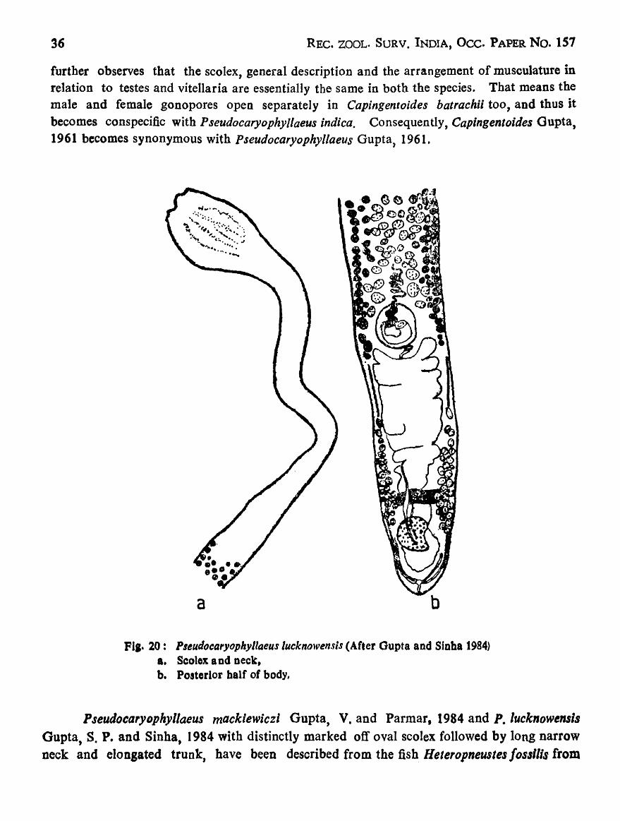
36 REC. ZOOL. SURV. INDIA, Occ. PAPER No. 157
further observes that the scolex, general description and the arrangement of musculature in relation to testes and vitellaria are essentially the same in both the species. That means the male and female gonopores open separately in Capingentoides batrachii too, and thus it becomes conspecific with P seudocaryophy/laeus indica. Consequently, Capingentoides Gupta, 1961 becomes synonymous with Pseudocaryophyllaeus Gupta, 1961,
a
Fla- 20: Pleudocaryophyllaeus lucknowensis (After Gupta and Sinha 1984) a. Scolex a nd neck, b. Posterior balf of body,
PseudocarYDphyllaeus mackiewiczi Gupta, V. and Parmar, 1984 and P. lucknowensls Gupta, S. P. and Sinha, 1984 with distinctly marked off oval scolex followed by long narrow neck and elongated trunk, have been described from the fish Heteropneustes /osIllis from

HAPEEZULLAH: Caryophyllidean Cestode Fauna of India 37
river Rapti at Gorakbpur and river Gomti at Lucknow respectively but without studying transverse sections. P. macklewlczi appears to be reported from very young specimens with very few testes (5-10 only). The two species have been differentiated from the type species P. indica Gupta, S. P. 1961 on the basis of variable characters which occur either due to age or state of contraction and relaxation at the time of fixation. Hence, both of them are also
considered as synonyms of Pseudocaryophyllaeus indica Gupta, S. P. 1961. Pseudocaryophyllaeus ritai Gupta, V. and Singh, 1984, has been described from the fishes Rita rita from Lucknow without studying the cross-sections. It also essentially resembles Pseudocaryphyllaeu3 indica Gupta, S. P, 1961 in scolex neck and general description. Therefore, it is also synonymised with P. indica.
Agarwal (1985), however, considers Pseudocaryphyllaeus indica Gupta, 1961 as belonging to the family Lytocestidae since on re-examination of the sections of this species he finds vitellaria cortical rather than medullary contray to the re-examination of the matelial by Mackiewicz (1981). However, till fresh information on muscle-vitellaria relationship becomes available in P. indica Gupta, 1961, it is tentatively kept in the family Caryophyllaeidae.
Family II. CAPINGENTIDAE Hunter
1929. Pseudolytocestinae Hunter, J. Parasit., 15: 185-192. 1930. Capiogentioae Huoter, Illinois bioi. Monogr. II, (1927), 186 pp. 1951. Capingentidae: Wardle .and McLeod. The Zoology o/Tapeworms. University of Minnesota
Press, Minneapolis, 780 pp.
Diagnosis: Resembling Lytocestidae in most characters. Vitellaria cortical (external to inner longitudinal muscles) only for one-third to one-half of their length while remainder lying in medulla (internal to inner longitudinal muscles) from where they
arise. Gonopores and ovary in last fifth of body. Uterine glands present.
f/'ype genus: Capingens Hunter, 1972.
Key to Indian genera~of Capingentidae
Scolex smooth, wide, truDcated or slightly convex, reduced; anterior extent of testicular field near anterior end. of body; ovary usually butterfly-or bowtie-shaped
Breviscolex Kulakovskaya, 1962. Scolex smooth, wide, slightly convex, not reduced; anterior extent of testicular field remaining restricted appreciably behind scolex; ovary shaped like an inverted A
,., ft· Adenosc,,/ex Fotedar, 1958.

38 REC. ZOOL. SURV. INDIA, OCC. PAPER No. IS7
Genus 3. Adenoscolex Fotedar
1958. Adenoscolex Fotedar, J. Helminth., 32 (1·2): 10. 1981. Adenoscolex: Mackiewicz, Himalayan J. Sci., 1 (1): 7.
Diagnosis: Scolex smooth, unspecilised, slightly widened, not marked off from rest of body. Neck absent. Male and female gonopore separate in posterior seventh of body length. External seminal vesicle absent; internal seminal vesicle present. Ovary entirely in medullary parenchyma, inverted A-shaped. Uterus not extending anterior to cirrus sac. Seminal receptacle present. Vitellaria mostly dumbbell-shaped, annularly arranged surrounding testes. Post-ovarian vitelline follicles present. Eggs with blunt protuberance near basal end. Parasites of cyprinid fishes.
India.
rI'ype species: Adenoscolex oreini Fotedar, 1958, in Oreinus sinuatus; Kashmir;
4. AdenoseoleJl oreiai Fotedar
(Fig. rll)
1958. Adenoscolex oreini Fotedar, J. Helminth •• 32 (1-2) : 10. 1981. Adenoscolex oreini : Mackiewicz, Himalayan J. Sci., 1 (1): 7. 1985. Adenoscolex oreini: Agarwal, Indian Rev. Life Sci., 5: 142.
Material examined: Nil.
Host-Oreinus sinuatns (Heckel), (Cyprinifonnes: Cyprinidae) J location-intestine; locality-Anantnag (Arapat stream), Kashmir; no. of specimens-several, with Transverse Sections.
Description: Body elongated, 38·00 in maximum lengtn, 2·0 in maximum width, anterior end truncated, posterior end blunt ; worms with testes and without eggs 18-0 X t ·3. Scolex smooth, unspecialised, slightly wider than body width, 1'0 X "leO, not marked off from body, profusely furnished with gland cells. Neck absent.
Testes ,. numerous, . ~ rounded or ,"oval, 0·15 .. 0·23 in diameter, larger than vitelline follicles, medullary, extending from a level some distance behind scolex to cirrus SaC. Vas deferens strongly coiled in front of cirrus sac. External seminal vesicle absent. Cirrus sac oval, 0·4-0·575 x 0·32S-0·375, enclosing internal seminal vesicle and ejaculatory duct, opening to exterjor on ventral surface as male gonopore.
Ovary basically H-shaped, with posterior ends of wings strongly bent inwards (but not fusing together) giving an appearance of lnv~~t~d 'A'~ win~s ~onn~cted by

8APIlZULLAH: CarYDphyllidean CestDde Pauna of india
a
b c
e' .' .... ",,-
;' ..... ) ..
Fig. 21: Adenoscolex ortin; (After Fotedar, 1958) a. Anterior par' of body, b. Posterior half of body, c. Cross-section of body, d. Testis, e. Vitelline follicles in cross-section.

40 REe. zooL. SURV. tNDIA, Occ. PAP£R No. 1s1
isthmus or commissure, entirely medullary, wings 1'9-2·2 long, 0'25-0'37 wide. Uterus well developed, compactly coiled behind ovarian isthmus, thrown in symmetrically transverse coils anterior to isthmus, posterior coils not surrounded by gland eells while anterior coils with thick coat of them, not extending beyond cirrus sac. Vagina almost a straight tube, ventral in disposition, joining terminally with uterus to form a short uterovaginal canal opening mid ventrally as female gonopore behind male gonopore. Common genital atrium absent. Seminal receptacle present anterior to ovarian isthmus. Shell gland complex well developed, situated behind isthmus.
Vitellarium follicular, foHicles irregular, dumbbel1~shaped, 0·075-0'105 in diameter, partly medullary and partIy cortical, annular in arrangement, extending from testicular level to posterior level of cirrus sac, then arranged loosely in a single row on each side of uterus terminating in front of anterior horns of ovarian wings. No vitelline follicles lateral to ovarian region. Postovarian set of vitelline fallicles present. Eggs ovoid, mature ones 64-7 S x 36-48 11m.
Remarks: The species was never reported again after its original report in 1958. It may be endemic restricted to the cypriniformes fishes of the hill streams of Kashmir. Fotedar (1958) has given a vivid and comprehensive account of this species.
Distribution: Anantnag (Kashmir); India.
Genus 4. Breviscole~:Kulakovskaya
1962. Breviscolex Kulakovskaya, Doklady Akademii Nauk SSSR, 143 : 386-388. 1986. Breviscolex Schmidt, eRe Handbook 0/ Tapeworm Identification, CRe Press. Inc Boca
RatoD, Florida .. 38.
Diagnosis: Scolex SDlooth, wide, truncated or slightly convex, reduced (very Short), not marked off from body behind. Neck absent. Male and Female gonopores separate. Common genital atrium apparently present in posterior part of middle third of body. Anterior level of testicular field near anterior end of body_ External seminal vesicle absent. Ovary usually butterfly- or bowtie-shaped. Uterus not extending anterior to cirrus sac. Seminal recepatacle present or not. Vitellaria extending from a little behind anterior testes to anterior ovarian wings, medial and lateral to testes. Postovarian vitellaria present. Parasites of cyprinid fishes. Russia, India.
Type species: B. orientalis Kulakovskaya, 1962 : in Hemlbarbus maculatus, ChiiogobiD czarskii; Arnur river basin, Siberia (Russia).

HAP!EZtJi..LAH: Ca,yophyllidean Cestode Fauna of India 41
Key to Indian species of Breviscolex
Number of testes 50-100 Number of testes 200-350
B. naldurgensis (Shinde et 01. 1987) n. comb. B. aurangabadensis (Shinde, 1970) n. comb.
s. Breviscolex auraogabadeosis (Shinde) n. comb.
(Figs. 22-24)
1970. Lytocestoides aurangabadensis Shinde. MarQlhwada Univ. J. Sci., 9 : 173. 1970. L. aurangabadens;s var. minor Shinde. Ibid, 9: 174. 1970. L. aurangabadensis var. minuta Shinde. Ibid, 9: 175. 1981. L. aurantabadensis: Mackiewicz, Himalayan J. Sci., 1 (1) : 6.
Material examined: Nil.
Host-Barbus kolus (Sykes), Lobe" calbasu (Hamilton), (Cypriniformes: Cyprinidae); location-intestine; localities-Godavari river at Paithan and Purna river (locality not given) in Maharashtra; no. of specimens-Six whole mounts; no cross-sections.
Description: Body elongated, 4·72-6· 5 long, 0·89-1·17 wide, slightly tapering posteriorly. Scolex reduced, wide, broadly rounded, unspecialised, broader than rest of body. Neck absent. Male and female gonopores separate on ventral surface in beginning of last third of body. Common genital atrium not known. Testes follicular, about 200-350 in number, 0·13-0-16 in diameter, medullary, distributed from base of scolex to cirrus sac. Vas deferens in front of cirrus sac. External and internal seminal vesicles not known. Cirrus sac 0-34-0·45 long, 0'21-0·28 wide, situated in begining of posterior third of body, enclosing '''inding ejaculatory duct and cirrus, opening ventrally as male gonopore.
Ovary bilobed, dumbbell-shaped, in posterior third of body ; ovarian lobes connected by short isthmus, 0·38-0·78 long, 0·17-0'53 wide. Uterine region short, not extending anterior to cirrus sac. Vagina joining uterus near its anterior end to form a short utero-vaginal canal opening midventrally as female pore slightly posterior to male pore. Shell gland complex behind ovarian isthmus. Seminal receptacle not known. Vitellaria annular, extending from anterior level of testes to ovary. Post-ovarian vitellaria present, follicular (originaUy described as testes). Pre- and post-ovarian vitelline follicles connected by vitellaria lateral to ovarian lobes.
Intrauterine eggs 50-60 x 30-32 11m.
CF 6

42 llftc. ZOoL. SuaVe IHDIA, OCC. PAPER No. 1S7
Remarks: Mackiewicz (1981) has examined the material of this species. He found that the specimens were "decomposed~' and ·compressed", and due to poor condition
Fi,. 22: Breviscolex Qurangabadensis (Shindo) Entire worm (After Shindo, 1970).
of the material he was unable to determine its genus. From Figs. 22, 23 & 24 it can be very easily judged that the posterior fon-shaped pseudostructure was formed and in the specimen of Fig. 23 the follicles of testes were pushed in the scolex region due to crushing only during processing. Mackiewicz (in a personal communication dated September 3,

HAPBB'ZULLAH: Carlophyilidean Cestode Fauna of India 43
1985) informed me that all testes are preovarian; there are no postovarian testes, instead there are post-ovarian vitelline follicles in the postovarian region. The follicular nature of
F-. 23' L·vtoc·stoides Qurangabadensis minor Shlnde I. • .., ~
Bntire worm. (After Shinde, 1970).
post ovarian vitellaria gives the clue that the t>reovarian vitellaria m\lst also be folUculaf
rathor tban ~anul.r,

44 REC. ZOOL. SURV. INDIA, Oce. PAPER No. 151
Shinde (1970) did not cut cross .. sections of the body due to which the position of the inner longitudinal muscle layer in relation to vitellaria is difficult to ascertain, thus inhibiting the determination of the family of the species. As the author has mentioned that the vitellaria are cor-tical he kept his species in tb~ family Lytocestidae f The original description
Pia. 24: Lytocestoides aurangDbad,,,,,, mtnuttl Sblade Botire worm (Aftor Shlnde, 1970).
of the species is not very reliable. The species may not belong to the family Lytocestidae and genus LylocestQides because its anterior part is not narrower than the rest of the body. On the contrary, the scolex is reduced, wide and broadly rounded and the body tapers graduaIly towards posterior end only rish t from wide scolex. In LytticestDldes the soole~ is

HAP'EIlZULLAH: Caryophyllidean Cestode Fauna of India 4S
narrow and conical, and body tapers on both sides. It is quite probable that the vitellaria may be partly internal to the inner longitudinal muscle layer surrounding testes and partly outside it. The characters like reduced and widened scolex, the body tapering gradually posteriorly right from anterior end, testes extending to near anterior end of body, presence of postovarian follicles of vitellaria and butterflly-shaped ovary suggest the genus BreviscDlex Kulaskovskaya, 1962 in the family Capingentidae. As is the case with the genus Breviscolex, the present species has also been recovered from a cyprinid fish. Attempt should be made to restudy it by collecting fresh material from the type host and locality. Till then the species LytDcestoidea aurangabadensis Shinde, 1970 and LytocestDides naldurgensis Shinde et al., 1987 are tentatively kept in the genus Breviscolex under the family Capingentidae.
6. Breviseolex naldorgensis (Shinde, Mohekar, Jadav, and Hafeezullah) n. comb. (Figs. 2S-26)
1987. Lytocestoides naldurgens;s Sbinde, Mobeker. Jadav and Hafeezullab. Bull, zool. Surv. India, 8 (1-3): 198.
1987. L. mackiewicz; Sbinde. Mobekar, Jadav and Hafeezullab, Ibid,8 (1-3): 199, (n. 81D.).
Material examined: Nil.
Host-Cirrhina mrigala (Hamilton), MrigaJ, (Cypriniformes: Cyprinidae); locationintestine; locality-Naldurg; (Dist. Osmanabad, Maharashtra); no. of specimens-10+ 15; no cross-sections.
Description: Body elongated, broad anteriorly, gradually tapering posteriorly, 4-77. 6-00 long, 1·00-1·55 wide. Scolex short, wide, broadly rounded unspecialised, 0·66 long, 1·50- t ·55 wide at base, sbape variable. Neck! absent. Male and female gonopores on ventral surface in anterior part of second half of body. No common genital atrium.
Testes 50-85 in number, 0-10-0-19 in diameter, in central medulla in a single field. extending from base of scolex to cirrus sac. Vas deferens in front of cirrus sac. External seminal vesicle absent. Cirrus sac small, about 0·30 in diameter, posterior to middle of body, enclosing ejaculatory duct and cirrus, opening as male pore ventrally.
Ovary dumbbell .. or butterfly-shaped, lobes connected by narrow isthmus, follicular, medullary, in posterior region of body. 0·33-0·53 long, 0·54-0·77 wide. Uterine region comparatively short, uterus glandular. not extending anterior to cirrus sac. Vagina joining uterus to form a sbort uterovaginal canal opening ventrally posterior to male pore. Seminal receptacle absent. Vitellaria follicular J follicles smaller tQan testes, Post-ovarian vitelline

46 REC. ZOOL. SURV. INDIA, OCC. PAPER No. lS1
follicles present. Pre- and post-ovarian vitelline follicles continuous lateral to ovary. Intrauterine egg numerous, 56-67 x 33-44 J.l.m.
Remarks: It is not clear from the study of Lytocestoides naldurgensls and L. macklewiczi whether the two species occurred in the same population or they were recovered sepapately. The former bas been differentiated from the latter in baving less number of testes (50-55 vs 80-85) and in that the lateral vitelline follicles are in a single row rather than two.
4)G-.I ..
: .~.-.. 0.- •• . -.o •• ,p_ •• _ '-~ ... -~ .-&- , ,.~ .. _,.l. ".- -. ~ .. -.. .
• QiIIIII' •••• .. .. .. -.- .. CP- 0' •• •• •• ~ -. ·0· •. (1fI
e
Fig. 2S: Breviscolex naldurgens;s (ShiDde II al.) Entire worm. (After ShlDdo It til., 1987)
These differences are considered mearge and not morphologically pronouced. These characters may be variable if large populations are studied. Therefore L. macklewi~z' has been considered as a synonym of L. naldurgnesis.
The comments regarding familly and genus allocations of this species as given under L. aurangabadensis are ap:pliQable in this case also t

BAPltzuu..AH: Caryophyilidean Cestode Fauna of India
D;Itrlbutllln: Naldurg (Maharashtra State).
::~~~ . •• ® ~ •• :: o,f..J ~ • • _ r:tIo •
• • 'Wi' JOn C C!!D • •• ®~~~ • . - ~~. ~ .. ::0 CU" :-
.- ~E!)I'(j!\ •• . - ~ ~ .. :. @ e> :: ::Oe~: - '-" ~ .. .. ~ . •• ®G!> • .. ~ . •• ~- r.- • • _ "II ~.
:: e G :-.e .(iJ) • •• @e-.. (i).r.:o. : :-.. ~ . .. c· .• e 0 Q:-........ ~ . •• 0. ~_
". . · · .. · ·
Fig. 26: Lytocestoides mackiewiczi Shinde et 01 .• Entire worm. (After Shinde et a/. J 1987).
Family III. L YTOCESTIDAE Hunter
1927. Lytocestinae Hunter J. Parasl., 14: 16·26.
47
1931. Bovieninae FuhrmaoD, In: Handbuch der Zoologie (W. Kukenthal and T. Krumbach, eds.), pp. 141-416.
19S2. Lytocestidae: Wardle and McLeod. The Zoology of Tapeworms. University of Minnesota Press. Minneapolis. 780 pp.
19S9. Lallidae Johri, Z. Parasitenkd., 19: 368-374. 1980. Djombanginae Salpute and Agarwal, Proc. Indian Acat. Parasit., 1: 13-16.
DIagnosis: Vitellaria cortical being external to inner longitudinal muscle layer, annuaJarly arranged around it and medullary testes in prea-uterine region, occasionally

REC. ZOOL. SURV. iNDIA; JOCC. PAPER :NO. lS?
lateral; gonopore and ovary usually in last quarter of body; ovary basically wing- or H-shaped, ovarian arms cortical while isthmus medul1ary ; uterine glands present.
Type genus: Lytoceslus Cohn, 1908.
Key to Indian genera of Lytocestidae
1. Post-ovarian vitellaria present Lytocestoides Baylis, 1928. Post-ovarian vitellaria absent .,. 2.
2. Scolex specialised with a terminal pseudobothrium or introvert ; uterine coils reaching nearly as far forward as testes Djombangia Bovien, 1926. Scolex un specialised ; uterine coils not reaching as far forward as testes ... 3.
3. Vitellaria lateral to testes in cross-section Vitellaria annular i. e. surrounding testes
Genus S. Lytocestus Cohn
Bovienia Fuhrmann, 1931. •.. Lytocestus Cohn, 1908.
1908. Lytocestus Cohn, Centra/b/. Bakteriol. Parasitenkd., 46: 134-139. 1961. Lucknowia Gupta, Proc. helminth. Soc. Washington, 28 (1) : 38.
Diagnosis: Scolex un specialised, distinct or not, not broader than rest of body. Neck formed or not. In cross-section through testicular zone, inner longitudinal muscle layer forming a ring around testes; outer longitudinal muscle layer also forming a ring internal to nuclear layer of sub-cuticula. Male and female gonopores on ventral surface closely one behind other. No common genital atrium. Longitudinal extent of uterus at most one-third that of testicular field, usually much less, not extending anterior to cirrus sac. External seminal vesicle absent. Ovary bilobed or H-shaped; ovarian follicles cortical,
only ovarian commissure and proximal portions of ducts being medullary. Uterine glands present. Vitellaria cortical, surronnding testes in testicular zone. Post-ovarian vitellaria absent. Serrinal receptacle present or not. Parasitic in Mormyriformes. Cypriniformes and Siluriformes. Hong Kong, Burma, India, Singapore, Mollucus, Sudan, Chad.
Type species: Lytocestus adha~rens Cohn, 1908 ; in Clarias /uscus ; Hong Kong.

BAPIlZULLAH: CarYDplayllidean CestDde Fauna of India 49
Key to Indian species of Lytocestus
1. Scolex knob-like, neck long and slender 2. Scolex stumpy, fiat or pointed; neck indistinct or a constrictioD, very short 3.
2. Ovary wing-like; posterior extent of vitellaria well beyond posterior to female gonopore; seminal receptacle absent ... L. birmanicus Lynsdale, 1956.
Ovary H-shaped; posterior extent of vitellaria restricted anterior to female gonopore ; seminal receptacle present ... L. iongicollis Rama Oevi, 1973.
3. Body robust; uterus well developed with very thick coat of gland cells; ovary wing-like with very wide wings L. indicus (Moghe, 1925).
Body not at all robust; uterus normally developed with only thin coat of gland cells; ovary H-sbaped with narrow wings ... 4.
4. Vitelline follicles three to four times smaller than size of testes; anterior half or third of body attennuated and usually filiform or ribbon-shaped; scolex variable in shape, usually narrow and pointed in front ... L. filiformis (Woodland, 1923).
Vitelline follicles only a little smaller than size of testes; anterior half of body flat, not filiform; scolex stumpy and bluntly rounded in front L. fossilisi
7. Lytocestus birmanicus Lynsdale
(Fig. 27)
1956. Lytoceslus birmanicus Lynsdale, J. Helminth., 30 (2-3) : 88.
(Gupta, 1961).
1988. Lytocestus birmanicus: Chakravarty and TandoD. Indian J. Helminth. (N. S.), S (1) : 43.
Material Examined: Two specimens loaned by Dr. V. TandoD.
Host-Claria, batrachus (L.), Walking catfish, (Siluriformes: Clariidae); locationintestine; locality-Guwabati (Assam) but collected from Shillong market (Meghalaya); no. of specimens-362; collected by R. Chakravarty and V. TandoD, Northeast Hill University, Shillong, Meghalaya.
Description: Body elongated, flattened, 5·28-16·36 long, 0'5-1·45 in maximum width at level of cirrus sac. Scolex globular to lanceolate, 0·46-1·18 long, unspecialised, disctinctly marked off from neck behind, Neck present, narrow, 1·32 .. 5·47 long, Anterior part of trunk (main body) devoid of any organs.
CF 7

so Roc. ZOOL. SURV. INDIA, OCC. PAPER No. 157
Testes 170-384 in number, med uIlary, oval or spherical, 0·06-0'21 x 0 ·02-0·11, extending from a short distance behind anterior viteUaria to cirrus sac. Cirrus sac oval, opening
as male gonopore in beginning of last 1/7tb of body.
a b
FII.27: Lylocestus birmanicus Lynldale. (Drawn from a specimen loaned by Dr. V. Tandon). a. Scolex and neck, b. Posterior half of body, c. Cross-section of body tbroulb tosticula, resion (After LYDsdale, 1956).

RAPBEZULLAH: Caryophyllidean CestDde Fauna 1)/ India 51
Ovary wing-like, near posterior end of body; wings cortical, follicular, connected by medullary band-like isthmus, 0·33-1·25 long, extending posteriorly to some distance short of posterior end of body. Shell gland complex posterior to ovarian isthmus, inconspicuously small. Uterus in a number of loose coils, glandular, extending up to cirrus sac. Vagina a straight tube, joining distalmost part of uterus to form utero-vaginal canal. Utero-vaginal canal opening immediately behind male gonopore. No seminal receptacle. Vitellarium follicular, follicles transversely elongated, 0·04-0-14 x 0·02-0·10, smaller than testes, cortical, annularly arranged outer to inner longitudinal muscle layer, extending posteriorly beyond level of utero-vaginal pore. Post-ovarian set of vitelline follicles absent. Intra-uterine eggs
oval, SO-70x 20-30 lim.
Remarks: Lytocestus birmanicus was originally described by Lynsdale (1956) from the intestine of the fish Clarias batrachus (L.) from Rangoon, Burma. Chakravartyand Tandon (1989) recorded it from the same host fish from Guwahati (Assam). Johri (1959)
considered Lytocestus a/estesl Lynsdale, 1956 from the fish host Alestes nurse (Ruppell) (Cypriniformes: Characinidae) from river Nile at Khartoum, Sudan, synonymous to LytCJcestus birman/cuI Lynsdale, 1956 from the fish host Clarias batrachus (L.) (Siluriformes: Clariidae) from Rangoon on the basis of similarities in the position of the genital apertures, ovary, extent of uterus and other structures including the size of eggs, disregarding the basic morphological differences in their scoleces and size and number of testes in them. In L. birmanicul the scolex is well defined, muscular, and knob-like distinctly marked off from the narrow, slender and long neck behind, while in L. aleltes; the scolex is basically ill-defined and ribbon-like with little trace of a constricted neck. According to Mackiewicz (1962), these scolex differences can not be regarded as artifacts of fixation but they represent basic morphological differences. Additionally, the number of testes in L. birmanicus is half as much as in L. a/estesi owing to smaller in size in tke latter. l'hus, both the species are quite distinct from each other.
The record of occurrence of this species from one and the same fish host in north ... eastern region of India, adjoining to the type locality Rangoon ( Burma ), is understandable.
Mackiewicz ( 1962), on re-examination ,counted 8S to 95 testes in L. birmanicus whereas Chakravarty and Tandon (1989) recorded 170-384 testes in it but they are comparatively smaller in size_ The posterior extent of vitellaria has been originally described to be at the level of uterovaginal pore whereas in Chakravarty and Tandon's fig. 9 the vitellaria has been shown to extend beyond that level posteriorly. Further, the follicles of vitellaria and ovary ha ve . been shown in the same figure not to meet each other whereas originally they have been shown to be continuous.
Distribution: Ransoon ( Burma ) and Guwahati ( India ).

52 REe. ZOOL. SURV. INDIA, OCC. PAPER No. 157
8. Lytocestos filiformis ( Woodland) Fuhrmann and Baer
( Fig. 28 )
1923. Caryophyllaeus /iliformis Woodland, Quart J. microsc. Sci. (N. S.), 67: 447. 1925. Lytocestus /ili/ormis: Fuhrmann and Baer, Proc. zool. Soc. London (1925) : 79-100. 1956. Lytocestus alestes; Lynsdale, J. Helminth., 30 (2-3) : 92. 1962. Lytocestus /iliformis: Mackiewicz, Rev. Suisse zool.~ 69 : 735.
Material examined: Two specimens loaned by Dr. V. Tandon.
Host-Clarias batrachus (L.), Walking catfish, (Siluriformes: Clariidae); locationintestine; locality-Guwahati ( Assam ) but collected from Shillong market (Meghalaya); no. of specimens-209 : collected by Chakravarty and V. Tandon, North-east Hill University, Shillong, Meghalaya.
Description: Body elongated, flattened, anterior half or third attenuated to a pointed apex, broadest in posterior half, hind end bluntly rounded, 5'94-33'00 long, 0'59-1'65 in maximum width in posterior half. Scolex smooth, not distinctly marked off from neck behind, variable in shape, may be flat or pointed. Neck present, long, slender, 1'98·12'54 long, gradually widening into trunk or main body_
Testes 232-532 in number, spherical or oval, 0·04 .. 0'14 x 0'01-0'08, medullary, extending from behind neck to cirrus sac. Vas deferns indistinct. Cirrus sac oval, in posterior part of body, enclosing coiled ductus ejaculatorius, opening on midventral body surface as male gonopore.
Ovary H- shaped; arms follicular, cortical, 0'53-1-5 long, connected by medullary band shaped isthmus, posterior horns extending to extreme posterior end of body leaving practically no post .. ovarian region, anterior horns reaching midlevel of uterine coils. Shell gland inconspicuously smal1, posterior to ovarian isthmus. Uterine region glandular, very short as compared to testicular region, extending much beyond anterior horns of ovary up to cirrus sac. Vagina joining near its distal end to form a short utero-vaginal canal, opening as female gonopore just behind male gonopore at about 0'73-2'1 from poeterior end of body. Genital atrium absent. Seminal receptacle absent. Vitellaria follicular, foHicles smaller (three to four times) than testes, 0-02-0-07 X 0'01-0-04, spherical or oval in shape, cortical, annularly arranged around testes, extending posteriorly up to anterior horns of ovary and becoming lateral only in region of anterior half of uterus. Post-ovarian vitelline follicles abs~nt. Bg~s ov~l, 30-~O x 10-30 ~m. Excretory pore terminal.

HAFEIZULLAH: CarlDphyllidean Cestode Fauna of India 53
Remarks: Lynsdale (1956) differentiated her species Lytocestus alestest from L,'fJCIstru fillformis (Woodland), both from river Nile, Sudan, mainly in egg sizes and different host fishes. Mackiewicz (1962),observes that "Eggs (in utero) vary in their size and
shape with respect to fixatives and their position in ~uterus." He further observes that the
a
. .. . .. • (§:!:) dD ~ •• •
. • ' f(:;1D'd1Jj .. .- .' • . .... ... ~ ,. ~~~ •. cJ:> ~ ~'f • .. ,,~.~ . • ~ #,-,..; .......
.. " • t::J,<:[S2.. ~ ,. • t/<:n-~' .... '
• • v~ .~ , .. '.' ( ~.' . • • • t . , . , .. ., .. • • • • • •
• • . , •• «
• • •
c Pig. 28: Lytocestus filiformis (Woodland) (Drawn from a specimen loaned by Dr. V. Tandon).
aAb. Scolex and Dock, c. Postorior half of body.
fish MDrm"w cashlve L. (Mormyriformes : Mormyridae), the host of L. fillfDrmis and the fish Alutes ,,",se (Ruppell) (Cypriniformes : Characinidae), the host of L. aleatesi, both
Gam.e from the same river NUe and region Su4an and both are bottom feeders~ On ~

54 REC. ZQOL. SURV. INDIA, OCC. PAPER No. IS1
comparasion of both the species, he concludes that "they have the same body conformations, scolex type, testes and vitellaria relationship, size and ovary type", meaning thereby that there are no pronounced morphological differences between them. Thus, L. alestesi Lynsdale, 1956 is a synonym of L. flli/ormis tWoodland, 1923).
Chakravarty and Tandon (1989) described that vitellaria "form a crescent around the testes", a character uncharacteristic of the genus Lytocestus. They are perhaps in .. advertently in error. Actually, the vitellaria form a ring around the testes. However, they
tend to become lateral in posttesticular region.
From the basic morphological characters like body conformation, scolex type, testes and vitellaria relationship, position of genital apertures, ovary type and extent of uterus, it appears that Chakravarty and Tandon (1989) have correctly identified theip specimens as Lytocestus filiformis (Woodland, 1923). This species has been originally described from the fish Mormyrus caschive L. (Mormyriformes: Mormyridae) from the river Nile in Sudan and later reported from Alestes nurse (Ruppell) (Cypriniformes: as Lytocestus alestest Lynsdale, 1956 from the same river and from the same region. Its record from an entirely different host Clarias batrachus L. (Siluriformes: Clariidae) and from a different zoogeographical region appears difficult to explain. However, its distribution is extended from Ethiopian region to Oriental region also.
9. Lytocestus fossilisi (Gupta)
(Figs. 29-33)
1861. Lucknowiafossilis; Gupta, S. P., Proc. helminth. Soc. Washington, 28 (1): 39. 1973. Caping~ntoides moghe; Pan de, Indian J. Zoot., 14 (3) : 223. 1975. Lytocestusfossilis Singb, Dr. B. S. Chauhan Comm, Vol., : 79 .. 82. 1984. Lytocestoides fossi/is Kanth, Sinha and Srivastava, Indian J. Helminth. (n. s.), 1 (1 .. 2) : 26. 1985. Capingentoides gorakhnathi Agarwal and Siogh, Indian J. Helminth. (n. s.), 2 (1-2): 81.
Material examined: Nil.
Host-Heteropneustes fossilis (Bloch) ; Stinging cat .. fish, (Siluriformes : Heteropneus
tidae), Clarias batrachus (L.), Walking cat-fish, (Siluriformes: Clariidae), Chan"a strlatus
(Bloch), Striped snakehead, (Channiormes: Channidae) ; location-intestine; localities-Lucknow (river Gomti). Gorakhpur, Bania (U.P.), Darbhanga (Bihar), and Kathmandu (river Bagmati, Nepal), no. of specimens-several mature and some immature worms; collected by respective authors.

~ULLAH: Caryophyllidean Cestode Fauna of India S5
Description: Body elongated flat 5'8-6'78 long 1'13-1'3 in maximum width ", , maiD body usuaUy tapering into neck and scolex. Scolex stumpy or slightly differentiated into an oval structure, anterior end bluntly rounded, narrowel.' than neck and much so than
a
c b
Fig. 29: Lytocestus fossiIisi (Gupta). (After Gupta, 1961). a& b. Scolex variations,
c. Posterior part of body.
main body, unspecialised, 0'348-0"59 long, 0"21-0'48 wide. Neck present, constricted. usually not so, 0'522-1'218 long, 0'365-0'73 wide at base. Male and female gonopores separate opening closely mid ventrally in last seventh of body length. No common genital
atrium.

REC. ZOOL. SURV. tNDIA, bee. PAPER No. 15'1
Testes rounded or oval, numerous, medullary, extending from a level posterior to anterior most vitelline follicles to posterior level of cirrus sac. Vas deferens convoluted
Fig. 30: Capingentoides moghe; Pandey Entire worm. (After Pandey 1973).
in front of cirrus sac. External seminal vesicle absent. Cirrus sac ovoid, large 0'34-0'43 long, 0·27 .. 0·31 wide, enclosing convoluted ejaculatory duct and cirrus, opening outside as male gonopore.
Ovary H-shaped, in posterior region of body; arms strongly follicular, lateral, cortical, connected by bandshaped medullary isthmus or commisure ; follicles of anterior horns mayor may not be continuous with those of vitellarium; follicles of posterior horns

.r£EZULLAH: Caryophyl/idean Cestode Faurta of India 57
mayor may not be intermingling near posterior end of body. Vitellaria follicular cortical annular (i.e. surrounding testes), follicles smaller than testes, extending fro~ a levei
b a
Fig. 31: Lytocestus fossilis Singh. (After Singh 1975) a. Anterior pard of body, b. Posterior part of body.
anterior to testicular level to anterior level of follicular ovary. No post-ovarian set of vitelline follicles. Uterus laterally coiled, post-commissural coils descending to excretory bladder, pre--commissural coils reaching sl ightly posterior to cirrus sac, never extending anterior to it. Uterine glands present. Vagina a narrow tube, mid ventral to uterus,
CF 8

sa ltEC. ZOOL. SURV. INDIA, Oce. PAPER No, IS'
JOIning anteriorly with uterus to form a short utero-vaginal canaJ, opening as female gonopore on ventral surface slightly behind male gonopore. No seminal receptacle. Shell
a
c b
Fig. 32: Capingentoides gorakhnathl Agarwal and SloSb 1985 (After Agarwal and Singh 1985).
a. Entire worm; b. Scolex. c. Posterior part of body.
gland complex large, on ventral side of ovarian commissure or posterior to it. Eggs oval, 17-18x 10·11~m, unfUamented.

IlAPlmzULLAH: CarYDphyllidean Cestode Fauna of India S9
Remarks: The species has been described from whole mounts. Transverse sections were not cut. Mackiewicz ( 1981 b) has re-examined the material. According to him the ovary is H-shaped (instead of band-shaped) whose lateral arms are strongly follicular and anteriorly extend up to posterior level of vitellaria and posteriorly reach
a b
FiS. 33: Lytoceslodies fossilis Kaoth et al. (After Kloth et al., 1984). a. Anterior part of body, b. Posterior-part of body.
near posterior end of body. There are no postovarian vitelline follicles. These observations obviously indicate that anterior follicles of the ovary may be continuous with those of vitellaria and posterior ovarian follicles were mistook as vitelline follicles. Mackiewicz (1981 b) further observes that jntrauterine eggs are nonfilamentous and the vitelline follicles are annular (i.e. surrounding testes). He has cautioned that "'the systematic status of this genus (Lucknowia Gupta, 1961) should be re-examined in the light of the above observations".
Lack of transverse sections of LucknDwia !Dssilisi Gupta, 1961 makes it difficult to know the relationship of vitelline glands with respect to inner longitudinal muscles

60 REC. ZOOL. SURV. INDIA, OCC. PAPER No. lS7
and hence the determination of its rami ly also becomes difficult. However, annular and cortical vitellaria suggest the family Lytocestidae for the genus Lucknowia as Gupta (1961) has already done. Regarding the status of the genus Lucknowia in the family Lytocestidae, one has to take into account the redescription of fossills; Gupta, 1961 as presented above. The characters like unspecialised scolex which is not broader than remainder of body, cortical and annular prevarian vitellaria, absence of postovarian vitelline follicles, separate (although closely apposed) male and female genital pores, absence of common genital atrium, uterine coils not extending anterior to cirrus sac and presence of uterine gland cells suggest the genus Lytoces.tus Cohn, 1908. Thus, Lucknowla Gupta, 1961 falls as a synonym of Lytocestus Cohn, 1908 and consequently, the species becomes Lytocestus fossilisl (Gupta, 1961) D. comb.
Transverse sections of all the species in the synonymic list were not studied but their bodies are similar consisting of usually a stumpy scolex distinctly narrower than the following short neck and a widened and long main body. All of them have been recovered mainly from Heteropneustes ,fossilis except Capingentoides gorakhnathl which is from Clarias batrachus and Caplngentoides moghel Pande, 1973 which has been described from an accidential host Channa slriatus. They all look identical to Lytocestus fossilist (Gupta, 1961) being essentially the same as redescribed above.
Distribution: Uttar Pradesh: Lucknow, Gorakhpur and BaHia; Bihar: Darbhanga J Nepal: Kathmandu.
10. Lytocestos indicDs (Moghe)
(Fig. 34-3S)
1925. Caryophyl/aeus indicus Moghe, Parasitology 17 : 222. 1926. Lytocestus indicus : Woodland, Proc. zool. Soc. London, 1926 : 56. 1930. Lytocestus indlcus : Mebra, Proc. 17th Indian Sci. Congr. Ass. (Abstracts) : 241. 1931. Lytoceslus Indicus: Moghe, Parasitology, 23: 84. 1961. Pseudolyloceslus c/ar/ae Gupta, Proc. helmint", Soc. Washington, 28 (1) : 43. 1981b. Pseudolytoestus clar/ae: Mackiewicz, Himalayan J. Sci., 1 (1) : 7. 1985. Pseudolylocestus clariae: Agarwal, Indian Rev. Life Sci., 5 : 145. 1988. Lytocestus marathwadaens/s Shinde and Phad, Riv. Parasslt., (1986), 47 (2): 295. 1989. Introvertus chauhani Pandey and Tiwari, Dr. B. S. Chauhan Comm. Yol. : 141. 1990. Pseudolytocestus thapari Gupta and Parmar, Indian J. Helminth., 42 (1): 28.
Material examined: Host-C/arias batrachus (L.), Walking Cat-fish, (Cypriniformes: Clariidae); location-intestine; localities-Guwahati Diamand Harbour Calcutta t , ,. , number of specimens-numerous,

HAP'EEZULLAH: Caryophyllidean Cestode Fauna of India 61
Description: Body robust, elongated, flat, 27 -0-40·0 long, 2·05-4·4 in maximum width. Scolex stumpy, bluntly rounded anteriorly, much narrower than rest of body,
b
Fia. 34: Lytocestus indicus (Moghe). 8. Entire worm, (After Moghe. 1931). b. Posterior half of body. (After Rama Devi, 1973), c~ Cross section of body. (After Mogbe,1931).
unspecialised (i.e. bothria, introvert, furrows or grooves absent), 2-65-3-0 long, 1-3 wide in middle. Neck present, a constriction or very short and indistinct. Main body slishtly

62 REe. ZOOL. SuaVe INDIA, OCC. PAPER No. \57
tapering posteriorly with rounded end. Male and female gonopores mid .. ventral in last seventh or eight of body length. Genital atrium absent.
Testes about 212-600 in number, 0'13-0'19 in diameter, medutIary, extending from a short distance behind anterior most vitelline follicles to cirrus sac. Vas deferens a much
b Fig. 3SA: Pseudolytocestus clariae Gupta. (After Gupta, 1961).
a. Scolex, b. Posterior part of body.
convoluted narrow tube in front of cirrus sac. External and internal seminal vesicles absent. Cirrus sac prominent, large with thick muscular wall, oval or conical bell-shaped, 0'50·1'04 long, 0'6-0'7 wide, enclosing a coiled and muscular ejaculatory duct, opening as male pore by a slit-like aperture on ventral surface of body,
Ovary bilobed, wing-like, in posterior part of body; lobes cortical, irregular in shape, consisting of numerous loosely packed follicles, connected by medullary bagpipe-1 ilte transverse isthmus; shell sland complex posterior to ovarian isthmus. Vasina.

HAFEEZULLAH: CarYDphyllidean Cestode Fauna 0/ india
narrow and slightly wavy tube, running ventrally along midline, dialating at anterior end. No seminal receptacle. Uterus with thick coat of glandular cells, thrown in lateral coils, extending posteriorly beyond shell gland complex, anteriorly opening into dialated terminal part of vagina to form a short utero-vaginal canal. Utero-vaginal canal opening as female pore a bit posterior to male pore. Vitellaria follicular, cortical, follicles surrounding
Fig. 3SB: Introvertus chauhani Pandey and TiwarI. Entire worm. (After Pandey and Tiwari, 1989).
testes, smaller than latter in size, extending from behind neck to almost level of uterovaginal pore, remaining restricted much anteriorly from ovarian arms. No post-ovarian vitelline follicles. Intra-uterine eggs ova), 60-88 X 20-44 ~m, embryonated.

64 REC. ZOOLi SURV. tNDIA, OCC. PAPER No. IS7
Osmoregulatory canals 8 pairs (2 marginal, 2 central and 4 median), joining to form excretory vesicle near posterior end; pore terminal.
Remarks: Moghe (1925) obtained this species from the stomach of the siluroid fish Clarias batrachus. He (1931) further mentioned that the worms appear as small knobs attached to the rugose wall of the stomach. Rama Devi (1973) mentions that "Mature and gravid worms make tunnels through the mucous and epithelial layers of intestinal wall and finally are to be found in the serose layer as a white speck.- The worms are obtained by rupturing intestinal wall. Niyogi, Gupta and Agarwal (1984) observe that it is a tissue parasite which peneterates through the mucosa to various depths in intestinal wall. Niyogi and Agarwal (1989) observe that Lytocestus indicus peneterates "through the mucosa and submucosa to lie close to musculosa, exceptionally reaching the serosa but never perforating it". Lytocestus indicus is normally a duodenal parasite of Clarias batrachus which is not reported from any other siluroid fish as yet. It occurs in single infection or with some other caryophyllid. It is found almost round the year.
Mackiewiez (1981b) persoqaIly examined the single specimen of Pseudolytocestus clariae Gupta, 1961 described from the fish Clarias batrachus from Brahmaputra river at Guwahati, Assam. He observes that the specimen is contracted, the testes are more than twice the size of the vitelline follicles and the ovary is foHicuIar, not compact as drawn. No sections were cut by Gupta (1961). He further points out that the genus Pseudolytocestus Hunter, 1927 is found in the Nearctic region in the fish family Catostomidae, and its report from the oriental region from a divergent fish family Clariidae is unexplainable. Agarwal (1985) considers Pseudolytocestus clariae a possible synonym of Lytocestus indicus. On the basis of nature (stumpy) of scolex, absence of post-ovarian set of vitelline follicles, testes larger in size than vitelline follicles and wing-like or bowtieshaped follicular ovary (not H-shaped i.e. elongated and narrow wings with jsthmus in almost Dlid-region of ovarian wings as is 'found in Pseudolytocestus) in Pseudolytocestul clariae, I am inclined to concur with Agarwal (1985). PseudDlytocestus thapari Gupta, V. and Parmar, 1990 from Clarias batrachus from river Gomati at Lucknow is identical to Pseudolytocestus clariae except a slight variation in the anterior extent of uterine coils.
Pandey and Tiwari (1989) described lntrovertus chauhani from the intestine of the fish Clarias batrachus from Faizabad (U.P.). Quite obviously, it is a case of misidentification and erroneous description of the material. The species is nothing but Lytocestus indicus (Mogbe).
Sinha and Rashid-un-Nisa (1981), Chakravarty and Tandon (1989) and Hasnian and Sabay (1989) have reported morphological anomaly involving duplication of body in this species. Ahmad and Sanaullah (1977) reported its occurrence in districts Bogra (Sherpur), Dacca (Demra, Maridder beel and Tongi), Mymensingh (Oauripur), Noakhali (Torabganj,

J.JAPEBZULLAH: Caryophyllidean Cestode Pauna of India 6S
Ramgati), Rangpur (Vakir beel, Gaibandha) and Sylhet (Shalutaker beel) from Bangladesh.
Distribution: Nagpur (Maharashtra), Raipur (Madhya Pradesh), Lucknow, Mainpuri and Faizabad (U. P.), Visakhapatnam and Hyderabad (A. p.), Guwahati (Assam), lamshedpur (Bihar), widely found in West Bengal, and districts Bogra, Dacca, Mymensingh, Noakhali, Rangpur and Sylhet (Bangladesh).
11. Lytocestus longicollis Rama Devi
(Fig. 36)
1973. Lytocesills longicollis Rama Devi, J. Helminth., 47 (4): 416. 1980. Lyloceslus longicol/is: Sbinde and Desbmukh, Riv. Parassit., 41 (2) : 211. 1989. Lytocestus longicollis: Cbakravarty and Tandoo, Indian J. Helminth. (N. S.), 5 (1) : 46.
Material examined: One without scolex, loaned by Dr. Veena Tandon.
Host: Clarias batrachus (L.) Walking cat-fish, (Family Clariidae) ; location-intestine; localities-Visakhapatnam district, Nagpur and Guwahati ; no. of specimens-173 + 1+141.
Description: Body long, narrow, 10'S-20·0 long, 0·5-0'84 on maximum width. Scolex unspecialised, variable in shape, spatulate or oblong or swollen pear-shaped, 0·94-1·78 long, 0·48-0·97 wide. Neck present, narrow, long, S·36-7·6x 00 17-0-51. Trunk or main body tapering posteriorly, 4 0 48-9.5 long. Male and female gonopores opening on ventral surface in last tenth or eleventh of entire body length. Common genital atrium absent.
Testes spherical or broadly oval, 0-10-0·16 in dialneter, 105-140 in number, medullary, extending from base of neck to cirrus sac. Vas deferens much convoluted. External and internal seminal vesicles absent. Cirrus sac rounded or oval, 0'24-0·31 long, 0·16-0·23 wide, enclosing long and convoluted ductus ejaculatorius, opening midventraUy as male pore.
Ovary H-sbaped, situated in posterior region of body, lobes cortical, ovarian isthmus medullary, lobes 0·46-0, 78 in length, closely packed with follicles. Uterus glandular, thrown in lateral coils, extending upto posterior level of cirrus sac. Vagina a narrow tube, straight or slightly convoluted, joining uterus to form a short utero-vaginal canal ; utero-vaginal canal opening as female pore slightly posterior to male pore ventrally. Receptaculum seminis oval, 0-16 X 0·06, anterior to ovarian isthmus. Shell gland complex and common vitelline duct posterior to isthmus. Vitellarium follicular, cortical, surrounding testes, extending from base of neck to anterior tip of ovary, follicles measuring 0'039- 0-07 in diameter. Post" ovarian set of vitelline follicles absent. Follicles of vitellarium and those of ovarian lobes mayor may not be continuous_
CF 9

66 lEe. t.OOL. SURV. tNDIA, OCC. PAPER No. IS7
a
c
d e
Pig. 36: Lytocestus longlcollis Rama Devi (Aftor Rama Devi, 1973,. a. Scolox and neck, b. Main body. c. Soolex enlarged, d. POlterior balf of body I ~. Croll-aectioD of body.

HAFIEZULLAH: Caryophyllidean Cestode Fauna of India
Eggs oval, 92-109 x 42-55 l1m.
67
Osmoregulatory canals 4 to 5 paas, uniting posteriorly to form small excretory vesicle; excretory pore terminal.
Remarks: Lytocestus longicol/is is characterised by a long, narrow and delicate neck and presence of a seminal receptacle anterior to isthmus. Shinde and Desbmukh (1980) and Chakravartyand Tandon (1989) also reported it from the same host from Nagpur and Guwahati respectively without studying cross-sections.
The present author studied a whole mount of the species by the courtesy of Dr. Veena Tandon. The flattening and staining are excellent, so much as the distributions of testes and vitelline follicles are very clearly and distinctly seen. This author finds that vitelJine follicles are cortical and lateral beyond any shadow of doubt, although Chakravarty and Tandon (1989) describe ·Vitellaria cortical, in a ring around the testes". r~o follicles of vitellaria are present medial1y. In that case their material could be Bovienia serlalis (Fuhrmann, 1931) only. It is quite possible that they might be having a mixed populations of Lytoceatus longicollis and Bovienia serialis.
Distribution: Visakhapatnam district (A. p.), Nagpur (Maharashtra) and Guwahati
(Assam).
Genus 6. DjombaDgia Bovien
1926. Djombangia Bovieo, Vidensk. Medd. Dansk naturh. Foren., 82: 167-181.
Diagnosis: Scolex globular or roughly· triangular, with a terminal introvert (no sucker or loculi), profusely provided with glandular cells, specially concentrated below apex in circular or semilunar fasbion. Male and female gonopores separate. Common genita~ atrium present on ventral surface near posterior end of body. Testes in lateral medulla. from neck to ovary. External seminal vesicle absent. Ovary bilobed connected by commissure or isthmus, entirely medullary. Uterus in median medul1a, extending as far as anterior level of testes or vitellaria, turning back and joining vagina immediately posterior to cirrus sac to form utero-vaginal canal. Vitellaria entirely cortical, lateral. No post-ovarian vitellaria. Receptaculum seminis absent. Eggs small, smooth, embryonated.
Parasitic in siluroid fishes. Java, India.
Type species: Djombangia Penetrans Bovien, 1926, in Clarlaa batrachus; Brantas river
near DJombang in East lava,

68 REC. ZOOL. SURV. INDIA, OCC. PAPER No. 157
12. Djombangia penetrans Bavien
(Fig. 37)
1926. Djombangia penetrans Bovien, Vidensk. Medd. Dansk Naturh. Foren., 82: 167-181, 1974. Djombangia indica Satpute and Agarwal, Indian J. Expt/. Bioi., 12 (4) : 373. 1977. Djombangia cabal/ero; Sabay and Sabay, Excerta Parasit%gica en memoria del doctor
Eduardo Cabal/ero Y CabelJero. Instituto de Biologia Publicaciones Especials 4. Maxico : 374. 1980. Djombangia indica: Satpute and Agarwal, Proc. Indian Acad. ParasUol., 1 (2): 13. 1985. Djombangia c/ariae Kundu, Bhattacharya anb Datta, Bull. zoot. Surv. India, 7 (2-3) : lSI.
Material examined: Host-Clarias batrachus (L.), Walking cat-fish, (Siluriformes: Clariidae), Hetoropnewtes fossilis (Bloch), Stinging cat-fish, (Siluriformes: Heteropneustidae); location-duodenum; localities-Bongaon and MaIda (West Bengal); no. of specimens-l +4, on 5 slides; col1ected by D. K. Kundu and party on 23.5.79 and 20.3.84 respectively, Z.S.I. Reg. Nos. W 7476/1 and W 7610/1-W 7613/1 ; six specimens on 3 slides from Nagpur (Maharashtra) collected by M. HafeezuUah and party on 25.7.1991; 2 specimens from Guwahati loaned by Dr. V. Tandon.
Description: Body milky white when freshly recovered from host, divisib~e into scolex, neck and flat trunk measuring about 9·00-1S·00 long, 3'50-5'50 wide (body proper or trunk about 7'5-11'S mm long), posterior end broadly rounded. Segment thick with deep denticulations due to contraction after treatment with fixative. Scolex 0·533-1'033 long. 0.88-1 0788 wide, globular or roughly triangular, distinctly marked off from rest of body, with a terminal introvert (without sucker or loculi), profusely provided with glandular cells below entire margin, speciany concentrated below apex in semilunar or circular fashion without association of any muscle fibers and staining dark giving false impression of feeble sucker. Neck narrow. distinctly separating scolex from trunk, short or long depending upon state of contraction or relaxation when fixed, usually 0'602-1·247 long, O'S 16-00 86 wide. Main body or trunk flattened; flesby, some anterior most space free from any system organs.
Testes numerous, rounded or ovoid. 0'103-0'165 in diameter, larger than vitelline follicles, strewen in lateral medulla, extending from some distance behind neck to ovarian lobes. Vas deferens tubular, narrow, coiled. External and internal seminal vesicles absent. Cirrus sac globular or oval, 0·731-1·243 long, 0'516-0·721 wide, distinctly discernible in front of ovarian isthmus near posterior end of body i enclosing winding ejaculatory duct, opening as male gonopore in common genital atrium on ventral surface.
Ovary dumbbell-sbaped, bilobed; lobes 0·70S·0·962 long, 0·344-0·S59 wide, follicular, connected by a strip of isthmus 0·516-0·729 acros~~ situated in :posterior part

BAFEIZULLAH: CarYDphyllidean Cestode Fauna of India
b
e
~.:52i;~ ' .. , .... ".-"" . ) ......... .
f
a 9
Fig. 37: Djombangia penetrens Bovien. a. Entire: worm, (After Satpute and Agarwal, 1980),
b. c. d. Scolex shapes, e. Cross-section of scolex showins sland COllI,
f. Cross-section of body J
8. E,g with onchol~here,
69

70 REC. ZOOL. SURV. INDIA, OCC. PAPER No. Is1
of body in medullary zone. Ootype and Mehlis' gland complex posterior to ovarian isthmus. Uterus extending in median medulla to almost anterior level of testes, turning back and joining vagina immediately posterior to cirrus sac to form a short utero-vaginal canal. Uterovaginal canal opening as a female pore in common genital atrium behind male pore ventrally. Vagina a long cana1, connecting oviduct bebind isthmus and utero-vaginal canal. Seminal receptacle absent. Vitellaria spherical to ovoid, 0·86-0-132 in dianleter, smaller than testes. cortical, extending from about anterior level of testes to ovarian lobes. Post-ovarian set of vitelline follicles absent.
Eggs thin-sheUed, 68-94x 43-51 11m, smooth (no spines) embryonated; onchasphere with three pairs of hooks, middle one being largest.
Remarks: Bovien (1926) described Djombangia penetrans to possess a small terminal sucker on the scolex. Fuhrmann (1931) checked and found it to be a glandular structure instead of a true sucker. Mackiewicz (1972) verified and found Fuhrmann's study to be correct. Satpute and Agarwal (1974, 1980) described a feeble sucker at the tip of scolex in their species Djombangia indica which is obviously a misinterpretation of congregation of glandular cells there. Sahay and Sahay (1977) found a groove or slitlike invagination in apical region of scolex, but the Bongaon, Maida (West Bengal) and Nagpur (Maharashtra) materials show that such an invagination (a pseudostructure) may be an artifact of fixation. Bovien (1926) mentioned that the eggs of D. penetrans have fine spines on the surface. Loser (1965) states that they are formed due to the deposition of the secretion of the glandular part of the uterus during the passage of the eggs through it. Sahay and Sahay (1977) found the surface of the egg~ in their species D. caballero; from Hetero .. pneusles fossilis from Chhotanagpur (Bihar State) as smooth (definitely unspined). Satpute and Agarwal (1974) mentioned tbe eggs in their species D. indica as smooth and without fine spines but when they (1980) published the detailed description of their species from the same material they stated the eggs to be having 3 f.t long spines. Kundu et. at. (1985) also did not find minute spines on the eggs of their species D. clariae. Kundu's ~aterial from MaIda also does not have spines on the eggs. Chakravarty and Tandon (1985) mention the presence of fine spines on the eggs of their material of D. penetTans from Guwahati (Assam State). Mackiewicz (1972) observes that opercles of caryophyllid eggs (measuring 9-16 p, in various species) are difficult to see even with the best optical equipments when they are in utero. That is the reason of confusion that some authors described the caryophyllid eggs as non-operculate while others mention them as operculate. So is also the case with fine egg spines measuring 3 II long only. Therefore, the presence or absence of egg spines in Djombangia is interpretational and disputed. Consequently such characters should not be taken into consideration for distinguishing species. Mackiewicz (1972) further observes that seminal vesicle is a transient structure which may be seen when 1Ule4 with sperms but it ma~ not be seen when exhausted of them,

NAPEEiULLAH: Caryophyllidean Cestode Fauna oj India 71
Disregarding the consideration of the above mentioned structures which are in nature interpretational, disputed, transient or variable, D. ceballeroi, D. indlcus and D. clariae are not morphologically distinguishable from the type species D. penetfans. Thus, the former three species were considered as synonyms of D. penetrans Bovien, 1926 by Hafeezullah (1986), and the same position is maintained.
DistributiDn : East Java; India: Assam, W. Bengal, Bihar, Maharashtra and Madhya Pradesh.
Genus 7. Bovienia Fuhrmann
1931. Bovienia Fuhrmann, Handbuch der Zo%gie (W. Kukentbal and T. Krumbanch, eds.) Bd. 2: 141-416.
1963. Crescentovitus Murhar, Parasitology, S3 : 414. (n. syn.). 1980. Introvertus Satpute and Agarwal, Proc. Indian Acad. Parasitol., 1 (2) : 17 (n. syn.).
Diagnosis: Scolex variable in shape, elongated conical, truncated at anterior end or slightly swollen, usually poorly defined, not wider than main body. Main body gradually tapering into scolex. Neck presents long. Male and female gonopores separate, closely apposed, in last quarter of body on ventral surface. No common genital atrium. Testes medullary, surrounded by a ring of internal longitudinal muscles. External seminal vesicle absent. Cirrus unspined. Pre-ovarian vitelline follicles cortical, lateral (in a crescent on each side in cross-section), extending posteriorly to anterior horns of ovarian arms or short of that level. No post-ovarian vitellaria. Ovary basically H-shaped, occasionally posteriormost follicles from posterior ovarian horns intermingling so as to give it a shape like inverted A; ovarian arms cortical connected by medullary isthmus, extending up to midlevel of pre-ovarian uterine extent. Uterine extent more than twice to four and a half times longer than post-ovarian space, not extending anterior to cirrus sac. Seminal receptacle present or not. Parasites of siluroid and clupeioid fishes. Java, India, Pakistan.
Type species: Bovienia serialis (Bovien, 1926) Fuhrmann, 1931 ; in Clarias batrachua; Java, India.
Key to Indian species of Bovienia
1. Posterior extent of vitellaria restricted to region of cirrus sac or level of female genital pore; pre-isthmus uterine extent only somewhat longer than post-isthmus space; anterior horns of ovary equal or slightly longer than posterior horns
B. serialis (Bovien, 1926).
Posterior extent of vitellaria extending far behind female genital pore; pre-isthmus uterine extent twice to four times longer than post.isthmus space 2.

2.
REC. tOOL. SURV. iNDIA, aCC. PAPER. 'NO. IS'
Length of anterior ovarian horns more than three times that of posterior ones; Seminal receptacle present ... ... B. llishai Zaidi & Khan, 1976.
Length of anterior ovarian horns almost one and a balf times more than that of-posterior ones; seminal receptacle absent ... B. bilocula (Murhar, 1963).
13. Bovienia bUocula (Murhar) u. comb.
(Figs. 38.44)
1963. Crescentovitus hi/ocus Murhar, Parasitology, S3 : 414. 1971. Pseudcapingenloides indica Verma, Indian J. Helminth., 23 (1) : 72. 1971. Capingenloides singhi Verma, Indian J. Helminth., 23 (I) : 76. 1980. Capingentoides heleropneusli Gupta, S. P. and Sinha, Indian J. Helminth., (1979), 31
(I) : 65. 1984. Pseudocapingenloides cameron; Gupta, S. P. and Sinha, Indian J. Helminth., 36 (1) : 78. 1986. Capingentoides!otedari Gupta, V. and Parmar, Indian J. Helminth., (1985), 37 (1): 31. 1990. Pseudocapingentoides gomali; Gupta, V. and Parmar, Indian J. Helminth .• 41 (1) : 2S.
Material examined: Nil.
Host-Heteropneustes fossilis (Hamilton), Stinging cat-fish, (Siluriformes : Heteropneustidae); Clarias batrachuJ (L.), Walking cat-fish, (Siluriforlnes : Clariidae); location-intestine; locality-Lucknow (river Gomati); no. of specimens-about 160
mature + 2 immature_
Description: Body elongated, about 20-00-48·00 long, 1·40 .. 2·32 in maximum width in region of uterus, anteriorly gradually tapering into scolex. Scolex variable in shape, elongated conical or truncated at anterior end or sljghtly swollen, usually poorly defined, narrower than body, followed by neck; neck long, not marked off from main body behind; main body cylindrical being more or less of same width except in utero-ovarian zone from where abruptly slightly decreasing in width to blunt rounded posterior end, anteriorly tapering into scolex.
Male and female genital pores separate, closely apposed, 4·12-6·72 from posterior end i.e. 1/7th to I/Sth of body length from hind end. Common genital atrium absent.
Testes about 175, 0·05-0·09 in diameter rounded to ovoid medul1ary surrounded , " by a complete ring of inner longitudinal muscle layer, extending from a little behind anterior vitellaria to cirrus sac. Vas deferens loosely convoluted tube in fornt of cirrus sac in medulla. External seminal vesicle absent. Cirrus sac ova), 0·40-0·70 long, 0·23-0·61 wide, at 4-08-6·91 fronl posterior end of body, enclosing bell-shaped winding ejaculatory duct.

IlAnIiULLAH: Caryophyllidean Cestode Fauna of india 73
a
d
b
e
f
c
FiS. 38: Bovienia locula (Murhar). (After Murhar, 19(3) a. Scolex, b. Portion of anterior part of body_ c. Posterior half of body t d. Cross-secti on of body t e. Cross-section through opening of cirrus sac, f. Cross-section through ovarian isthmus.
CF 10

14 REe. ZOOL. SURV. INDIA, Oce. PAPER No. IS'
Ovary H-shaped; arms follicular, lateral, connected by medullary band-shaped commissure or isthmus at 0'78-1'8S from posterior end of body I anterior horns reaching almost midlevelof pre-commissural uterine extent, posterior horns reaching posterior
a
Fig. 39: P$eudocopingentoides indica Verma (After Verma, 1971). a. Scolex and Deck, b. Posterior balf of body.
end of body, occasionally posteriormost ovarian follicles intermingling to give it a shape like inverted A and also giving false impression of presence of post-ovarian vitelline follicles. Vitelline follicles spherical or oval in outline, cortical, arranged laterally in a crescent (in cross-section) extending from a bit anterior level of testes to almost midlevel of pre-commissural uterine extent, sometimes continuous with the follicles of anterior ovarian horns. No post·ovarian median vitelline follicles. Ootype large oval chamber recoiving oviduct, common vitelline duct and she]] gland cells. Uterus thrown

HAPEBZULLAH: Caryophyllidean Cestode Fauna Df India 7S
into close lateral coils, pre-commissural uterine extent more than twice to four and a half times of post-commissural space, not extending anterior to cirrus sac. Vagina slightly undulating narrow tube running in median line on ventral side of body, joining uterus terminally forming short utero-vaginal canal. Seminal receptacle absent.
Eggs operculated, oval, 30-40x 21-25 llm.
Excretory pore terminal.
Remarks; The genus Pseudocapingenloides Verma, 1971 is characterised by
.. ~ .. . . . . . / !.:::'.:
:.:. :1.' : ., ... , ... I, ~ .-,.
" ..... :' ... • • t ., : .. : .. :-
". '. \ .' ,.: ~ ... :: r~ '/' •• I: ';,,:: . . .
'I.'! ·::t • 1', " ',' ,,' ! ;' e'l.
,. I'" • ~ .: : . :,:! r · J #. =- ~ • ;. !e ••.••.• : :.~.e.: . ,. .... ..
, .- ... . I .~.' •• ~ , ...... " ....
, •••• A .. '. ~~ . . .... : a
Fig. 40: Capingenioidts singhl Verma (After Verma. 1971). a. Scolex and neck,
b, c & d. Scolex shapes.
having main body gradually tapering into neck and scolex not marked off one from the other, scolex unspecialised, poorly defined, not wider than body, pre-commissural uterine

76 REe. ZOOL. SURV. INDIA, OCC. PAPER No. 157
extent very long being more than twice to four and a half times longer than post-commissural body space or uterine extent almost twice the length of anterior ovarian horns removing genital pores much anteriorly. The genus was placed in the family Capingentidae without studying cross· sections of its material and without ascertaining muscle .. vitellaria relationship. In all the above listed six species of the genus Capingentoldes Gupta,
• • • • . ...
'
00 eO.) 0 0 • •• • 0 0 0
.,It 0 0 0
"v~~ .. >ol. 0 eo ... ~r • t..O.o OlD 0 0 o~ •
.., 0 o~. o ~°oO
!O
b
Fia. 41: Capingentoides heteropnellsli Gupta and Sinha (Aftor Gupta aDd Sinha, 1980). a. ADtorior part of body, b. Posterior half of body.
1961 and PseudDcaplngentoldes Verma, 1971 the vitelline follicles have been described to be spherical to oval in outline and disposed lateral to the testicular field. As the vitelline folli91es are not lobed, irresular or ~lon8ate in outline~ th' aenua P'fudocapl""ntoidel

HAFEEZULLAH: CaryDphyllidean Cestode Fauna of India 77
does not seem to belong to the family Capingentidae. The description that the testes are bounded by vitelline follicles laterally is ambiguous. In the absence of the knowledge of the inner longitudinal muscles, this could be a condition of Lytocestidae
a
FII. 42: Pseudocapingentoides cameroni Gupta and Sinha (Aftor Gupta and Sinha, 1984). a. Anterior part of body J
b. Posterior balf of body.
as well as CaryophyUaeidae. In Pseudocapingentoides indica Verma, 1971, P. came,,,nl Gupta, S.P. and Sinha, 1984 and P. gomatii Gupta, V. and Parmal', 1990 the male and female gonopores open separately but they are very close to each other. This fact itself is strong evidence that Capingentoides slnghl Verma, 1971, Caplngentelde$

78 Roc. ZOOL. SURV. INDIA, OCC. PAPER No. 157
heteropneustl Gupta, S.P. and Sinha, 1980 and C. fotedar' Gupta, V. and Parmar, 1986 must also be having separate male and female gonopores instead of a single common gonopore which is not supported by any midsagittal section. The investigator is misled because the male and female gonopores are very closely apposed to each other and the expelling eggs cover up the female pore. Further, the vitellaria do Dot extend from anterior level to posterior end of body. This is evident from Figs. 41 b, 42 band 43b of
FiS· 43: Capingtntoldes fotedari Gupta and Parmar (After Gupta and Parmar, 1986), a. Anterior part of body, b. Posterior half of body.
CQplngen toides heteropneustl, PseudDcoplngentDldes camerDni and CaplngentDtdes JDtedarl respectively in which the posterior vitelline follicles do not become continuous with the follicles of the anterior horns of the ovary. Similarly, there are no post--ovarian vitelline follicles. In fact, they are the follicles of the posterior horns of the ovary which occasionally intermingle at the posterior end of the body giving the ovary 'U' or inverted 'A' shape,
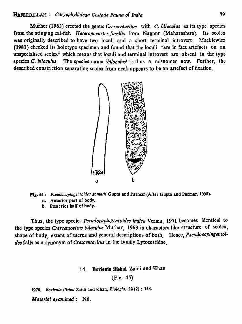
HArEEiULLAH: Caryophyllidean Cestode Fauna of India 19
Murher (1963) erected the genus CrescentDvilus with C. hi/oculus as its type species from the stinging cat.fish Heteropneustes fossilis from Nagpur (Maharashtra). Its scolex was originally described to have two loculi and a short terminal introvert. Mackiewicz (1981) checked its holotype specimen and found that the loculi "are in fact artefacts on an unspecialised scolex" which means that loculi and terminal introvert are absent in the type species C. bllocu/w. The species name 'blloculus' is thus a misnomer now. Further, the described constriction separating scolex from neck appears to be an artefact of fixation.
a b
Fil. 44: Pseudocapingentoides gomalii Gupta and Parmar (Aftor Gupta and Parmar, 1990). a. Anterior part of body, b. Posterior balf of body.
Thus, the type species Pseudocapingentoides Indica Verma, 1971 becomes identical to the type species Crescentovitus hiloculus Murhar, 1963 in characters like structure of scolex, shape of body, extent of uterus and general descriptions of both. Hence, P seudocapingento;. des falls as a synonym of Crescentovitus in the family Lytocestidae.
14. Bovienia ilishai Zaidi and Khan
(Fig. 45)
1976. Bovienia ilishai Zaidi and Khan, Bi%gia, 22 (2): IS8.
Material examined: Nil.

so REc. ZOOL. SuaVe INDIA, Oce. PAPER No. 157
Host-Macr"ra illsha (Hamilton) syn. of Hilsa ilisha (Hamilton-Buchanan), Hilsa sbad, (Clupeiformes: Clupeidae); location-intestine; locality-Ghulam Mohammad Barrage, Karachi, Pakistan ; no. of specimens-two worms, no cross-section.
a b
Fig. 45: Bovienia ilishai Zaidi and KhaD (After Zaidi and Khan, 1976). a. Bntire worm. b. Posterior half of body (enlarged).

HAl'!!ZULLAH: Caryophyliidean Cestode Fatlna of india 81
Description: Body elongated, tapering anteriorly, pointed at posterior end, 15·082-1~:972 long, 0·582-0·59 wide. Scolex not marked off from neck, narrower than body, uDspecialised. Neck long, narrow. About anterior one-fifth of body devoid of any organ system. Positions of musculatures not known as cross-sections not studied. Male and female gonopores separate, at about beginning of last one-fifth of body. Common genital atrium not known.
Testes numerous, rounded, 0-039-0-0 i9 in diameter, extending in about middle threefifth of body length in median field, remaining post-eriorly restricted to level of vas deferens. Vas deferens strongly convoluted behind testicular field. Cirrus sac muscular, enclosing sinuous ejaculatory duct and cirrus opening as male gonopore on ventral surface.
Ovary H-sbaped, in posterior part of body ; ovarian arms 1·74-1·9510ng, 0·112-0-114 wide, follicular, connected by transverse isthmus; anterior ovarian horns much longer (about 2-3 times) than posterior ones. Uterus closely coiled in median field between female gonopore and level of posterior ends of ovarian arms, pre-isthmus uterine extent almost twice or more longer than anterior horns of ovary, never extending anterior to cirrus sac. Large seminal receptacle present near ovarian isthmus, (illustrated, not mentioned in description). Vitellaria follicular, cortical, lateral on each side, extending from slightly anterior to anterior level of testicular region to anterior horns of ovary, follicles smaller than size of testes. Post-ovarian vitellaria absent.
Eggs 31-36 X 20-23 11m.
Remarks: Serial transverse sections through various regions of body were not cut and studied and therefore position of parenchymal muscles could not be known. This inadequacy affects the determination of the family of the present species. I have simply concurred with Zaidi and Khan (1976) in placing this species in the family Lytocestidae. Caryophyllid cestodes generally parasitise fresh water teleosts belonging to the Orders
cypriniformes and Siluriformes but the report of this species from a migratory fish (living in marine, brakish and fresh water) belonging to the order Clupeiformes is quite unusual. The host fish Hllsa llisha (= Macrura illsha) appears to be an accidental host.
1 S. BovieDia lerialis (Bovien) Fuhrmann
(Figs. 46-48)
1926. CaryophyllQ~us serialls BovioD, Vidensk. Medd. Dansk. nature Forening: Kobenhaum, 82: 157-181.
CF 11
1931. Bovienia serio/is: FubrmODD, Handbuch de,. Zoologie (W. Kukenthal and T. Krumbacb, eds.). Bd. 2: 141·416.

82 REe. ZQOL. SURV. INDIA, Occ. PAPER No. I~
1972. Bovienia strialis: Mackiewicz and Murhlr, J. Parasit., 46 (4) : 401. 1980. Introvertus raipurensis Satpute and Agarwal, Proc. Indian Acad. Parasit., 1 (2): 17. 1982. Lucknowia indica Niyogi. Gupta and Agarwal, Proc. Indian Acad. Parasit., 3 (1-2): 17.
Material examined: Nil.
Host-Clarias batrachus (L.), Walking cat-fish, (Siluriformes : Clariidae) ; locationintestine; localities-Nagpur, Ramtek (Maharashtra), Durg, Raipur (Madhya Pradesh). Varanasi (Uttar Pradesh) and Guwahati (Assam); no. of specimens-ISS gravid and immature worms; of these 33 gravid and 7 immature ones were studied, 11 were sectioned~. Holotype in Helm. Coli., Institute de Zoologie, NeuchateI, Switzerland; 2 whole mounts in USNM Helm. ColI. No. 72139; 2 whole mounts in BMNH Helm. CoIl. Nos. 1972 1'3, 1 and 2.
Description: Body elongated, narrow, tapering anteriorly, posterior and pointed, about 14'00-3S'00 long, 0'8-1'6 in maximum width, scolex not distinctly demarcated from neck, unspecialised, 0,2-0'4 wide, not broader than body. Neck present, Jong, narrow. Male and female gonopores in beginning of last eight of body length on ventral surface. Common genital atrium probably absent.
Testes ovoid or spherical, 213-479 in number, 0·OS-0·2S in diameter, medullary, extending from slightly behind anterior most vitellaria to coils of vas deferens, surrounded by a ring of inner longitudinal muscles. Vas deferens a much convoluted narrow tube. External and internal seminal vesicles absent. Cirrus sac ovoid, 0'S-0'8 'in diameter, anterior to ovary, enclosing convoluted ejaculatory duct and unspined cirrus, opening on ventral body surface as male gonopore.
Ovary H-shaped, situated near posterior end of body ; ovarian arms 1·2-2-3 long, follicular, cortical, connected by transverse medullary isthmus, occasionally follicles of posterior horns of ovary intermingling near posterior end to give ovary an inverted 'A' shape. Uterus thrown in lateral coils in median field, not extending anterior to cirrus sac, pre-isthmus uterine extent somewhat longer than anterior horns of ovary. Vaginal tube narrow, straight. Utero-vaginal canal short, opening as female gonopore close behind male gonopore. Seminal receptacle large, well developed, immediately anterior to ovarian isthmus. Vitelline foilicles cortical, external to inner longitudinal muscles in two lateral rows, pre-ovarian, extending from a level slightly anterior to anterior most testes to region of cirrus sac or female gonopore, follicles 0·06-0·23 in diameter. Postovarian vitelline follicles absent.
Bggs 37 .. 42 X 28-31 ~m, smooth, operculate.
Osmoregulatory canals 10-13 at mid-region of body, joining to form excretory vesicle near posterior end; pore terminal.

HAPIEZULLAH: Caryoph)'llidean Cestode Fauna of India
e
c
b
d f
Pia. 46: Bov;enia seriaUs (Bovlen). (After Mackiewicz and Murhar. 1972), 8. Bntire worm, b. Scolex:sbapes, c. Mlddle:part: of body, d. Posterior balf of body, e. Cross-section tbrougb realon of testes, f. Cross-section throuah resioD of ovary.
83

84 Roc. ZOOL. SuaVe INDI~, Oce. PAPER. No. lS7
Remarks: Bovien (1926) originally described this species from a single whole specimen and some fragments of others in the genus Caryophy//aeus. He collected the material from the cat-fish Clar/as batrachus (L .. ) from Java. Fuhrmann (1931) made it the
c a
b
d e
f
Fig. 47: Introvertus reipurensis Satpute and Alarwai (Alter Satpute aDd A.arwIl, 198Gb). 8. Scolex and neck. b. Middle part of body, c. Posterior part of body, d. Cross-section through testicular field, e. Cross .. sectioD ,hrough cirrus lac, f. Cross .. section tbroush ovary.

HAFEEZULLAH: Caryophyllidean Cestode Fauna of India 8S
type species of his new genus Bovlenia but he did not formulate a generic diagnosis. Yamaguti (1959) first and Schmidt (1970) later on attempted to give a diagnosis of the genus but not without gross errors. Mackiewicz and Murhar (1972) not only gave a correct diagnosis of Bovlenia but also redescribed B. serialis completely and correctly on the basis of material recovered from the type host from Java, Malaysia and India, and thus removed all confusions about it.
Satpute and Agarwal (1980) proposed a new genus lntrovertus and described the type species 1. ralpurensl, on the basis of the material recovered from the fish Clarlas
a b c d
FiS .. 48: Lucknowia indica Satputo and Agarwal (After Satpute and Agarwal. 1980a). a. Scolex and neck,
b&c. Middlo parts af body. d. Posterior part of body.
batrachua examined at Raipur (Madbya Pradesh). The scolex of this genus has been characterised to be "'ovoid globular or pear-shaped with a pair of longitudinal grooves and a terminal introvert". Their Fig. 2 suggests that some of the specimens were partially decomposed and flaccid. As Mackiewicz (l98lb) observed in the case of Crescentovltus biloculus Murhar, 1963 that -the loculi in fact are artifacts on an uDspecialised scolex", the same seems to be true witb the scolex. of Intr(lvertus raipurensis too. This is corroborated

86 Roc. ZOOL. SURV. INDIA, Oce. PAPER No. 157
by the action of Schmidt (1986) in synonymising Intfovertus Satpute and Agarwal, 1980 with Lucknowia Gupta, 1961 (although I do not agree with this synonymy on some different grounds) and characterising scolex as unspeciaIised for both. Thus, the two longitudinal grooves and terminal introvent on the scolex of 1. raipurensis appear to be pseudostructures and non-existent being artifacts of fixation. Niyogi, Gupta and Agarwal (1982) described Lucknowla indica from the same host Clarlas batrachus and from the same locality of Raipur. Its scolex in Fig. 1 has been shown slightly swoDen as in Bovienia serialis from the same host and locality while in Fig. 2 the scolex is stumpy. It means that the shape of scolex in L. indica is variable. From the foregoing discussion it becomes fairly clear that the scoleces in 1. raipurensil and L. indica (both from the same host and locality) are unspecialised althougb swollen. Further, as has been observed by Mackiewicz (1981b) in the case of Lucknowia fDssilis Gupta, 1961, it seems there are no post-ovarian vitelline follicles in 1. faipurensis and L. indica too. In fact, they are the follicles of the ovary. Post-ovarian follicles of vitellaria are therefore absent in these two species. Once t~specialised nature of scolex in I. raipurensis and absence of post-ovarian vitelline follicles are established, there remains no pronounced morphological differences between these two species and Bo, ienia serlalls. Thus, Introvertus falls as a synonym of Bovienia. Consequently, I. faipurenlis and L. indica also become synonyms of B. serialis.
Genus 8. L,tocestoic1es Baylis
1928. Lytocestoidies Baylis, Ann. Mag. nat. Hlsl., sorios 10, 1 : 552-562.
Diagnosis: Scolex short, roughly conical, not broader than rest of body, unspecialised. Neck absent. Male and female gonopores separate. Common genital atrium present in last fourth of body length. Testes mejullary, between scolex and uterus. Vitelline follicles regular in shape, cortical, superficial, forming a continuous layer surrounding testes and continuing laterally to post-ovarian region. Ovary bowtie-shaped, bilobed. Uterus relatively short, not extending anterior to cirrus sac. Excretory canals, at least in posterior region of body, internal to vitellaria. Parasiti<Nn cyprinoid and siluroid fishes. Africa, India.
Type sp~cles: Lytocegto'JeJ ta"Jl'lylk 'e Blylis. 1928, in A.festeJ sp. ; Kiranclo in Tanganyika,

HAFEEZULLAH: Caryophyllidean Cestode Fauna Df I"dia
16. Lytocestoides paithaneDsis Shinde and Deshmukh
(Fig. 49)
81
1975. Lylocesloide pailhanensis Shinde and Deshmukb, Marathwada U niv. Nat. Sci. J., 14 (7) : 233. 1981b. L. pailhanensis: Mackiewicz, Himalaya" J. Sci. 1 (1) : 6. 1985. L. pailhanensis : Agarwal, Indian Rev. Lile. Sci., 5: 143.
Material examined: Nil.
Host-Pseudeutropius taakree (Sykes), (Siluriformes : Schilbeidae) ; location-intestine; locality-Paithan (river Godavari, Maharashtra) ; no. of specimen-one.
Fia.49 I Lytocestoides paithanensis Sbinde and Desbmukb (After Sbindo and Deshmukb, 1975).

88 Roc. ZOOL. SURV. INDIA, bee. PAPER :No. 1S'
Description: Body elongated, conical anteriorly, broadened in middle, slightly tapering posteriorly to conical end, measuring 2'5 x 0'79. Scolex conical, not marked off from body, unspecialised. Neck absent. Male and female gonopores separate, opening on ventral surface j n middle region. Common genital atrium apparently
present.
Testes follicular, medullary, about 400 in number, 0'025·0'061 in diameter, extending from base of scolex to cirrus sac. Vas deferens almost in middle of body, winding. External and internal selninaJ vesicles apparently absent. Cirrus sac situated in middle region of body, 0·16 x 0'066, opening by male pore ventrally.
Ovary medullary, dumbbell- or butterfly-shaped, bilobed, lobes connected by very shorl and narrow isthmus, situated in posterior region of body. Uterine extent twice length of post-isthmus space (not shown figure). Vagina thin, narrow, winding tube. No seminal receptacle. Utero-vaginal opening as female pore closely behind male pore. Vitellarium follicular, follicles smaner than testes, cortical, lateral, post-ovarian follicles of vitellarium present. Pre-ovarian and post-ovarian follicles of vitellarja connected by follicles lateral to lobes of ovary_
Remarks: Mackiewicz (198tb) has examined the solitary specimen of the present species. He could not determine its genus because of its bad and decomposed condition. Moreover, the material has been inadequately studied and ill-described since some important characters could not be made known and there are certain selfcontradictions in the discription. Attempt should be made to coneet some more material from the type host and type locality and the species carefuIJy reappraised since it has been described on the basis of a single specimen which is immature without the development of uterus and eggs. The vitellarium has been presumed to be follicular rather than granular because the post-ovarian vitellarium is follicular. Since the species looks akin to Lytocestoides tanganyikae Baylis, 1928 in shape and general details, it is retained in the genus Lytocestoides Baylis 1928 tentatively.
Lytocestoides paithanensis differs from the type species L. tanganylkae mainly in the position of genital pores being almost in middle of body and longer extent of pre-isthmus uterus (supposed).

&urDzULLAH':' ~dryDphyllldean Cestode Fauna of India'
SPECIES OF UNCERTAIN SYSTEMATIC POSITION
Caryophyllaeos kashmirensis Mehra Species inguirenda
1930. Caryophyl/aeus kashmirensis Mehra. Proc. 17th Sci. Congr. Ass. : 247. 1972. Caryophyl/aeus kalhmirensis: Mackiewicz. Exptl. Parasit., 31 : 486, 1981 b. Caryophy/laeus kashmirensis: Mackiewicz: Rima/yan J. Sci •• 1 (1): 4. 1985. Caryophyllatus kashmirensis : Agarwal, Indian Rev. Life. Sci., 5: 142.
Material examined: Nil.
Host-Schizothorax micropogan Heckel, (Cypriniformes : Cyprinidae); locationintestine; locality-near Srinagar, Kashmir; no. of specimens-several.
Description: Body elongated, 30,0-60,0 long, t·O- 2,0 wide at level of male gonopore situated in last eight to tenth of body length. Scolex slightly wider than rest of body, smooth, unspecialised. Vas deferens strongly convoluted. Pre-ovarian vitelline follicles cortical, external to inner longitudinal muscles, annular]y arranged surrounding testes. Post-ovarian vitelline follicles present, medulIary in disposition. Ovary entirely medullary. Uterus much convoluted, with thick coat of uterine glands, not extending anteriorly beyond female gonopore. Vagina a straight tube, joining terminally with uterus to from a short utero-vaginal canal opening ventrally as female gonopore. Genital atrium not known. Seminal receptacule not known.
Eggs 45-55 x 30-30 (lm.
Osmoregulatory canals 10-13.
Remarks: The account of Caryophyllaeus kashmirensis 'Mehra, 1930 is available only in the abstract from. Its full description was never published. That is why the necessary illustrations are not available. Mehra (1930) did not mention the outline (whether irregular or smooth) of the vitelline follicles in his abstract and clearly stated that they are cortical in disposition being external to inner longitudinal muscles but placed his species in the genus CaryophyUaeus Gmelin, 1790 in the family CaryophyIlaeidae. This is a serious self-contradiction. According to the stated cortical position of pre-ovarian vitelline follicles, the species ought to have been placed in the family Lytocestidae. Mehra was apparently misled by medullary disposition of post-ovarian vit. follicles in placing it in the genus Caryophyl/aeus. Moreover, information on the Dumber and extent of testes, position of cirrus sac, external and internal seminal vesicles, shape and anterior extent of ovary, seminal receptacle and genital atrium is Dot at all available. Knowledge on these characters is necessary to formulate a definite
CF 12

90 REC. ZOOL. SURV. INDIA, Oce. PAPER No. 151
diagnosis of Caryopnyllaeus kashmirensis. Non-availability of information on these important characters made Mackiewicz (1972) declare it as species inquirenda. He (1981) further suggested that fresh material should be collected from the type host and type locality, carefulIy studied and compared with Adenoscolex oreini Fotedar, 1958 which is reported from the fish Orienus sinuatus commonly found in the hin streams of Kashmir.
Taking clue from the described distribution of pre. ovarian vitelline follicles to be external to the inner longitudinal muscles, Agarwal (1985) included Ca1yophyllaeu8 ka.shmirensis in the family Lytocestidae. Further, presuming the AdenoscQlex orei"i Fotedar, 1958 (a1so described from a hill stream fish of Kashmir) to be identical to Caryophyllaeus kashmirensis, he suggested that "c. kashmirensis be considered synonymous to A. oreini". There does not seem to be good justification in this action or suggestion since Adenoscolex Fotedar, 1958 (type species A. oreini) is a very well established genus in the family Capingentidae, and Agarwal (1985) himself considers C. Kashmirensis to belong to the family Lytocestidae. Mackiewicz (1981b) has rightly suggested that fresh material of C. koshmirensis should be collected from the type host from Kashmir and its correct and complete description should be furnished first, and only then any action can be taken regarding its systematic position vis-a-vis A. oreini. TiU then C. kashmirensis should best be considered species inquirenda or a species of uncertain systematic position. Probably Mackiewicz thinks that Mehra (1930) might have erred in studying the position of vitelline follicles in re1ation to the inner longitudinal muscles because he has himself observed in the abstract that the species (C. kashmirensis) apparently connects the two genera Carlophyllaeus and Lytocestus. From this discussion at least this much becomes clear that Caryophyllaeus orein; Mehra, 1930 does not belong to the genus Caryophy//aeus.
DISCUSSION
No concerted efforts have been made to collect and study caryophyllid cestodes in India. They have been reported only from a fe~ places. A vast area still remains to be explored for these worms. Altogether 40 species under 14 genera have been reported from Indian region, including one species each from Nepal and Pakistan. Lytocestus filiformis was original1y described from Sudan (Africa), but it has been recorded from the north-eastern region of India also. The Indian caryophyllids represent three of four families, namely, Caryophyllaeidae, Capingentidae and Lytocestidae. The family Balanotaeniidae is known from Australian region only.

HArEBZULLAH: Ca" ophyllidean Cestode Fauna of India 91
Prof. 1.S. Mackiewicz of the State University of New York at Albany, USA, bas personally examined most of the described Indian caryophyllid species. He (1981b) found that some materials are in such a condition that their family and genera are difficult to be determined while others have been inadequately and erroneously studied and described. He further remarks that the taxonomy of described Indian caryophyUids is far from satisfactory and a clear picture of the systematics of the Indian caryophyllid fauna does not emarge.
These observations and remarks of Prof. J.S. Mackiewicz prompted me to revise the taxonomy of described species of Indian caryophyllids and straighten it which is in utter confusion. With th~s aim in view I tried to seek the co-operation of some of the Indian authors for examining their reported materials, but the same was not extended for reasons best known to them excepting Dr. V. Tandon. So, I had to depend on the existing literature on the group as well as my own collection to study intraspecific variations. As a result, most of the diagrams of species, reliable or not, used in this work have been taken from original sources. Due to inadequacies in the description of species some tentative taxonomic decisions had to be taken.
While going through the literature critically I could understand at least five main reasons for unsound foundation of the taxonomy of Indian caryophyllids. Firstly, the materials perhaps were not processed for collection according to the standard technique due to which overflattening and crushing occured. In many cases probably late fixation caused creeping in of artefacts of fixation. Secondly, barring a few cases, cross-sections and mid~ sagittal sections of body were not cut leading to erroneous family and genus determinations. Thirdly, perhaps suitable stain was not chosen to stain the whole specimens and differentia. tion atler overstaining was not dextrously done making it difficult to distinguish follicles of testes, vitel1aria and ovary one from the other. Fourthly, use of cytological studies of these organs were not at aU employed along with the general morphological and anatomical studies. Fifthly, in many cases even the morphological and anatomical studies were d~plorably inadequate and inaccurate leading to repeated descriptions of many species under new names and under different genera. Moreover, there is no dearth of examples in which new species have been described utilising the pseudo-characters which develop due to the state of contraction and relaxation at the time of fixation.
From the study of my own caryophyl1id co1Jections from the cat-fishes Clarias balrachua and Heteropneustes tossi/is, I have found it very difficult in the whole mounts to make out number of gonopores, presence or absence of common genital atrium and whether the vitellaria is lateral or annular, medullar, or partly medullary and partly cortical. I have also found variations in scolex shape in the specimens of a single population. In one and the same population it has also been detected that the posterior extent of vitalline fol1icles may OJ' may ~ot be continuous with those of the anterior horns of tb~ ovary, an4 tl:t~ follicles of

92 REe. ZOOL. SURV. INDIA, Oce. PAPER No. 151
posterior ovarian horns may extend up to the posterior end of the body, occasionally intermingling from both sides giving the false impression of post .. ovarian vitelline follicles. This intraspecific variation is ver y common misleading to the investigators.
The biology, specially the development and life-history, has not at all been worked out. Our knowledge on the biology of Indian caryopbyUid is rather cipher. The pressing need to have a good and sound understand ing of the taxonomy of these cestodes is to make adequate and accurate studies on morphology, anatomy and intraspecific variations employing correct techniques of collection and staining of material well supplemented by crosssections and mid-sagitta] sections.
As a result of the study of 40 species of caryophyllids under 14 genera, the valid species and genera have been found to be 16 and 8 respectively with one species, Caryophyllaeus kashmirensis, of uncertain systematic position, Mackiewicz (1972) considered it as species inquirenda as its inadequate account is available only in abstract form. However, its resemblances to Adenoscolex oreini, also from Kashmir, are unmistakable.
India is a vast country with numerous benthic feeding Siluriformes and Cypriniformes freshwater fishes, and with about 67 species of freshwater oligochacte annelids known so far belonging to the families Tubicidae, Naididae and Aelosomatidae. These facts are indicative that many more caryophyllids are yet to be di$covc;red froto Indi~.
HOST-PARASITE LIST
Host
Order Clupeiformes
Family Clupeidae Hi/sa i1isha (Harn.-Buch.) Syn. Macrura ilisha (Ham.)
Order Channiformes
Family Channidae Channa striatus (Bloch)
Order Cypriniformes
Family Cobi tidae LepidQcephalur guntea (Ham.)
Parasite
BDvlenla illshal Zaidi and Khan
LytocestUl /ossllisl (Gupta)
Paraca'1ophyllaeus lepfdecephall (Kundu)

BAFE!ZULLAH: CaryDphyllidean Cestode Fauna of India
Host
Family Cyprinidae Barbus kolus (Sykes) Cirrhina mrigala (Ham.) Labeo calbasu (Ham.) Oreinus sinuatus (Heckel) Ostiobrama cotio (Ham.)
Order siluriformes
Family Bagridae Rita rita (Ham.)
Family Clariidae Clarias batrachus (L.)
Family Heteropneustidae Heteropneustes fossilis (Bloch)
Family Schilbeidae
PseudeutrDpius taakree (Sykes)
Parasite
Brevicolus aurangabadensis Shinde Breviscolex naldurgensis Shinde et al. Breviscolex aurangabadensis Shinde Adenoscolex oreini Fotedar Paracaryophyllaeus ostiobramensis
(Gupta and Sinha)
Pseudocaryophyllaeus indica Gupta
Bovienia serialis (Bovien) DjDmbangia Penetrans Bovien Lytocestus birmanicuJ Lynsdale Lytocestu$ fillformis (Woodland) Lytocestus indicus (Moghe) Lytocestus IDngicol/is Rama Devi P seudocaryophyllaeus indica Gupta
Bovienia bllocula (Murhar) Djombangia penetrans Bovien Lytocestus fossilisi (Gupta) Pseudocaryophyllaeus indica Gupta
Lytocesloides paithanensis Shinde and Deshmukh
93

94 REe. ZOOL. SURV. INDIA, Occ. PAPER No. 157
PARASITE-HOST LIST
(Species marked with asterisk are synonyms)
Parasite
Family Caryophyllaeidae Leuckart
Genus Paracaryophyllaeus Kulakovskaya P. lepidocephali (Kundu) P ostiobramensis (Gupta & Sinha)
Genus Pseudocaryophyllaeus Gupta P. indica Gupta
*Capingentoides batrachil Gupta * P seudDcaryophyllaeus lucknowensis
Gupta & Sinha *P. mackiewlczi Gupta & Parmar
*p. rita; Gupta & Singh
Family Capiogentidae Hunter
Genus Adenoscolex Fotedar A. oreini Fotedar
Genus Breviscolex Kulakovskaya
B. aurangabadensis (Shin de)
B. naldurgens;s Shinde et all
* Lytocestoides mackiewicz; Shinde et al.
Family Lytocestidae Hunter
Genus Bovienia Fuhrnlann B. hilocula (Murhar) *Capingentoldes fOledar;
Gupta & Parmar *0. heteropneusti Gupta & Sinha. *C. sinuhl Verm'l
Host
LepidDcephalur guntea (Ham.) Ostiobrama colio (Ham.)
Clarias batrachus (L.) Cia ria, batrachus (L.) Heteropneustes fossilis (Bloch)
HeterDpneustes fossilis (Bloch) Rita rita (Ham.)
Oreinus sinuatus (Heckel)
Barbus kQlus (Sykes) Labeo calbasu (Ham.)
Clrrhlnus mrigala (Ham.)
Clrrhlnus mrigala (Ham.)
Heteropneustes f"ssilis (Bloch)
Clarlas batrachus (L.) Heteropneustes fossilis (Bloch) lIeterDpneustes !"ssills (Bloch)

lttarazULLAH: Caryophyilidean C,stode Fauna oj India
Parasite
~;:Pseudocapingentoides cameroni Gupta and Sinha
*P. gomalii Gupta and Parmar *P. indica Verma Bovienia ilishai Zaidi and Khan
Bovienia serialis (Bovien) *Introvertus raipurensis
Satpute and Agarwal
* Lucknowia indica Niyogi el al.
Genus Djombangia Bovien D. penetrans Bovien
*D. caballeroi Sahay and Sahay * D. clariae Kundu et all *D. Indica Satpute and Agarwal
Genus Lytocestoides Baylis L. paithanensis Shinde and
Deshmukh
Genus Lytocestus Cohn L. birmanicus Lynsdale L. filiformis (Woodland) L. fossilisi (Gupta)
*Capingentoides gorakhnathi Agarwal and Singh
*C. moghei Pandey * Lytocestoides f Dssilis Kanth
et al. Lytocestus fossili~ Singh
L,tocestus indicus (Moghe) * Introvertus chauhani Pandey and
Tiwari
*LytDcestus marathwadaensls Shinde & Phad
*Pseudolytocestus clariae Gupta *p thapari Gupta and Parmar
Lytocestus longicDllis Rama Devi
Host
Heteropneustes jossilis (Bloch) Heteropneustes fossilis (Bloch) Heteropneustes fassilis (Bloch) Hilsa ilisha (Harn.-Buch.) Syn. Macrura ilisha (Ham.) Clarias batrachus (L.) Clarias batrachus (L.)
Clarias batrachus (L.)
Clarias batrachus (L.) Heteropneustes fossilis (Bloch) Clarias batrachus (L.) Clarias batrachus (L.)
Pseudeutropius taakree (Sykes)
Clarias batrachus (L.) Clarias batrachus (L.) Heteropneustes fossilis (Bloch) Clarias batrachus (L.)
Channa striatus (Bloch) Heteropneustes fossilis (Bloch)
Heteropneustes rossilis (Bloch) Clarias batrachus (L.)
C larias batrachus (L.)
Clarias batrachus (L.)
G larias batrachus (L.) Clarias batrachus (L.) Clarias batrachus (L.)
95

96 kEe.' ZOOL. SuaVe iNblA, Occ. PAPER No. 1S7
SUMMARY
A comprehensive account of the morphology, anatomy and life cycles of the Caryophyllidea in general and systematic account of Indian caryopbyUids in particular have been provided. The taxonomy of the Indian caryophyllid cestodes known so far consisting of 40 species (including one each from Nepal and Pakistan) under 14 genera and 3 families was in utter confusion. This was largely due to inadequate, and faulty descriptions of species and mostly without cutting the cross-sections and mid-sagittal sections. The taxonomy of these forms has been revised largely on the basis of literature bringing down the numbers to 16 species under 8 genera with one species of uDcertain systematic position. Reasons have been given for the belief that many more species remain undiscovered in Iudia. Host-parasite and parasite-host lists have also been furnished.
ACKNOWLEDGEMENTS
The author is thankful to Dr. A. K. Ghosh, Director, Zoological Survey of India, Calcutta, for providing laboratory and library facilities. He is indebted to Dr. Veena Tandoo, North-east Hill University, Shilloog, for loaning some material for study and comparison. He gratefully acknowledges the help extended by Prof. J. S. Mackiewicz, State University of New York at Albany, USA, Dr. Sundecp Kumar Malhotra, University of Allahabad, Dr. Ashwini Kumar Gupta, Ravishanker University, Raipur, Dr. G. B. Shinde, Marathwada University, Aurangabad, and Dr. Veena TandoD, by way of providing some much needed literature. The author is also thankful to Shri Shuvajit Chakrabarti for making a few drawings.
REFERENCES
Agarwal, S. M. 1985. CaryopbyUaeids and caryophyUidiasis In India. Indian Rey. Life Sci., 5: 139-161.
Agrawal, N. and Singh, H. S. 1985. Cestode fauna of district Gorakhpur. Part I. On a rare cestode, CapingentDides gorakhnathl D. sp. [ndlan J. Helminth (".s.), 2 (1 & 2) : 81-84.

ifAPBJZULLAH: Ca'YDphyllidea,,. Cestode Fauna of India 97
Ahmed, A. T. A. and Sanaullah, M. 1977. Studies on the distribution of some metazoan parasites of HeterDpneustes fossUls (Bloch) and Clarias batrachus (L.) in Bangladesh. Bangladesh J. Zool., 5 (2) : 117-123.
Bovien, P. 1926. Caryopbyllaeidae from Java. Vidensk. Medd, Dansk naturh. Foremlng: KDbenhau1I, 81: 157-181.
Calentine, R, and Ulmer M. 1961. Khawia iowensis n. sp. (Cestoda: Caryophyllaeidae) from Cyprinns carpio L. in Iowa. J. Paras;t., 47: 795-805.
Chakravarty, R. and Tandon. V. 1989. On the present status of Caryophyllidea with a report on some caryophyUid infections in the freshwater catfi~h Clarias batrachus (L.) in north·east India and a record of an anomalous form. Indian J. Helminth. (n.s. ),( 1988), 5 (1): 37 .. 54.
Donges, 1. and Harder, W. 1966. N esolecithus aJricanus D. sp. (Cestodaria, Amphilinidea) aus dem coelom von Gymnarcus nilodeus Cuvier 1829 (Teleostei). Z. Parasitenkd., 28 : 12S-141.
Dubinina, M. N. 1982. Parasitic Helminths. Class Amphilinida (Platyhelminthes). Akad. Nauk. SSSR Tr. ZODl. Inst., Leningrad, 100: 1-143.
Fotedar, D. N. 19S8. On a new caryophyllaeid cestode, Adenoscolex oreini gen. et. sp. nov. from freshwater fish in Kashmir, and a note on some related genera. 1. Helminth .• 32 (1-2): 1-16.
Fiihrmann, O. 1931. Ordung der Unterklasse dec Cestoda: Pseudophyllidea, 289-334 in Kukentha/'s Handbuch der Zoologie,2: 141-416.
Gupta, S. P. 1961. Caryophyllaeids (Cestoda) from fresh-water fishes of India. Proc. helminth. Soc. Washington, 28 (1): 38-50.
Gupta, S. P. and Sinha, N. 1980. On a new caryophyllaeid Caping~ntoides heteropneusti n. sp. from the intestine of a freshwater fish Heteropneustes fossilis (Ham.) from Lucknow. Indian J. Helminth., (1979), 31 (1): 65-68.
Gupta, S. p. and Sinha, N. 1984. On three new species of caryophyllaeids from fresh water fishes of Lucknow. Indian 1. Helminth., 36 (1) : 73-80.
Gupta, V. and Parmar, S. 1984. On a new caryophyllaeid Pseudocaryophyllaeus mackiewiczl sp. nov. from the intestine of a freshwater fish, Hete;opnetlstes Jossilis (Ham.) from Gorakhpur, U.P. Indian J. Helminth., (1982), 34 (2) : 136-138.
Gupta, V. and Parmar, S. 1986. Cestode parasite of vertebrates. Capingentoides fotedar; sp. nov. from a freshwater fish Clarias batrachus (Linn.) from Lucknow. Indian J. Helminth .• (1985),37 (1): 31-35.
CF 13

98 Roc. iOOL. SURV. tNDIA, Oce. PAPER No. 157
Gupta, V. and Parmar, S. 1990. On the new caryophyllaeids from river Gomati, Lucknow,
Uttar Pradesh. Indian J. Helminth., 42 (1) : 25-30.
Gupta, V. and Singh, S. R. 1984. On a new species Pseudocaryophyl/aeus rita; sp. nov. (Family Caryophyllaeidae) from the intestine of a fresh-water fish, Rita rita, from river Gomati at Lucknow, U.P. Indian 1. Helminth., (1983), 3S (1): 11-14.
Hafeezullah, M. 1986. Status of the species of DjDmbangia Bovien, 1926 (Cestoidea: Caryophyllidea: Lytocestidae). Ree. zDDI. Surv. India, 83 (3 & 4): 121-125.
Hasnain, M. and Sahay, U. 1989. Duplication of somatic region in a LytDcestus species.
Potedar Commemoration Volume: 120-124.
Hunter, G. W. III. 1930. Studies on the CaryophyJlaeidae of North America. I/linois
bioi. MonDgr' J 11 : 186 pp,
lohri, G. N. 19S9. On a remarkable new caryophyllaeid cestode, Hunteroides mystei gen. et. sp. nov. from a freshwater fish in Delhi State. Z. Parasitenkd., 19 : 368-374.
Kanth, L. K., Sinha, K. P. and Srivastava, L. P. 1984. The morphology of Lytocestoides fossilis n. sp. (Cestoda: CaryopbyUidea) from the intestine of Heleropneusles fossilis
(Bloch). Indian J. Helminth. (n. s.), 1 (1 & 2) : 26-29.
Kundu, D. K. 1985. On a new species of the genus LYlocestoides Baylis. 1928 (Cestoidea: Caryopbyllidea: Lytocestidae) from a cobitid fish, JJepidocephalus guntea (Ham.) from West Bengal. Bull. zDDI. Surv. India, 7 (2-3): 285-290.
Kundu, D. K., Bhattacharya, S. B. and Datta, I. B. 1985. Djombangia clariae n. sp. (Cestoidea; Caryophyllidea: Lytocestidae) from a siluroid fish in West Bengal. Bull.
zool. Surv. India,7 (2-3): 151-154.
Lynsdale, J. A. 1956. On two new species of Lytocestus from Burma and the Sudan respectively. J. Helminth.) 30 (2/3): 87-96.
Mackiewicz, S. 1962. Systematic position of Carlophyllaeus /uhrmanni Szidat, 1937 and Lytocestus a/estesi Lynsdale, 1956 (Cestoidea: CaryophyUidea). Rev. Suisse Zool., 69 : 729-735.
Mackiewicz, J. S. 1972. CaryophylJidea (Cestoidea) : A review. Exper. Parasit., 31 : 417 .. 512.
Mackiewicz, S. 1981a. CaryophyUidea ( Cestoidea): Evolution and classification. Adv. Parasilol., 19: 139-206.
Mackiewicz J.S. 1981h. Synoptic review of the CaryophyUidea (Cestoidea) of India, Pakistan and Nepal. Himalayan I. SCi., 1 (1): 1-14.

HArEEzULLAH: Caryophyllidean Cestode Fauna of India 99
Mackiewicz, S. 1982, CaryopbyUidea (Cestoidea): Perspective. Parasitology, 84 (2): 397-417.
Mackiewicz, J. S. and Blair, D. 1978. Balanotaeniidae fame n. and Balanotaenia newguinenlis sp. n. (Cestoidea: Caryophyllidea) from Tandanus (Siluriformes: Plotosidae) in New Guinea. 1. Helminth., 52: 199-203.
Mackiewicz, J. S. and B. M. Murhar, 1972. Redescription of Bovienia serialis (Bovien, 1926) (Cestoidea: CaryopbyUidea) from the catfish Clarias batrachus (L.) in India. 1. Helminth., 46 (4): 399 .. 405.
Mehra, H. R. 1930. On a new species of Caryophyllaeus O. F. Muller from Kashmir with remarks on Lytocestus indicus (Moghe) 1925. Proc 17th Indian Sci. Cong. Ass., : 247.
Moghe, M. A. 1925. Caryophyllaew indicus n. sp. (Trematoda) from the catfish ClaTias batrachus (L.). Parasitology, 17 : 232-235.
Moghe, M. A. 1931. A supplimentary description of Lytocestus indicus Moghe (Syn. Caryophyllaeus indicus Moghe, 1925), Cestoda. Parasitology, 23 : 84·87.
Murhar, B. M. 1963. Crescentovitus biloculus gen. nov., sp. nov., a fish cestode (CaryophylIaeidae) from N agpur, India. P arasitolDgy, 53: 413-418.
Nama, H. S. 1979. On the occurrence of Crescentovitus biloculus Murhar, 1963 (Cestoidea:
CaryophyUaeidae) in Rajasthan. Indian 1. Parasit., Lucknow, 3 (6) : 171.
Niyogi, A.J Gupta. A. K. and Agarwal, S. M. 1982. Morphology of Lucknowia indica sp. nov. (Lytocestidae : Caryophyllidea). Proc. Indian Acad. Parasit., 3: 17-22.
Pandey, K. C. 1973. Studies on some cestodes from fishes, birds and mammals. Indian J.
ZDDt., 15: 221-226.
Pandey, P. N. and Tiwari, S. K. 1989. On a new cestode Introvertus chauhani n. sp. from Clarlas batrachus. Dr. B.S. Chauhan Comm. Vol.: 141-142.
Pritchard, M. H. and Kruse, G. O. 1982. Tee Collection and Preservation of Animal Parasites. University of Nebraska Press, Lincoln, 141 pp.
Ramadevi, P. 1973. Lytocestus longicolli, sp. nov. (Cestoidea: CaryophyUidea) from the
catfish Clarlas batrachul (L.) in India. 1. Helminth., 47 : 415-420.
Sabay, S. N. and Sabay, U. 1977. On a new caryophyllid cestode, Djombangia caballeroi sp. nov. from freshwater fish Heteropneusles fossilis in Cbotanagpur with an amendation of the generic character. Excerta Parasitologica en Memoria del Doctor Edwordo Caballero Y Caballero, Maxico. 4: 371-376.
Satpute, L. R. and Agarwal, S. M. 1974. Diverticulosis of the fish duodenum infested with
cestodes. Indian J. Exper. Bioi., 13 : 313-37'.

too REC. ZOOL. SURV. INDIA, Oce. PAPER No. 157
Satpute, L. R. and Agarwal, S. M. 1980a. Morphology and systematics of Djombangia indica Satpute and Agarwal, 1974 causing diverticulosis of duodenum of Clarias Batrachus. Proc. Indian Acad. Parasit., 1: 13-16.
Satpute, L. R. and Agarwa], S. M. 1980b. Introvertus rc'lipurensis g~n. nov., a fish cestode (Cestoda: CaryophyUidea: Lytocestidae) from Raipur, India. Pr(Jc. Indian A cad. Parasite 1 : 17-19.
Shinde, G. B. 1970. A new species and two new varieties of the genus LytocestDides Baylis, 1928 in fresh\vater fishes in Maharashtra, India. Marathwada Univ. J. Sci., 9 : 173·178.
Shinde, G. B. and Deshmukh, R. A. 1975. On a new species of LytocestDldts Baylis, 1928 from a freshwater fish in Maharashtra, India. Nat. Sic. J. Marathwada Univ., 14 : 233-236.
Shinde, G. B. and Deshmukh, R. A. 1980. Description of two species of the genus Lytocestus Cohn, 1908 (Cestoda: Cotyloda : CaryophyUidea) from a freshwater fish. Riv. Parassit., 41 (2) : 209-214.
Shinde, G. B., Mohekar, A. D., Jadhav, B. V. and Hafeezullah, M. 1987. Two new species of the genus Lytocestoldes Baylis, 1928 (Cestoidea: Cestoda: Caryophyllidea: Lytocestidea) from a fresh water fish in Maharashtra, India. Bull. zool. Sur,. India, 8 (1-3): 197-201.
Shinde, G. B. and Phad, A. N. 1988. On a new cestode Lytoceslus marathwadaensls from freshwater fiish. Riv. Parassit. 47 (2): 295-298.
Sinha, S. S. and Rashid-un-Nisa, 1981. An anomaly in caryophyllid cestode, Lytocestw indlcus Moghe, 1931. Indian J. Parasit., 5 (2): 197-198.
Singh, S. S. 1975. On Lytocestus fossilis D. sp. (Cestodea : Lytocestidae) from Heteropnerutes fossills from Nepal. In: Dr. B. S. Chauhan CODmmemoratlon Volume: 79-82.
Szidat. L. 1937. Archigetes R. Leuckart 1878, die progenetische larva ciner fur Europa neuen Caryophyllaeiden-Gattung Biacelabulum Hunter 1927. Zool. Anz., 119: 166- t 72.
Verma, S. L. 1971. Helminth parasites of fresh·water fishes. Part I. On two caryophUaeids from fresh·water fishes of Lucknow. I"dian 1. Helmlnlh., 23 : 71·80.
Wardle, R. A. and McLeod, J. A. 1952. rJ'he Zoology of 'l'apewDrms. University of Minnesota Press, Minneapolis, 780 pp.
Wardle, R. A., McLeod, J. A. and Radinovsky, S. 1974. Advances in the Zoology ./ rI'apeworms, 1950·1970. University of Minnesota Press, Minneapolis, 274 p~,
HAPEEZULLAH: Caryophyl/idean Cestode Fauna of India 101
Woodland, W. N. F. 1923. On some remarkable new forms of Caryophyllaeidae from the Anglo-Egyptian Sudan, and a revision of the families of the Cestodaria. J. microsc. Sci. (N. S,), 67: 435-472.
Woodland, W. N. F. 1926. On the genera and possible affinities of the CaryophyUaeidae : A reply to Drs. O. Fuhrmann and J. O. I. Baer. Proc. zool. Soc, London,: 49-69.
Woodland, W. N. F. 1937. Some cestodes from Sierra Leone. I. On Wenyonia 'ongicauda sp. n. and Proteocephalus bivitellatus sp. n. Proc. zool. Soc. LDndon, (1936): 931-937.
Yamaguti, S. 1959. SystenlQ Helminthum. Vol. 11. The Ceslodes of Vertebrates. Interscience Publishers. Inc. N. Y., 860 pp.
Zaidi, D. A. and Khan, A. 1976. Cestodes of fishes from Pakistan, BlIDgla,21 (2) :
1'7-179.


![FEASIBILITY STUDY REGARDING RE …faunaofindia.nic.in/PDFVolumes/records/113/01/0001-0024.pdfSANYAL et al. : Feasibility Study Regarding Re-introduction of Pygmy Hog 3 IUCN, 1998].](https://static.documents.pub/doc/80x56/5b0dfe5d7f8b9ab7658d43ba/feasibility-study-regarding-re-et-al-feasibility-study-regarding-re-introduction.jpg)


