To determine whether expression of the XPD/ERCC2 repair gene intrichothiodystrophy (TTD) group D cells could restore mutagenesis characteristics of repair-proficient cells, we compared the UV mutagenesis ofnormal cells, TTD group D cells, and TTD group D cells retrovirallytransduced by the wild-type XPDIERCC2 gene (TTD + ERCC2 cells). We
first verified the expression of the XPD protein, correction of UV cellsurvival, and DNA repair ability of TTD + ERCC2 cells. UV-inducedmutations were studied using the pR2 shuttle vector. The addition of theXPD/ERCC2 gene in TTD cells led to a significant but partial decrease ofmutation frequency compared with the parental TTD cells. Types ofmutations of TTD + ERCC2 cells get closer to those observed in normalcells (i.e., a reduction of multiple mutations). New hotspots appeared and
some disappeared in the complemented line, suggesting that hotspot distribution is particular to each cell line and cannot be correlated with therepair status of the cells. In conclusion, the expression of the XPD/ERCC2
repair gene completely corrected UV hypersensitivity and almost all typesof mutations of TTD group D cells, whereas hypermutagenesis was partially corrected.
INTRODUCTION
The NER3 system is the fundamental mechanism of cell protectionagainst the effects of various types of DNA damage induced bygenotoxic agents. Deficiency in NER has been associated with ‘LTDand XP, two clinically distinct human syndromes inherited as anautosomal recessive trait (for review, see Refs. 1—3).In the XPsyndrome, persistence of unrepaired DNA damage produced by exposure to UV light is associated with photophobia and an extremelyhigh level of skin tumors in sun-exposed sites. In contrast to XPpatients, lID patients do not have an increased frequency of skincancers (4, 5) and show sulfur-deficient brittle hair and mental andphysical retardation (6). Approximately 50% of lTD patients exhibitphotophobia and a marked sensitivity to sunlight. The clinical photosensitivity in TFD patients is usually associated with an increase ofboth cellular UV sensitivity (7, 8) and UV-induced mutability (9, 10).The NER defect of photosensitive lTD patients has been assigned tothree genetic complementation groups. The large majority of cases(>90%) fall into the same group as XP-D and together represent theTFD/XP-D group (1 1—13). One family was found to belong to XP-Bgroup (14) and another kindred constitutes a distinct NER deficientgroup, lID-A (15).
In several TLD/XP-D patients, molecular analysis of the XPD/ERCC2 gene showed point mutations and deletions leading to aminoacid substitutions or to a truncated XPD protein (16—18).The XPD/
Received 5/7/96; accepted 10/3/96.The costs of publication of this article were defrayed in part by the payment of page
charges. This article must therefore be hereby marked advertisement in accordance with18 U.S.C. Section 1734 solely to indicate this fact.
I This work was supported by the Ligue Française Nationale Contre Ic Cancer, the
ERCC2 gene encodes a 760-amino acid protein with a predictedmolecular weight of 86,900 (19). The purified XPD protein wasshown to possess an AlP-dependent DNA 5'—@3'helicase activity(20) and to be associated with the basic transcription factor II (IFIIH)complex (21).
The introduction of the functional XPD/ERCC2 cDNA in ‘LTDandXP-D cells, thanks to plasmidic (22—25)or retroviral vectors (26) ledto the restoration of wild-type levels of UV sensitivity and DNArepair. Moreover, UV hypermutability of XP-D cells was partiallycorrected in two XPD/ERCC2-transformed XP-D cell lines after plasmid transfer (24).
To examine whether the complementation of ‘LTD/XP-Dcells withthe wild-type XPD/ERCC2 gene also concerned UV mutagenesis, weused a TTD/XP-D cell line previously characterized in our laboratorywith respect to its UV-C mutagenesis properties (10). In the presentstudy, this cell line was transduced with the LXPDSN retroviral vectorcarrying the wild-type XPD/ERCC2 repair gene as described previously (26) and was called here the lID + ERCC2 cell line. We firstverified, after UV irradiation, the correction of UV hypersensitivity bycolony-forming ability and the restoration of the wild-type DNArepair system by UDS. Then, the pR2 shuttle vector carrying the lacZ'target gene was used to compare UV-C mutagenesis characteristics of‘LTD/XP-D,‘LTD+ ERCC2, and normal cell lines. We found that theexpression of the XPD/ERCC2 gene in the TFD/XP-D cells led to amutation frequency showing intermediate values between the TTDparental line and the normal line. In addition, the types of mutationsfound in TTD + ERCC2 cell line were closer to those in normal cellline than to those in parental T'TD cell line.
MATERIALS AND METHODS
Cells and Cell Transduction. Human fibroblasts were cultured in Eagle'sMEM supplemented with 10%FCS, 2.5 @tg/mifungizone, and antibiotics [100units/ml penicillin, 100 @g/m1streptomycin, and 0.25 @g/mlhygromycin B(Sigma) for the TTD and TTD + ERCC2 cell lines].
We used the human SV4O-transformedMRC-5V1 DNA repair-proficientfibroblast line (27), provided by Dr C. Arlett (Brighton, England). The 5V40LT-antigen-transformed TTD1VI-LAS-KMT11 cloned fibroblast cell line(TTD/XP-D cells) has been established in our laboratory from a skin biopsy ofa 9-month-old French TTD patient (10). The retrovirus producer cell line
‘I'-CRIP-LXPDSN (26) was used to transfer the XPD/ERCC2 cDNA into the
TFD1VI-LAS-KMT11 cells. Preconfluent ‘I'-CRIP-LXPDSNcells weregrown for 24 h in fibroblast medium. This conditioned medium containing thevirus was filtered through 0.22 @Mnitrocellulose membrane and added to
TFD1VI-LAS-KMT11 medium with 8 ,.@g/mlpolybrene (Sigma) for 48 h.Transduced cells were selected for 15 days with 1 mg/ml neomycin-analog
G4l8 (Life Technologies, Inc.). G418-resistant cells were seeded in 10-cmPetri dishes to clonal density. All 12 isolated clones were UV resistant and
were tested for their efficiency to replicate the pR2 shuttle vector, leading tothe selection of the complemented TFD1VI-LAS-KMT11-D10 clone, called
here the lTD + ERCC2 cell line. Mutagenesis assays on this cell line wereperformed during the month immediately following the G4I8-selection.
Cell Survival Study and Unscheduled DNA Synthesis. Cell survivalafterUV-C treatment was measured by the colony-forming ability procedure. Growing cells were UV irradiated under a germicidal lamp (254 nm) with a dose rate
of 0.36 J . m2@ ‘and then plated at low density (100 cells per 10-cm Petridish) to measure their colony-forming ability (28).
5450
Recovery of Normal DNA Repair and Mutagenesis in Trichothiodystrophy Cellsafter Transduction of the XPD Human Gene'
Claire Marionnet, Xavier Quilliet, Annie Benoit, Jacques Armier, Alain Sarasin,2 and Anne Stary
Laboratory of Molecular Genetics, UPR 42 Centre National de la Recherche Scientifique IFCI, Institut de Recherches sur le Cancer, BP8-94801 Villejuij France
UV MUTAGENESISOF rm CELLS COMPLEMENTED BY THE XPD GENE
Analysis of repair synthesis after UV-C irradiation was carried out by theUDS method, as described elsewhere (10).
Western Blotting. Westernblot analysis was carriedout using 50 @gofproteins as described previously (26). The monoclonal anti-XPD antibody,generously provided by Dr J. M. Egly (Strasbourg, France), was raised againstamino acids 749—759 at the carboxy-terminal end of the human XPD protein.
The intensities of the bands were quantitated using a Samba IPS 4.02 (Alcatel)bioimage analyser.
Plasmid UV Treatment, Transfection, and Mutational Assay. The pR2shuttle vector contains the SV4O replication origin that allows its replication in
human cells in the presence of the SV4OLT-antigen. The pR2 plasmid alsoharbors the pBR322 replication origin and the kanamycin resistance gene,which allows the selection ofthe pR2 plasmid in bacteria, and the l68-bp lacZ'gene, which is used as target gene for mutation analysis (10). The pR2 plasmidwas irradiated with 254-nm UV light using a germicidal lamp at the dose rateof 2 J . m2@ s@'. Transfections of 5—10 @g,according to the cells, ofUV-treated or untreated pR2 vectors into the three human cell lines wereperformed for 6 h using the calcium phosphate precipitation method (29). Five
@.Lgofunirradiated p205-KMT11plasmid (30) were cotransfected with the pR2plasmid into MRC-5V1 and TTD + ERCC2 fibroblasts. Cells were thenincubated for 2—3days, according to the ability of the cells to replicate theplasmid, in medium containing 100 @LMZnC12 and 1 @.LMCdCl.,, which allows
an overexpression of the 5V40 LT-antigen gene carried by the p205-KMT1Ivector and leads to a very efficient replication of the pR2 plasmid inside thehuman cells. lTD cells were not cotransfected with the p2O5KMTll plasmid
because these cells already contained the vector (10), and the pR2 plasmid wasefficiently replicated.
Replicated plasmid DNA was recovered from the human cells by a smallscale alkaline lysis procedure (30), and the DNA preparations were then treated
with DpnI restriction enzyme to degrade any input plasmid still carrying thebacterial methylation patterns. Rescued plasmids were used to transform therecombination-deficient E. coli DH5aMCR (Bethesda Research Laboratories)
plated on selective medium (31). The white or light blue bacterial coloniesindicating an inactivated lacZ' gene were isolated as described previously (10).
Sequence analysis of the lacZ' gene was performed by the chain elongationtermination method on double-strand DNA using Sequenase 2.0 kits (UnitedStates Biochemical Corp.) using a specific primer.
Statistical Study. Differences in proportions were tested by the t@test orby exact tests. A result was considered nonsignificant when its associated Pvalue was above 0.05. Hotspot determination was carried out according to thePoisson law at a probability of less than 1%, as described previously (10).
RESULTS
Cell Characterization
XPD Protein. Expression of the XPD protein was monitored byWestern blot hybridization in the ITD1VI-LAS-KMI1 1 fibroblasts(called here T@FDcells) in the TTDIVI-LAS-KMII l-DlO fibroblastclone that contains the human wild-type DNA repair XPD/ERCC2gene (called here lTD + ERCC2 cells) and in MRC-5Vl repairproficient human fibroblasts (normal cells; Fig. 1). The bands detectedwith the monoclonal anti-XPD antibody in proteins from normal andT@D+ ERCC2cells were locatedat the expected position of 87 kDa.
kDa@
Fig. 1. Western blot analysis of the XPDIERCC2 protein in normal, TTD, andlTD + ERCC2 cells. Fifty pg of protein were used for electrophoresis. The XPD proteinwas detected using a monoclonal antibody raised against the carboxy-terminal part of theprotein. Lane I, normal cells; Lane 2, TTD cells; Lane 3, lTD + ERCC2 cells. Theunique band observed migrates at the expected value for the XPD protein.
10 15
UV DOSE (J.m2)
Fig. 2. UV-C cell survival of normal (•).lTD (i±).and lTD + ERCC2 (A) humancell lines. After UV irradiation of growing cells, UV cell survival was measured bycolony-forming ability. The cloning efficiencies of the three unirradiated cell lines were94,85,and 81%, respectively.
Densitometric scans showed that the amount of the XPD proteinisolated from TTD + ERCC2 cells was 2-fold higher than the amountisolated from normal cells. The protein was not detected in lTDfibroblasts.
Cell Survival and DNA Repair. Normal, lTD. andlID + ERCC2 cells were tested for determining their colony-forming ability after UV irradiation (Fig. 2). We observed that therrD + ERCC2cell line showedan increasedcell survivalfollowingUv irradiationcomparedwithitsparentallTD cell lineandalsoa cellsurvival similar to normal cells.
The analysis of the DNA repair capacity of the lTD + ERCC2cells performed by UDS was similar to that of normal cells andshowed a striking higher level of DNA repair compared with lTDcells (Fig. 3). These results indicated that the lID + ERCC2 cell linerecovered the two essential properties of repair-proficient cells (i.e.,Uv cell survivaland DNArepair ability).
Plasmid Mutagenesis
Plasmid Mutation Frequency. The ratioof the numberof bacterial colonies containing the inactivated lacZ' gene to the total numberof bacterial colonies defined the mutation frequency on the lacZ'gene.
The spontaneous mutation frequency was low in the three cell lines,ranging from 0.8 X l0@ to 7.9 X l0@ (Table 1). However, thesemutation frequencies were about 10-fold higher in the ‘lTD+ ERCC2cell line and in the lID cell line than in the normal cell line(P < 0.05). Thus, both the ‘LTD+ ERCC2 and lTD cell lines had ahigher mutation background than the normal cell line.
Fig. 4 shows that the mutation frequency increased linearly with theUv dose(rangingfrom100to 1000J . m2) in all threecell lines.Atall UV doses, the lTD + ERCC2 cell line showed a lower mutationfrequency compared with the parental lTD cell line. However, themutation frequency of the lID + ERCC2 cells was still higher thanfor normal cells. For example, at a plasmid treatment with 1000J . m2, the mutation frequency of the Ti']) + ERCC2 cell line(58 X l0@) was significantly lower than for the ‘LTDcell line
Cell line Colonies Mutants Mutation frequency (Xl0@)Normal
26643 20.75‘lTD+ ERCC2 22550 135.76―1-rD
13988 117.86―a
The value is statistically different from that found for normal cells, with P < 0.05inFisher's
exact test. There is no statistical difference between ‘lTDand ‘lTD+ ERCC2celllinesin a @2test.
mutations (91% for lID + ERCC2 cells, 84% for lTD cells, and97% for normal cells). In addition, a large part of the mutatedplasmids showed single mutations (Fig. 5). Normal andlTD + ERCC2 cells had similar levels of mutated plasmids withsingle mutations (83 and 76% of the independent vectors with basesubstitutions, respectively), whereas lTD cells exhibited a significanfly lower level of plasmids with single base substitutions (69%)than normal cells (P = 0.01 ; Fig. 5).
The proportion of multiple mutants was significantly higher in lTDcells (20%) compared with normal cells (8%; P = 0.01) and with‘LTD+ ERCC2 cells (1 1%; P = 0.01 ; Fig. 5).
The types of single, tandem and multiple base substitutions areshown in Table 2. With all three cell lines, every type of basesubstitution was observed. A majority of transitions were found (69—78%), and most of them were G:C—@A:Ttransitions (63—73%).A:T—+C:Gtransversions were very rare for all three cell lines (1—3%).No significant difference between lTD and normal cells was found inthe proportion of each type of transition and each type of transversion.The addition of the wild-type XPD/ERCC2 gene into ‘LTDcells didnot change the type of UV-induced base substitution: no significantdifference could be observed between lTD + ERCC2 cells and lTDcells or between ‘LTD+ ERCC2 cells and normal cells.
The CC—@lTtandem base substitution, a signature of UV-inducedlesions (32, 33), constituted a large part of tandem base substitutionsinduced by UV-C for all three cell lines: 50% of tandem substitutionswere CC—@lTfor TT'D + ERCC2 cells, 69% for ‘LTDcells, and 64%for normal cells. No statistical difference was found between the threecell lines in a@ test.
Table 3 shows the frequency of spontaneous or UV-induced mutants that contained DNA rearrangements. This type of mutation hasbeen shown to be characteristic of lTD mutagenesis (10). For thethree cell lines, no statistical difference was found between sponta
5452
UV MUTAGENESIS OF lTD CELLS COMPLEMENTED BY THE XPD GENE
(130 X l0@; P = 0.0001) and higher than for the normal cell line(26 X l0@; P = 0.0001).
Mutation Analysis. Mutations inactivating the lacZ' gene were allcharacterized by DNA sequencing. We separated the mutated plasmids into two classes according to their nature, i.e. , a mutated plasmidcontaining point mutations on one hand and a mutated plasmid contaming DNA rearrangements on the other hand. Point mutationsinclude either one base (single mutation), two or three adjacent bases(tandem mutation), or several bases distant from each other (multiplemutation) or else frameshifts. DNA rearrangements were constitutedof insertion, deletion or duplication of three or more nucleotides.
Iwo hundred six independent mutated plasmids from thelTD + ERCC2 cell line, 177 independent mutated plasmids from theTfD cell line, and 108 independent mutated plasmids from normalcell line were recovered. We found that the UV-induced mutatedplasmids rescued from the three cell lines contained mostly point
200
150
100
50
I
0
500 750 1000
Uv DOSE(J.m2)Fig. 4. Mutation frequency of UV-irradiated shuttle vector pR2 propagated in normal
(•),‘I-rD(tx), and‘fl'D+ ERCC2(A) cell lines.The frequencycorrespondsto thenumber of mutants determined by their color (white or light blue) and, after sequencing,divided by the total number of kanamycin-resistant bacterial colonies. Data points, meanvalues from 5—24independent transfection experiments; bars, SE. (Bars not shown aresmaller than the symbols.)
0 10 20 30
I.Ll0@
IUv DOSE(J.m2)
Fig. 3. Level of UDS induced by increased doses of UV-C in normal (I), Tl'D (1k),and ‘fl'D+ ERCC2 (A) cell lines. UDS was quantified by the number of grains pernucleus as a function of UV dose. Bars, SE.
Table I Spontaneous frequency of mutated plasmids following replication of theuntreated pR2 vector in human cell lines
The untreated pR2 shuttle vector was transfected into normal, lTD. and1-rD + ERCC2 cell lines. Two to 3 days after replication, the plasmids recovered fromhuman cells were used to transform indicator bacteria plated on kanamycin medium tomeasure mutations inactivating the lacZ' gene. The number of independent transfectionswas S for ‘lTD+ ERCC2, 19 for ‘LTD.and 24 for the normal cell line.
UV MUTAGENESI5 OF lTD CELLS COMPLEMENTED BY THE XPD GENE
82.9(87)
75.91(142)
69. isI (103)
Fig. 5. Nature of independent UV-induced mutatedplasmids containing point mutations after replicationof the pR2 shuttle vector in normal (D),1-rD + ERCC2 (•)and ‘LTD(9) cell lines. Fornormal and TfD + ERCC2 cells, the pR2 plasmid wastreatedwith 250—1000J â€m̃2. For lTD cells, thepR2 plasmid was treated with 100—1000J . m2. Thenumbers given at the top of the histogram columnscorrespond to the percentage of UV-induced plasmidswith point mutations. The number of mutants is givenin parentheses. a, different from normal cells;P = 0.01. b, different from TTD cells; P = 0.01.
There are no statistical differences, for any type of base substitution mutations,between the three cell lines in a x2 test.
No. of mutations (%)
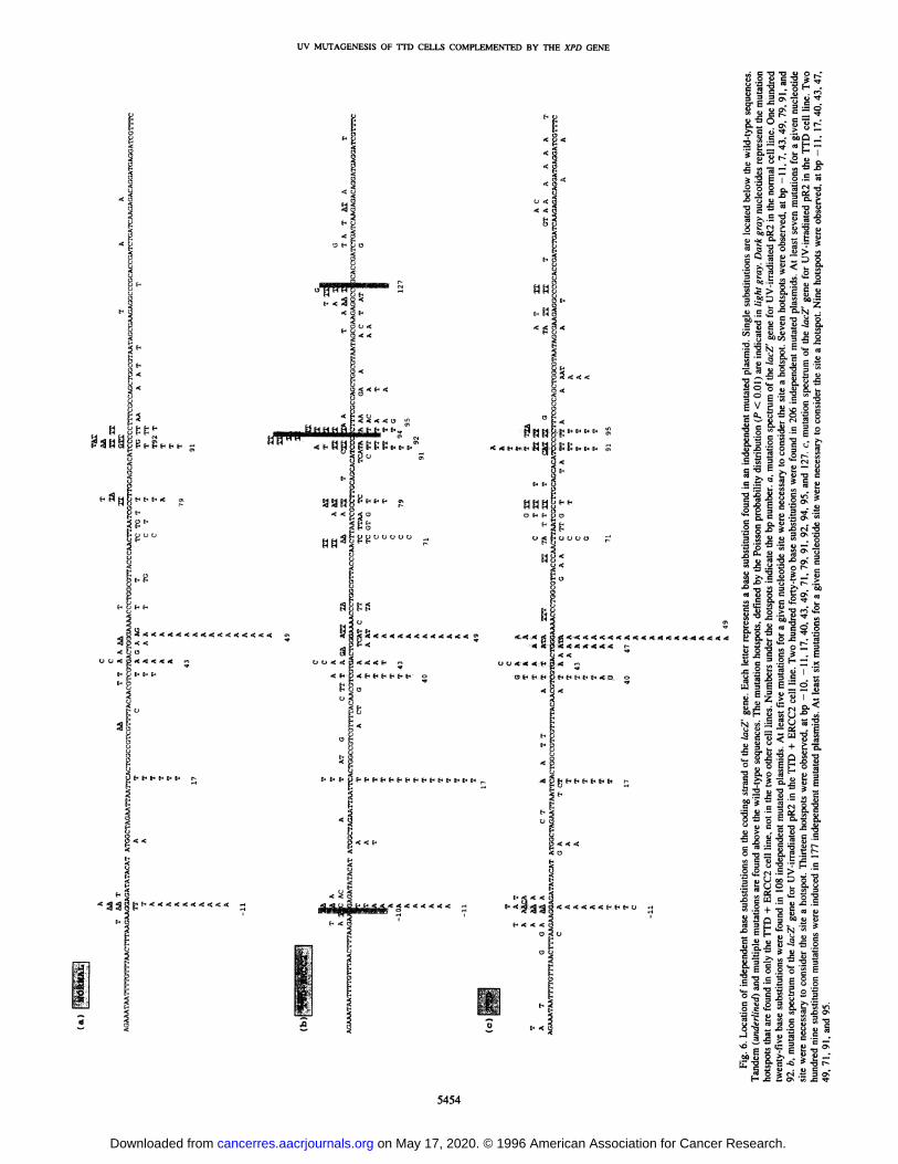
In the lTD + ERCC2 cell line, most of the hotspots found onlyin lTD cells were still present (40, 71, and 95 bp). Hotspots at
positions 79 and 92 were found in lTD + ERCC2 cells and innormal cells but not in lTD cells. Three new hotspots appeared inthe lTD + ERCC2 cell line at positions —10, 94, and 127 (Fig. 6,dark gray nucleotides).
DISCUSSION
The lTD syndrome is an autosomal recessive disorder characterized by brittle hair with a reduced sulfur content. Clinical photosensitivity is present in about 50% of lTD patients but is not associatedwith an elevated frequency of cancers. The fibroblasts from theselTD patients exhibit features of hypersensitivity to killing by UV-Cand reduced UDS similar to cells from patients with XP-D andUV-hypermutability (4, 10). The introduction of the wild-type XPD/ERCC2 gene by a plasmidic vector into ‘LTD/XP-Dcells restored anormal colony-forming ability of cells following UV-C exposure anda normal DNA repair capacity as well (23).
Here, we showed that some mutagenesis characteristics of repairproficient cells were restored in TID/XP-D cells complemented bythe wild-type XPD/ERCC2 gene. We used a clone isolated from thehuman ‘LTDIVI-LAS-KMTll fibroblast cell line transduced with aretroviral vector carrying the wild-type XPD/ERCC2 repair gene
Table 3 Frequency of mutated plasmids containing DNA rearrangements followingreplication of the untreated or UV-treated pR2 vector in human cell lines
aThepR2plasmidwastreatedwithdosesfrom250to1000J.m2.b pR2 plasmid was treated with doses from 100 to 1000 J . m2.
5453
neous and UV-induced frequency of rearranged mutants. This suggests that DNA rearrangements were not the consequence of UVirradiation. For the untreated or UV-treated pR2 plasmid, the frequency of mutants with rearrangements obtained with lTD andT@FD+ ERCC2 cells was at least 9-fold higher than that obtained withnormal cells (P < 0.01).
Mutation Spectra. The distributions of all UV-induced base substifurious found in the !acZ' gene using ‘LTD+ ERCC2, ‘LTD.and normalcell lines are shown in Fig. 6. We observed that almost all UV-inducedbase substitutions occurred at dipyrimidine sites (95% in thelTD + ERCC2 cells, 93% in the lTD cells, and 97% in normal cells).
The base substitutions were not scattered along the gene but formedseveral hotspots (Fig. 6, !ight gray nuc!eotides) that were determinedby the Poisson probability distribution (P < 0.01%). We found 13hotspots for ‘LTD+ ERCC2 cells, 9 hotspots for ‘LTDcells, and 7hotspots for normal cells. Five hotspots were common to the three celllines; they are located at positions —11, 17, 43, 49, and 91 . All ofthese hotspots did not share a common primary sequence.
Uv MUTAGENESISOF lTD CELLS COMPLEMENTED BY THE XPD GENE
(‘LTD+ ERCC2 cells). This clone recovered a UV cell survivalsimilar to that of normal cell line (Fig. 2), and the defect in UDS afterUv treatment was corrected as compared with the parental lID cellline (Fig. 3).
We showed that the XPD protein was expressed in the normal cellline and was mostly expressed in the ‘LTD+ ERCC2 cell line (Fig. 1).This result was also found by Carreau et a!. (26), who obtained goodsignals corresponding to XPD protein, when using transduced G418-selected XP-D primary fibroblasts. These authors did not detect theexpression of the XPD protein in repair-proficient untransformedprimary skin fibroblasts, whereas in another study the protein could bedetected in repair-proficient SV4O-transformed fibroblasts (24). Wefailed to detect the XPD protein in the ‘LTDcell line (Fig. 1).Takayama et a!. (18) studied the nucleotide sequence of the XPD/ERCC2 cDNA from the 1TD1VI patient from whom the ‘LTDcellswe used derived (10). They showed that one allele of the geneexhibited a C—@Gtransversion at bp 1459 leading to a Leu-46l-to-Valsubstitution and a 45-bp deletion at bp 2224—2268 leading to a15-amino acid deletion (amino acids 716—730)near the C-terminalpart of the protein. The authors also found a point mutation on theother allele, which led to a protein having Arg-722 changed intoTrp-722. Since the monoclonal antibody we used has been raisedagainst amino acids 749—759ofthe XPD protein, we can consider thatin our TTD cell line, the protein expression was too low to be detectedunder our stringent conditions or that conformation of the mutatedprotein would not allow a good presentation of the epitope.
Recently, Cleaver et a!. (34) showed that overexpression (abouttwo-fold) of the XPA gene in XP-A cells or in repair-competent cellsincreased the UV survival of the cells to levels above those of normalcells. Thus, they concluded that a correlation exists between DNArepair capacity and level of XPA protein expression. However, a goodcorrelation with UV cell survival but not with repair capacity has beenshown in two XP-D clones expressing the XPD protein at differentlevels (24). In our case, the lTD + ERCC2 cell line showed a levelof the XPD protein higher than in the normal cell line. However, theUv cell survival of the lTD + ERCC2 cells was not higher than thatof normal cells, whereas the UDS in the lTD + ERCC2 cells wasslightly increased at the UV dose of 20 J . m2 (Figs. 2 and 3). Hence,overexpression of the XPD protein does not seem to induce a repairlevel higher than that observed in repair-proficient cells.
The sequence analysis of approximately 500 independent mutatedplasmids from lTD + ERCC2, lID, and repair-proficient cellsrevealed that the major UV-induced type of mutation was single basesubstitution (Fig. 5). This finding was already shown in numerouspublished works using either shuttle vectors or endogenous genes (fora review, see Ref. 35).
In the three cell lines, a large majority of G:C—@A:Ttransitions(Table 2) were found, in agreement with earlier investigations, inwhich the G:C—@A:Ttransition was the major type of UV-inducedbase substitution (for a review, see Ref. 35). Classes of UV-inducedbase substitutions were similar between lTD + ERCC2 and normalcell lines, as we already found between lTD and normal cells (10).Thus, the expression of the XPD protein in ‘lTDcells did not changethe proportion of base substitutions.
In previous works (10, 36), we noticed that UV mutagenesis of thelTD cell line was characterized, in part, by multiple base mutations.Here, we showed that expression of the wild-type XPD protein afteraddition of the XPD/ERCC2 cDNA into the ‘LTDcells led to asignificant decrease of multiple mutants to a level similar to that ofnormal cells (Fig. 5). In turn, the level of single mutated plasmids thatwas lower in lTD cells (69%) than in normal cells (83%), wasincreased to a normal level (76%) after the introduction of the XPD/ERCC2 repair gene. The process leading to multiple base substitu
tions, which is still not understood, was overcome by the addition ofthe XPD/ERCC2 gene. It is interesting to note that GözUkaraet a!.(24) showed that the frequency of post-UV-mutated plasmids recovered with multiple base substitutions was lower in XP-D cells than innormal cells and remained low in the cell line transfected with theXPD/ERCC2 gene.
The lTD + ERCC2 cells were characterized by a lower mutationfrequency than the parental ‘LTDcell line after UV irradiation (Fig. 4).Despite this result, the plasmid mutability of the lTD + ERCC2 cellline was not lowered to values similar to those of normal cells, as onecould have expected from the expression of the wild-type XPDprotein. The clonal G4l8 selection of the LXPDSN-transduced cellline and mutagenesis assays performed immediately after this selection permit us to rule out the hypothesis that among the cells thatreplicate the pR2 plasmid, some may have lost the expression of the
XPD protein. In addition, Quilliet et a!. (37) recently showed thelong-term reversion of DNA repair-deficient primary fibroblasts bythe retroviral transduction of the XPD/ERCC2 gene. We can supposethat the mutation frequency would be inherent in the lTD + ERCC2and lTD parental cell lines. lTD and lTD + ERCC2 cells showedsimilar spontaneous mutation frequencies, 10-fold higher than the
mutation frequency of normal cells (Table 1). Most of the spontaneousmutants contained DNA rearrangements, and we showed that mutantswith DNA rearrangements obtained after UV irradiation could be dueto spontaneous lesions, since their frequencies do not vary with theUV dose (Table 3). The level of spontaneous mutants was not decreased to that of normal cells in the lTD + ERCC2 cells (Table 3).Thus, the introduction of the XPD/ERCC2 gene in the lTD cell linedid not decrease the frequency of spontaneous mutations. This suggests that the intrinsic mutation background of lTD andlTD + ERCC2 cell lines should be independent of the NER system.
Concerning the distribution of mutation hotspots, Tornaletti andPfeifer (38) suggested that hotspots could be generated by a slowDNA repair of lesions. In our case, the addition of the wild-typeXPD/ERCC2 repair gene in lTD cells failed to restore a pattern ofmutation hotspot distribution similar to that observed in normal cells.This suggests that slow repair in ‘LTDcells was not, or not only, thecause of mutation hotspots. This result was also found for XP-D (24)and XP-A cells (39). It is possible, as suggested by Seetharam et a!.(40), that some differences in the distribution of the UV-inducedmutation hotspots may be due to cellular factors.
In conclusion, the introduction of the wild-type XPD/ERCC2 DNArepair gene in a TTDIXP-D cell line corrected most consequences ofa DNA repair defect in the lTD cell line. It restored normal UV cellsurvival, normal DNA repair system, and, at least in part, mutagenesischaracteristics of repair-proficient cells. Indeed, it allowed, qualitatively, the restoration of UV-induced mutations similar to those observed in normal cells. Quantitatively, the mutation frequency of thelTD + ERCC2 cell line was considerably lower than that of the lTDparental cell line, although it did not reach the normal range. It ispossible that the parental lTD cell line, due to its state of transformation, had acquired intrinsic high mutagenic properties, which willlead to a still high mutation frequency found even after the correctionof the DNA repair defect. Finally, the location of mutation hotspotsfound for the lTD + ERCC2 cells was unique to this cell line and notfully similar to that of the normal cell line we used as a control.
ACKNOWLEDGMENTS
The authors thank Drs. A. Gentil and T. Magnaldo for critical reading of themanuscript; Drs. D. Biard and M. Carreau and N. Dumaz for their help inWestern blot analysis; and Dr. J. M. Egly for providing the anti-XPD mono
UV MUTAGENESIS OF lTD CELLS COMPLEMENTED BY THE XPD GENE
21 . Schaeffer, L., Moncollin, V., Roy, R., Staub, A., Mezzina, M., Sarasin, A., Weeds,G., Hoeijmakers,J. H. J., and Egly. J-M. The ERCC2/DNArepair protein isassociated with the class II BTF2TI'FIIH transcription factor. EMBO J., 13: 2388—2392, 1994.
22. Flejter, W. L., McDaniel, L. D., Johns, D., Friedberg, E. C.. and Schultz, R. A.Correction of xeroderma pigmentosum complementation group D mutant cell phenotypes by chromosome and gene transfer: involvement of the human ERCC2 DNArepair gene. Proc. Nail. Acad. Sci., USA, 89: 261—265,1992.
23. Mezzina, M., Eveno, E., Chevalier-Lagente, 0., Benoit, A., Carreau, M., Vermeulen,W.,Hoeijmakers,J. H.J., Stefanini,M.,Lehmann,A.R.,Weber,C. A.,andSarasin,A.CorrectionbytheERCC2geneof UV-sensitivityandrepairdeficiencyphenotypein a subset of trichothiodystrophy cells. Carcinogenesis (Lond.), 15: 1493—1498,1994.
24. GözUkara, E. M., Parris, C. N., Weber, C. A., Salazar, E. P., Seidman, M. M.,Watkins, J. F., Prakash, L.. and Kraemer, K. H. The human DNA repair gene, ERCC2(XPD),correctsultraviolethypersensitivityand ultraviolethypermutabilityof ashuttle vector replicated in xeroderma pigmentosum group D cells. Cancer Res., 54:3837—3844, 1994.
25. Carreau, M., Eveno, E., Quilliet, X., Chevallier-Lagente, 0., Benoit, A., Tanganelli,B.,Stefanini,M.,Vermeulen,W.,Hoeijmakers,J. H.J.,Sarasin,A.,andMezzina,M.Development of a new and easy complementation assay for DNA repair deficienthuman syndromes. Carcinogenesis (Lond.), 16: 1003—1009,1995.
26. Carreau, M., Quilliet, X., Eveno, E., Salvetti, A., Danos, 0., Heard, J. M., Mezzina,M., and Sarasin,A. Functionalretroviralvectorfor a genetherapyof XerodermaPigmentosum group D patients. Hum. Gene Ther., 6: 1307—1316,1995.
27. Huschtscha, L I., and Holliday. R. Limited and unlimited growth of SV4O-transformed cells from human diploid MRC-5 fibroblasts. J. Cell. Sci., 63: 77—99,1983.
28. Lehmann, A. R., Kirk-Bell, S.. Arlett, C. F., Harcourt, S. A., de Weerd-Kastelein,E. A.,Keijzer,W.,andHall-Smith,P. Repairof ultravioletlightdamagein a varietyof human fibroblast cell strains. Cancer Res., 37: 904—910,1977.
29. Wigler, M., Pellicer, A., Silverstein, S., and Axel, R. Biochemical transfer of singlecopy eucaryotic genes using total cellular DNA as donor. Cell, 14: 725—731,1978.
30. Stary. A., and Sarasin, A. Simian virus 40 (SV4O) large T antigen-dependent amplification of an Epstein-Barr virus-SV4O hybrid shuttle vector integrated into the humanHeLa cell genome. J. Gen. Virol., 73: 1679—1685, 1992.
31 . Hanahan, D. Studies on transformation of Escherichia coli with plasmids. J. Mol.Biol., 166: 557—580,1983.
32. Bredberg, A., Kraemer, K. H., and Seidman, M. M. Restricted ultraviolet mutationalspectrum in a shuttle vector propagated in xeroderma pigmentosum cells. Proc. Nail.Acad. Sci., USA, 83: 8273—8277,1986.
33. Dumaz, N., Stary, A., Soussi, T., Daya-Grosjean, L., and Sarasin, A. Can we predictsolar ultraviolet radiation as a causal event in human tumors by analysing themutation spectra of the p53 gene? Mutat. Ret., 307: 375—386,1994.
34. Cleaver, J. E., Charles, W. C., McDowell, M. L., Sadinski, W. J., and Mitchell, D. L.Overexpression of the XPArepair gene increases resistance to ultraviolet radiation inhuman cells by selective repair of DNA damage. Cancer Res., 55: 6152—6160,1995.
35. Sage, E. Distribution and repair of photolesions in DNA: genetic consequences andthe role of sequence context. Photochem. Photobiol., 57: 163—174,1993.
36. Madzak, C., Armier, J., Stary. A.. Daya-Grosjean. L, and Sarasin, A. UV-inducedmutations in a shuttle vector replicated in repair deficient trichothiodystrophy cellsdifferwiththosein genetically-relatedcancerpronexerodermapigmentosum.Carcinogenesis (Lond.), 14: 1255—1260,1993.
38. Tornaletti, S., and Pfeifer, G. P. Slow repair of pyrimidine dimers at p53 mutationhotspots in skin cancer. Science (Washington DC), 263: 1436—1438,1994.
39. Levy, D. D., Saijo, M., Tanaka, K., and Kraemer, K. H. Expression of a transfectedDNArepairgene(XPA)in xerodermapigmentosumgroupA cellsrestoresnormalDNArepairand mutagenesisof UV-treatedplasmids.Carcinogenesis(Land.),16:1557—1563,1995.
40. Seetharam, S., Kraemer, K. H., Waters, H. L., and Seidman, M. M. Mutationalhotspot variability in a ultraviolet-treated shuttle vector plasmid propagated in xeroderma pigmentosum and normal human lymphoblasts and fibroblasts. J. Mol. Biol.,212: 433—436,1990.
5456
REFERENCES
1. Hoeijmakers, J. H. J. Human nucleotide excision repair syndromes: molecular cluesto unexpected intricacies. Eur. J. Cancer, 30A: 1912—1921,1994.
2. Kraemer, K. H., Levy, D. D., Parris, C. N., GdzUkara, E. M., Moriwaki, S., Adelberg,S., and Seidman, M. M. Xeroderma pigmentosum and related disorders: examiningthe linkage between defective DNA repair and cancer. J. Invest. Dermatol., 103:96—101. 1994.
3. Ma. L., Hoeijmakers, J. H. J., and Van der Eb, A. J. Mammalian nucleotide excisionrepair. Biochim. Biophys. Acts. 1242: 137—164.1995.
4. Sarasin, A. The paradox of DNA repair-deficient diseases. The Cancer J.. 4: 233—237,1991.
5. Stary, A., and Sarasin, A. The genetic basis of xeroderma pigmentosum and trichothiodystrophy syndromes. Cancer Surv., 26: 55—171,1996.
6. Itin, P. H., and Pittelkow, M. R. Trichothiodystrophy: review of sulfur-deficientbrittle hair syndromes and association with the ectodermal dysplasias. J. Am. Acad.Dermatol., 22: 705—717,1990.
7. Van Neste, D., Caulier, B., Thomas. P., and Vasseur, F. PIBIDS/Tay's syndrome andxeroderma pigmentosum. J. Am. Acad. Dermatol., 12: 372—373,1985.
8. Sarasin, A.. Blanchet-Bardon, C., Renault, G., Lehmann, A., Arlett, C., and Dumez,Y. Prenataldiagnosisin a subsetof trichothiodystrophypatientsdefectivein DNArepair. Br. J. Dermatol.. 127: 485—491, 1992.
9. Lehmann, A. R., Arlett, C. F., Broughton, B. C., Harcourt, S. A., Steingrimsdottir, H.,Stefanini, M., Malcolm, A., Taylor, R., Natarajan, A. T., Green, S., King, M. D.,MacKie, R. M., Stephenson, J. B. P., and Tolmi, J. L. Trichothiodystrophy, a humanDNA repair disorder with heterogeneity in cellular response to ultraviolet light.Cancer Res., 48: 6090—6096, 1988.
10. Marionnet, C., Benoit, A., Benhamou, S., Sarasin, A., and Stary, A. Characteristics ofUV.induced mutation spectra in human XP-D/ERCC2 gene-mutated xeroderma pigmentosum and trichothiodystrophy cells. J. Mol. Biol., 252: 550—562,1995.
I 1. Stefanini, M., Lagomarsini, P., Arlett. C. F.. Borrone, C., Crovato, F., Trevisan, G.,Cordone, G.. and Nuzzo, F. Xeroderma pigmentosum (complementation group D)mutation is present in patients affected by trichothiodystrophy with photosensitivity.Hum.Genet., 74: 107—112,1986.
12. Broughton. B. C., Lehmann, A. L., Harcourt, S. A., Arlett, C. F., Sarasin, A., Kleijer,W. J., Beemer. F. A., Naim. R., and Mitchell, D. L. Relationship between pyrimidinedimers, 6-4 photoproducts, repair synthesis and cell survival: studies using cells frompatients with trichothiodystrophy. Mutat. Res., 235: 33—40,1990.
13. Stefanini, M., Lagomarsini, P., Giliani, S., Nardo, T., Bona, E., Peserico, A., Kleijer,w. j., Lehmann,A. R.,andSarasin,A. Geneticheterogeneityof theexcisionrepairdefect associated with trichothiodystrophy. Carcinogenesis (Land.). 14: 1101—1105,1993.
14. Vermeulen, W., van Vuuren, A. J., Chipoulet, M., Schaeffer, L., Appeldoorn, E.,Weeds, G., Jaspers, N. G. J., Priestley, A., Arlett, C. F., Lehmann, A. R., Stefanini,M., Mezzina,M., Sarasin,A., Bootsma,D., Egly,J-M.,and Hoeijmakers,J. H. J.Three unusual repair deficiencies associated with transcription factor BTF2 (TFIIH).Evidence for the existence of a transcription syndrome. Cold Spring Harbor Sym.Quant. Biol., UX: 317—329,1994.
15. Stefanini, M., Vermeulen, W., Weeds. G. A., Giliani, S., Nardo. T., Mezzina, M.,Sarasin, A., Harper, J. I., Arlett, C. F.. Hoeijmakers, J. H. J., and Lehmann, A. R. Anew nucleotide excision repair gene associated with the disorder trichothiodystrophy.Am. J. Hum. Genet., 53: 817—821,1993.
16. Broughton. B. C., Steingrimsdottir. H., Weber, C. A., and Lehmann, A. R. Mutationsin the xeroderma pigmentosum group D DNA repair/transcription gene in patientswith tnchothiodystrophy. Nat. Genet.. 7: 189—194, 1994.
17. Mondello, C.. Nardo, T., Giliani. S.. Arrand, J. E., Weber, C. A.. Lehmann, A. R.,Nuzzo, F., and Stefanini, M. Molecular analysis of the XP-D gene in Italian familieswith patients affected by trichothiodystrophy and xeroderma pigmentosum group D.Mutat. Res., 314: 159—165,1994.
18. Takayama. K., Salazar, E. P., Broughton, B. C., Lehmann, A. R., Sarasin, A.,Thompson. L. H., and Weber, C. A. Defects in the DNA repair and transcription geneERCC2(XPD)in trichothiodystrophy.Am.J. Hum.Genet.,58: 263—270,1996.
19. Weber, C. A., Salazar, E. P., Stewart, S. A., and Thompson, L. H. ERCC-2: cDNAcloning and molecular characterization of a human nucleotide excision repair genewith high homology to yeast RAD3. EMBO J.. 9: 1437—1447,1990.
20. Sung, P.. Bailly, V., Weber, C., Thompson, L. H., Prakash, L., and Prakash, S. Humanxeroderma pigmentosum group D gene encodes a DNA helicase. Nature (Land.), 365:852—855,1993.