21
| Date post: | 22-Dec-2015 |
| Category: |
Documents |
| Upload: | vernon-stanley |
| View: | 221 times |
| Download: | 0 times |
Rectal Examination
Anatomy I
The rectum is the curved lower, terminal segment of large bowel.
It is about 12 cms long and runs along the concavity of the sacrum.
Anterior to the lower 1/3 of the rectum lie different structures in men and women
Anatomy II
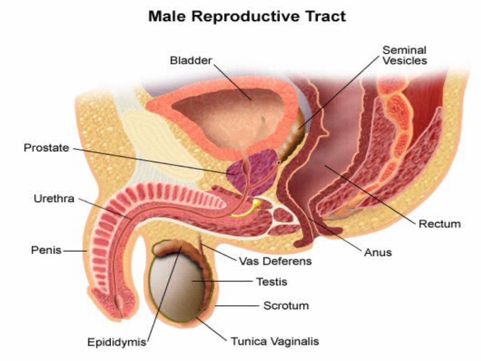
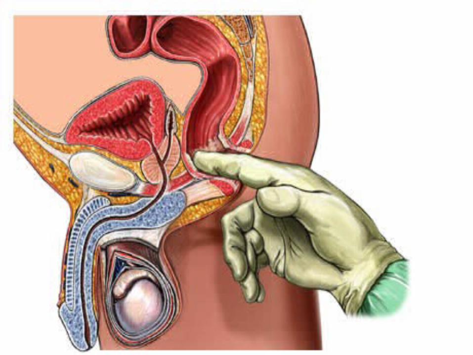
In men, anterior to the lower 1/3 of the rectum lie the prostate, bladder base and seminal vesicles.
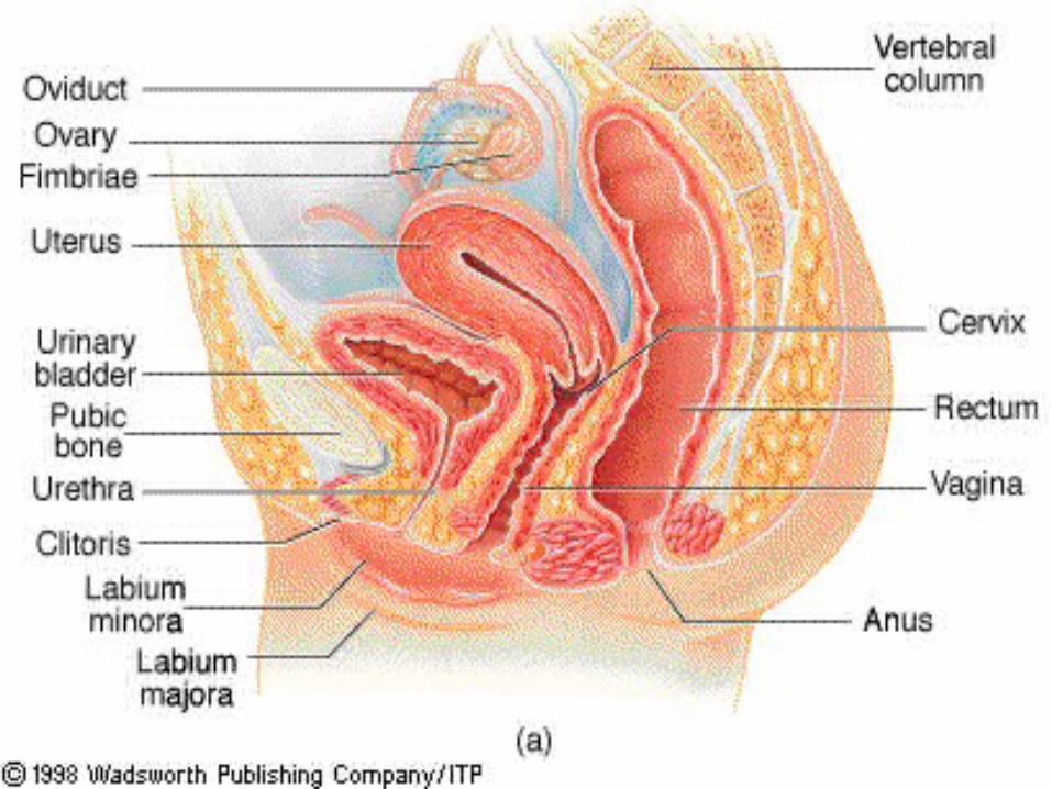
In women, anterior to the lower 1/3 of the rectum lies the vagina. At the tip of the examining finger it may be possible to feel cervix and even a retroverted Uterus
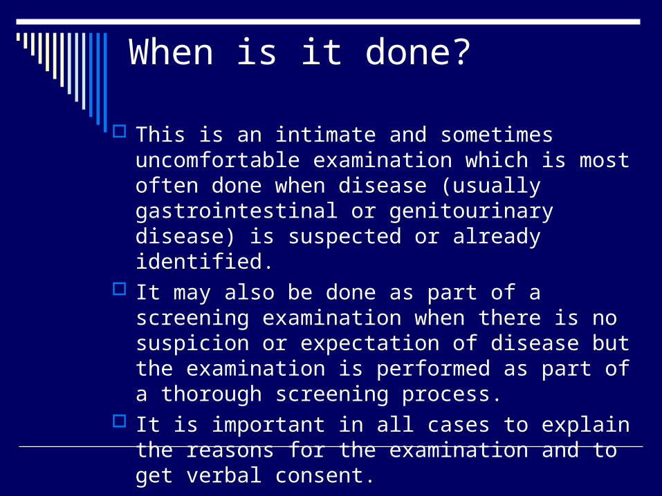
When is it done?
This is an intimate and sometimes uncomfortable examination which is most often done when disease (usually gastrointestinal or genitourinary disease) is suspected or already identified.
It may also be done as part of a screening examination when there is no suspicion or expectation of disease but the examination is performed as part of a thorough screening process.
It is important in all cases to explain the reasons for the examination and to get verbal consent.

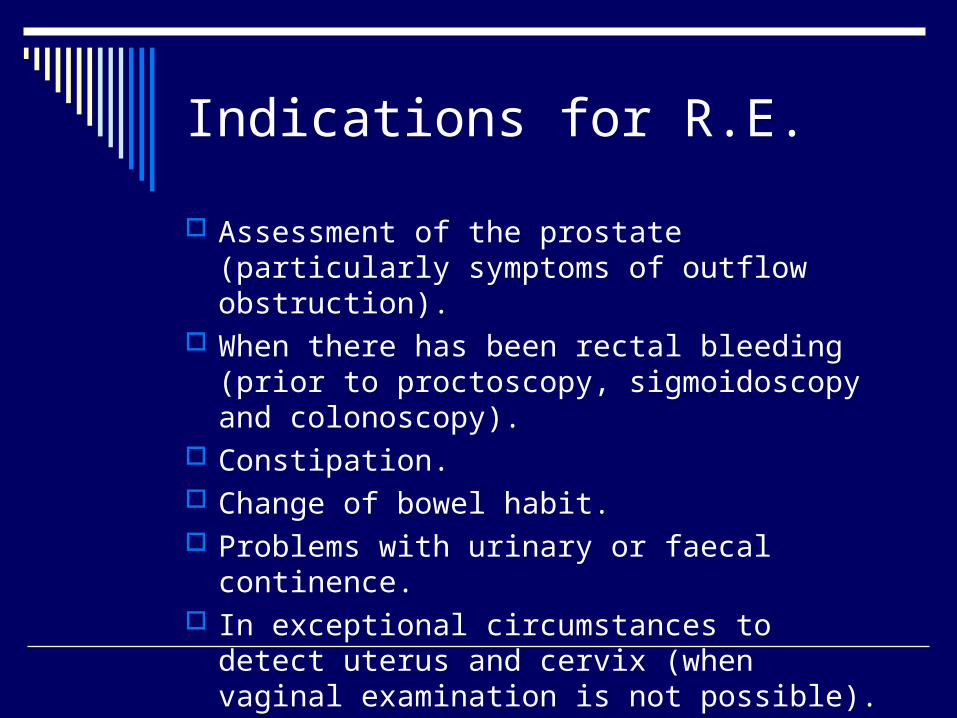
Indications for R.E.
Assessment of the prostate (particularly symptoms of outflow obstruction).
When there has been rectal bleeding (prior to proctoscopy, sigmoidoscopy and colonoscopy).
Constipation. Change of bowel habit. Problems with urinary or faecal continence. In exceptional circumstances to detect uterus
and cervix (when vaginal examination is not possible).
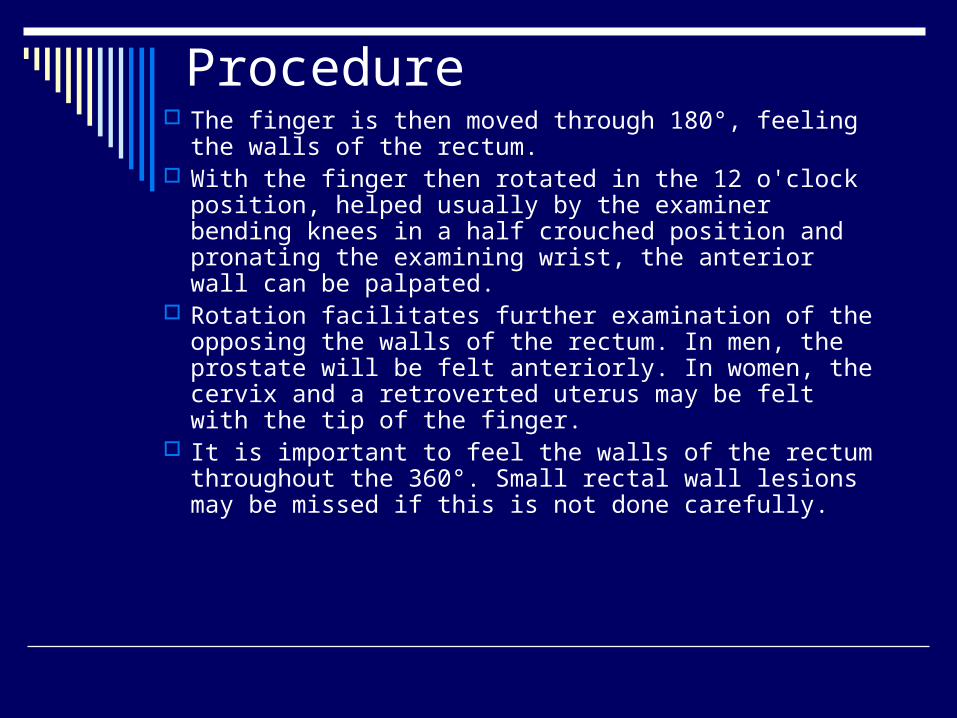
Procedure The finger is then moved through 180°, feeling the
walls of the rectum. With the finger then rotated in the 12 o'clock position,
helped usually by the examiner bending knees in a half crouched position and pronating the examining wrist, the anterior wall can be palpated.
Rotation facilitates further examination of the opposing the walls of the rectum. In men, the prostate will be felt anteriorly. In women, the cervix and a retroverted uterus may be felt with the tip of the finger.
It is important to feel the walls of the rectum throughout the 360°. Small rectal wall lesions may be missed if this is not done carefully.
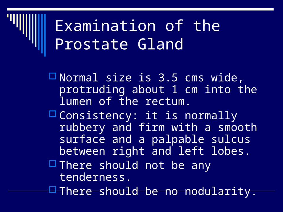
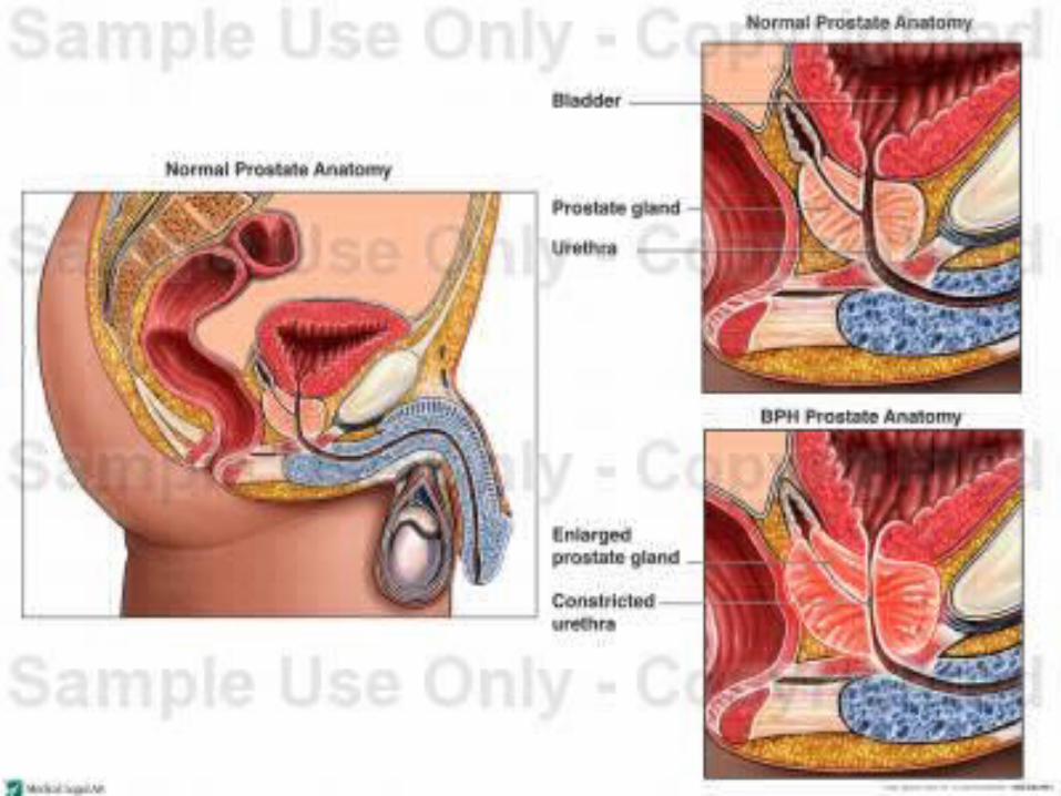
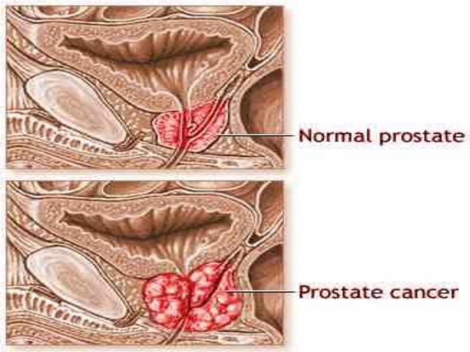
Examination of the Prostate Gland
Normal size is 3.5 cms wide, protruding about 1 cm into the lumen of the rectum.
Consistency: it is normally rubbery and firm with a smooth surface and a palpable sulcus between right and left lobes.
There should not be any tenderness. There should be no nodularity.
http://beta.medicalvideos.us/videos-354-Rectal-Examinations
http://beta.medicalvideos.us/videos-2539-Proctoscope-Medical-Examination-of-the-Rectum

External Inspection Skin disease. Skin tags Genital warts Anal fissures Anal fistula External haemorrhoids Rectal prolapse Skin discolouration with Crohn's disease External thrombosed piles
Internal Inspection
Simple piles (but best examined at proctoscopy)
Rectal carcinoma Rectal polyps Tenderness Diseases of the prostate gland Malignant or inflammatory conditions of
the peritoneum (felt anteriorly)
Contraindications
Imperforate Anus Unwilling patient Immunosuppressed patient Absence of anus following surgical
excision Stricture Moderate to severe anal pain Prolapsed thrombosed internal
hemorroids

THANKS FOR LISTENING

21


