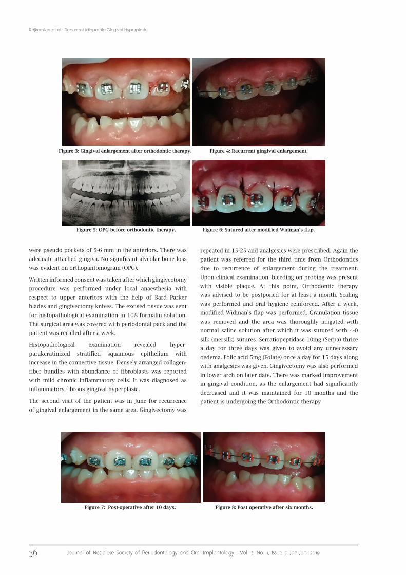
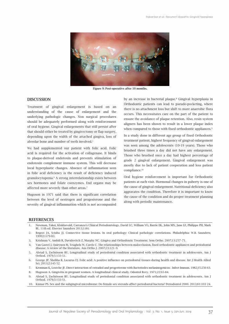
35 Journal of Nepalese Society of Periodontology and Oral Implantology : Vol. 3, No. 1, Issue 5, Jan-Jun, 2019 Recurrent Idiopathic Gingival Hyperplasia Case Report ABSTRACT Increase in size of the gingiva is termed as gingival enlargement. Most common type of gingival enlargement is inflammatory, which his caused due to plaque accumulation and improper oral hygiene maintenance. Orthodontic therapy can often lead to failure to improve oral hygiene. This case describes a recurrent, progressive gingival enlargement of a 19 year old female orthodontic patient in which gingivectomy was performed and repeated, which subsequently failed. Hence modified Widman’s flap was performed with medical supplements. Periodic periodontal check up is required in orthodontic cases to control the gingival inflammation. Patient compliance is also very important in such cases. There should be proper co-operation between the Orthodontist and Periodontist for successful treatment of gingival hyperplasia. Patients with such conditions should be carefully monitored and checked to avoid the recurrence and avoid further progression into chronic periodontitis. Keywords: Gingival enlargement; hyperplasia; orthodontic patient; recurrent. INTRODUCTION Gingival enlargement or overgrowth is a clinical descriptive term for increases in size of gingiva. 1 Most commonly, gingival enlargement can be due to plaque accumulation, poor oral hygiene, inadequate nutrition, or systemic hormonal stimulation. 2 Gingival enlargements are also seen in blood dyscrasias like leukaemia and thrombocytopenia. 1 Idiopathic gingival enlargement is a rare type of gingival enlargement that has no definite cause. Inability to properly clean the teeth surfaces clean and presence of plaque is considered as one of the main factors responsible for the development of gingivitis as orthodontic brackets and elastics might interfere with effective removal of plaque. 3 There is also shift in the composition of bacteria due to orthodontic treatment as it can increase the bacterial retention. 4 Gingival enlargement in orthodontic patient can lead to pseudo pocket, where the hyperplasia can lead to deep artificial pockets and this has been related to shift to more anaerobic flora like B. intermedius, Spirochetes, B. forsythus, T. denticola, P. nigrescens, etc. 5 This is a case report of an unusual case of a non-syndromic recurrent idiopathic gingival enlargement. Dr. Junima Rajkarnikar, 1 Dr. Bikash Veer Shrestha, 2 Dr. Santhosh Kumar 3 1 Department of Periodontology, Nepal Medical College, Kathmandu, Nepal; 2 Department of Orthodontics, Nepal Medical College, Kathmandu, Nepal; 3 Department of Periodontology, Manipal College of Dental Sciences, Manipal Acadamy of Higher Education, Manipal, Karnataka, India. CASE REPORT A 19 year old female patient from Besigaun, Jorpati was referred to the department of Periodontics, Nepal Medical College Teaching Hospital from the department of Orthodontics of the same for the treatment of gingival enlargement. Her first visit to the Department of Periodontics was in May 2018 during which Phase I therapy was completed with oral prophylaxis and oral hygiene instructions. Chlorhexidine mouthwash 0.2% (Hexidine) 10ml BD was given for seven days. She gave the history of gingival enlargement which was evident since few years. Gingival tissue was pale pink, enlarged, firm, fibrotic and mild inflammatory. It was painless and there was no difficulty in speech and mastication. There was no history of drugs intake, no mental or physical disorder and no systemic disease present. Family history was non contributory. So the patient was recalled for gingivectomy after a week. On her second visit, oral examination revealed fibrotic gingival enlargement involving upper and lower anteriors with grade II gingival enlargement involving the papilla and marginal gingiva (Bokenkamp Index - 1994). There J Nepal Soc Perio Oral Implantol. 2019;3(5):35-7 Correspondence: Dr. Junima Rajkarnikar Department of Periodontics, Nepal Medical College, Kathmandu, Nepal. email: [email protected]Citation Rajkarnikar J, Shrestha BV, Kumar S. Recurrent Idiopathic Gingival Hyperplasia. J Nepal Soc Perio Oral Implantol. 2019;3(5):35-7. Figure 1: Gingival enlargement prior to orthodontic therapy. Figure 2: Extraoral view.
Transcript
35Journal of Nepalese Society of Periodontology and Oral Implantology : Vol. 3, No. 1, Issue 5, Jan-Jun, 2019
Recurrent Idiopathic Gingival Hyperplasia
Case Report
ABSTRACTIncrease in size of the gingiva is termed as gingival enlargement. Most common type of gingival enlargement is inflammatory, which his
caused due to plaque accumulation and improper oral hygiene maintenance. Orthodontic therapy can often lead to failure to improve
oral hygiene. This case describes a recurrent, progressive gingival enlargement of a 19 year old female orthodontic patient in which
gingivectomy was performed and repeated, which subsequently failed. Hence modified Widman’s flap was performed with medical
supplements. Periodic periodontal check up is required in orthodontic cases to control the gingival inflammation. Patient compliance is
also very important in such cases. There should be proper co-operation between the Orthodontist and Periodontist for successful treatment
of gingival hyperplasia. Patients with such conditions should be carefully monitored and checked to avoid the recurrence and avoid further
3. Krishnan V, Ambili R, Davidovitch Z, Murphy NC. Gingiva and Orthodontic Treatment. Sem Ortho. 2007;13:257–71.
4. Van Gastel J, Quirynen M, Teughels W, Carels C. The relationships between malocclusion, fixed orthodontic appliances and periodontal disease: A review of the literature. Aus Ortho J. 2007;23:121–9.
5. Alstad S, Zachrisson BU. Longitudinal study of periodontal condition associated with orthodontic treatment in adolescents. Am J Orthod. 1979;5:133-51.
6. George JP, Shobha R, Lazarus FJ. Folic acid: A positive influence on periodontal tissues during health and disease. Int J Health Allied Sci. 2013;2:145-52.
7. Kronman K, Loseche JF. Direct interaction of estradiol and progesterone with Bacteriodes melaninogenicus . Infect Immun. 1982;35:256-63.
8. Hugoson A. Gingivitis in pregnant women. A longitudinal clinical study. Odontol Revy. 1971;22:65-84.
9. Alstad S, Zachrisson BU. Longitudinal study of periodontal condition associated with orthodontic treatment in adolescents. Am J Orthod. 1979;5:133-51.
10. Kumar PS. Sex and the subgingival microbiome: Do female sex steroids affect periodontal bacteria? Periodontol 2000. 2013;61:103 24.
Rajkarnikar et al : Recurrent Idiopathic Gingival Hyperplasia
DISCUSSION
Treatment of gingival enlargement is based on an
understanding of the cause of enlargement and the
underlying pathologic changes. Non surgical procedures
should be adequately performed along with reinforcement
of oral hygiene. Gingival enlargements that still persist after
that should either be treated by gingivectomy or flap surgery,
depending upon the width of the attached gingiva, loss of
alveolar bone and number of teeth involved.1
We had supplemented our patient with folic acid. Folic
acid is required for the activation of collagenase. It binds
to plaque-derived endotoxin and prevents stimulation of
endotoxin complement immune system. This will decrease
local hyperplastic changes. Absence of inflammation seen
in folic acid deficiency is the result of deficiency induced
granulocytopenia.6 A strong interrelationship exists between
sex hormones and folate coenzymes. End organs may be
affected more severely than other areas.7
Hugoson in 1971 said that there is significant correlation
between the level of oestrogen and progesterone and the
severity of gingival inflammation which is not accompanied
by an increase in bacterial plaque.8 Gingival hyperplasia in
Orthodontic patients can lead to pseudo-pocketing, where
there is no attachment loss but shift to more anaerobic flora
occurs. This necessitates care on the part of the patient to
ensure the avoidance of plaque retention. Also, resin system
aligners has been shown to result in a lower plaque index
when compared to those with fixed orthodontic appliances.9
In a study done in different age group of fixed Orthodontic
treatment patient, highest frequency of gingival enlargement
was seen among the adolescents (10-19 years). Those who
brushed three times a day did not have any enlargement.
Those who brushed once a day had highest percentage of
grade 2 gingival enlargement. Gingival enlargement was
mostly due to lack of patient cooperation and less patient
compliance.10
Oral hygiene reinforcement is important for Orthodontic
patients at each visit. Hormonal changes in puberty is one of
the cause of gingival enlargement. Nutritional deficiency also
aggravates the condition. Therefore it is important to know
the cause of the condition and do proper treatment planning


![Endometrium presentation - Dr Wright[1] · Endometrial Hyperplasia Simple hyperplasia Complex hyperplasia (adenomatous) Simple atypical hyperplasia ... Progression of Hyperplasia](https://static.documents.pub/doc/80x56/5b8a421e7f8b9a50388bc13d/endometrium-presentation-dr-wright1-endometrial-hyperplasia-simple-hyperplasia.jpg)


