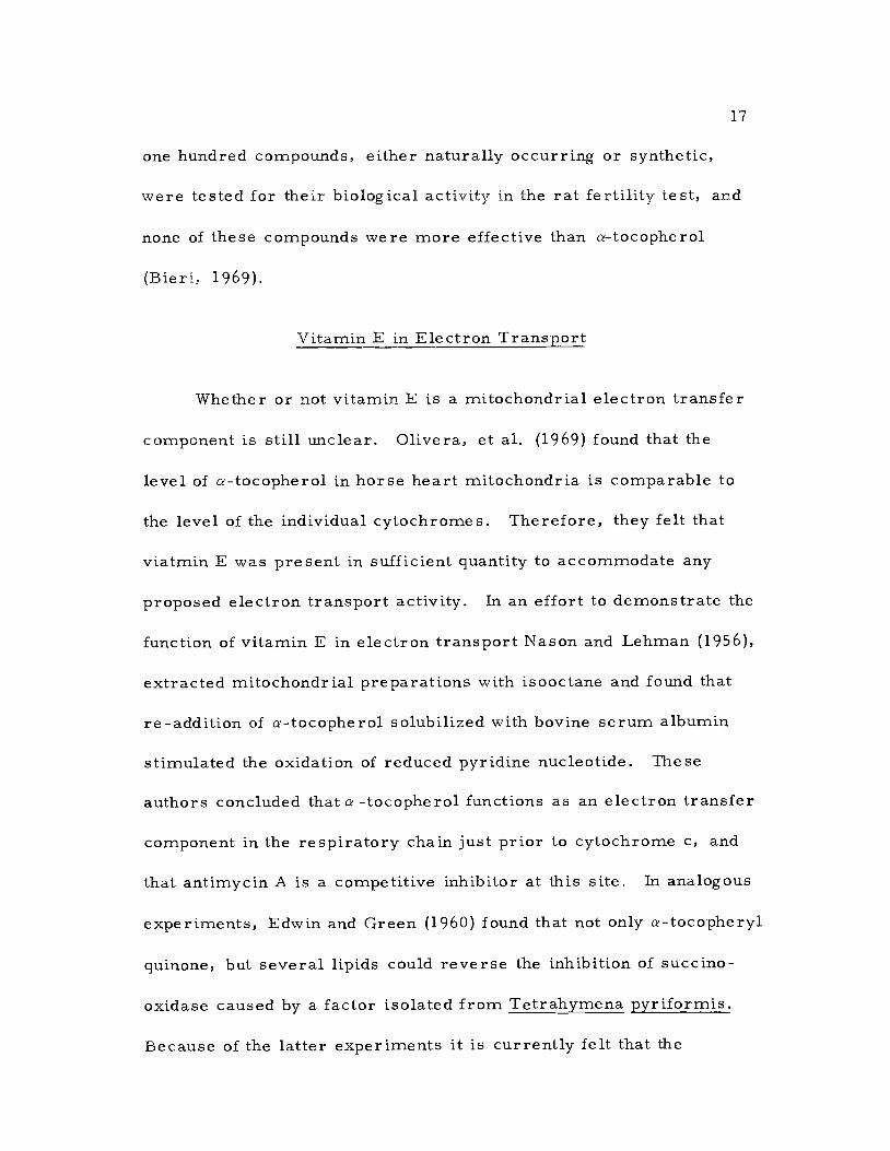
AN ABSTRACT OF THE THESIS OF Richard Charles Sicher, Jr. for the degree of Doctor of Philosophy in Botany (Plant Physiology) presented on June 28, 1976 Title: THE EFFECTS OF A MUTATION WITHIN VITAMIN E BIOSYNTHESIS UPON THE DEVELOPMENT AND FUNCTION OF THE PHOTOSYNTHETIC APPARATUS Abstract approved: Redacted for Privacy (NormaK' I. Bishop) A new photosystem-II mutant of the green alga Scenedesmus obliquus D3, strain PS-28, has been shown to lack a-tocopherol (vitamin E). The photosynthetic activity of dark grown samples of PS-28 is about 20% of the wild-type control. Culturing the mutant at low light intentisites (104 ergs/sec-cm 2) stimulates photosynthetic activity by as much as 3 fold. Mutant PS-28 has a high relative fluorescence which lacks the variable yield component, but the levels of plastoquinone A, cytochrome b-559 (H. P. ), and chlorophyll are nearly normal. This evidence suggests that the nature of the muta- tion in PS-28 is not pleiotropic, but occurs at a specific site, in the vitamin E biosynthetic pathway. In both mixotrophic and heterotrophic samples of the mutant photosynthesis can be destroyed by exposure of the cells to high intensity irradiation (106 ergs/sec-cm 2). This photoinactivation is proportional to the intensity of the irradiation, and does not occur if
Transcript
AN ABSTRACT OF THE THESIS OF
Richard Charles Sicher, Jr. for the degree of Doctor of Philosophy
in Botany (Plant Physiology) presented on June 28, 1976
Title: THE EFFECTS OF A MUTATION WITHIN VITAMIN E
BIOSYNTHESIS UPON THE DEVELOPMENT AND FUNCTION
OF THE PHOTOSYNTHETIC APPARATUS
Abstract approved: Redacted for Privacy(NormaK' I. Bishop)
A new photosystem-II mutant of the green alga Scenedesmus
obliquus D3, strain PS-28, has been shown to lack a-tocopherol
(vitamin E). The photosynthetic activity of dark grown samples of
PS-28 is about 20% of the wild-type control. Culturing the mutant
at low light intentisites (104 ergs/sec-cm 2) stimulates photosynthetic
activity by as much as 3 fold. Mutant PS-28 has a high relative
fluorescence which lacks the variable yield component, but the levels
of plastoquinone A, cytochrome b-559 (H. P. ), and chlorophyll are
nearly normal. This evidence suggests that the nature of the muta-
tion in PS-28 is not pleiotropic, but occurs at a specific site, in the
vitamin E biosynthetic pathway.
In both mixotrophic and heterotrophic samples of the mutant
photosynthesis can be destroyed by exposure of the cells to high
intensity irradiation (106 ergs/sec-cm 2). This photoinactivation is
proportional to the intensity of the irradiation, and does not occur if
treatments are performed anaerobically; thus, the damage to the
photosynthetic process occurs via a photodynamic mechanism.
a-Tocopherol, a-tocopheryl acetate or synthetic antioxidants, such
as nordihydroguaiaretic acid and N, N' -diphenyl-p-phenylenediamine,
when added to the growth medium neither stabilize the mutant against
photoinactivation nor reverse the mutation syndrome.
The capacities for hydrogen photoreduction, the production of
a 518 nm light-induced absorbancy change and PMS-mediated photo-
phc.,sphorylation are only moderately affected by the mutation. Also,
the above mentioned processes do not appear to be influenced by
exposure of the cells to damaging intensities of white light. Con-
trarily, the rates of hydrogen photoproduction and anaerobic glucose
photoassimilation are below normal inthemutant, and these processes
show strong sensitivities to irradiation. The ferricyanide or DCPIP
Hill reactions (Photosystem-11) in contrast to the ascorbate -DC PIP
to methylviologen photoreduction (Photosystem-1) are not observed
in chloroplasts prepared from the mutant. Summarized, these
findings indicate that the mutant has a partially impaired photo-
system-II which is sensitive to high intensity irradiation treatments,
and a fully functional photosystem-I which is stable to irradiation.
The lipid and fatty acid complement in irradiated and unirrad-
iated samples of PS-28 were compared to similar samples of the
wild-type, and in no case was any difference noted between the
mutant and the parent strain. Furthermore, several photosystem-II
mutants, possessing limited photosynthetic capacities, but having
normal levels of a -tocopherol were also found to be susceptible to
photoinhibition by high intensity irradiation treatment. These
results indicate that a -tocopherol does not function as a general
membrane antioxidant for the photosynthetic process.
The levels of vitamin E were analyzed during the greening of
mutant C -2A'. In dark grown cells of C-2.A' the level of
a-tocopherol is equivalent to that of the wild-type. After greening,
the level of a-tocopherol in the mutant is equivalent to that of mixo-
trophic samples of the wild-type. Contrarily, the level of plas-
toquinone A is at a minimum in dark grown cells of C-2A' and is
synthesized in parallel with the onset of photosynthesis during
greening. These observations suggest that the role of a-tocopherol
in photosynthesis is different than that of plastoquinone A, which is
a known electron transport intermediate.
A thorough consideration of the above information rules out a
role for vitamin E in photosynthetic electron transfers or phosphory-
lations. The data do not support the conclusion that vitamin E func-
tions in the chloroplast as a general membrane antioxidant. This
suggests that toc ,plierol must either function as a site specific
antioxidant or as a structural component in or near the photosystem-
II chloroplast subunit.
The Effects of a Mutation Within Vitamin E BiosynthesisUpon the Development and Function of the
Photosynthetic Apparatus
by
Richard Charles Sicher, Jr.
A THESIS
submitted to
Oregon State University
in partial fulfillment ofthe requirements for the
degree of
Doctor of Philosophy
June 1977
APPROVED:
Redacted for PrivacyProfessor of Botany and PlaAPathology
in charge of major
Redacted for PrivacyChairman of Department of Botany and Plant Pathology
Redacted for Privacy
Dean of Graduate School
te- thesis is 'presented June 28, 1976
Typed by Susie Kozlik for Richard Charles Sicher, Jr.
ACKNOWLEDGMENTS
I would like to thank my major professor, Dr. Norman Bishop,
for his support and encouragement during the period when these
studies were being conducted, and for his valuable suggestions
concerning the preparation of this manuscript. Very special
appreciation is extended to Drs. Harold Evans, Don Reed, Irvin
Isenberg, and B. J. Verts for serving on my graduate studies
committee. Acknowledgments are also extended to Drs. Ralph
Quatrano and W. David Loomis for reading and editing this manu-
script. My friends and associates, Drs. Larry Jones and Gunnar
Oquist, were of invaluable assistance, and I also acknowledge the
technical help of Ms. Marianna Frick and Mr. James Wong.
I would like to express my gratitude to Mary Ellen Hood for
typing the early drafts of this thesis, and to my wife, Joan, for her
unwavering encouragement.
Financial support for these investigations was provided by
a Research Assistantship from the National Science Foundation
(GB-33925 and BMS-7518023).
TABLE OF CONTENTS
Chapter Page
I INTRODUCTION 1
Definition and Role of Photosynthesis 1
Photosynthetic Components and LipophilicQuinones of Biological Importance 6
Ubiquinone 8Napthoquinone 9Plastoquinone 12Tocopheryl Quinone 14Effects of a Vitamin E Deficiency 16Vitamin E in Electron Transport 17Vitamin E in Phosphorylation 20Vitamin E as a Membrane Stabilizer 22
vated charcoal and silicic acid filtered). The individual bands
corresponding to plastoquinone A and a-tocopherol were located with
0.01% Rhodamine B in ethanol, by fluorescence quenching under
47
ultraviolet light. These bands were scraped from the plates and
were eluted free of the adsorbent with redistilled CHC13. The
eluant was dried under a vacuum and the compounds were dissolved
in ethanol (3 ml) for quantitation.
The plastoquinone A concentrations were calculated from the
following equation, based on an a mM of 15:
total p.moles plastoquinone A = (A255) (0. 2005) (Vt/Va)
where A255 was the absorbancy of the reduced versus the oxidized
absorbance of plastoquinone A at 255 nm, Vt was the total volume
of the extract in CHC13, and Va was the volume of the extract
assayed.
Vitamin E was quantitated by the method of Emmerie-Engel
(Barr and Crane, 1971), adapted for spectrophotometry. The optical
densities obtained for 1 ml of the ethanolic solution of vitamin E
were converted to concentration (imo les) by the following equation:
total p.moles a -tocopherol = (3) (0. 35) (A520)
where 3 was the dilution factor correcting for the volume of the
sample assayed, 0. 35 was the conversion coefficient obtained from
a standard curve made with authentic a-tocopherol (Nutritional
Biochemical Corporation), and A520 was the absorbance observed
for the sample.
48
a-Tocopheryl quinone was isolated by the procedures des-
cribed by Barr and Crane (1971). Cells (2.0 ml PCV) were extracted
with warm methanol as above, and the extracts were dried under
reduced pressure. The dried samples were taken up in a minimal
volume of redistilled petroleum ether and were adsorbed onto a
column of acid alumina. The column was eluted sequentially with
100 ml each of 10% and 20% diethylether-petroleum ether. The final
elution of the column, 200 ml of 30% diethylether-petroleum ether,
contained the a-tocopheryl quinone. This fraction was reduced to
2 ml under vacuum and the sample was chromatographed on thin-
layer silicic acid plates (Silica gel G, Merck) with petroleum ether-
benzene-ethanol) (8:3:0.7). Quinones present in the chromatogram
were detected with 0.01% Rhodamine B as before, and the band
corresponding to a-tocopheryl quinone was eluted by filtration with
chloroform. The eluant was taken to dryness under reduced pres-
sure and the sample was dissolved in exactly 3 ml of ethanol. The
concentrations of a-tocophyl quinone (p,moles) were quantitated with
the following equation:
total p.moles a-tocopheryl quinone = (A262) (0. 1658) (Vt/Va)
where A262 was the optical density of the sample in 3 ml (oxidized
minus reduced), 0.1658 was a conversion coefficient based on an
49
extinction coefficient of 17.8 amM, Vt was the total sample volume,
and V awas the volume of the sample assayed.
50
IV. RESULTS AND DISCUSSION
General Characteristics, Photosynthesis, and Respiration
A mutant of the alga Scenedesmus obliquus, strain PS-28, was
induced by x-ray irradiation, and was isolated with standard tech-
niques (Bishop, 1971b). The mutant strain grows well on nitrate-
glucose-yeast extract medium (Kessler, Arthur, and Brugger,
1957), either in the dark or in the light, but is incapable of auto-
trophic growth on nonsupplemented nitrate medium. These findings
indicated that PS-28 could not grow under strictly photosynthetic
conditions, therefore, the mutation had affected the photosynthetic
apparatus.
Mutant PS-28 does not synthesize a-tocopherol (Bishop and
Wong, 1974). To investigate the function of a-tocopherol, in its
relation to photosynthesis, several secondary mutants were gener-
ated from PS-28 by further x-ray induction. These sub-mutants
were identified by pigment deficiencies and were isolated by visual
means. The subisolates were designated C-28-2, C-28-3, C-28-4,
etc. , and data obtained from certain of these are presented in
subsequent sections. It was observed that for both mixotrophic
and heterotrophic growth, the photosynthetic rate of the mutant was
approximately half that of the wild-type (Table 1). As expected the
photosynthetic activity of the wild-type improved when cultured
51
Table 1. Photosynthesis and respiration measurements of wild-typeScenedesmus and mutant strain PS-28.
Comparative rates of photosynthesis and respirationbetween wild-type Scenedesmus and mutant strain PS-28are expressed as the average of 5 independent observations.Measurements were performed with a Clark Ag -AgClelectrode in conjunction with a model KM Gilson oxygraph.For experimental details see Materials and Methods.
evolved/hr-20 p.1PCV, as determined at saturating intensities of red light (2.5 x10⁵ ergs/sec-cm2).
** Rate of respiration expressed as p.moles of 02 consumed/hr-20p.1 PCV.
photohetrotrophically (cf., Harvey, 1974). In contrast to the above,
the photosynthetic activity of the mutant was not enhanced upon trans-
fer to mixotrophic growth conditions (Bishop and Wong, 1974). The
respiratory rates of both hetertrophic and mixotrophic samples of
the mutant and the wild-type were virtually the same upon compari-
son. It should be noted that there are significant differences between
mixotrophic and heterotrophic cultures, and comparisons between
samples cultured by the two methods are only relative.
52
High Light Intensity Experiments
The photosynthetic responses of the mutant and wild-type
Scenedemus to high intensity irradiation were different (Figure 2).
When 2-day old dark grown cultures of both the mutant and wild-
type were exposed to high intensity illumination (106 ergs/sec-cm2),
the photosynthetic capacity of the wild-type increased and that of the
mutant decreased. The low light intensities utilized for mixotrophic
culturing (104 ergs/sec-cm 2) did not have a deleterious effect upon
the mutant, but at higher light intensities photodynamic damage
occurred,. The conclusion that the loss of photosynthetic activity
in mutant PS-28 during high intensity irradiation was of a photo-
dynamic nature stems from two lines of evidence. Firstly, the
response was intensity dependent (data not shown), and secondly,
photosynthetic decline did not occur when the irradiations are per-
formed in the absence of oxygen (Figure 3). These findings indicate
that a photosensitized form of oxygen (either singlet or free-radical)
is responsible for the deleterious effect of high intensity irradiation
upon the oxygen evolving apparatus of mutant PS-28.
To date two types of light sensitive mutants of Scenedesmus
have been described. The first type loses pigmentation (bleaches)
rapidly upon exposure to even weak light (Williams, 1971), and the
second type only bleaches after extended exposures to high intensity
0 400
0
200E
0
53
0 20 40TIME-minutes
60
Figure 2. Photosynthetic capacity of heterotrophic wild-typeScenedesmus (AA) and mutant PS-28 (0-0) during a timecourse expos .re to high intensity white light (1.0 x 10⁶ergs/sec-cm ). Data presented above are representativeof three independent experiments. For experimental detailssee Materials and Methods.
0
IBM
Pm*
0 20 40 60TIME minutes
Figure 3. Photosynthetic capacity of heterotrophic mutant PS-28 during a time course exposureto high intensity irradiation (1.0 x 10⁶ ergs/sec-cm2) under air-4% CO2 (0 0)or under nitrogen-4% CO2 (AA). For experimental details see Materials and Methods.
55
irradiation (Harvey, 1974). The chlorophyll concentration of mutant
PS-28 remains constant during high intensity light exposures for
several hours, therefore, the light sensitive nature of PS-28 re-
sembles that of the latter mutants described above. Photodamage
in light sensitive mutants which do not bleach rapidly occurs first in
the electron transport chain of the photosystem that has been mutated
(Harvey, 1974).
The results of experiments with two synthetic antioxidants,
N, N' -diphenyl-p-phenylenediamine (DPPD), and nordihydroguaiaretic
acid (NDGA), demonstrated that these two compounds could neither
reverse the mutation nor stabilize it against photoinactivation
(Figures 4 and 5). These two compounds were used successfully
to reverse certain of the effects of a vitamin E deficiency in animal
systems (Krisnamurthy and Bieri, 1962; Scott, and Stoewsand, 1961).
Additionally, several other substances were added to the growth
medium, including a-tocopherol and a-tocopheryl acetate without
detectable benefit (Bishop and Wong, 1974). In the latter experiments
it was not certain that vitamin E or its acetate derivative had pene-
trated the cell.
Photo reduction
The photochemical reaction performed by certain species of
anaerobically adapted algae, whereby CO2 is reduced to carbohydrate
30
56
r".
,..-c
ty. 20r
0N' IO-N0E
0 IMO
I I I I
30TIME minutes
60
Figure 4. Photosynthetic capacity of mixotrophic mutant PS-28(0-0) during a time course exposure to high intensityirradiation (1.0 x 10⁶ ergs/sec-cm2) cultured in thepresence of 10-5 M nordihydroguaiaretic acid (P____A).For experimental details see Materials and Methods.
30
a_20
0C\J
0 10
E
0
0 20TIME minutes
Figure 5. Photosynthetic capacity of mixotrophiexposure to high intensity irradiationof N, N' -diphenyl-p-phenylenediamineand Methods.
40
c mutant PS-28 (0-0) during a time course(1.0 x 10⁶ ergs/sec-cm2) cultured in the presence(iir---). For experimental details see Materials
58
by molecular hydrogen is known as photoreduction (Gaffron, 1940).
The reaction is catalyzed by the enzyme hydrogenase, which is
completely inhibited by even a trace of oxygen. The comparative
ability of the mutant grown heterotrophically to perform this
reaction is equal to that of the wild-type (Figure 6). Based upon the
hypothesis that photoreduction is strictly a photosystem-I driven
reaction (Bishop and Gaffron, 1962, Bishop, 1966) it was concluded
that mutant PS-28 has an impaired photosystem-II (because of the
low photosynthetic rates), and an intact photosystem-I. These con-
clusions are supported by data on photoreduction that were obtained
with poisoned and non-poisoned cells (Figure 8). The herbicide
DCMU is known to be a potent inhibitor of oxygen evolution (Bishop,
1958). It was observed that wild-type Scenedesmus when not treated
with DCMU rapidly inhibited the hydrogenase reaction via the pro-
duction of photosynthetic oxygen. Mutant PS-28 was incapable of
evolving enough photosynthetic oxygen to inhibit the hydrogenase
reaction, so that the sample that had not been treated with DCMU
behaved identically to the wild-type and PS-28 samples that were
treated with DCMU (Figure 8),
Mutant C-2A' followed a typical light saturation response of
photoreduction (Bishop and Senger, 1972a), whereas the light
intensity curve for mutant C-28-21 exhibited nonsaturating kinetics
(Figure 7). This was an indication that the capacity of the mutant
OEM
,KM
1.0 2.0INTENSITY x 10
5 ergs/sec-cm2Figure 6. Light intensity responses of photoreduction for heterotrophic wild-type Scenedesmus
(AA) and mutant strain PS-28 (0-0). For experimental details see Materials andMethods.
0 1.0 2.0INTENSITY x 10u ergs/sec cm2
Figure 7. Light intensity responses of photoreduction for mixotrophic Scenedesmus mutants rnC -2A? (1111-111), C-28-21 (t A), and C-28-18 (0-0). For experimental details seeMaterials and Methods.
I0
00
c -I
cv -20
-3
10 20
TIME minutes
30 40
Figure 8. Time course of photoreduction in white light (2. 5 x 10⁵ ergs/sec-cm2) for wild-typeScenedesmus with DCMU (0 0), without DCMU ( ), and for mutant strainPS-28 with DCMU (AA), without DCMU (A A). For experimental details seeMaterials and Methods.
62
for light absorption was impaired (Williams, 1971). The light
saturation curve of photoreduction for mutant C-28-18 indicated that
the mutant was partially blocked in photosystem-I (Figure 7). When
the saturated rates of photoreduction were corrected on a chloro-
CO2/min-iamole Chl.), the overall efficiency of mutant C-28-18
exceeded that of even the wild-type. Similar results were observed
by Williams (1971) for Scenedesmus mutant C-6E.
It was of interest to determine whether or not mutant PS-28
lost photosystem-I during high intensity irradiation. After 1 hour of
irradiation the photosynthetic ability of the mutant had been reduced
to 30% of the untreated control, but the ability for hydrogen photo-
reduction remained equal to that of the control. The photosynthetic
rate of the irradiated sample that had been incubated under hydrogen-
4% CO2 for 4 hours remained at 30% of the control, indicating that
dark repair had not occurred during anaerobiosis. These results
suggested that photosystem-I in mutant PS-28 was not damaged by
high intensity irradiation. Similar results were reported elsewhere
(Bishop and Wong, 1974).
Heat Treatment and Ultraviolet Irradiation
Although it is clear that the effects of ultraviolet irradiation
result in the destruction of plastoquinone, it is less certain that the
63
loss of plastoquinone is responsible for photosynthetic decline
(Mantai, Wong, and Bishop, 1970). Lichtenthaler and Tevini (1969)
demonstrated that ultraviolet irradiation also resulted in the rapid
reduction of a-tocopherol and neoxanthin. It is currently believed
that the effect of ultraviolet light on photosystem-II is general rather
than site specific (Jones and Kok, 1966a, 1966baYamashita and
Butler, 1968).
It was observed that the time necessary for photosynthesis to
be destroyed by ultraviolet irradiation was about 20 minutes for both
the mutant and the wild-type (Figure 9). These results indicated that
the inactivation of water photolysis by ultraviolet irradiation was not
influenced by the presence or absence of a-tocopherol.
Similar results were obtained in experiments on the heat treat-
ment of whole cells (Figure 10). The rate of photosynthetic decline
during the thermal inactivation of wild-type Scenedesmus and PS-28
was virtually equivalent. This result was unexpected because it was
assumed that the absence of vitamin E from the photosynthetic
lamellae would perhaps make them less stable to thermal inactivation.
Whether or not photosystem-II is the site of thermal inactivation in
these experiments remains to be determined; however, in chloroplast
particles of Scenedesmus it is well established that photosystem-II
is inhibited by brief exposures to 35 C temperatures (Stuart, 1971).
Figure 9.
I
4 8 12
TIME minutes16
Photosynthesis of heterotrophic wild-type Scenedesmus (AA) and mutant PS-28 (0-0)during a time course exposure to ultraviolet irradiation. For experimental details seeMaterials and Methods.
40
-c 30
00
0 20(.1
ON
E1. 10
0
65
0 3 6 9 12 15
minutes - 45C18
Figure 10. Photosynthesis of heterotrophic wild-type Scenedesmus(0-0) and mutant PS-28 (trE) during a time courseexposure to elevated temperatures (45 C). For experi-mental details see Materials and Methods.
66
Hydrogen Photoproduction
A second light-driven reaction that is common to several
species of anaerobically adapted algae is the evolution of hydrogen
(Gaffron and Rubin, 1942). Generally after exposure of the cells to
long periods of low oxygen tension hydrogen is evolved slowly in the
dark and quite rapidly in the light. Spruit (1958) suggested that the
hydrogen photoproduced by Chlorella was derived from the photolysis
of water. Later work by Kaltwasser, Stuart, and Gaffron (1969),
and Stuart and Kaltwasser (1970) suggested that hydrogen photo-
production was a strict photosystem-I response, and that electron
flow originated from an organic carbon pool, not from water.
However, Stuart and Gaffron (1971; 1972) after observing the drastic
inhibition of hydrogen photoproduction by DCMU amended their prior
hypothesis to include at least a partial contribution of photosystem-II
to the overall process. Basically, the photoevolution of hydrogen
is dependent upon respiratory carbon, photosystem-I and photosys-
tern-II, and the presence of the enzyme hydrogenase.
Hydrogen photoevolution by mutant PS-28 was greatly impaired
in comparison to the wild-type (Figure 11). This observation coupled
with the results obtained from experiments onhydrogenphotoreduction
suggested that mutant PS-28 had a functional photosystem-I, and a
functional hydrogenase, but was impaired in photosystem-II. This
67
t on
4 off
I0.51/1.1 H2/ ml
0 2 4I
0
TIME minutes
2
Figure 11. Hydrogen photoproduction by wild-type Scenedesmusand mutant strain PS-28, For experimental details seeMaterials and Methods.
68
conclusion is consistent with the evidence presented by Bishop,
Frick, and Jones (1975), who demonstrated that both photosystems
are required for maximal photoproduction in Scenedesmus.
Anaerobic Glucose Photoassimilation
The anaerobic photoassimilation of glucose is thought to be a
process which permits the in vivo measurement of photosystem-I
driven cyclic photophosphorylation (Tanner, Loos, and Kandler,
1966). Theoretically, the assimilation of one mole of glucose into
a starch polymer (Pratt and Bishop, 1968a) corresponds to the
utilization of three moles of ATP, and it is believed that no reducing
power is consumed by the reaction (Taylor, 1960). Senger (1970)
reported that DCMU did not inhibit anaerobic glucose photoassimila-
tion in unicellular green algae, but there is ample evidence to the
contrary (Tanner, Daschel, and Kandler, 1965).
The rate of anaerobic glucose uptake by mutant PS-28 is one-
third that of the wild-type (Figure 12), and the rates reported here
are comparable to those obtained by Pratt and Bishop (1968a).
Interestingly, the relative loss of glucose photoassimilation caused
by the mutation in PS-28 is very similar to the relative decline in
photosynthesis (see above). Wild-type cells exposed to high intensity
irradiation have an increased rate of glucose uptake (about 25%),
whereas the process is virtually eliminated in irradiated cells of
69
0
0 20 40 60TIME minutes
Figure 12. Anaerobic glucose photoassimilation by heterotrophicwild-type Sc ene de s mu s (0-0), mutant PS -28 (AA), andirradiated samples of wild-type (411-11) and mutant PS-28(A---4. For experimental details see Materials andMethods.
70
mutant PS-28 (Figure 12). These results suggest that anaerobic
glucose photoassimilation in Scenedemus is in part dependent upon a
functional photosystem-II. Evidence in support of this was provided
by the analysis of DCMU inhibition. Concentrations of DCMU greater
than 10-7 M inhibited glucose uptake by greater than 70% (Figure 13).
These data suggest that photosystem-II participates in the overall
process, but it remains necessary to demonstrate that DCMU does
not exert inhibitory effects on Photosystem-I. It already was sug-
gested that higher concentrations of DCMU inhibit Photosystem-I
(Tanner and Kandler, 1967), but this restriction does not apply to
this study.
In Vitro Photophosphorylation
Phenazine methosulfate (PMS) catalyzed ATP formation in
isolated chloroplasts is generally considered to be a photosystem-I
reaction (cf. , Hauska, McCarty, and Racker, 1970). Chloroplast
particles from both wild-type and mutant PS-28 strains of
Scenedemus perform cell-free cyclic photophosphorylation, and they
possess similar rates of overall activity (Figure 14). The rates of
PMS mediated photophysphorylation obtained in these experiments
are similar to the rates (15.0-20.0 Ilmoles/hr-mg Chl.) obtained
by Pratt and Bishop (1968a), but are extremely low when compared
to the rate of 600 p.moles/hr-mg Chl. obtained by Kamientzky and
80
60
40
20
0
0 .5 1.0 1.5 2.0 2.5
CONC. DCMU X 107(M)
71
Figure 13. Inhibition of anaerobic glucose photoassimilation bywild-type Scenedesmus with increasing concentrations ofof DCMU. For experimental details see Materials andMethods.
0
E4. 1.0
5.0
4.0
3.0
2.0
0
72
0 4 8 12
TIME minutes16
Figure 14. Time course rates of in vitro photophosphorylation bychloroplasts prepared from wild-type Scenedesmus (0-0)and mutant PS-28 (AA). For experimental details seeMaterials and Methods.
73
Nelson (1975) using whole lettuce chloroplasts.
The results of in vitro photophosphorylation experiments
indicate that phosphorylation was intact in the mutant and that the
lower rates of anaerobic glucose photoassimilation observed for
mutant PS-28 (see above) were not caused by an impairment in the
cyclic photophosphorylation mechanism. It is likely that the loss of
photosystem-II has influenced the decrease in glucose uptake.
Mutant 11, a photosystem-II mutant, was shown to possess a light
dependent glucose uptake equal to about one-half the wild-type rate
(Tanner, Zinecker, and Kandler, 1967). These data rule out any
function of vitamin E in photophosphorylation, which are in agreement
with the results obtained by Krogmann and Olivera (1962).
Fluorescence
The primary component of fluorescence when measured at
room temperature is of a constant yield, and probably emanates from
bulk absorbing chlorophyll. The second component of the fluorescence
yield is affected by changes in photosynthetic efficiency, and is be-
lieved to be directly related to the photosystem-II trapping center.
Duysens and Sweers (1963) proposed a mechanism to explain the
variable component of the fluorescence yield. In this scheme the
fluorescence is quenched by an hypothetical compound, Q, which lies
in the electron transport system between the photosystem-II and
74
photosystem-I trapping centers. When Q is in the oxidized form
fluorescence is quenched and when Q is reduced it becomes a non-
quencher. Therefore, when light of photosystem-II wavelengths is
absorbed electrons are stripped from water, Q is reduced, and the
fluorescence yield is increased. When light of photosystem-I wave-
lengths is absorbed QH is re-oxidized by P700, and the fluorescence
yield is quenched.
In contrast to the wild-type, mutant PS-28 has a very high
steady state fluorescence similar to the patterns observed for photo-
system-II mutants (Bishop and Wong, 1971). Neither photosystem-I
wavelengths (71 3 nm), nor photosystem-II wavelengths (650 nm)
induced a variable yield fluorescence, although the mutant ex-
hibited an as yet unexplained 650 nm light-off transient (Figure 15).
The fluorescence data indicate that mutant PS-28 is very dissimilar
to the wild-type, and behaves like a typical photosystem-II mutant.
Further evidence in support of this conclusion is that the mutant
lacks the 697 nm low temperature fluorescence emission band which
has been attributed to the photosystem-II trapping center (Bishop and
Wong, 1974).
Kessler (1966) demonstrated that the fluorescence of hydro-
gen adapted algae increased over that of normal aerobic cells,
however, if the cells were manganese deficient, then a decrease in
fluorescence was observed. Cheniae and Martin (1970) identified the
site of manganese function in photosynthesis to be on the oxidizing
7 6 5 4 3 2
TIME minutes
0
375 idzwcn
25.0
LL
12.5 ?_1--
-J
0
Figure 15. Comparison of the influence of photosystem-I (712 nm) and photosystem-II (650 nm)wavelengths of light on the variable yield fluorescence of wild-type Scenedesmus andmutant PS-28. For experimental details see Materials and Methods.
76
side of photosystem-II. Therefore, if the steady state fluorescence
of mutant PS-28 decreases during hydrogen adaptation it might be
concluded that the block in photosystem-II is on the oxidizing side.
The fluorescence level of hydrogen adapted wild-type cells is
about 40% higher than the aerobic control (Table 2). When air is
vigorously introduced to the sample the fluorescence level rapidly
returns to normal (data not shown). Adaptation to an hydrogen
environment does not increase or decrease the steady state fluores-
cence of mutant PS-28 (Table 2). The fluorescence behavior des-
cribed here was interpreted by Schreiber, Bauer, and Franck
(1971). The fluorescence quencher, Q, becomes reduced under
hydrogen adapted anaerobic conditions causing an increase in
fluorescence. The mutants of Scenedesmus which are blocked on
the reducing side of photosystem-II are unable to re-oxidize QH so
that fluorescence is maximal at all times (non-quenched). Exposing
photosystem-II mutants to DCMU or to reducing conditions (an
hydrogen environment) has no effect upon the steady state fluores-
cence. Therefore, with this interpretation it was concluded that
mutant PS-28 is blocked on the reducing side of photosystem-II.
The relative fluorescence of mutant PS-28 chloroplast particles
exceeded that of the wild-type (1.27/1.00), but the two to three fold
difference observed for whole cells no longer holds true (see Figure
15, and Table 2). These findings indicated that the preparation of
77
Table 2. Relative fluorescence measurements of aerobic andhydrogen adapted cells of wild-type Scenedesmus andmutant strain PS-28.Relative fluorescence is expressed in arbitrary units andthe results presented here are similar to those obtainedin 5 independent experiments. Two-day old heterotrophicsamples were gassed with either air-4% or hydrogen-4%CO2 for four hours prior to measurement. For experi-mental details see Materials and Methods.
chloroplasts from wild-type Scenedesmus lowered the coupling
efficiency between the two photosystems, thus increasing the
fluorescence yield. The relative fluorescence of both wild-type and
PS-28 chloroplast particles was reduced upon the addition of potas-
sium ferricyanide and was increased upon the addition of sodium
dithionite (Table 3). This finding established that the fluorescence
quencher was present in the mutant and that it was functional upon
exposure to external oxidants and reductants. This also was evi-
dence that mutant PS-28 was blocked on the reducing side of photo-
system-II, otherwide the responses observed in Table 3 would not be
expected.
The effect of PMS (or PMSH2) on fluorescence may be ex-
plained by two principal mechanisms. The quenching of fluorescence
may be caused by membrane conformational changes (Hauska, et al.,
78
Table 3. Relative fluorescence measurements of wild-typeScenedesmus and mutant PS-28 chloroplast particles.The relative fluorescence of wild-type Scenedesmus andmutant PS-28 chloroplast particles was expressed inarbitrary units, and is presented here in ratios as desig-nated below. These results were similar to those obtainedin 1 other experiment. For experimental details seeMaterials and Methods.
Relative'Fluorescence
plus2H2O
plus3NaS2O4
plus4K3Fe(CN)6
plus5PMS
PS-28 treatedWT treated 1. 27 1.20 1.52 0.54 1.14
PS-28 treatedPS-28 control 1.00 0.95 1.20 0.46 0.89
WT treatedWT control 1.00 1.02 1. 36 O. 43 0.801 Ratio of control to control. Reaction mixture contained 3 ml ofstandard reaction mixture (20 mM tricine-KOH, pH 7.5; 30 mMKC1; 0.4 M sucrose; and 1% w/v bovine serum albumin) andchloroplasts, 25 Kg chlorophyll.
2Same as in (1), but add 0.1 ml of distilled water.3Same as in (1), but add trace amounts of Na2S2O4.4Same as in (1), but add 0.1 ml of 25 mM K3Fe(CN)6.5Same as in (1), but add 0.1 ml of 0.1 mM phenazine methosulfate.
79
1970), or by a direct chlorophyll-PMS interaction (Homann, 1976).
The steady state fluorescence of both mutant PS-28 and wild-type
Scenedesmus was quenched by PMS (Table 3). A detailed interpreta-
tion of the PMS quenching phenomenon is necessary before the
significance of these experiments will be realized; however, it is
interesting that a photosystem-II mutant is sensitive to the presence
of PMS.
Chloroplast Photoreductions
There are three classes of chloroplast reactions that are of
importance to photosynthetic electron transport: 1) those that
require only photosystem-II activity; 2) those that require only
photosystem-I activity; and 3) those that require the activities of
both photosystems. Class (2) photoreductions do not use water as a
source of electrons, and are therefore different from the other two
classes. Bishop and Wong (1971), and Pratt and Bishop (1968a)
demonstrated that it was possible to categorize photosystem-I and
photosystem-II mutations of Scenedesmus through the application of
in vitro chloroplast reactions.
Water to ferricyanide is a class (1) photoreduction, and the
wild-type exhibits a typically high rate (Pratt and Bishop, 1968a).
The capacity of the mutant for this reaction is greatly diminished
(Table 4). (Negligible rates of potassium ferricyanide reduction are
80
Table 4. Chloroplast photoreductions of wild-type Scenedesmus andand mutant strain PS-28.Oxygen measurements were obtained with a Clark Ag-AgC1electrode in association with a Gilson model KM5oxygraph.Reactions were initiated with red light (2.5 x 10 ergs/sec-cm2). Data are given as an average of 3 independentobservations. For experimental details see Materials andMethods.
H20 -.+MV1
DC PIP- ascorb ate - MV2
H20
K3
Fe(CN)6
wild-type 107. 6 270.8 70.9
mutant PS-28 86.7 246.1 5. 0
Water to methylviologen. Reaction mixture contained 2 ml ofstandard buffer (20 mM tricine-KOH, pH 7.5, 30 mM KC1, 0.4 Msucrose, and 1% w/v bovine serum albumin) with 0.1 mM methyl-viologen, 0. 3 mM NaN3, and chloroplasts (90 p.g of chlorophyll persample). Data are expressed as p.moles of 02 consumed/hr-mg Chl.
2 DCPIP-ascorbate to methylviologen. Reaction mixture containedsame as in (1), but with 0.2 p.m DCPIP, 20 p.M Na+ascorbate, 2.0p.M DC MU, and 50 p.g/m1 catalase in place of NaN3. Data areexpressed as p.moles 02 consumed/hr-mg Chl.
3Water to ferricyanide. Reaction mixture contained the same as in(1), but 3.0 mM K3Fe(CN)6 is substituted for methylviologen andNaN 3.
Data are expressed as p.m.oles 02 evolved/hr-mg Chl.
81
expressed as < 5 p.moles per mg chlorophyll per hour because such
low rates are difficult to distinguish from background noise and light
independent changes in oxygen evolution.) Similar results were
obtained with a second class (2) photoreduction, the DC PIP (2,6-
dichlorophenol-indophenol)-Hill reaction, corroborating the fact that
chloroplast particles prepared from mutant PS-28 are completely
lacking detectable photosystem-II activity (data not shown). The
DC PIP and ferricyanide Hill reactions also are inactive when elec-
trons are provided to photosystem-II by the exogenous donor,
diphenylcarbizide.
The DCPIP-ascorbate to methylviologen chloroplast reaction
is a class (2) photoreduction, and the results indicate that photo-
system-I in both the mutant and the wild-type is highly functional
(Table 4). The rates observed here are comparable to the rates
observed by Harvey (1974). The data also are in agreement with in
vivo analyses which indicate that mutant PS-28 has a functional
photosystem-I, and an impaired photosysteni-II (see above).
The results of the water to methylviologen photoreduction
indicate that both the mutant and the wild-type have coupled photo-
systems (Table 4). This result was unexpected and was at odds with
the class (1) photoreductions which indicate that mutant PS-28
chloroplast particles cannot utilize water as an electron donor.
However, similar results were obtained by Cheniae and Martin (1970)
82
using chloroplast particles that were inactivated with tris -washing.
Because of these results it seems likely that substances other than
water can serve as an electron donor for the water to methylviologen
photoreduction. Therefore, data obtained from this chloroplast
reaction should not be weighted heavily.
518 nm Absorbancy Change
The light-induced spectral shift at 518 nm, which has been
variously attributed to carotenoids, chlorophyll b, and semi-reduced
quinones, was first described by Duysens (1954). Recent interest in
the 518 nm absorbancy change stems from the possible interrela-
tionship between it and the potential difference that is established in
the light across the chloroplast membrane (Junge and Witt, 1968).
Baltscheffsky (1969) presented evidence to support a direct relation-
ship between these spectral shifts in chromatophores of
Rhodopseudomonas spheroides and the membrane potential.
There was little difference in the magnitude of the 518 nm
absorbancy band obtained for both the wild-type and mutant PS-28
(Figure 16). This indicated that the respective ability of the mutant
to transpose a potential difference across the photosynthetic mem-
brane was not impaired by the loss of vitamin E. Pratt and Bishop
(1968b) observed similar results for a series of photosystem-II
mutants of Scenedesmus in a detailed analysis of the 518 nm
83
Figure 16. Light-induced absorbancy changes at 518 nm in cells of(1) wild-type Scenedesmus, (2) mutant PS-28, (3) irradi-ated wild-type, and (4) irradiated PS-28. Irradiationswere performed for 1 hr in a field of white light (1. 0 x10 ergs/sec-cm 2). For experimental details seeMaterials and Methods.
84
absorbancy change. They demonstrated detectable differences in the
kinetics of the 518 nm absorbancy change between photosystem-II
mutants and the wild-type, but the instrumentation employed in this
study was not capable of such high resolution.
Upon inactivating mutant PS-28 with high intensity irradiation,
the 518 nm electrochromic shift was dampened (Figure 16). Accord-
ing to the findings of Pratt and Bishop (1968b) this would indicate
that photosysterri-I was inactivated by the high intensity treatment.
However, the wild-type behaved similarly under the same treatment,
and it was observed that the magnitude of the 518 nm absorbancy
change was diminished in mixotrophically grown cells of
Scenedesmus (data not shown). These findings do not support the
concept that high intensity illumination adversely affects photo-
system-I in the mutant. The decrease in signal intensity of the 518
nm absorbancy change in samples exposed to light has not been
explained.
Methanolic Absorption Spectra
The comparative absorption spectra of methanolic extracts of
the wild-type and Scenedesmus mutants PS-28, C-28-21, C-28-18,
C-2A', and C-6D are presented below (Figures 17a, and 17b). The
latter four mutants possess less than one-tenth of the chlorophyll
content of the wild-type when cultured heterotrophically, but rapidly
400 500WAVELENGTH nm
Figure 17a. Comparative absorbancy spectra of methanolic extracts of heterotrophic wild-typeScenedesmus (), mutant C-2A' ( ), and C -6D (----). For experimental detailssee Materials and Methods.
600
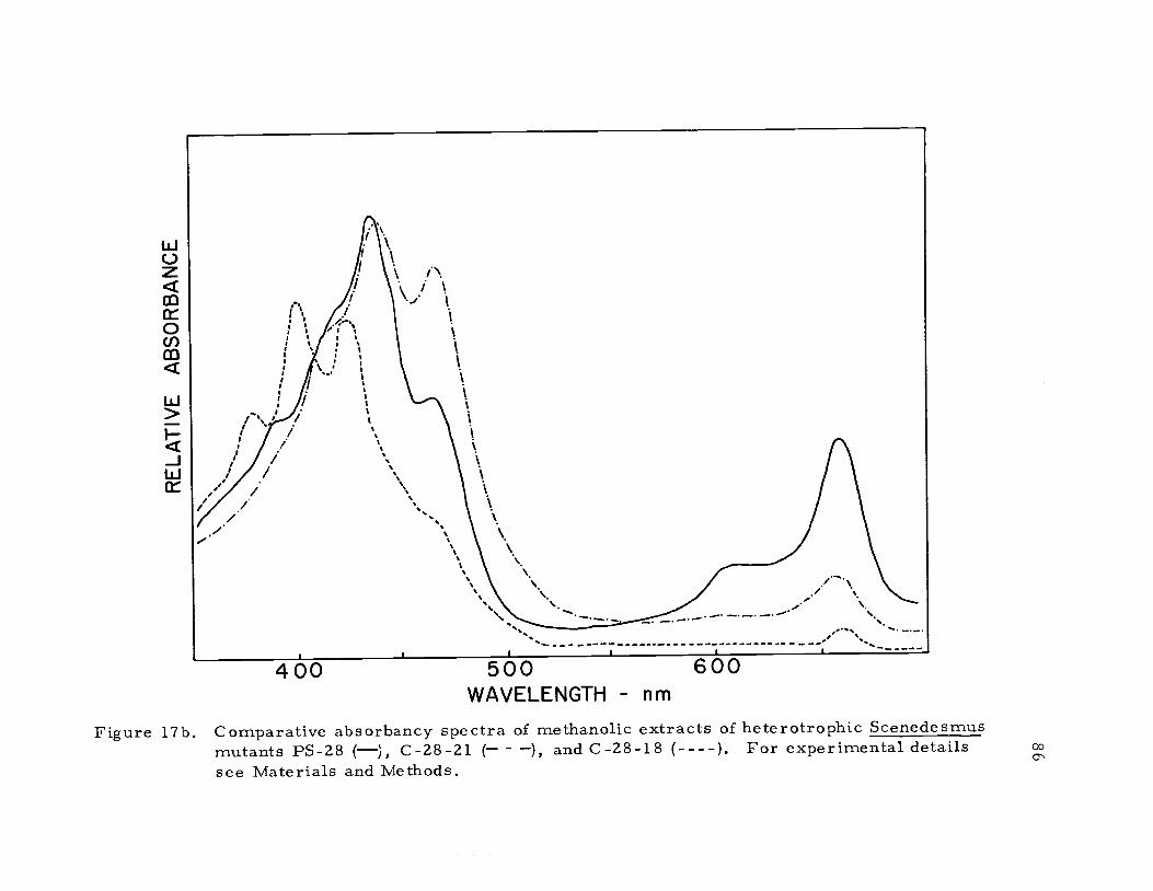
400 5 0 0 600WAVELENGTH nm
Figure 17b. Comparative absorbancy spectra of methanolic extracts of heterotrophic Scenedesmusmutants PS-28 (), C-28-21 ( ), and C-28-18 (----). For experimental detailssee Materials and Methods.
87
accumulate chlorophyll when transferred into the light. Williams
(3 97 1) analyzed the carotenoid composition of mutants C-6D and
C -2A'. Her data indicated that mutant C-6D when cultured in the
dark is characterized by a preponderance of acyclic carotenoids
(phytoene, phytofluene, and neurosporene) and had only a trace of
cyclic carotenoids. The block in carotenoid biosynthesis in mutant
C -6D resulted in a number of unusual peaks between 350 nm and
422 nm in the methanol absorption spectrum. In this study it was
concluded that mutant C-28-18 also was blocked in carotenoid
biosysthesis (Figure 17b). Williams (1971) demonstrated that the
carotenoid composition of mutant C -2A' was identical to the wild-
type, and that the mutation involved a block in chlorophyll biosyn-
thesis. Again by spectral comparison (Figure 17b) it was concluded
that the carotenoid composition of mutant C-28-21 was identical to
that of mutant C-2A'. By analogy the mutation in C-28-21 is also
involved in chlorophyll biosynthesis. Having an analogous series
of greening mutants with and without vitamin E allowed a study of
the influence of vitamin E on the greening process.
Greening Studies
After 24 hours of continuous illumination the amounts of
chlorophyll in mutant C -2A' has approached the normal wild-type
level (Figure 18). The maximum chlorophyll level of greening
88
8 16I
TIME hours24
Figure 18. Comparative chlorophyll synthesis during the greening of2-day old Scenedesmus mutants C-2A' (0-0), C-2A' plus
DCMU (111-0), C-28-21 (A E), and C-28-21 plus p.MDCMU (a---a.). For experimental details see Materialsand Methods.
89
cultures of C-28-21 is about one-third that of the wild-type. Reasons
for this will be presented below.
Cells of mutant C-28-21 were insensitive to the presence of
DCMU in the culture medium during greening, but cells of mutant
C-2A' that had been poisoned with DCMU stopped greening after 8-12
hours into the light period (Figure 18). The fact that the greening
curve of mutant C -2A' did not exhibit an inhibition by DCMU until
the 8th to 12th hour into the light period indicates that the greening
process in this mutant is divided into two phases. The first phase
is independent of photosynthesis, and the second phase is dependent
on photosynthesis (see below). Because mutant C-28-21 is a sub-
mutant of PS-28 it also is blocked in photosystem-II, and DCMU
which is a photosystem-II inhibitor has no influence upon the pattern
of greening (Figure 18).
The onset of photosynthesis during the greening process for
both mutants C -2A' and C-28-21 is given in Figure 19. The oxygen
evolving apparatus of mutant C -2A' is fully functional at 4 hours
into the light period, even though the chlorophyll level is virtually
the same as in dark grown cells. The photosynthetic rate of mutant
C-28-21 is about one-third that of the wild-type, and the maximum
rate is not achieved until after 8 hours of development (Figure 19).
The greening pattern of both mutants C -2A' and C -28 -21
was altered by light intensities greater than 104 ergs/sec-cm2
0
0
N 200E
0
0
I 1 I 1
4 8TIME- hours
12
Figure 19. Comparative development of photosynthesis during the greening of 2-day old Scenedesmusmutants C-2A' (0-0), and C-28-21 (AA). For experimental details see Materialsand Methods.
0
91
(Figure 20a and 20b). The amount of chlorophyll synthesized in 12
hours by mutant C-28-21 can be doubled over the normal level by
maintaining the light intensity below 103 ergs/sec-cm 2 throughout
the course of the experiment (Figure 20b). The specific influence of
light upon the early stages of the greening process in mutant C -2A'
has been discussed by Oh-hama and Senger (1975). They observed
that the controlling action of light in this first phase of greening was
complex but resulted in one principal thing, maintaining levels of
ALA (8-aminolevuinic acid) high enough to support chlorophyll
biosynthesis. The removal of light from the system arrested the
greening process at that point. Higher levels of light detrimental to
chlorophyll accumulation (Figure 20a and 20b), indicated that
mutants C-2A' and C-28-21 are photosensitive for at least up to the
12th hour of the light period. Further evidence in support of this
observation was that mutant C -2A' did not green, and in fact bleached
in a light field of 106 ergs/sec-cm 2 (Senger and Bishop, 1972a).
The antibiotics chloramphenicol and cycloheximide interfere
with chloroplast development and chlorophyll accumulation. Cyclo-
heximide is an inhibitor of protein synthesis by 80s ribosomes
(cytoplasmic protein synthesis), and chloramphenicol inhibits protein
synthesis by 70s ribosomes (plastid protein synthesis; cf. , Smillie,
et al., 1970). The greening patterns of mutants, C-2A' and C-28-21,
in the presence of these two inhibitors are presented below (Figure
6.0
4.0
2.0
00 8 16
TIME hours24
92
Figure 20a. Chlorophyll synthesis during the greening of 2-day oldsamples of Scenedesmus mutant C -2A' at two differentlight intensit ie s: 1.2 x 10⁴ (0-0) and 2.4 x 10³ (A--L)ergs/sec-cm2. Data are representative of five inde-pendent experiments. For experimental details seeMaterials and Methods.
3.0
2.0
1.0
0
93
0 8 16
TIME hours24
Figure 20b. Chlorophyll synthesis during the greening of 2-day oldsamples of Scenedesmus mutant C-28-21 at two differentlight intensities: 1.2 x 10⁴ (0-0) and 2.4 x 10³ (AA)ergs/sec-cm2. Data are representative of three inde-pendent experiments. For experimental details seeMaterials and Methods.
94
21a and 21b). Chloramphenicol (1 mg/ml) only partially inhibits
the greening of either mutant during mixotrophic development
phyll accumulation in either mutant (Figure 21b). Smillie, et al.
1970 and Kirk and Allen (1965) have described the effects of these
two antibiotics on chloroplast development, and the results of their
studies using Euglena were similar to the results presented here.
It was determined that the lack of vitamin E and the loss of photo-
system-II in C-28-21 did not influence the effect of the two protein
synthesis inhibitors on greening.
The greening data presented here for C-2A' and C-28-21 fully
supported the conclusions of previous authors (Bishop and Senger,
1972a; Senger and Bishop, 1972; Oh-hama and Senger, 1975). The
first stage of the greening process, which lasted up to 12 hours,
was dependent upon the mobilization of stored carbohydrate as an
energy source. The second stage of greening was dependent upon
photosynthesis (hence, light intensity) to support the energy require-
ments of the cell. These observations explained the following:
1) The greening of C-2A' was affected by DCMU poisoning in a
biphasic manner. This was not observed for C-28-21. 2) Low light
intensities were sufficient to trigger chlorophyll biosynthesis during
greening, but were not strong enough to cause photodamage to the
chloroplast in the early stages of development. 3) The inhibition of
0E 4.0
>-20_00 2.0
E
_J
0
0 8 16
TIME hours24
95
Figure 21a. Comparative chlorophyll synthesis during the greeningof 2-day old Scenedesmus mutants C -2A' (0-0), C-2A'plus 1 mg/m1 chloramphenicol ( -0), C-28-21 (AA),and C-28-21 plus 1 mg /ml chloramphenicol ( a). Forexperimental details see Materials and Methods.
4.0
20
0
96
0 8 16TIME hours
24
Figure 21b. Comparative chlorophyll synthesis during the greening of2-day old Scenedesmus mutants C-2A' (0-0), C-2A'plus 1 mg/m1 cycloheximide (111-11), C-28-21 (AA), andC-28-21 plus 1 mg /ml cycloheximide (A A). Forexperimental details see Materials and Methods.
97
protein synthesis prevented the mobilization of stored carbohydrates
and prevented greening in both early and late stages. The effect of
the total absence of vitamin E in C-28-21 only was apparent because
of the loss of photosystem-II activity.
Low Temperature Absorbance Studies
Cooling samples of algae to liquid nitrogen temperature (77 K)
allows resolution of the chlorophyll absorbance bands in the red
region of the spectrum ordinarily not possible at room temperature.
The low temperature spectrum of the wild-type reveals three peaks
at 677 nm, 670 nm, and 650 nm (Figure 22a). In a comparable
spectrum, the mutant, C-28-21, lacks the 650 nm absorbance band
(Figure 22b), which was attributed to chlorophyll b (Cho and
Govindjee, 1970). From this information it was concluded that mutant
C-28-21 cultured mixotrophically can not synthesize chlorophyll b.
This finding explained why the maximum chlorophyll levels of
greening samples of the mutant were lower than normal (see Figure
18), and why the light intensity response of photoreduction was
unusual (see Figure 7). The absence of chlorophyll b in mutant
C-28-21 might indicate that there is a genetic lesion in the manufac-
turer of the light-harvesting-pigment-protein. This protein, which
is the attachment site for all of the chlorophyll b in the chloroplast,
is virtually universal in higher plants, but is not essential for
absorbance 1 0.01
1
a
600 650 700WAVELENGTH nm
absorbance 0.0I
600 650 700WAVELENGTH nm
Figure 22. Low temperature absorbancy spectra of (a) mutant C-28-21, and (b) wild-typeScenedesmus. For experimental details -see Materials and Methods.
99
photochemical activity (Thornber and Highkin, 1974). Research is
currently being performed in our laboratory to demonstrate the
presence or absence of this pigment protein in mutant C-28-21.
Plastoquinone A and Vitamin E
Concentrations of a-tocopherol, plastoquinone A, and chloro-
phyll were measured in wild-type Scenedesmus and mutant PS-28
under different patterns of growth, and the results are presented in
Table 5. Evidence that mutant PS-28 lacked vitamin E was reported
elsewhere (Bishop and Sicher, 1974; Sicher and Bishop, 1975), and
the chromatographic techniques and identification methodology have
been reported in detail by Bishop and Wong (1974).
Photosynthetic membranes are sensitive to changes in the
physiological status of the cell; therefore, an analysis of the quinone
complement of the thylakoids was performed on normal wild-type
Scenedesmus under different patterns of growth. A comparison of the
vitamin E levels (p.moles a- tocopherol /ml PCV) grown hetero-
trophically and mixotrophically indicated that the cultures maintained
in the light had lower concentrations of vitamin E (Table 5). The
level of vitamin E (ilmoles a-tocopherol/ml PCV) measurable for
autotrophically grown cultures of wild-type Scenedesmus was at
least double the mixotrophic value (Table 5). These results were of
interest because they suggested that the presence of glucose in light
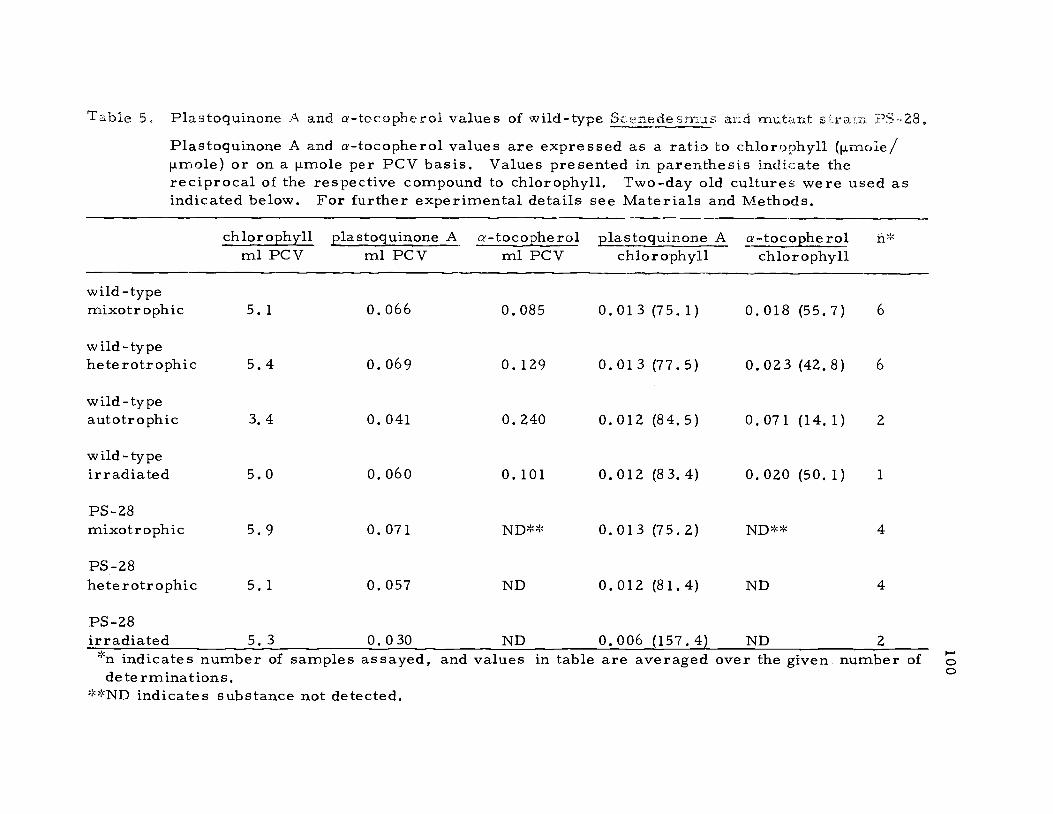
Table 5. Plastoquinone A and a-tocopherol values of wild-type S fledesmus and mutant s;,raln PS-28.Plastoquinone A and a-tocopherol values are expressed as a ratio to chlorophyll (p.moie/ilmole) or on a ilmole per PCV basis. Values presented in parenthesis indicate thereciprocal of the respective compound to chlorophyll. Two-day old cultures were used asindicated below. For further experimental details see Materials and Methods.
chlorophyll plastoquinone A a-tocopherol plastoquinone A a-tocopherol n*ml PCV ml PCV ml PCV chlorophyll chlorophyll
PS-28irradiated 5. 3 0. 0 30 ND 0. 006 (157.4) ND 2*n indicates number of samples assayed, and values in table are averaged over the given number ofdeterminations.
**ND indicates substance not detected.
101
grown cultures of Scenedesmus could substitute partially for the func-
cior of vitaminE. Comparisons between cultures grown either autotro-
phically, heterotrophically, or mixotrophically are only relative because
the chlorophyll concentration, cellvolume, and rates of cell division
were different in each instance. Despite the physiological differ-
ences between autotrophic and mixotrophic cultures, it is still con-
idered that cells grown by the former method have the highest
vitamin E levels (Hanigk and Lichtenthaler, 1975).
The concentration of plastoquinone A was equivalent in hetero-
trophic and mixotrophic cultures of both mutant PS-28 and wild-type
Scenedesmus (Table 5). This was a surprising result because it
was established previously that photosystem-II mutants in general
were characterized by low plastoquinone levels (Bishop and Wong,
1971; Smillie and Levine, 1963). This made PS-28 unique as far as
typical photosystem-II mutants were concerned. When the mutant
was exposed to high intensity irradiation the level of plastoquinone A
decreased in parallel with the loss of photosystem-II activity (Table
5). The photodynamic destruction of plastoquinone A also occurred
in mutant LS-41 (Harvey, 1974; Table 5).
As expected the chlorophyll to plastoquinone A and chlorophyll
to a-tocopherol values were at a minimum when measured in dark
grown cultures of mutant C -2A', but after 24 hours of greening the
two ratios approximated the normal mixotrophic values obtained for
102
the wild-type (Figure 23). The chlorophyll to plastoquinone A data
presented below for greening cultures of mutant C -2A' were similar
to the results presented by Bishop and Senger (1972a).
When the plastoquinone A and a-tocopherol data were expressed
on a packed cell volume (PCV) rather than on a chlorophyll basis, a
very different pattern was observed (Figure 24). The dark grown
cultures of mutant C-2A' possessed normal hetrotrophic values of
q-tocopherol, even in the virtual absence of chlorophyll. This situa-
tion appeared to be true for dark grown tissues or samples that have
etioplasts, but was not evident in those systems with proplastids
(Threlfall and Griffiths, 1967). After exposure of the cells to light
for 24 hours the vitamin E levels(p.rrioles a- tocopherol/ml PCV)
decrease to a value very near the normal mixotrophic level obtained
for the wild-type Scenedesmus (Figure 24). Lichtenthaler and
Grumbach (1975) observed similar changes in both the plastoquinone A
and vitamin E levels when 8-day old etiolated barley seedlings were
illuminated. Contrarily, Threlfall and Griffiths (1967) reported that
the levels of a -tocopherol remained constant in etiolated maize shoots
after exposure to light. Their (Threlfall and Griffiths, 1967)
plastoquinone A data were similar to the results presented below. A
more detailed analysis of the changes of vitamin E levels during the
development of the chloroplast would be highly desirable.
0
CHLOROPHYLL / PLASTOQUINONE-A
CHLOROPHYLL / a- TOCOPHEROLFigure 23. Chlorophyll to plastoquinone A and chlorophyll to a-tocopherol ratios (p.mole/p.mole of
Scenedesmus mutant C -2A' at different stages of greening. Chlorophyll (a + b)/plasto-quinone A (0-0); chlorophyll (a + b)/a-tocopherol (Cr A). For experimental details seeMaterials and Methods.
a_
E0.12
-0 0.08c
0a_
(5 0.04z0
0
104
0 8 16
TIME hours
24
Figure 24. Concentrations of plastoquinone A (p.mole/m1 PCV) anda-tocopherol (p.mole/m1 PCV) of Scenedesmus mutantC-2A' at different stages of greening. plastoquinone A(0-0) and a-tocopherol (AA). For experimental de-tails see Materials and Methods.
105
The results presented in Figure 24 indicate that plastoquinone A
and a-tocopherol are synthesized via independent pathways. If the
two lipophilic benzoquinones shared a common precursor, as has
been suggested by Eck and Trebst (1963), the curves in Figure 24
would be expected to follow similar kinetics. Incorporation studies
using 14C-mevalonate and 14C02
supported the independent pathway
tenet (Threlfall and Griffiths, 1967).
The molar ratio of chlorophyll to a-tocopheryl quinone in
hetrotrophic wild-type samples was 166.6. No trace of this substance
was detected in comparable samples of the mutant.
Ascorbic Acid (Vitamin C)
Values of vitamin C were obtained for both mutant PS-28 and
wild-type Scenedesmus cultured either heterotrophically or mixo-
trophically (Table 6). In both circumstances the ascorbic acid values
for the mutant lacking vitamin E were equivalent to those of the wild-
type. Additionally, an increase in the ascorbic acid values of light
grown versus dark grown cultures confirmed the now classic obser-
vation that vitamin C levels were affected by light, carbon dioxide,
and photosynthesis (Moldtmann, 1939). These results were interest-
ing because following the observation by Dam, et al. (1948) that large
doses of vitamin C prevented encephalomalacia and exudative diathesis
in the vitamin E deficient chick, Caputto, et al. , (1958) reported that
106
Table 6. Ascorbic acid levels of wild-type Scenedesmus and mutantstrain PS-28.
Chlorophyll and ascorbic acid levels are expressed aseither a ratio or on a p.mole per PCV basis. Values pre-sented in parenthesis indicate the reciprocal of the abscor-bic acid to chlorophyll ratio. Two-day old cells were usedand were cultured as indicated. Data presented below aresimilar to those obtained in 1 other experiment. Forexperimental details see Materials and Methods.
ild -typemixotrophic
PS-28mixotrophic
wild -typeheterotrophic
PS-28heterotrophic
chlorophyll (a + b) ascorbic acid ascorbic acidml PCV ml PCV chlorophyll (a + b)
5. 0 0.40 0.080 (12.5)
5.3 0.43 0.082 (12. 3)
5. 3 0.23 0.044 (22.5)
4.5 0.26 0.058 (17.1)
vitamin C was not synthesized in rats fed a diet deficient in a-toco-
pherol. Furthermore, Tappel (1962) predicted a synergistic relation-
ship between the presence of vitamin C in the cell, and the function of
vitamin E in preventing free-radical membrane damage. Noguchi,
Cantor, and Scott (197 3) pursued this line of investigation and
demonstrated that one reason for the lack of detectable ascorbic acid
in vitamin E deficient as compared to normal liver tissue, was that
reduced compounds such as ascorbic acid, gluthathionione, and re-
duced NADP+ were oxidized in vitro by enzymes of the microsomes
107
and mitochondria. The oxidation of vitamin C by hepatic liver cells
deficient in vitamin E lead to the formation of free-radicals, and
rnalonyldialdehyde, an end product of free-radical lipid peroxidation.
They (Noguchi, Cantor and Scott, 1974) demonstrated that a-tocopherol
and the enzyme glutathione peroxidase would prevent the oxidation
of ascorbic acid by vitamin E deficient chicks.
Because the ascorbic acid levels in mutant PS-28 were not
depleted it was assumed that the complete absence of vitamin E in
Scenedesmus grown either in the dark or in the light did not lead to
free-radical damage (and the subsequent disappearance of vitamin C).
This observation indicated one of two situations exist in mutant
PS-28, either there is a mechanism in Scenedesmus other than
vitamin E that eliminates or prevents free-radical damage, or more
likely, the vitamin C oxidizing system observed in hepatic cells does
not occur in Scenedesmus.
Lipids and Fatty Acids
Several species of the Cyanophyceae are known to lack poly-
enoic fatty acids (Holton, et al. , 1968), and vitamin E (Hirayama,
1967). The photosynthetic lamellae of higher plants have very high
concentrations of polyunsaturated fatty acids and vitamin E
(Lichtenthaler and Park, 1963). These facts suggested a relationship
108
between the fatty acid composition of the photosynthetic membrane
and the occurrence of vitamin E.
Two dimensional thin layer chromatographic analyses were
performed on whole cell lipid extracts of heterotrophic PS-28 and
wild-type Scenedesmus (Figures 25a and 25b). The lipid composition
of the two algal strains was determined to be qualitatively equivalent.
These results implied that even in the complete absence of vitamin E,
the lipid component of the photosynthetic membrane of dark grown
cells was not under stress due to free-radical damage.
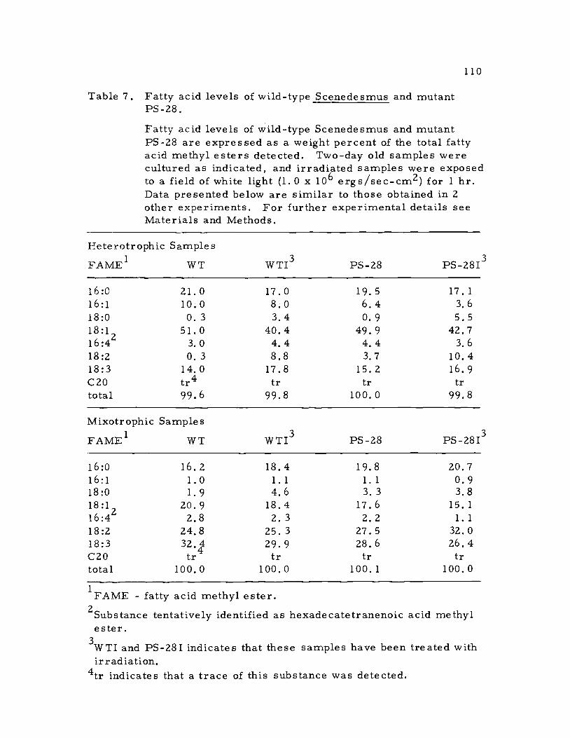
The above conclusion was tested further via an in depth anal-
ysis of the fatty acid compositions of the two algal strains. The
fatty acid composition of heterotrophic and mixotrophic samples of
the wild-type and mutant were examined, and compared to like
samples from these cultures that were treated with high intensity
irradiation (Table 7). In each instance the fatty acid composition of
mutant PS-28 was comparable to that of the wild-type, and further-
more, the fatty acid profile presented below for mixotrophic cells
closely correlated with the results presented by Klenk, et al. (1963)
for autotrophic samples of Scenedesmus. The photo-heterotrophic
cultures of both the wild-type and mutant exhibited large increases
in the size of the octadecatrienoic (18:3) and octadecadienoic (18:2)
fatty acid pools over the dark grown cultures. These fatty acids are
the predominant acyl esters of lipids which are abundant in the
T' oPoDG pE
0 PC
PI
2a
Figure
0 10 00 DG P E
PC
API
172
CIMG
b
25. Two dimensional thin layer chromatographic analysis of a (1:1) CHC13-CH3OH extract of(a) mutant PS-28, and (b) wild-type Scenedesmus. Solvent direction 1 consisted of CHC13-CH3OH-7 N NH4OH (97. 5:37:6), and solvent direction 2 consisted of CHC13-CH3OH-CH3CO2-H20 (85:12.5:12.5:2). Lipids were detected with iodine vapor and were identifiedwith Supelco standards. MG-monogalactosyl diglyceride, DG-digalactosyl diglyceride,PE-phosphatidyl ethanolamine, PC -Phosphatidyl choline, PI-phosphatidyl inositol. Lipiddegradation spots and pigmented regions have been deleted. For experimental details seeMaterials and Methods.
0
110
Table 7. Fatty acid levels of wild-type Scenedesmus and mutantPS-28.
Fatty acid levels of wild-type Scenedesmus and mutantPS-28 are expressed as a weight percent of the total fattyacid methyl esters detected. Two-day old samples werecultured as indicated, and irradiated samples were exposedto a field of white light (1.0 x 10⁶ ergs/sec-cm2) for 1 hr.Data presented below are similar to those obtained in 2other experiments. For further experimental details seeMaterials and Methods.
from studies on PS-28 indicate that this concept may be correct.
The principle evidence suggesting that vitamin E and related
substances function in photosynthesis as electron transport carriers
comes from extraction and re-addition experiments (Bishop, 1959;
Trebst, 1963). It was found that freeze-dried chloroplasts extracted
with various organic solvents lost their capacity for the Hill reaction,
however, partial restoration of photochemical activity was obtained
upon re-addition of the crude extract, or by addition of plasto-
quinone.
Subsequent and more detailed analyses have produced far more
complicated results. It was found that photosystem-I activity could
be inactivated by extraction, and activity could be restored with
different quinones (Henninger and Crane, 1963). In one instance
photochemical activity was restored to freeze-dried chloroplast that
had been extracted with acetone, a procedure that removed 90% of
the chlorophyll among other substances, with mixtures of p-tocopherol
122
quinone and a-tocopheryl quinone or plastoquinone A. These mix-
tures were more efficient in restoring the Hill reaction to acetone
extracted chloroplasts than plastoquinone A or B alone, and the
overall rate of activity obtained exceeded that of the unextracted
freeze-dried chloroplasts, (Dilley, Henninger, and Crane, 1963).
These results were interpreted as evidence that plastoquinones
and possibly other quinones function at specific sites in the electron
transport chain. However, this interpretation was not supported by
spectrophotometric evidence (Stiehl and Witt, 1968), and because
of the non-specific nature of extraction and re-addition experiments
it was not known that the quinone has returned to its original site
in the membrane. Perhaps the re-addition of an isoprenoid quinone
to the extracted membranes partially restores a structural require-
ment, thus allowing photochemical activity.
Data obtained from PS-28 did not support the suggestion that
vitamin E participates in photophosphorylation. Hydrogen photo-
reduction, which is an ATP requiring reaction, is normal in the
mutant (Figure 7). Secondly, the rates of PMS-mediated photo-
phosphorylation obtained with chloroplasts prepared from PS-28 and
wild-type Scenedesmus were comparable (Figure 14). Although the
rates of anaerobic glucose photoassimilation were decreased in
PS-28 (Figure 13), evidence presented here, Figure 14, and else-
where (Tanner, Daschel and Kandler, 1965), suggested that this was
123
due to impaired activities of photosystem-II and not of photophos-
phorylation.
It is possible that a-tocopherol is a structural lipid. The
lipids of the photosynthetic membrane are divided into two specific
types according to the lipoprotein fluid mosaic model of Singer
(1974). The first type, boundary lipids, are closely associated with
the surfaces of globular membrane proteins. The second type,
matrix lipids, occur in the membrane in molecular bilayers and
are freely translational (hence, membrane fluidity). This feature
allows the membrane to change conformations and still retain attach-
ment sites for affiliated proteins. a-Tocopherol does not appear to
function as a general lipid antioxidant, and for this reason it probably
performs a more specific function, i.e. , as a boundary lipid.
There are three major polypeptide groupings in the chloroplast,
and these are associated with the photosystem-I complex, the photo-
system-II complex, and the light-harvesting-pigment-protein complex.
Photosystem-I functions normally in PS-28, therefore, it is not
likely that it has any connection with vitamin E. This finding con-
tradicts the proposals of Baszynski (1974) who demonstrated that
a-tocopherol restored photosystem-I activity to acetone extracted
freeze-dried spinach chloroplasts.
Several chloroplast dissociation procedures demonstrated that
two main subchloroplast particles are obtained by fractionation
124
(Anderson, 1975). The first subchloroplast particle was enriched
in photosystem-I and the second was enriched in both photosystem-
II and the light-harvesting-pigment-protein complex. In fact, prior
to the recognition of the existence of the chlorophyll alb, 1:1, light-
harvesting -pigment-protein complex it was believed that chlorophyll
b was associated with photosystem-II. There is every indication
that the light-harvesting-pigment-protein complex in vivo is closely
associated with photosystem-II. Therefore it is likely that a-toco-
pherol is associated with either the photosystem-II complex, the
light-harvesting-pigment-protein complex, or with the resultant
subchloroplast aggregate. In a brief and early investigation
Lichtenthler (1969) found that vitamin E was enriched in the photo-
system-II complex upon chloroplast fractionation but that the photo-
system-I particles contained a small portion of the compound. In
the light of the recent advances in this field a more detailed and
modern investigation of this subject would be desirable.
125
BIBLIOGRAPHY
Alien, C. F. and P. Good, 1971. Acyl lipids in photosyntheticsystems. In: Methods in enzymology. Vol. 23, ed. by A. SanPietro. New York, Academic Press. p. 523 -547.
Almquist, H. J. 1937. Crystals with vitamin K potency. Nature140:25-26.
Anderson, J. M. 1975. The molecular organization of chloroplastthylakoids. Biochimica et Biophysica Acta 416:191-235,
Anderson, S. S. , I. G. Lyle, and R. Paterson. 1976. Electrontransfer across membranes using vitamin K1 and coenzymeQ10 as carrier molecules. Nature 259:147-148.
Arnon, D. I. , M. B. Allen, and F. R. Whatley. 1954a. Photosyn-thesis by isolated chloroplasts. Nature 174:394 -396.
Arnon, D. I. , F. R. Whatley, and M. B. Allen. 1954b. Photosyn-thesis by isolated chloroplasts. II. Photosynthetic phosphory-lation, the conversion of light into phosphate bond energy.Journal of the American Chemical Society 76:6 324-6 329.
Arnon, D. I. and A. A. Horton. 1963. Site of action of plasto-quinone in the electron transport chain of photosynthesis. ActaChemica Scandanavica 17 :1 35 -1 39.
Asano, A., A. F. Brodie, A. F. Wagner, P. E. Wittreich, and K.Folkers. 1962. The new synthetic 6-chromanyl phosphate ofvitamin K1(20) and its behavior in an enzymatic system fromMycobacterium ablei. Journal of Biological Chemistry 237:PC241 1 -2412.
Avron, M. 1960. Photophosphorylation by swiss-chard chloroplasts.Biochimica et Biophysica Acta 40:257 -272.
Baltscheffsky, M. 1969. Energy conversion linked changes ofcarotenoid absorbance in Rhodospirillum rubrum chromato-spores. Archives of Biochemistry and Biophysics 130:646-652.
Bas sham, J. A. 1965. Photosynthesis: the path of carbon. In:Plant biochemistry, ed. by J. A. Bonner and J. E. Varner.New York, Academic Press. p. 875-902.
126
Barr, R. and F. L. Crane. 1971. Quinones in algae and higherplants. In: Methods in enzymology. Vol. 23, ed. by A. SanPietro. New York, Academic Press. p. 372-408.
Barr, R., M. D. Henninger, and F. L. Crane. 1967. Comparativestudies on plastoquinone. II. Analysis for plastoquinones A,B, C, and D. Plant Physiology 42:1246-1254.
Baszynski, T. 1974. Effect of a-tocopherol on reconstitution ofphotosystern I in heptane-extracted spinach chloroplasts.Biochimica et Biophysica Acta 347:31-35.
Baxter, J. G. , C. D. Robeson, J. D. Taylor, and R. W. Lehman.1943. Natural - and -y -tocopherols and certain esters ofphysiological interest. Journal of the American ChemicalSociety 65:918-924.
Bennoun, P. and R. P. Levine. 1967. Detecting mutants that haveimpaired photosynthesis by their increase in fluorescencelevel. Plant Physiology 43:1284-1287.
Benson, A. A. 1963. The plant sulfolipid. Advances in LipidResearch 1:387-394.
Bertsch, W. , J. R. Azzi, and J. B. Davidson. 1967. Delayed lightstudies on photosynthetic energy conversion. I. Identificationof the oxygen-evolving photoreaction as delayed light emitterin mutants of Scenedesmus obliquus. Biochimica et BiophysicaActa 143:129-143.
Berzborn, R. J. and N. I. Bishop. 197 3. Isolation and propertiesof chloroplast particles of scenedesmus obliquus D3 with highphotochemical activity. Biochimica et Biophysica Acta 292:700-714.
Bieri, J. G. 1969. Biological activity and metabolism of N-sub-stituted tocopheramines: implications on vitamin E function.In: The fat soluble vitamins, ed. by H. J. DeLuca and J. W.Suttie. Madison, University of Wisconsin Press. p. 307-316.
Bishop, N. I. 1958. The influence of the herbicide DCMU on theoxygen-evolving system of photosynthesis. Biochimica etBiophysica Acta 27:205-206.
127
Bishop, N. I. 1959. The reactivity of a naturally occurring quinone(Q-255) in photochemical reactions in isolated chloroplasts.Proceedings of the National Academy of Sciences 45:1696-1702.
Bishop, N. I. 1966. Partial reactions of photosynthesis and photo-reduction. Annual Review of Plant Physiology 17:185-208.
Bishop, N. I. 1971a. Photosynthesis: the electron transport systemof green plants. Annual Review of Biochemistry 40:197 -226.
Bishop, N. I. 1971b. Preparation and properties of mutants:Scenedesmus. In: Methods in enzymology. Vol. 23, ed. byA. San Pietro. New York, Academic Press. p. 130 -143.
Bishop, N. I. 1972. Whole cell and chloroplast reactions of algalmutants deficient in cytochrome f(552). In: Proceedings ofthe second international congress on photosynthesis. Vol. 1,ed. by G. Forti, M. Avron and A. Melandri. The Hague, Dr.W. Junk, N. V. Publishers. p. 459-481.
Bishop, N. I. 197 3. Analysis of photosynthesis in green algaethrough mutation studies. In: Photophysiology. Vol. VIII,ed. by A. C. Griese. New York, Academic Press. p. 65-96.
Bishop, N. I., M. Frick, and L. W. Jones. 1975. Photohydrogenproduction in normal and mutant forms of various green algae:the requirement for photosystem II. Plant Physiology 56:9(Suppl.).
Bishop, N. I. and H. Gaffron. 1962. Photoreduction at X 705 milin adapted algae. Biochemical and Biophysical ResearchCommunications 8:471-476.
Bishop, D. H. L. , K. P. Pandya, and H. K. King. 1962. Ubiqui-none and vitamin K in bacteria. Biochemistry Journal 8 3:606-614.
Bishop, N. I. and H. Senger. 1972. The development of structureand function in chloroplasts of greening mutants of Scenedesmus,II. Development of the photosynthetic apparatus. Plant andCell Physiology 1 3:9 37-95 3.
Bishop, N. I. and R. Sicher. 1974. The loss of photosystem II inan algal mutant lacking vitamin E. Federation Proceedings33:1 329 (Abstr. ).
128
Bishop, N. I. and J. Wong. 1971. Observations on photosystem-IImutants of Scenedesmus: pigments and proteinaceous compon-ents of the chloroplasts. Biochimica et Biophysica Acta 234:433-445.
Bishop, N. I. and J. Wong. 1974. Photochemical characteristicsof a vitamin E deficient mutant of Scenedesmus obliquus.Berichten Deutsche Botanische Gesellscheaft 87:359-371.
Bowers, G., W. L. Ogren, and R. H. Hageman. 1971. Phos-phoglycolate production catalyzed by ribulose diphosphatecarboxylase. Biochemical and Biophysical ResearchCommunications 45:716-722.
Brand, J., D. W. Korgmann, and F. L. Crane. 1971. A lipidrequirement for photosystem-I activity in heptane-extractedspinach chloroplasts. Plant Physiology 47:135-138.
Brodie, A. F. 1965. Naphoquinones in electron transport. In:Biochemistry of quinones, ed. by R. A. Morton. London,Academic Press, p. 355-404.
Bunyan, J. A., T. Diplock, and J. Green. 1967. Effects of vitaminE deficiency on total polyunsaturated fatty acids in rats andchicks. British Journal of Nutrition 21:217-222.
Butler, W. L., B. Epel, and R. P. Levine. 1972. A spectroscopicanalysis of low fluorescent mutants of Chlamydomonasreinhardti blocked in their water-splitting oxygen-evolvingapparatus. Biochimica et Biophysica Acta 275:395-400.
Caputto, R., P. B. McCay, and M. P. Carpenter. 1958. Require-ments of Mn++ and Co ++ for the synthesis of ascorbic acid byliver extracts of animals deprived of tocopherol. Journal ofBiological Chemistry 233:1025-1029.
Carabello, T., F. Lien, C. Canes, and J. Bird. 1971. Effect ofvitamin E deficiency on mitochondrial energy transfer.Federation Proceedings 30:639 (Abstr. ).
Carpenter, M. D. 1966. Polyenoic fatty acids of rat testis, theeffects of a-tocopherol. Federation Proceedings 25:764(Abstr. ).
129
Carr, N. G., G. Exell, V. Flynn, M. Halloway, and S. Talukdar.1967. Minor quinones in some Myxophyceae. Archives ofBiochemistry and Biophysics 120:503 -507.
Cheniae, G. M. and I. F. Martin. 1968. Site of manganese functionin photosynthesis. Biochimica et Biophysica Acta 15 3:819-837.
Cheniae, G. M. and I. F. Martin. 1970. Sites of function ofmanganese within photos'y-stem-II. Roles in oxygen evolutionand system-II. Biochimica et Biophysica Acta 197:219 -239.
Cho, F. and Govindjee. 1970. Low temperature (4-77° K) spec-troscopy of Chlorella; temperature dependence of energytransfer efficiency. Biochimica et Biophysica Acta 216:151-161.
Clark, V. , G. Kirby, and A. Todd. 1958. Oxidative phosphoryla-tion: a chemical approach using quinol phosphates. Nature181:1650-1652.
Cooper, T. G., D. Filmer, M. Wishnick, and M. D. Lane. 1969.The active species of "CO2" utilized by ribulose diphosphatecarboxylase. Journal of Biological Chemistry 244:1081 -1083.
Corwin, L. M. 1965. Further studies on the regulation of succinateoxidation by vitamin E. Journal of Biological Chemistry240:34-38.
Crane, F. L. 1959. Internal distribution of coenzyme Q in higherplants. Plant Physiology 34:128 -131.
Crane, F. L. 1962. Quinones in lipo-protein electron transportsystems. Biochemistry 1:510-517.
Crane, F. L. , B. Ehrlich, and L. P. Kegel. 1960. Plastoquinonereduction in illuminated chloroplasts. Biochemical andBiophysical Research Communications 3:37-40.
Dam, H. 1942. Vitamin K, its chemistry and physiology. Advancesin Enzymology 2:285-324.
130
Dam, H. , I. Kruse, I. Piange, and E. Sondegaard. 1948. Influ-ence of dietary ascorbic acid, nordihydroguaraiaretic acidand cystine on vitamin E deficiency symptoms in chicks.Biochimica et Biophysica Acta 2:501 -513.
Das, B. C., M. Lounasman, C. Tendile, and E. Lederer. 1965.Mass spectrometry of plastoquinones: the structure ofplastoquinones A, B, C and D. Biochemical and BiophysicalResearch Communications 21:318-322.
Dilley, R. and F. L. Crane. 1963. Subcellular distribution ofa-tocopherol in spinach and lilac leaf tissue. Plant Physiology38:452-456.
Dilley, R. A. and F. L. Crane. 1964. Light dependent conversionof a-tocopheryl-quinone and plastoquinone-D in Spinaciaoleracea chloroplasts. Plant Physiology 39:33-38.
Dilley, R. A., M. D. Henninger, and F. L. Crane. 1963. Evidencefor the role of several quinones in the electron transportsystem of chloroplasts. In: Photosynthetic mechanisms ofgreen plants. Washington, D. C. p. 273-277. (NationalResearch Council. Publication 1145).
Doleiden, F. H., S. R. Fahrenholtz, A. A. Lamola, and A. M.Trozzolo. 1974. Reactivity of cholesterol and some fatty acidstoward singlet oxygen. Photochemistry and Photobiology20:519-521.
Duysens, L. N. M. 1954. Reversible changes in the absorptionspectrum of Chlorella upon irradiation. Science 120:35 3-354.
Duysens, L. N. M. and H. E. Sweers. 1963. Mechanism of twophotochemical reactions in algae as studied by means offluorescence. In: Studies on microalgae and photosyntheticbacteria, ed. by the Japanese Society of Plant Physiologists.Tokyo, University of Tokyo Press, p. 353-372.
Eck, H. V. and A. Trebst. 1963. Uber die Konstitution einesweiteren Plastochinons and seines Dimeren aus Kastauien-blattern. Zeitschrift fur Naturforschung, ser. B, 18:446-451.
131
Edwin, E. E. and J. Green. 1960. Reversal by a-tocopherol andother substances of succinoxidase inhibition produced by aTetrahymena pyriformis preparation. Archives of Bio-Chemistry and Biophysics 87:337-338.
Epel, B. and W. L. Butler. 1972. A spectroscopic analysis of ahigh fluorescent strain of Chlamydomonas reinhardi. Bio-physical Journal 12:922-928.
Evans, H. M. and. K. S. Bishop. 1923. Existence of a hithertounknown dietary factor essential for reproduction. Journalof the American Medical Association 81:889-892.
Fahrenholtz, S. R., F. H. Doleiden, A. M. Trozzolo and A. A.Lamo la. 1974. On the quenching of singlet oxygen bya-tocopherol. Photochemistry and Photobiology 20:505 -509.
Foote, C. S. 1968. Photosensitized oxygenations and the role ofsinglet oxygen. Accounts of Chemical Research 1:104-110.
Foote, C. S. , Ya-Yen Ching, and G. G. Geller. 1974. Chemistryof singlet oxygen. XVIII. Rates of reaction and quenching bya-tocopherol of singlet oxygen. Photochemistry and Photo-biology 20:511-513.
Foote, C. S. and R. W. Denny. 1968. Chemistry of singlet oxygenquenching by p -carotene. Journal of the American ChemicalSociety 90:6233-6235.
Foote, C. S. and S. Wexler. 1964. Singlet oxygen, a probableintermediate in photosensitized autooxidations. Journal of theAmerican Chemical Society 86:3880-3881.
Frenkel, A. 1954. Light induced phosphorylation by cell-freepreparations of photosynthetic bacteria. Journal of theAmerican Chemical Society 76:5568-5569.
Gaffron, H. 1940. Carbon dioxide reduction with molecularhydrogen in green algae. American Journal of Botany 27:273-283.
Gaffron, H. and J. Rubin. 1942. Fermentative and photochemicalproduction of hydrogen in algae. Journal of GeneralPhysiology 26:219-240.
132
Green, J. 1969. Vitamin E and the biological antioxidant theory.In: The fat soluble vitamins, ed. by H. F. DeLuca and J. W.Suttie. Madison, University of Wisconsin Press, p. 293-305.
Green, J. , A. T. Diplock, J. Bunyan, D. McHale, and I. R.Muthy. 1967. Vitamin E and stress I. Dietary unsaturatedfatty acid stress and the metabolism of a-tocopherol in the rat.British Journal of Nutrition 21:69-78.
Green, J. P. , E. Sondegaard, and H. Dam. 1956. Intracellulardistribution of vitamin K in beef liver. Biochimica et Bio-physica Acta 19:182-183.
Griffiths, W. T. 1966. Plastoquinone B -- some structural studies.Biochemical and Biophysical Research Communications 25:596-602.
Hanigk, H. and H. K. Lichtenthaler. 1975. The kinetics of thyla-koid prenyl lipid formation and oxygen evolution in Scenedesmuscultures grown under photo-autotrophic and photo-hetero-trophic conditions. In: Proceedings of the third internationalcongress on photosynthesis. Vol. III, ed, by M. Avron.Amsterdam, Elsevier. p. 2021-2028.
Harvey, G. W. 1974. Characteristics of two noncarotenogeniclight sensitive mutants of Scenedesmus obliques D3. Ph. D.thesis. Corvallis, Oregon State University. 94 numb. leaves.
Hatch, M. D. and C. R. Slack. 1968. A new enzyme for the inter-conversion of pyruvate and phosphopyruvate and its role in theC4 dicarboxylic acid pathway of photosynthesis. BiochemistryJournal 106:141-146.
Hatch, M. D. and C. R. Slack. 1970. Photosynthetic CO2-fixationpathways. Annual Review of Plant Physiology 21:141-162.
Hatch, M. D., C. R. Slack, and H. S. Johnson. 1967. Furtherstudies on a new pathway of photosynthetic carbon dioxidefixation in sugar cane and its occurrence in other plant species.Biochemistry Journal 102:417-422.
Hauska, G., R. E. McCarty, and E. Racker. 1970. The site ofphosphorylation associated with photosystem-I. Biochimicaet Biophysica Acta 197:206-218.
133
Heath, R. L. and L. Packer. 1965. Effect of light on lipidperoxidation in chloroplasts. Biochemical and BiophysicalResearch Communications 19:716-720.
Heath, R. L. and L. Packer. 1968a. Photoperoxidation in isolatedchloroplasts. I: kinetics and stoichiometry of fatty acidperoxidation. Archives of Biochemistry and Biophysics 125:1 89 -198.
Heath, R. L. and L. Packer. 1968b. Photoperoxidation in isolatedchloroplasts. II: role of electron transport. Archives ofBiochemistry anu Biophysics 125:850-857.
Henninger, M. D., H. N. Bhagavan, and F. L. Crane. 1965.Comparative studies on plastoquinones. I. Evidence of threequinones in the blue-green alga, Anacystis nidulans. Archievesof Biochemistry and Biophysics 110:69-74.
Henninger, M. D. and F. L. Crane. 1963. Restoration of photo-reductase activities in acetone-extracted chloroplasts byplastoquinones and tocopheryl-quinones. Biochemistry 2:1168-1171.
Henninger, M. D. and F. L. Crane. 1964. Isolation of plastoqui-nones C and D from spinach chloroplasts. Plant Physiology39: 598-602.
Henninger, M. D. and F. L. Crane. 1967. Electron transport inchloroplasts. III. The role of plastoquinone C. Journal ofBiological Chemistry 242:1155-1159.
Hilditch, T. P. 1950. Autoxidation of oleic (or other monethenoidlong -chain) esters. Nature 166:558 -559.
Hirayama, 0. 1967. Lipids and lipoprotein complex in photo-synthetic tissue. II. Pigments and lipids in the blue-greenalga Anacystis nidulans. Journal of Biochemistry 61:179-185.
Hoekstra, W. G. 1975. Biochemical function of selenium and itsrelation to vitamin E. Federation Proceedings 34:2083-2089.
Holden, M. 1965. Chlorophylls. In: Chemistry and biochemistryof plant pigments, ed. by T. W. Goodwin. New York,Academic Press. p. 461-488.
134
Holton, R. W. , H. H. Becker, and T. S. Stevens. 1968. Fattyacids in blue-green algae, possible relationships to phylogeneticposition. Science 160:545-547.
Homann, P. 1976. Interaction of N-methylphenazinium methyl-sulfate with the thylakoids of illuminated chloroplasts in thepresence of 3- (3,4 -dichloropheny1)-1,1 -dime thylure a. PlantPhysiology 57:387-392.
Howell, S. H. and E. N. Moudrianakis. 1967. Function of thequantosome" in photosynthesis: structure and properties of
a membrane-bound particle active in the dark reactions ofphotophosphorylation. Proceedings of the National Academyof Sciences 58:1261-1269.
Hulanicka, D., J. Erwin, and K. Block. 1964. Lipid metabolismin Euglena gracilis. Journal of Biological Chemistry 239:2778-2786.
Issodores, A. and H. A. Mattill. 1951. The biological activityof a-tocopheryl-hydroquinone in rats. Journal of BiologicalChemistry 188:313-316.
Jaffee, M. and P. L. Harris. 1943. The biological potency of thenatural tocopherols and certain derivatives. Journal of theAmerican Chemical Society 65:925-927.
Jones, L. W. and N. I. Bishop. 1976. Similtaneous measurementof oxygen and hydrogen exchange from the blue-green algaAnabaena. Plant Physiology 57:659-665.
Jones, L. W. and B. Kok. 1966a. Photoinhibition of chloroplastreactions. I. Kinetics and action spectra. Plant Physiology41:1037-1043.
Jones, L. W. and B. Kok. 1966b. Photoinhibition of chloroplastreactions. IL Multiple effects. Plant Physiology 41:1044-1049.
Junge, W. and H. T. Witt. 1968. On the ion transport system ofphotosynthesis -- investigations on a molecular level.Zeitschrift fur Naturforschung, ser. B, 23:244.
Kaltwasser, H. , T. S. Stuart, and H. Graffron. 1969. Light-dependent hydrogen evolution by Scenedesmus. Planta89:309-322.
135
Kamienietzky, A. and N. Nelson. 1975. Preparation and proper-ties of chloroplasts depleted of chloroplast coupling factor 1by sodium bromide treatment. Plant Physiology 59:282-287.
Karrer, P. and H. Fritzsche. 1938. Die niedrigeren homologendes a-Tocopherols. Helvetica Chimica Acta 21:1234-1240.
Kegel, L. P. and F. L. Crane. 1962. Vitamin K1 in chloroplasts.Nature 194:1282.
Kegel, L. P., M. D. Henninger, and F. L. Crane. 1962. Two newquinones from chloroplasts. Biochemical and BiophysicalResearch Communications 8:294-298.
Kessler, E. 1969. Effect of manganese deficiency on fluorescencein algae adapted to hydrogen. In: Progress in photosynthesisresearch. Vol. II, ed. by H. Metzner. Tubingen, H. Lapp,Jr. Publisher. p. 9 38-942.
Kessler, E., W. Arthur, and J. E. Brugger. 1957. The influenceof manganese and phosphate on delayed light emission,fluorescence, photoreduction, and photosynthesis in algae.Archives of Biochemistry and Biophysics 71:326-335.
Kirk, J. T. 0., and R. L. Allen. 1965. Dependence of chloroplastpigment synthesis on protein synthesis: effect of actidione.Biochemical and Biophysical Research Communications21:523 -531.
Klenk, E., W. Kniprath, D. Eberhagen, and H. P. Koof. 1963.Uber die Ungesattigten Fettsauren der Fellstoffe von suffwasserand meresalagen. Zeitschrift fur Physiologische Chemie334:43-51.
Klingenberg, M., A. Muller, P. Schmidt-Mende, and H. T. Witt.1962. Changes in abosrption during photosynthesis in the ultra-violet spectrum. Nature 194:379-380.
Kok, B., H. J. Rurainski, and O. U.H. Owens. 1965. The reducingpower generated in photoact I of photosynthesis. Biochimicaet Biophysics Acta 109:347-356.
Kortschack, H. P., C. E. Hartt, and G. 0. Burr. 1965. Carbondioxide fixation in sugar cane leaves. Plant Physiology 40:209 -21 3.
136
Krinsky, N. I. 1966. The role of carotenoids as protective agentsagainst photosensitized oxidations in chloroplasts. In:Biochemistry of the Chloroplasts. Vol. 1, ed. by T. W.Goodwin. New York, Academic Press p. 423-430.
Krisnamurthy, S. and J. Bieri. 1962. Dietary antioxidants asrelated to vitamin E function. Journal of Nutrition 77:245-253.
Krogmann, D. W. 1961. A requirement for plastoquinone inphotosynthetic phosphorylation. Biochemical and BiophysicalResearch Communications 4:275-277.
Krogmann, D. W. and E. Olivera. 1962. The specificity ofplastoquinone as a cofactor in photophosphorylation. Journalof Biological Chemistry 237:3292-3295.
Laetsch, W. M. 1968. Chloroplast specialization in dicotyledonspossessing the C4-dicarboxylic acid pathway of photosyntheticCO2 fixation. American Journal of Botany 55:875-883.
Lavorel, J. and R. P. Levine. 1968. Fluorescence properties ofwild-type Chlamydomonas reinhardi and three mutant strainshaving impaired photosynthesis. Plant Physiology 43 :1049-1055.
Lawson, D. E. M., E. I. Mercer, J. Glover, and R. A. Morton.1960. Biosynthesis of ubiquinone in the rat. BiochemistryJournal 74:38P.
Lee, D. J. W. and M. McC. Barnes. 1969. The effects of vitaminE deficiency on the total fatty acids of rat tissue. BritishJournal of Nutrition 23:289-293.
Lester, R. L. and Crane, F. L. 1959. The natural occurrence ofcoenzyme Q and related compounds. Journal of BiologicalChemistry 234:2169-2175.
Levine, R. P. 1969. The analysis of photosynthesis using mutantstrains of algae and higher plants. Annual Review of PlantPhysiology 20:523-540.
137
Lichtenthaler, H. K. 1969. Localization and functional concentra-tions of lipoquinones in chloroplasts. In: Progress in photo-synthesis research. Vol. I, ed. by H. Metzner. Tubingen,H. Lapp, Jr. Publisher. p. 304-344.
Lichtenthaler, H. K. and K. H. Grumbach. 1975. Observationson the turnover of thylakoids and their prenyl lipids inHordeum vulgare L. In: Proceedings of the third internationalcongress on photosynthesis. Vol. III, ed. by M. Avron.Amsterdam, Elsevier. p. 2007-2016.
Lichtenthaler, H. K. and R. B. Park. 1963. Chemical compositionof chloroplast lamellae from spinach. Nature 198:1070-1072.
Lichtenthaler, H. K. and M. Tevini. 1969. Die Wirkung von UV-Strahlen auf die Lipochinon-Pigment-Zusammensetzungisolierter Spinatchloroplasten. Zeitschrift fur Naturforschung,ser. B, 24:764-769.
Mantai, K. E. 1968. The effects of ultraviolet irradiation on thephotosynthetic apparatus. Ph. D. Thesis. Corvallis, OregonState University. 120 numb. leaves.
Mantai, K. E. and N. I. Bishop. 1967. Studies on the effects ofultraviolet irradiation on photosynthesis and on the 520 nmlight-dark difference spectra in green algae and isolatedchloroplasts. Biochimica et Biophysica Acta 131:350-356.
Mantai, K. E., J. Wong, and N. I. Bishop. 1970. Comparisonstudies on the effects of ultraviolet irradiation on photosyn-thesis. Biochimica et Biophysica Acta 197:257-266.
Martius, C. 1961. Recent investigations on the chemistry andfunction of vitamin K. In: CIBA foundation symposium onquinones in electron transport, ed. by G. E. Wolstenholmeand C. M. O'Connor. London, Churchill, Ltd. p. 312-326.
Mason, K. E. 1954. The tocopherols. VII. The effects ofdeficiency. In: The vitamins: chemistry, physiology,pathology. Vol. III, ed. by W. H. Sebrell, Jr. and R. S.Harris. New York, Academic Press. p. 514-562.
Mattill, H. A. 1931. Antioxidants and the autoxidation of fats.Journal of Biological Chemistry 90 :141 -151.
138
Menke, W. 1961. Uber das Lamellarsystem des Chromatoplasmasvon Cyanophyceen. Zeitschrift fur Naturforschung, ser. B,16:543-546.
Mills, G. C. 1957. Hemoglobin catabolism, part I. Gluthationeperoxidase, an erythrocyte enzyme which protects hemo-globin from oxidative breakdown. Journal of BiologicalChemistry 229:189-195.
Mills, G. C. 1960. Gluthathione peroxidase and the destruction ofhydrogen peroxide in animal tissues. Archives of Biochem-istry and Biophysics 86:1-11.
Mills, G. C. and H. P. Randall. 1958. Hemoglobin catabolism,part II. The protection of hemoglobin from breakdown in theintact erythrocyte. Journal of Biological Chemistry 232:589-596.
Moldtmann, H. G. 1939. Untersuchen uber den Ascorbinsauregehaltder Pflanzen in seiner abhangigkeit von inneren and ausserenFaktoren. Planta 30:297-342.
Molenaar, I. , J. Vos, and F. A. Hommes. 1972. Effect of vitaminE deficiency on cellular membranes. Vitamins and Hormones20:45-82.
Myers, J. 1974. Conceptual developments, 1924-1974. PlantPhysiology 54:420-426.
Nason, A. and I. R. Lehman. 1956. The role of lipids in electrontransport. II. Lipide cofactor replaceable by tocopherolfor the enzymatic reduction of cytochrome c. Journal ofBiological Chemistry 198:511-530.
Nichols, B. W. and R. S. Appleby. 1969. The distribution andbiosynthesis of arachidonic acid in algae. Phytochemistry8:1907-1915.
Noguchi, T. , A. H. Cantor, and M. L. Scott. Mode of action ofselenium and vitamin E in the prevention of exudativediathesis in chicks. Journal of Nutrition 103:1502-1511.
139
Oh-hama, T. and H. Senger. 1975. The development of structureand function in chloroplasts of greening mutants of Scenedes-mus . III. Biosynthesis of 5 -aminolevulinic acid. Plant andCell Physiology 16:514-526.
Olcott, H. S. 1935. Vitamin E. III. Evidence for the presence ofa hydroxyl group. The biological utilization of esters. Ab-sorption spectrum. Journal of Biological Chemistry 110:695-701.
Olivera, M. M. , W. B. Weglicki, A. Nason, and P. P. Nair. 1969.Distribution of a -tocopherol in beef heart mitochondria.Biochimica et Biophysica Acta 180:98-11 3.
Patterson, E. L. , R. Milstrey, and E. L. R. Stokstad. 1957.Effect of selenium in preventing exudative diathesis in chicks.Proceedings of the Society for Experimental BiologicalMedicine 95:617-620.
Pennock, J. F. , R. A. Morton, D. E. M. Lawson, and D. L.Laidman. 1962a. Quinones and related compounds in fishtissue. Biochemistry Journal 84:6 37-640.
Pennock, J. F., G. Neiss, and H. R. Mahler. 1962b. Biochemi-cal studies on the avian embryo. 5. Ubiquinone and someother saponifiable lipids. Biochemistry Journal 85:5 30-5 37.
Pratt, L. H. and N. I. Bishop. 1968a. Chloroplast reactions ofphotosynthetic mutants of Scenedesmus obliquus. Biochimicaet Biophysica Acta 15 3:664-674.
Pratt, L. H. and N. I. Bishop. 1968b. The 520 nm light-inducedabsorbance change in photosynthetic mutants of Scenedesmus.Biochimica et Biophysica Acta 162:369-379.
Pumphrey, A. M. and E. K. Redfearn. 1960. The kinetics ofubiquinone reactions in heart-muscle preparations. Bio-chemistry Journal 76 :6 1 -64.
Rabinowitch, E. I. 1945. Photosynthesis and related processes.Vol. 1. New York, Interscience. p. 62.
Redfearn, E. R. 1965. Plastoquinone. In: Biochemistry ofquinones, ed. by R. A. Morton. London, Academic Press.p. 149-181.
140
Roe, J. H. and C. A. Kuether. 1943. The determination of ascor-bic acid in whole blood and urine through the 2, 4- dinitrophenyl-hydrazine derivative of dehydroascrobic acid. Journal ofBiological Chemistry 147:399-408.
Rotruck, J. T. , A. L. Pope, H. E. Gunther, A. B. Swanson,D. G. Hafeman, and W. G. Hoekstra. 1973. Selenium:biochemical role as a component of glutathione peroxidase.Science 179:588-589.
Ruben, S. 1943. Photosynthesis and phosphorylation. Journal ofthe American Chemical Society 65:279-282.
Schreiber, U. , R. Bauer, and U. F. Franck. 1971. Chlorophyllfluorescence induction in green plants at oxygen deficiency.In: Proceedings of the second international congress onphotosynthesis. Vol. 1, ed. by G. Forti, M. Avron, and A.Melandri. The Hague, Dr. W. Junk, N. V. Publishers.p. 459-481.
Scott, G. 1965. Atmospheric oxidation and anti-oxidants.Amsterdam, Elsevier. p. 528.
Scott, M. L. 1969. Studies on vitamin E and related factors innutrition and metabolism. In: The fat soluble vitamins,ed. by H. F. DeLuca and J. W. Suttie. Madison, Universityof Wisconsin Press. p. 355-374.
Scott, M. L. and G. S. Stoewsand. 1961. A study of the ataxiasof vitamin A and vitamin E deficiencies in the chikc. PoultryScience 40:1517-1523.
Schwarz, K. 1962. Vitamin E, trace elements and sulphydrylgroups in respiratory decline. Vitamins and Hormones20:46 3 -484.
Schwarz, K. 1965. Role of vitamin E, selenium, and relatedfactors in experimental nutritional liver diseases. Federa-tion Proceedings 24:58-69.
Schwarz, K. and C. M. Foltz. 1958. Factor 3 activity of seleniumcompounds. Journal of Biological Chemistry 233:245-253.
141
Senger, H. 1970. Charakterisierung einer Synchronkultur vonScenedesmus obliquus, ihrer potentiellen Photosynthese-leistung and des Photosynthesesquotienten wahrend desentwicklung cyclus. Planta 90:243-266.
Senger, H. and N. I. Bishop. 1972a. The development of structureand function in chloroplasts of greening mutants of Scenedes-mus . I. Formation of chlorophyll. Plant and Cell Physiology1 3:6 33-649.
Senger, H. and N. I. Bishop. 1972b. Changes in fluorescence andabsorbance during synchronous growth of Scenedesmus. In:Proceedings of the second international congress on photo-synthesis. Vol. 1, ed. by G. Forti, M. Avron, and A.Melandri. The Hague, Dr. W. Junk, N. V. , Publishers.p. 677-687.
Sicher, R. and N. I. Bishop. 1975. The effects of high intensityirradiation on an algal mutant deficient in vitamin E. PlantPhysiology 56:10 (Suppl.).
Singer, S. J. 1974. The molecular organization of membranes.Annual Review of Biochemistry 4 3:805-8 33.
Smillie, R. M. , D. G. Bishop, G. C. Gibbons, D. Graham, A. M.Grieve, J. K. Raison, and B. J. Reger. 1970. Determina-tion of the sites of synthesis of proteins and lipids of thechloroplast using chloramphenicol and cycloheximide. In:Autonomy and biogenesis of mitochondria and chloroplastsed. by N. K. Boardman, A. W. Cinnae, and R. M. Smillie.Amsterdam, North Holland Publishing Co. p. 422-433.
Smillie, R. M. and R. P. Levine. 1963. The photosyntheticelectron transport chain of Chlamydomonas reinhardi.Components on the triphosphopyridine nucleotide-reductivepathway in wild-type and mutant strains. Journal of BiologicalChemistry 238 :4058 -4062.
Smith, L. I., L. J. Spillane, and I. M. Kolthoff. 1942. Thechemistry of vitamin E. XXXV. The behavior of tocopherolsat the dropping mercury electrode. Journal of the AmericanChemical Society 64:447-451.
142
Sondegaard, E. 1967. Selenium and vitamin E interrelationships.In: Selenium in biomedicine, ed. by 0. H. Muth. Westport,American. p. 365-413.
Spruit, C. J. P. 1958. Similtaneous photoproduction of hydrogenand oxygen by Chlorella. Mededelingen van de LandbonWhogeschool to Wageningen 58:1-17.
Stevens, B. , R. D. Small, Jr., and S. R. Perez. 1974. Thephoto-peroxidation of unsaturated organic molecules. XIII.Singlet oxygen quenching by a -tocopherol. Photochemistryand Photobiology 20:515-518.
Stichl, H. H. and H. T. Witt. 1968. Die kurzzeitigen ultraviolettenDifferenzspektren bei der Photosynthese. Zeitschrift furNaturforschung, ser. B, 23 :220 -224.
Stuart, T. 1971. Hydrogen production by photosystem-I ofScenedesmus: effect of heat and salicylaldoxime on electrontransport and photophosphorylation. Planta 96:81-92.
Stuart, T. S. and H. Gaffron. 1971. The kinetics of hydrogenphotoproduction by adapted Scenedesmus. Planta 100:228-243.
Stuart, T. S. and H. Gaffron. 1972. The mechanism of hydrogenphotoproduction by several algae. Planta 106:101-112.
Stuart, T. S. and H. Kaltwasser. 1970. Photoproduction ofhydrogen by photosystem-I of Scenedesmus. Planta 91:302-313.
Sure, B. 1924. Dietary requirements for reproduction. II.. Theexistence of a specific vitamin for reproduction. Journal ofBiological Chemistry 58 :693 -709.
Tanner, W. , L. Daschel, and 0. Kandler. 1965. Effects of DCMUand antimycin A on photoassimilation of glucose in Chlorella.Plant Physiology 40:1151-1156.
Tanner, W. and 0. Kandler. 1967. Die abhangigkeit dur Adaptationder Glucose -Aufnahme von der oxydativen and der photo-synthetischen Phosphorylierung bei. Zeitschrift furPflanzenphysiologie 58:24- 32.
143
Tanner, W., E. Loos, and 0. Kandler. 1966. Photoassimilationof glucose by Chlorella in monochromatic light of 658 and 711fr. In: Currents in photosynthesis, ed. by J. B. Thomasand J. C. Goedheer. Rotterdam, A. D. Donker Publisher.p. 243-271.
Tanner, W. , U. Zinecker, and 0. Kandler. 1967. Die anaerobePhotoassimilation von Glucose by Photo syntheses- Mutantenvon Scenedesmus, Zeitschrift fur Naturforschung, ser. B,22:358-359.
Tappel, L, 1962. Vitamin E as the biological lipid antioxidant.Vitamins and Hormones 20:493-510.
Tappel, A. L. 1965. Free radical lipid peroxidation damage andits inhibition by vitamin E and selenium. Federation Pro-ceedings 24:73-76.
Taylor, F. J. 1960. The absorption of glucose by Scenedesmusquadricata. II. The nature of the absorptive process.Proceedings of the Royal Society of London, ser. B. 151:483-496.
Thornber, J. P. 1975. Chlorophyll-proteins: light-harvesting andreaction center components of plants. Annual Review ofPlant Physiology 26:127-158.
Thornber, J. P., R. P. F. Gregory, C. A. Smith, and J. Leggett-Bailey. 1967a. Studies on the nature of the chloroplastlamella. I. Preparation and some properties of two chloro-phyll-protein complexes. Biochemistry 6:391-396.
Thornber, J. P. and H. R. Highkin. 1974. Composition of thephotosynthetic apparatus of normal barley leaves and a mutantlacking chlorophyll b. European Journal of Biochemistry41:109-116.
Thornber, J. P., J. C. Stewart, M. W. C. Hatton, and J. Leggett-Bailey. 1967b. Studies on the nature of chloroplast lamellae.II. Chemical composition and further physical propertiesof two chlorophyll-protein complexes. Biochemistry 6 :2006-2014.
144
Threlfall, D. R. and W. T. Griffiths. 1967. Biosynthesis ofterpenoid quinones. In: Biochemistry of the chloroplasts.Vol. II, ed. by T. W. Goodwin. London, Academic Press.p. 255-271.
Threlfall, D. R., W. T. Griffiths, and T. W. Goodwin. 1965.Isolation of two analogues of plastoquinone from senescentleaves of tobacco. Biochimica et Biophysica Acta 102:614-618.
Trebst, A. 1963. The role of benzoquinones in the electrontransport system. Proceedings of the Royal Society ofLondon, ser. B, 157:355-366.
Van Neil, C. B. 1931. On the morphology and physiology of thepurple and green sulphur bacteria. Archiv fur Mikrobiologie3 :1 -112.
Wang, R., F. P. Healey, and J. Myers. 1971. Amperometricmeasurement of hydrogen evolution by Chlamydomonas.Plant Physiology 48:108-110.
Warburg, 0. and W. Luttgens. 1944. Experiment zur assimilationKohlensaure. Naturwissenschaften 32:161.
Weaver, E. C. , and N. I. Bishop. 1963. Photosynthetic mutants,separate electron paramagnetic resonance signals ofScenedesmus. Science 140:1095-1097.
Weier, T. E., C. R. Stocking, W. W. Thomson, and H. Dreyer.1963. The grana a structural unit in chloroplasts of mesophyllof Nicotiana rustica and Phaseolus vulgaris. Journal ofUltrastructure Research 8:122-143.
Wessels, J. S. C. 1954. Investigations into some aspects of theHill reaction. Ph. D. Thesis. Leiden, University of Leiden,55 numb. leaves.
Whatley, F. R. , M. B, Allen, and D. I. Arnon. 1959. Photosyn-thesis in isolated chloroplasts. VII. Vitamin K and ribo-flavin phosphate as cofactors of cyclic photophosphorylation.Biochemica et Biophysica Acta 32:32-46.
1 45
Williams, J, L. 1971. A study of the carotenoids of wild-type andmutant strains of the alga Scenedesmus obliquus. Master'sThesis Corvallis, Oregon State University. 66 numb. leaves.
Witt, H. T. , A. Muller, and B. Rumberg. 1963. Electron-trans-port system in photosynthesis of green plants analyzed bysensitive flash photometry. Nature 197:987-991.
Yamashita; T. and W. L. Butler. 1968. Photoreduction and photo-phosphorylation with tris-washed chloroplasts. PlantPhysiology 43:1978-1986,
Zalkin, H. and A. L. Tappel. 1960. Studies of the mechanism ofvitamin E action. IV. Lipide peroxidation in the vitamin Edeficient rabbit, . Archives of Biochemistry and Biophysics88 :113 -117.
Zelitch, I, 1966. Increased rate of net photosynthetic carbondioxide uptake caused by the inhibition of glycolate oxidase.Plant Physiology 41:162 3-16 31.
Zelitch, I. 1974. The effect of glycidate, an inhibitor of glycolatesynthesis, on photorespiration and net photosynthesis.Archives of Biochemistry and Biophysics 163 :367 -377,
Zelitch, I 1975. Improving the efficiency of photosynthesis.Science 188:626 -6 33.