93 Journal of Refractive Surgery Volume 23 January 2007 REPORTS Reduced Best Spectacle-corrected Visual Acuity from Inserting a Thicker Intacs Above and Thinner Intacs Below in Keratoconus Colin C.K. Chan, MD, FRANZCO; Brian S. Boxer Wachler, MD ABSTRACT PURPOSE: To report a case of decreased best spectacle-correct- ed visual acuity (BSCVA) 2 months after Intacs implantation. METHODS: A 33-year-old woman with keratoconus and contact lens intolerance underwent Intacs surgery in the left eye at anoth- er institution. Two segments were used—a thinner one (0.25 mm) below the cone and a thicker one (0.35 mm) above the cone. RESULTS: Two months postoperatively, the patient presented to our practice with BSCVA reduced from 20/20 to 20/30. The superior Intacs segment was explanted, the inferior segment was exchanged for a thicker one (0.35 mm), and collagen cross-linking with ribofla- vin treatment was performed. This resulted in visual, topographic, and refractive improvement with BSCVA returning to 20/20. CONCLUSIONS: Single inferior segment Intacs may be more appro- priate for paracentral and peripheral cones. Collagen cross-linking may help cause further flattening. Using asymmetrical segments, with the thicker segment above the cone, may increase distortions and result in loss of BSCVA. [J Refract Surg. 2007;23:93-95.] A number of studies have been published with different Intacs configurations: single, double, asymmetrical, symmetrical, thicker above, and thicker below. Most studies report an improvement in best spectacle-corrected visual acuity (BSCVA) in the majority of patients but the degree to which this occurs is highly variable. Part of the problem is an imperfect understanding of corneal and Intacs biome- chanics. Our own experience and the results of our published study 1 have led us to believe that a single segment thicker Intacs placed below produces the best results for peripheral or paracentral cones. We present a patient who had a reduction in BSCVA as a result of undergoing double asymmetrical Intacs insertion with the thicker segment above. CASE REPORT A 33-year-old woman was evaluated at an out- side institution for consideration of Intacs (Addition Technology, Des Plaines, Ill) implantation. She was diagnosed with keratoconus 5 years previously and had been wearing gas permeable contact lenses since. Her father also had keratoconus. For 2 years she ex- perienced increasing contact lens intolerance, and the lenses would fall out of her eyes on a regular basis. Artificial tears were not helpful. Because the patient had an active lifestyle, she found her symptoms to be disturbing. Her health was good and she was taking no medications. Manifest refraction on initial examination was 6.75 1.25 130, with BSCVA of 20/20 in the right eye and 8.75 1.50 130 with BSCVA of 20/20 in the left eye. Central corneal thickness readings were 494 and 493 μm in the right and left eyes, respectively. Pupil size was 5.9 mm and 6.1 mm in the right and left eyes, respectively. Corneal topography showed para- central cones in both eyes, displaced inferiorly with steepest Ks of 47.50 diopters (D) at 114° and 47.90 D at 104° (right and left eyes, respectively). Slit-lamp ex- amination showed mildly ectatic corneas with no scar- ring. Fundus examination was normal. Because the left eye was dominant, Intacs implantation was planned for this eye. The surgical technique consisted of making an inci- sion in the 130° axis of astigmatism based on manifest refraction. Incision depth was 70% and a mechani- cal dissector was used to create the channels. Asym- metric segments were inserted—0.35 mm superiorly and 0.25 mm inferiorly. The technique of placing the thicker segment superiorly and thinner segment inferi- orly has been described previously by other surgeons and is based on the theory that it will shift the cone centrally towards the optical center. A single suture was placed at the incision site. Two months postoperatively, the patient presented to our practice with deteriorating vision in the left eye. She had not been fitted with a soft contact lens in the in- terim. Manifest refraction was 8.50 2.50 79 yield- ing 20/30, representing a loss of 2 lines of BSCVA. Slit- lamp examination revealed a loose suture and healed double Intacs segments. Corneal topography showed an inferior paracentral cone (Fig 1). Our proposed mechanism for loss of BSCVA was that the thicker superior segment caused excessive flatten- ing above the cone (an area relatively flatter preopera- tively). We explanted the 0.35-mm superior segment, From Boxer Wachler Vision Institute, Beverly Hills, Calif. Dr Boxer Wachler is a paid consultant for Addition Technology, Des Plaines, Ill, and owns the trademark for C3-R. Dr Chan has no finan- cial interest in the materials presented herein. Correspondence: Brian S. Boxer Wachler, MD, 465 N Roxbury Dr, Ste 902, Beverly Hills, CA 90210. Tel: 310.860.1900; Fax: 310.860.1902; E-mail: [email protected]Received: January 18, 2006 Accepted: May 2, 2006 Posted online: September 15, 2006 JRS0107REPORTS.indd 93 JRS0107REPORTS.indd 93 1/2/2007 12:34:38 PM 1/2/2007 12:34:38 PM
Transcript
93Journal of Refractive Surgery Volume 23 January 2007
R E P O R T S
Reduced Best Spectacle-corrected Visual Acuity from Inserting a Thicker Intacs Above and Thinner Intacs Belowin Keratoconus
Colin C.K. Chan, MD, FRANZCO; Brian S. Boxer Wachler, MD
ABSTRACT
PURPOSE: To report a case of decreased best spectacle-correct-ed visual acuity (BSCVA) 2 months after Intacs implantation.
METHODS: A 33-year-old woman with keratoconus and contact lens intolerance underwent Intacs surgery in the left eye at anoth-er institution. Two segments were used—a thinner one (0.25 mm) below the cone and a thicker one (0.35 mm) above the cone.
RESULTS: Two months postoperatively, the patient presented to our practice with BSCVA reduced from 20/20 to 20/30. The superior Intacs segment was explanted, the inferior segment was exchanged for a thicker one (0.35 mm), and collagen cross-linking with ribofl a-vin treatment was performed. This resulted in visual, topographic, and refractive improvement with BSCVA returning to 20/20.
CONCLUSIONS: Single inferior segment Intacs may be more appro-priate for paracentral and peripheral cones. Collagen cross-linking may help cause further fl attening. Using asymmetrical segments, with the thicker segment above the cone, may increase distortions and result in loss of BSCVA. [J Refract Surg. 2007;23:93-95.]
A number of studies have been published with different Intacs confi gurations: single, double, asymmetrical, symmetrical, thicker above, and
thicker below. Most studies report an improvement in best spectacle-corrected visual acuity (BSCVA) in the majority of patients but the degree to which this occurs is highly variable. Part of the problem is an imperfect understanding of corneal and Intacs biome-chanics. Our own experience and the results of our published study1 have led us to believe that a single segment thicker Intacs placed below produces the best results for peripheral or paracentral cones. We present a patient who had a reduction in BSCVA as a result of
undergoing double asymmetrical Intacs insertion with the thicker segment above.
CASE REPORTA 33-year-old woman was evaluated at an out-
side institution for consideration of Intacs (Addition Technology, Des Plaines, Ill) implantation. She was diagnosed with keratoconus 5 years previously and had been wearing gas permeable contact lenses since. Her father also had keratoconus. For 2 years she ex-perienced increasing contact lens intolerance, and the lenses would fall out of her eyes on a regular basis. Artifi cial tears were not helpful. Because the patient had an active lifestyle, she found her symptoms to be disturbing. Her health was good and she was taking no medications.
Manifest refraction on initial examination was �6.75 �1.25 � 130, with BSCVA of 20/20 in the right eye and �8.75 �1.50 � 130 with BSCVA of 20/20 in the left eye. Central corneal thickness readings were 494 and 493 µm in the right and left eyes, respectively. Pupil size was 5.9 mm and 6.1 mm in the right and left eyes, respectively. Corneal topography showed para-central cones in both eyes, displaced inferiorly with steepest Ks of 47.50 diopters (D) at 114° and 47.90 D at 104° (right and left eyes, respectively). Slit-lamp ex-amination showed mildly ectatic corneas with no scar-ring. Fundus examination was normal. Because the left eye was dominant, Intacs implantation was planned for this eye.
The surgical technique consisted of making an inci-sion in the 130° axis of astigmatism based on manifest refraction. Incision depth was 70% and a mechani-cal dissector was used to create the channels. Asym-metric segments were inserted—0.35 mm superiorly and 0.25 mm inferiorly. The technique of placing the thicker segment superiorly and thinner segment inferi-orly has been described previously by other surgeons and is based on the theory that it will shift the cone centrally towards the optical center. A single suture was placed at the incision site.
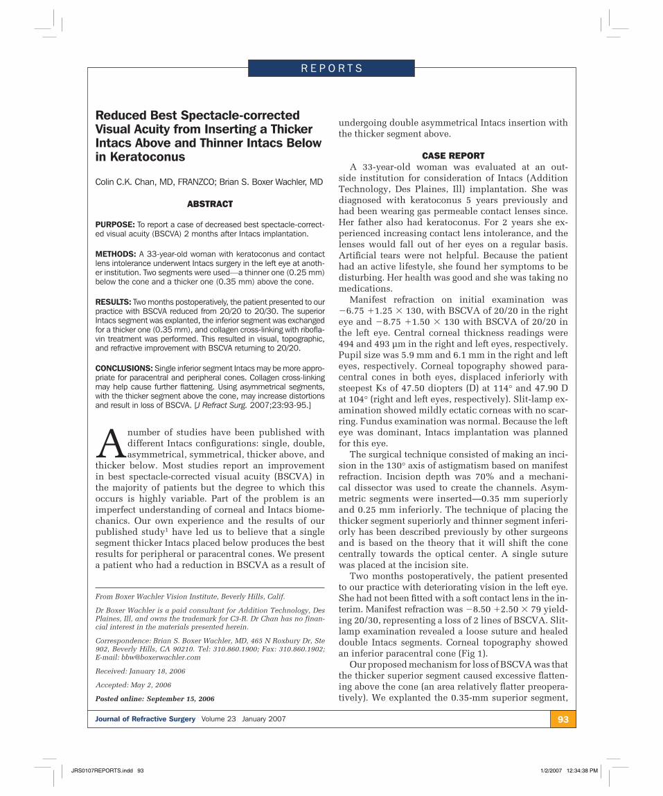
Two months postoperatively, the patient presented to our practice with deteriorating vision in the left eye. She had not been fi tted with a soft contact lens in the in-terim. Manifest refraction was �8.50 �2.50 � 79 yield-ing 20/30, representing a loss of 2 lines of BSCVA. Slit-lamp examination revealed a loose suture and healed double Intacs segments. Corneal topography showed an inferior paracentral cone (Fig 1).
Our proposed mechanism for loss of BSCVA was that the thicker superior segment caused excessive fl atten-ing above the cone (an area relatively fl atter preopera-tively). We explanted the 0.35-mm superior segment,
From Boxer Wachler Vision Institute, Beverly Hills, Calif.
Dr Boxer Wachler is a paid consultant for Addition Technology, Des Plaines, Ill, and owns the trademark for C3-R. Dr Chan has no finan-cial interest in the materials presented herein.
Correspondence: Brian S. Boxer Wachler, MD, 465 N Roxbury Dr, Ste 902, Beverly Hills, CA 90210. Tel: 310.860.1900; Fax: 310.860.1902; E-mail: [email protected]
Figure 1. Corneal topography of the left eye shows an inferior paracentral cone (lower left). Topography shows improve-ment after Intacs superior segment explan-tation, exchange of inferior segment, and collagen cross-linking treatment (upper left). Difference map showing marked flat-tening of the cone at 3 months after surgery (right).
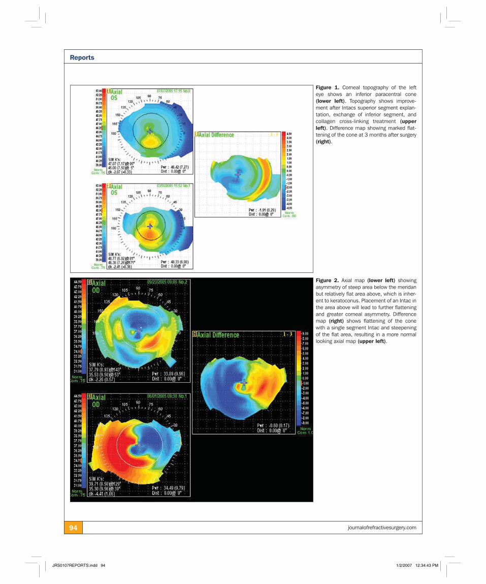
Figure 2. Axial map (lower left) showing asymmetry of steep area below the meridan but relatively flat area above, which is inher-ent to keratoconus. Placement of an Intac in the area above will lead to further flattening and greater corneal asymmetry. Difference map (right) shows flattening of the cone with a single segment Intac and steepening of the flat area, resulting in a more normal looking axial map (upper left).
95Journal of Refractive Surgery Volume 23 January 2007
Reports
and the 0.25-mm inferior segment was exchanged for a 0.35-mm segment. The Intacs procedure was com-bined with corneal collagen cross-linking with ribo-fl avin (C3-R) on the same day, as we have found this to augment fl attening by the Intacs (Chan CK, Sharma M, Boxer Wachler BS, unpublished data, 2005). These procedures led to a marked topographic and refractive improvement (see Fig 1) at 3 months postoperatively. The manifest refraction was �5.25 �0.75 � 150, which returned BSCVA to 20/20. The difference map (see Fig 1) shows resolution of induced astigmatism. The pa-tient was fi tted with a soft contact lens and is satisfi ed with the result.
DISCUSSIONIn the United States, Intacs have been used for kera-
toconus since 1999 but controversy remains regard-ing the best technique for placement of segments. It has been our experience that a single segment works best for paracentral and peripheral cones, whereas a symmetrical double segment works best for central cones.1 Our theory is that in peripheral keratoconus, the cone is oriented around a meridian of astigmatism, and there is a relatively steep area below the meridian and a relatively fl at area above (Fig 2). Placement of a segment above the meridian in the fl at topographic area exacerbates the topographic power asymmetry by causing further fl attening. We have observed a unique coupling effect with implantation of a single Intacs segment in that superior steepening occurs, which im-proves corneal symmetry (see Fig 2). We describe this to our patients as a “beanbag effect”; that is, sitting on or fl attening one end of the beanbag results in the other end popping up. Alio et al2 recently demonstrated that using a single segment for inferior keratoconus and a double segment for keratoconus, which extends above the 180° axis, is effective.
We have also found that C3-R augments the effect of inferior segment Intacs (Chan CK, Sharma M, Boxer Wachler BS, unpublished data, 2005). Wollensak et al3 demonstrated that fl attening occurs with collagen cross-linking alone. We believe the primary reason for our patient’s improvement in BSCVA and corneal shape was the Intacs exchange, but C3-R may have added to the topographic and refractive improvements, along with the superior segment explantation.
The idea of using a thinner segment under the cone and a thicker one above to displace the cone towards the optical center is an intriguing idea. Asymmetrical segments in the opposite confi guration (thicker seg-ment below cone and thinner segment above cone) have been used previously and have shown to be ef-fective.4-6 To our knowledge, the results of the tech-nique used in this report (thicker segment above cone and thinner segment below cone) have not been pub-lished in the literature.
In our patient, prior placement of a thicker segment above the cone resulted in loss of BSCVA and increase in manifest cylinder. Alio et al7 recently reported an-other case where this approach resulted in loss of BSCVA and increased keratometric values, which was subsequently remedied by exchanging the two segments. There are a number of possible reasons why this technique may not produce optimal results: 1) shifting the cone centrally will shift an irregular part of the cornea into the visual axis. If the cone was a sym-metrical sphere, this would have the effect of inducing myopia, but as it is an irregular shape, it will likely worsen distortions; and 2) the topography of a cone has two areas—a steep area below and a fl at area above. Placing a segment in the upper half will further fl atten an already fl at area. Placing the thicker segment will fl atten it further still and heighten the asymmetry.
REFERENCES 1. Sharma M, Boxer Wachler BS. Comparison of single-segment
and double-segment Intacs for keratoconus and post-LASIK ec-tasia. Am J Ophthalmol. 2006;141:891-895.
2. Alio JL, Artola A, Hassanein A, Haroun H, Galal A. One or 2 Intacs segments for the correction of keratoconus. J Cataract Refract Surg. 2005;31:943-953.
3. Wollensak G, Spoerl E, Seiler T. Ribofl avin/ultraviolet-a-in-duced collagen crosslinking for the treatment of keratoconus. Am J Ophthalmol. 2003;135:620-627.
4. Boxer Wachler BS, Christie JP, Chandra NS, Chou B, Korn T, Nepomuceno R. Intacs for keratoconus. Ophthalmology. 2003;110:1031-1040.
5. Hellstedt T, Makela J, Uusitalo R, Emre S, Uusitalo R. Treating keratoconus with intacs corneal ring segments. J Refract Surg. 2005;21:236-246.
6. Colin J, Cochener B, Savary G, Malet R, Holmes-Higgin D. INTACS inserts for treating keratoconus: one-year results. Ophthalmology. 2001;108:1409-1414.
7. Alio JL, Shabayek MH. Intracorneal asymmetrical rings for keratoconus: where should the thicker segment be implanted? J Refract Surg. 2006;22:307-309.
Corneal Haze Following PRK With Mitomycin C as a Retreatment Versus Prophylactic Use in the Contralateral Eye
Marcelo V. Netto, MD; Maria Regina Chalita, MD; Ronald R. Krueger, MD, MSE
ABSTRACT
PURPOSE: To report photorefractive keratectomy (PRK) treated with mitomycin C (MMC) for previous corneal haze in one eye and PRK with MMC to prevent corneal haze formation in the fellow eye.
METHODS: A 40-year-old woman underwent PRK with MMC to treat previous corneal haze (secondary to previous PRK without MMC) for residual refractive error of �0.50 �0.25 � 165 in the left eye and PRK with MMC to prevent corneal haze in the right eye.
RESULTS: Postoperative slit-lamp examination revealed no haze in the right eye, but continued mild haze in the left eye.
CONCLUSIONS: Treatment with PRK and MMC for previous cor-neal haze is not as effective as primary PRK with MMC in pre-venting postoperative corneal haze formation. [J Refract Surg. 2007;23:96-98.]
Excimer laser photorefractive keratectomy (PRK) is a popular refractive procedure proven to be ef-fective and safe for the correction of refractive
errors.1 However, postoperative corneal subepithelial scarring is a challenging complication following PRK, especially after high myopic corrections.2 Stromal wound healing modulators have been used to mini-mize corneal haze formation, but limited effi cacy is reported.3,4
Topical mitomycin C (MMC) was recently intro-duced as an adjunctive therapy for the treatment of
corneal haze after PRK.5,6 We describe a 40-year-old patient who underwent repeat PRK with MMC appli-cation to treat corneal haze formation following previ-ous PRK in one eye, and MMC to prevent corneal haze after PRK in the contralateral eye, which resulted in different outcomes.
CASE REPORTA 40-year-old woman presented at the Cole Eye In-
stitute, Cleveland, Ohio for refractive surgery to cor-rect manifest refractive error of �8.00 �2.50 � 89 and �8.75 �1.75 � 91 in the right and left eyes, respec-tively. Best spectacle-corrected visual acuity (BSCVA) was 20/20 in both eyes. Cycloplegic refraction was �7.50 �2.50 � 92 in the right eye and �8.25 �2.00 � 90 in the left eye. Preoperative keratometric readings were 43.25/45.12 @ 04 in the right eye and 43.00/44.75 @ 180 in the left eye. Ultrasonic pachymetry showed central corneal thickness of 545 µm in the right eye and 551 µm in the left eye. Corneal topography was normal in both eyes. Colvard infrared pupillometer (Oasis Medical, Glendora, Calif) showed pupil size of 5.2 mm in the right eye and 5.0 mm in the left eye. All preoperative tests were normal and there was no signifi cant medical or ocular history. However, slit-lamp examination revealed anterior base-ment membrane dystrophy in both eyes.
The patient underwent uneventful PRK for distance correction in the left eye with the LADARVision 4000 (Alcon Laboratories Inc, Ft Worth, Tex) in January 2002. Alcohol removal of epithelium was completed and stromal photoablation was performed using a 6.0-mm optical zone with ablation depth of 96 µm. Post-operatively, the patient was treated with prednisolone acetate 1% (Predforte; Allergan, Irvine, Calif), ketoro-lac tromethamine 0.5% (Acular, Allergan), and cipro-fl oxacin 0.3% (Ciloxan, Alcon Laboratories Inc) four times a day and a bandage contact lens (Sofl ens 66; Bausch & Lomb, Rochester, NY) was applied. The epi-thelial defect healed in 5 days and the bandage contact lens was removed. Ciloxan and Acular were discontin-ued 1 week postoperatively and Predforte was tapered over the next 3 weeks.
Two weeks after surgery, uncorrected visual acuity (UCVA) and BSCVA was 20/30 and 20/20, respectively, with residual refractive error of �0.50 �0.25 � 165. Four weeks after PRK, the patient developed central subepithelial corneal haze (3�/4�) and BSCVA was 20/30 with manifest refraction of �1.25 �0.50 � 175. Refractive error stabilized after 5 months, and the patient’s left eye was surgically enhanced with PRK and an MMC 0.02%–soaked corneal sponge was ap-plied intraoperatively for 2 minutes, followed by copi-ous irrigation with balanced salt solution. The previ-
From Cole Eye Institute, The Cleveland Clinic Foundation, Cleveland, Ohio (Netto, Chalita, Krueger); the Department of Ophthalmology, University of São Paulo (Netto); and the Department of Ophthalmology, Federal University of São Paulo (Chalita), São Paulo, Brazil.
The authors have no proprietary interest in the materials presented herein.
Correspondence: Ronald R. Krueger, MD, MSE, Dept of Refractive Surgery, Cole Eye Institute, The Cleveland Clinic Foundation, 9500 Euclid Ave, Cleveland, OH 44195. Tel: 216.444.8158; Fax: 216.445.8475; E-mail: [email protected]
97Journal of Refractive Surgery Volume 23 January 2007
Reports
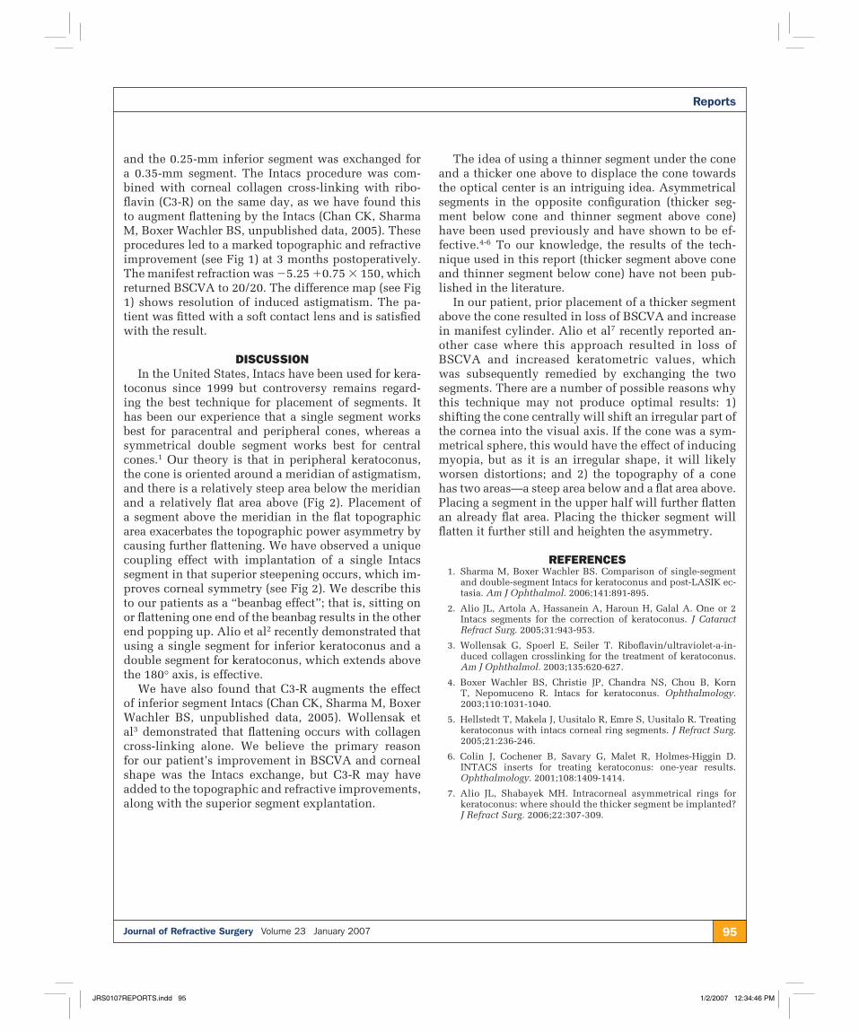
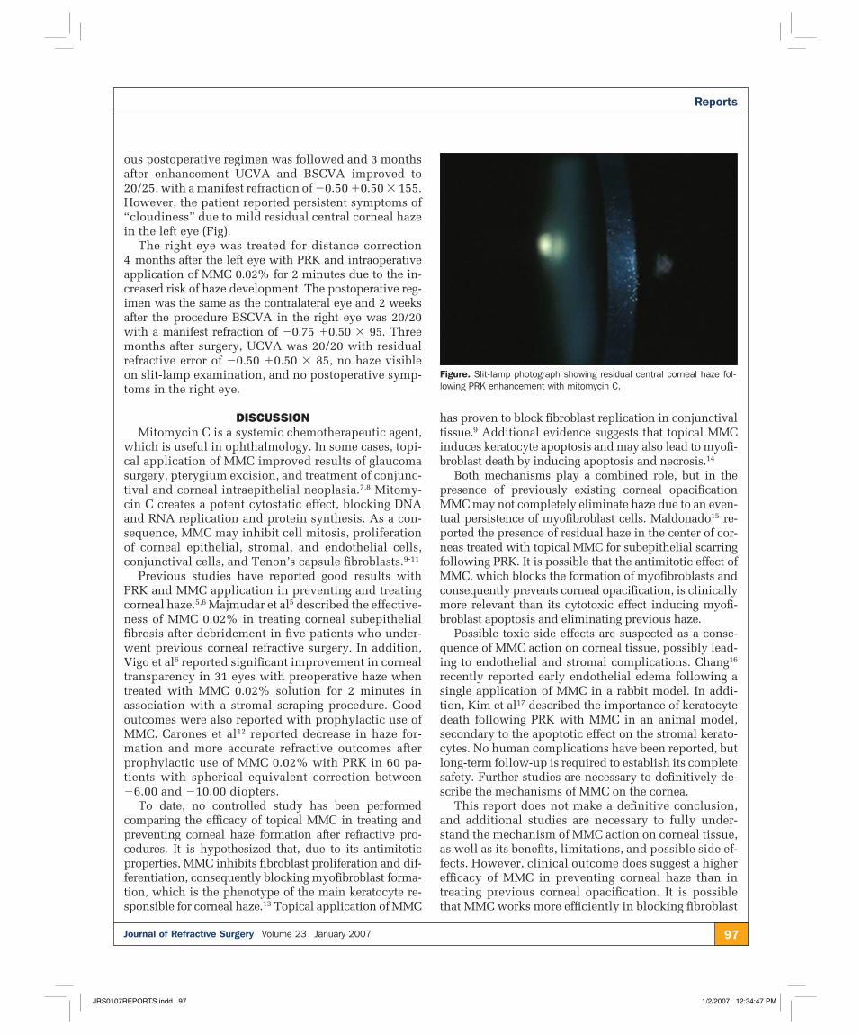
ous postoperative regimen was followed and 3 months after enhancement UCVA and BSCVA improved to 20/25, with a manifest refraction of �0.50 �0.50 � 155. However, the patient reported persistent symptoms of “cloudiness” due to mild residual central corneal haze in the left eye (Fig).
The right eye was treated for distance correction 4 months after the left eye with PRK and intraoperative application of MMC 0.02% for 2 minutes due to the in-creased risk of haze development. The postoperative reg-imen was the same as the contralateral eye and 2 weeks after the procedure BSCVA in the right eye was 20/20 with a manifest refraction of �0.75 �0.50 � 95. Three months after surgery, UCVA was 20/20 with residual refractive error of �0.50 �0.50 � 85, no haze visible on slit-lamp examination, and no postoperative symp-toms in the right eye.
DISCUSSIONMitomycin C is a systemic chemotherapeutic agent,
which is useful in ophthalmology. In some cases, topi-cal application of MMC improved results of glaucoma surgery, pterygium excision, and treatment of conjunc-tival and corneal intraepithelial neoplasia.7,8 Mitomy-cin C creates a potent cytostatic effect, blocking DNA and RNA replication and protein synthesis. As a con-sequence, MMC may inhibit cell mitosis, proliferation of corneal epithelial, stromal, and endothelial cells, conjunctival cells, and Tenon’s capsule fi broblasts.9-11
Previous studies have reported good results with PRK and MMC application in preventing and treating corneal haze.5,6 Majmudar et al5 described the effective-ness of MMC 0.02% in treating corneal subepithelial fi brosis after debridement in fi ve patients who under-went previous corneal refractive surgery. In addition, Vigo et al6 reported signifi cant improvement in corneal transparency in 31 eyes with preoperative haze when treated with MMC 0.02% solution for 2 minutes in association with a stromal scraping procedure. Good outcomes were also reported with prophylactic use of MMC. Carones et al12 reported decrease in haze for-mation and more accurate refractive outcomes after prophylactic use of MMC 0.02% with PRK in 60 pa-tients with spherical equivalent correction between �6.00 and �10.00 diopters.
To date, no controlled study has been performed comparing the effi cacy of topical MMC in treating and preventing corneal haze formation after refractive pro-cedures. It is hypothesized that, due to its antimitotic properties, MMC inhibits fi broblast proliferation and dif-ferentiation, consequently blocking myofi broblast forma-tion, which is the phenotype of the main keratocyte re-sponsible for corneal haze.13 Topical application of MMC
has proven to block fi broblast replication in conjunctival tissue.9 Additional evidence suggests that topical MMC induces keratocyte apoptosis and may also lead to myofi -broblast death by inducing apoptosis and necrosis.14
Both mechanisms play a combined role, but in the presence of previously existing corneal opacifi cation MMC may not completely eliminate haze due to an even-tual persistence of myofi broblast cells. Maldonado15 re-ported the presence of residual haze in the center of cor-neas treated with topical MMC for subepithelial scarring following PRK. It is possible that the antimitotic effect of MMC, which blocks the formation of myofi broblasts and consequently prevents corneal opacifi cation, is clinically more relevant than its cytotoxic effect inducing myofi -broblast apoptosis and eliminating previous haze.
Possible toxic side effects are suspected as a conse-quence of MMC action on corneal tissue, possibly lead-ing to endothelial and stromal complications. Chang16 recently reported early endothelial edema following a single application of MMC in a rabbit model. In addi-tion, Kim et al17 described the importance of keratocyte death following PRK with MMC in an animal model, secondary to the apoptotic effect on the stromal kerato-cytes. No human complications have been reported, but long-term follow-up is required to establish its complete safety. Further studies are necessary to defi nitively de-scribe the mechanisms of MMC on the cornea.
This report does not make a defi nitive conclusion, and additional studies are necessary to fully under-stand the mechanism of MMC action on corneal tissue, as well as its benefi ts, limitations, and possible side ef-fects. However, clinical outcome does suggest a higher effi cacy of MMC in preventing corneal haze than in treating previous corneal opacifi cation. It is possible that MMC works more effi ciently in blocking fi broblast
Figure. Slit-lamp photograph showing residual central corneal haze fol-lowing PRK enhancement with mitomycin C.
replication, thereby avoiding their formation, than in eliminating previously formed myofi broblasts, es-pecially among cells located more posteriorly in the stroma. Our fi ndings are consistent with preliminary results of a randomized experimental study performed in rabbits comparing the effi cacy of topical MMC in preventing corneal haze formation versus treatment of previous corneal haze.18
REFERENCES 1. Rajan MS, Jaycock P, O’Brart D, Nystrom HH, Marshall J. A
long-term study of photorefractive keratectomy: 12-year follow-up. Ophthalmology. 2004;111:1813-1824.
2. Hersh PS, Stulting RD, Steinert RF, Waring GO III, Thompson KP, O’Connell M, Doney K, Schein OD. Results of phase III ex-cimer laser photorefractive keratectomy for myopia. The Sum-mit PRK Study Group. Ophthalmology. 1997;104:1535-1553.
3. Thom SB, Myers JS, Rapuano CJ, Eagle RC Jr, Siepser SB, Gomes JA. Effect of topical anti-transforming growth factor-beta on cor-neal stromal haze after photorefractive keratectomy in rabbits. J Cataract Refract Surg. 1997;23:1324-1330.
4. Corbett MC, O’Brart DP, Marshall J. Do topical corticosteroids have a role following excimer laser photorefractive keratecto-my? J Refract Surg. 1995;11:380-387.
5. Majmudar PA, Forstot SL, Dennis RF, Nirankari VS, Damiano RE, Brenart R, Epstein RJ. Topical mitomycin-C for subepithe-lial fi brosis after refractive corneal surgery. Ophthalmology. 2000;107:89-94.
6. Vigo L, Scandola E, Carones F. Scraping and mitomycin C to treat haze and regression after photorefractive keratectomy for myopia. J Refract Surg. 2003;19:449-454.
7. Akarsu C, Onol M, Hasanreisoglu B. Effects of thick Tenon’s capsule on primary trabeculectomy with mitomycin-C. Acta Ophthalmol Scand. 2003;81:237-241.
8. Oguz H. Mitomycin C and pterygium excision. Ophthalmology. 2003;110:2257-2258.
9. Lee JS, Oum BS, Lee SH. Mitomycin C infl uence on inhibition of cellular proliferation and subsequent synthesis of type I col-lagen and laminin in primary and recurrent pterygia. Ophthal-mic Res. 2001;33:140-146.
10. Watanabe J, Sawaguchi S, Fukuchi T, Abe H, Zhou L. Effects of mitomycin C on the expression of proliferating cell nuclear antigen after fi ltering surgery in rabbits. Graefes Arch Clin Exp Ophthalmol. 1997;235:234-240.
11. Pinilla I, Larrosa JM, Polo V, Honrubia FM. Subconjunctival injection of low doses of mitomycin C: effects on fi broblast pro-liferation. Ophthalmologica. 1998;212:306-309.
12. Carones F, Vigo L, Scandola E, Vacchini L. Evaluation of the prophylactic use of mitomycin-C to inhibit haze formation after photorefractive keratectomy. J Cataract Refract Surg. 2002;28:2088-2095.
13. Mohan RR, Hutcheon AE, Choi R, Hong J, Lee J, Mohan RR, Ambrosio R Jr, Zieske JD, Wilson SE. Apoptosis, necrosis, pro-liferation, and myofi broblast generation in the stroma following LASIK and PRK. Exp Eye Res. 2003;76:71-87.
14. Kim TI, Tchah H, Lee SA, Sung K, Cho BJ, Kook MS. Apoptosis in keratocytes caused by mitomycin C. Invest Ophthalmol Vis Sci. 2003;44:1912-1917.
15. Maldonado MJ. Intraoperative MMC after excimer laser surgery for myopia. Ophthalmology. 2002;109:826.
16. Chang SW. Early corneal edema following topical application of mitomycin-C. J Cataract Refract Surg. 2004;30:1742-1750.
17. Kim TI, Pak JH, Lee SY, Tchah H. Mitomycin C-induced reduc-tion of keratocytes and fi broblasts after photorefractive keratec-tomy. Invest Ophthalmol Vis Sci. 2004;45:2978-2984.
18. Netto MV, Mohan RR, Sinha S, Sharma A, Gupta PC, Wilson SE. Effect of prophylactic and therapeutic mitomycin C on cor-neal apoptosis, cellular proliferation, haze, and long-term kera-tocyte density in rabbits. J Refract Surg. 2006;22:562-574.
Corneal Ectasia After Hyperopic LASIK
J. Bradley Randleman, MD; Christopher S. Banning, MD; R. Doyle Stulting, MD, PhD
ABSTRACT
PURPOSE: To report two cases of corneal ectasia, which devel-oped after hyperopic LASIK.
METHODS: Preoperative pellucid marginal corneal degeneration was observed in patient 1. Patient 2 had no preoperative risk factors.
RESULTS: Patient 1, a 47-year-old man, developed corneal ec-tasia in his right eye 6 months after unilateral hyperopic LASIK. Preoperative manifest refraction was �2.00 �1.50 � 178 in the right eye and �1.00 sphere in the left eye. Corneal thickness was 585 µm and 575 µm (right and left eye, respectively). Preopera-tive topography of the right eye demonstrated inferior steepening in the far periphery, suggestive of early pellucid marginal corneal degeneration. Patient 2, a 35-year-old man, developed corneal ectasia in his right eye �3 years after bilateral LASIK. Preopera-tive manifest refraction was �2.50 sphere and �3.25 sphere (right and left eye, respectively), and corneal thickness was 556 µm in both eyes. Preoperative topography was normal in both eyes with no evidence of asymmetry, steepening, or irregularity.
CONCLUSIONS: Corneal ectasia can occur after hyperopic LASIK in patients with or without recognized preoperative risk factors. Although uncommon, patients with pellucid marginal corneal degeneration can have hyperopic refractions and are at high risk for developing corneal ectasia after LASIK. [J Refract Surg. 2007;23:98-102.]
From the Department of Ophthalmology, Emory University and Emory Vision, Atlanta, Ga.
This study was supported by Research to Prevent Blindness Inc, New York, NY; and the National Institutes of Health (Core Grant P30 EYO6360), Bethesda, Md.
The authors have no proprietary interests in the materials presented herein.
Correspondence: J. Bradley Randleman, MD, 1365 B Clifton Road NE, Ste 4500, Atlanta, GA 30322. Tel: 404.778.4530; Fax: 404.778.4002; E-mail: [email protected]
99Journal of Refractive Surgery Volume 23 January 2007
Reports
Corneal ectasia after myopic LASIK was fi rst reported in 1998 and has been well docu-mented in the literature.1-4 Identifi ed risk fac-
tors include high myopia, low preoperative corneal thickness, low residual stromal bed thickness, and abnormal topographies, including forme fruste kera-toconus3,4 and pellucid marginal corneal degenera-tion.5,6 This is the fi rst report in the literature of cor-neal ectasia occurring after hyperopic LASIK. Two cases of ectasia after hyperopic LASIK are presented: one case occurred in a patient with no recognized risk factors, and the other occurred in a patient with topographic fi ndings of pellucid marginal corneal degeneration.
CASE REPORTS
PATIENT 1A 52-year-old man presented in 2005 requesting sur-
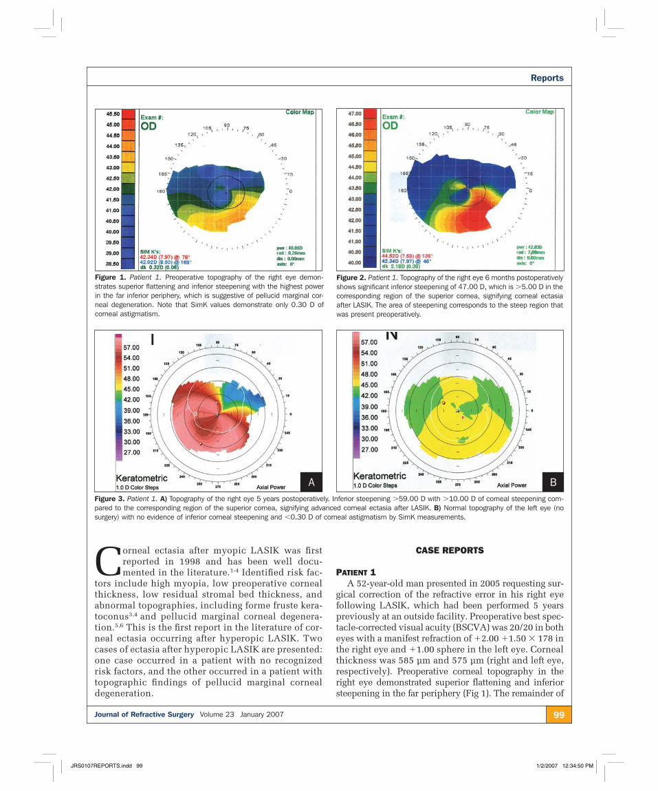
gical correction of the refractive error in his right eye following LASIK, which had been performed 5 years previously at an outside facility. Preoperative best spec-tacle-corrected visual acuity (BSCVA) was 20/20 in both eyes with a manifest refraction of �2.00 �1.50 � 178 in the right eye and �1.00 sphere in the left eye. Corneal thickness was 585 µm and 575 µm (right and left eye, respectively). Preoperative corneal topography in the right eye demonstrated superior fl attening and inferior steepening in the far periphery (Fig 1). The remainder of
Figure 1. Patient 1. Preoperative topography of the right eye demon-strates superior flattening and inferior steepening with the highest power in the far inferior periphery, which is suggestive of pellucid marginal cor-neal degeneration. Note that SimK values demonstrate only 0.30 D of corneal astigmatism.
Figure 2. Patient 1. Topography of the right eye 6 months postoperatively shows significant inferior steepening of 47.00 D, which is �5.00 D in the corresponding region of the superior cornea, signifying corneal ectasia after LASIK. The area of steepening corresponds to the steep region that was present preoperatively.
Figure 3. Patient 1. A) Topography of the right eye 5 years postoperatively. Inferior steepening �59.00 D with �10.00 D of corneal steepening com-pared to the corresponding region of the superior cornea, signifying advanced corneal ectasia after LASIK. B) Normal topography of the left eye (no surgery) with no evidence of inferior corneal steepening and �0.30 D of corneal astigmatism by SimK measurements.
the examination was normal. The patient underwent un-eventful LASIK in the right eye in December 2000 with the VISX excimer laser (VISX, Santa Clara, Calif). No oth-er specifi c surgical details were available for review.
Six months postoperatively, the patient complained of blurred vision in the right eye. Uncorrected visual acuity (UCVA) was 20/70, BSCVA was 20/25 with a manifest refraction of �0.25 �3.25 � 006, and topog-raphy showed signifi cantly increased inferior corneal steepening (Fig 2). The patient was diagnosed with corneal ectasia after LASIK in 2002 and fi tted with a rigid gas permeable contact lens in the right eye.
The patient fi rst presented to our institution in October 2005 seeking alternatives to the contact lens correction of his right eye. At this time, BSCVA was
20/80 in the right eye with a manifest refraction of �8.00 �7.75 � 140. Central corneal thickness mea-sured 532 µm and 573 µm (right and left eye, respec-tively) by ultrasound pachymeter. Topography demon-strated advanced corneal steepening in the right eye, and was normal in the unoperated left eye (Fig 3). Con-focal microscopy revealed a fl ap thickness of 154 µm and a residual stromal bed of 290 µm at the thinnest point inferonasally. The patient was informed of the fi ndings and elected to continue wearing the rigid gas permeable contact lens.
PATIENT 2A 35-year-old man presented for refractive surgery
evaluation in June 2000 at an outside facility. Preop-
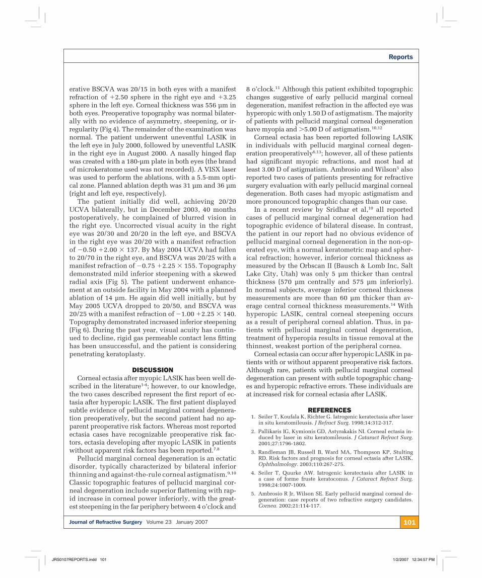
Figure 4. Patient 2. Preoperative topography of the A) right and B) left eyes was normal with no evidence of steepening, irregularity, or asymmetry between eyes.
A B
Figure 5. Patient 2. Topography of the right eye 4 years postoperatively shows increased inferior steepening with a skewed radial axis. The steep-est point inferiorly is �47.00 D with approximately 4.00 D of increased corneal steepening compared to the corresponding region in the superior cornea, signifying early corneal ectasia after LASIK.
Figure 6. Patient 2. Topography of the right eye 5 years after initial surgery and 1 year after enhancement shows increased inferior corneal steepening with more pronounced skewing of the radial axis. The steep-est point inferiorly is �49.00 D with �8.00 D of increased corneal steepening compared to the corresponding region of the superior cornea, signifying progression of corneal ectasia.
101Journal of Refractive Surgery Volume 23 January 2007
Reports
erative BSCVA was 20/15 in both eyes with a manifest refraction of �2.50 sphere in the right eye and �3.25 sphere in the left eye. Corneal thickness was 556 µm in both eyes. Preoperative topography was normal bilater-ally with no evidence of asymmetry, steepening, or ir-regularity (Fig 4). The remainder of the examination was normal. The patient underwent uneventful LASIK in the left eye in July 2000, followed by uneventful LASIK in the right eye in August 2000. A nasally hinged fl ap was created with a 180-µm plate in both eyes (the brand of microkeratome used was not recorded). A VISX laser was used to perform the ablations, with a 5.5-mm opti-cal zone. Planned ablation depth was 31 µm and 36 µm (right and left eye, respectively).
The patient initially did well, achieving 20/20 UCVA bilaterally, but in December 2003, 40 months postoperatively, he complained of blurred vision in the right eye. Uncorrected visual acuity in the right eye was 20/30 and 20/20 in the left eye, and BSCVA in the right eye was 20/20 with a manifest refraction of �0.50 �2.00 � 137. By May 2004 UCVA had fallen to 20/70 in the right eye, and BSCVA was 20/25 with a manifest refraction of �0.75 �2.25 � 155. Topography demonstrated mild inferior steepening with a skewed radial axis (Fig 5). The patient underwent enhance-ment at an outside facility in May 2004 with a planned ablation of 14 µm. He again did well initially, but by May 2005 UCVA dropped to 20/50, and BSCVA was 20/25 with a manifest refraction of �1.00 �2.25 � 140. Topography demonstrated increased inferior steepening (Fig 6). During the past year, visual acuity has contin-ued to decline, rigid gas permeable contact lens fi tting has been unsuccessful, and the patient is considering penetrating keratoplasty.
DISCUSSIONCorneal ectasia after myopic LASIK has been well de-
scribed in the literature1-4; however, to our knowledge, the two cases described represent the fi rst report of ec-tasia after hyperopic LASIK. The fi rst patient displayed subtle evidence of pellucid marginal corneal degenera-tion preoperatively, but the second patient had no ap-parent preoperative risk factors. Whereas most reported ectasia cases have recognizable preoperative risk fac-tors, ectasia developing after myopic LASIK in patients without apparent risk factors has been reported.7,8
Pellucid marginal corneal degeneration is an ectatic disorder, typically characterized by bilateral inferior thinning and against-the-rule corneal astigmatism.9,10 Classic topographic features of pellucid marginal cor-neal degeneration include superior fl attening with rap-id increase in corneal power inferiorly, with the great-est steepening in the far periphery between 4 o’clock and
8 o’clock.11 Although this patient exhibited topographic changes suggestive of early pellucid marginal corneal degeneration, manifest refraction in the affected eye was hyperopic with only 1.50 D of astigmatism. The majority of patients with pellucid marginal corneal degeneration have myopia and �5.00 D of astigmatism.10,12
Corneal ectasia has been reported following LASIK in individuals with pellucid marginal corneal degen-eration preoperatively6,13; however, all of these patients had signifi cant myopic refractions, and most had at least 3.00 D of astigmatism. Ambrosio and Wilson5 also reported two cases of patients presenting for refractive surgery evaluation with early pellucid marginal corneal degeneration. Both cases had myopic astigmatism and more pronounced topographic changes than our case.
In a recent review by Sridhar et al,10 all reported cases of pellucid marginal corneal degeneration had topographic evidence of bilateral disease. In contrast, the patient in our report had no obvious evidence of pellucid marginal corneal degeneration in the non-op-erated eye, with a normal keratometric map and spher-ical refraction; however, inferior corneal thickness as measured by the Orbscan II (Bausch & Lomb Inc, Salt Lake City, Utah) was only 5 µm thicker than central thickness (570 µm centrally and 575 µm inferiorly). In normal subjects, average inferior corneal thickness measurements are more than 60 µm thicker than av-erage central corneal thickness measurements.14 With hyperopic LASIK, central corneal steepening occurs as a result of peripheral corneal ablation. Thus, in pa-tients with pellucid marginal corneal degeneration, treatment of hyperopia results in tissue removal at the thinnest, weakest portion of the peripheral cornea.
Corneal ectasia can occur after hyperopic LASIK in pa-tients with or without apparent preoperative risk factors. Although rare, patients with pellucid marginal corneal degeneration can present with subtle topographic chang-es and hyperopic refractive errors. These individuals are at increased risk for corneal ectasia after LASIK.
REFERENCES 1. Seiler T, Koufala K, Richter G. Iatrogenic keratectasia after laser
in situ keratomileusis. J Refract Surg. 1998;14:312-317.
2. Pallikaris IG, Kymionis GD, Astyrakakis NI. Corneal ectasia in-duced by laser in situ keratomileusis. J Cataract Refract Surg. 2001;27:1796-1802.
3. Randleman JB, Russell B, Ward MA, Thompson KP, Stulting RD. Risk factors and prognosis for corneal ectasia after LASIK. Ophthalmology. 2003;110:267-275.
4. Seiler T, Quurke AW. Iatrogenic keratectasia after LASIK in a case of forme fruste keratoconus. J Cataract Refract Surg. 1998;24:1007-1009.
5. Ambrosio R Jr, Wilson SE. Early pellucid marginal corneal de-generation: case reports of two refractive surgery candidates. Cornea. 2002;21:114-117.
6. Fogla R, Rao SK, Padmanabhan P. Keratectasia in 2 cases with pellucid marginal corneal degeneration after laser in situ ker-atomileusis. J Cataract Refract Surg. 2003;29:788-791.
7. Amoils SP, Deist MB, Gous P, Amoils PM. Iatrogenic keratectasia after laser in situ keratomileusis for less than �4.0 to �7.0 diop-ters of myopia. J Cataract Refract Surg. 2000;26:967-977.
8. Klein SR, Epstein RJ, Randleman JB, Stulting RD. Corneal ecta-sia after laser in situ keratomileusis in patients without appar-ent preoperative risk factors. Cornea. 2006;25:388-403.
12. Tzelikis PF, Cohen EJ, Rapuano CJ, Hammersmith KM, Laibson PR. Management of pellucid marginal corneal degeneration. Cornea. 2005;24:555-560.
13. Klein SR, Muller LT, Uwaydat S, Konowal A, Goosey J, Moran J, Candal EM, Majmudar PA, Epstein RJ. Poor visual outcomes following corneal refractive surgery in patients with clinical-ly unsuspected pellucid marginal degeneration. Presented at: The American Academy of Ophthalmology Annual Meeting; November 18, 2003; Anaheim, Calif.
14. Liu Z, Huang AJ, Pfl ugfelder SC. Evaluation of corneal thick-ness and topography in normal eyes using the Orbscan corneal topography system. Br J Ophthalmol. 1999;83:774-778.
Removal of an Intracorneal Hydrogel Implant for Hyperopia After LASIK
Urs Vossmerbaeumer, MD, MSc; Klaus Ditzen, MD; Jost B. Jonas, MD
ABSTRACT
PURPOSE: To report a case of intracorneal hydrogel lens implan-tation for hyperopia after repeat LASIK surgery.
METHODS: A 34-year-old man underwent intracorneal lens im-
plantation following two LASIK procedures for correction of hy-peropia.
RESULTS: The decentered intracorneal lens was removed due to ocular pain and infl ammation, epithelial ingrowth under the corneal fl ap, and high order aberrations. Pain and infl ammation resolved, and corneal stability was regained �6 months after re-moval of the lens.
CONCLUSIONS: Intracorneal lenses may require explantation if previous laser ablative procedures fail to correct refractive errors. [J Refract Surg. 2007;23:102-104.]
Whereas corneal procedures for correction of myopia have reached a high level of safety, predictability, and effi cacy, good clinical
outcome for hyperopic patients continues to be prob-lematic. This is not attributed to the safety of the inter-ventions, but rather to the optimum design of ablation profi les yielding a stable and predictable refractive outcome.1 In addition, the techniques used continu-ously undergo investigational efforts, which has led to a number of proposed procedures and devices. Re-fractive challenges include predictably steepening the corneal profi le. Implantation of intracorneal lenses has been suggested as an alternative to excimer laser abla-tion. The idea of placing a refractive device within the corneal stroma dates back to almost half a century.2 It has recently taken a fresh impetus with the develop-ment of a transparent, high-water-content hydroxyethyl methacrylate (Nutrapore; Anamed Inc,* Lake Forest, Calif) lens that has proven to be well tolerated intra-corneally.3,4 A lenticule-shaped implant is made from this hydrogel and placed under a LASIK fl ap. Adhe-sion forces ensure a stable position centered in the op-tical zone without further fi xation. Additional support is given to the central and near peripheral cornea to increase corneal curvature and thus enhance refractive power. This device is proposed for the correction of hyperopia and for enhancement of previous refractive stromal procedures, such as LASIK.
Earlier experimental investigations and clinical studies report safety and tissue compatibility.2,5 We present a case that describes the clinical course of a patient who received an intracorneal lens after repeat LASIK.
CASE REPORTA 34-year-old man underwent LASIK on the right
eye for hyperopia of �3.25 diopters (D) (astigmatism �0.25 D @ 150°). A superior hinged fl ap was created, using a standard microkeratome, and excimer laser ablation was performed, but no subjective improve-ment was noted postoperatively. Retrospectively, it is
From the Department of Ophthalmology, Faculty of Clinical Medicine Mannheim, University of Heidelberg, Mannheim, Germany (Vossmerbaeumer, Jonas); and Private Eye Care Practice, Weinheim, Germany (Ditzen).
The authors have no proprietary interest in the materials presented herein.
103Journal of Refractive Surgery Volume 23 January 2007
Reports
not possible to identify the reason for the procedure’s refractive failure, as technical problems with the set-tings of the laser machine were not obvious. With the refractive effect absent, no evidence was found for re-sulting ocular problems. Weeks later, a second LASIK procedure was performed following a re-lift of the initial corneal fl ap. Again, it did not yield a relevant refractive change. Because the refractive effect was un-satisfactory, the corneal fl ap was re-lifted once more and an intracorneal lens (PermaVision, Anamed Inc) was implanted. After re-lifting the fl ap with a round microspatula, the bed was rinsed with 0.9% sodium chloride and the device laid onto the corneal stromal surface within the bed by sliding sidewards from the specially provided spoon. Centration was secured by subjective medial position relative to the patient’s mi-otic pupil to align the optical center of the lens with the optical axis of the eye. Finally, the fl ap was laid back onto the stromal surface and fl attened over the bed with the lens. Decentration of the intracorneal lens occurred 1 week postoperatively, which necessitated re-lifting of the fl ap a third time to reposition the lens.
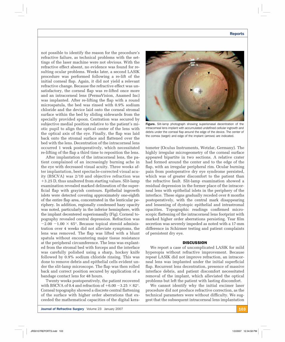
After implantation of the intracorneal lens, the pa-tient complained of an increasingly burning ache in the eye with decreased visual acuity. Three weeks af-ter implantation, best spectacle-corrected visual acu-ity (BSCVA) was 2/10 and objective refraction was �3.25 D, thus unaltered from starting values. Slit-lamp examination revealed marked delineation of the super-fi cial fl ap with grayish contours. Epithelial ingrowth islets were detected covering approximately one-eighth of the entire fl ap area, concentrated in the lenticular pe-riphery. In addition, regionally condensed hazy opacity was noted, particularly in the inferior hemisphere, with the implant decentered superionasally (Fig). Corneal to-pography revealed central depression. Refraction was �2.00 �1.00 � 65°. Because topical steroid adminis-tration over 4 weeks did not alleviate symptoms, the lens was removed. The fl ap was lifted with a blunt spatula without encountering major tissue resistance at the peripheral circumference. The lens was explant-ed from the stromal bed with forceps and the interface was carefully polished using a sharp, hockey knife followed by 0.9% sodium chloride rinsing. This was done to remove debris and epithelial cells evident un-der the slit-lamp microscope. The fl ap was then rolled back and correct position secured by application of a bandage contact lens for 48 hours.
Twenty weeks postoperatively, the patient recovered with BSCVA of 0.4 and refraction of �6.00 �3.25 � 82°. Corneal topography showed a discrete central fl attening of the surface with higher order aberrations that ex-ceeded the mathematical capacities of the digital kera-
tometer (Oculus Instruments, Wetzlar, Germany). The highly irregular microgeometry of the corneal surface appeared bipartite in two sections. A relative crater had formed around the center and to the edge of the fl ap, with an irregular peripheral rim. Ocular burning pain from postoperative dry eye syndrome persisted, which was of greater discomfort to the patient than the refractive fault. Slit-lamp examination showed a residual depression in the former place of the intracor-neal lens with epithelial islets in the periphery of the interface. These signs gradually receded over 8 months postoperatively, with the central mark disappearing and lessening of dystopic epithelial and intrastromal opacities. Topographic readings confi rmed micro-scopic fl attening of the intracorneal lens footprint with marked higher order aberrations persisting. Tear fi lm secretion was severely impeded as noted with a 17-mm difference in Schirmer testing and patient complaints of persistent dry eye.
DISCUSSIONWe report a case of uncomplicated LASIK for mild
hyperopia without refractive improvement. Because repeat LASIK did not improve refraction, an intracor-neal lens was implanted under the initial superfi cial fl ap. Recurrent lens decentration, presence of massive interface debris, and patient discomfort necessitated removal of the implant, which alleviated the optical problems but left the patient with lasting discomfort.
We cannot identify why the initial excimer laser procedure did not produce refractive correction, as the technical parameters were without diffi culty. We sug-gest that the subsequent intracorneal lens implantation
Figure. Slit-lamp photograph showing superionasal decentration of the intracorneal lens implant with accumulated undefined cellular ingrowth and debris under the corneal flap around the edge of the device. The center of the cornea (target) and edge of the implant (arrows) are indicated.
failed because calculation of the needed lens power could not be made with suffi cient precision due to stro-mal haze and previous LASIK interventions. Decentra-tion of an intracorneal lens has been previously report-ed,6 but does not impede the general notion of safety, predictability, and effi cacy of such a device. It could be speculated that previous intrastromal ablation and repeated lifting of the fl ap had interfered with its bio-mechanical stability, leading to less tight intrastromal adhesion. Additional patient interference by rubbing of the eye cannot completely be ruled out. The optical results from decentration of the lens can be compared to those arising from decentered ablation zones in laser ablative operations, with the difference being the rim of the lens further adds to secondary optical straying. In this case, opacities crystallized at the circumference of the lens, thus worsening visual acuity.
The degree of lasting surface irregularities is some-what surprising and may be attributable to a number of different factors. First, repeated lifting of the fl ap with possible exertion of some mechanical stress, particu-larly upon the outer margin, is thought to compromise the biomechanical stability. Second, the insertion and recentration of the lens into this jeopardized environ-ment may not contribute positively to the formation of a geometrically even corneal surface. Third, the efforts to remove sticky corneal debris from either side of the stromal bed may have acted as an additional ablative procedure. Furthermore, neuronal axon sprouting to repair the denervated fl ap seems to have been severely impeded by the fi vefold lifting of the lamella, and pos-sibly by the intracorneal lens itself. It remains unclear why a depression of the central corneal surface per-sisted, even with the intracorneal lens in place.
Based on observations in this case, objections re-garding tolerability of the methacrylate lens in the stro-ma can not be argued. However, the issues that arose
in this patient relating to effi cacy and safety of the de-vice merit consideration. Recurrent lens decentration, interface debris and cell accumulation, and refractive failure with massive subjective discomfort raises the question whether implantation of the device into a non-virginal interface may be appropriate.7 Also, re-sidual aberrations after removal of the device may not be attributed to the device itself, but rather stem from the entire sequence of surgical, laser ablative, and re-correctional procedures. Complications may have occurred due to the series of manipulations, which traumatically altered corneal integrity. Intracorneal lenses may evoke a diffi cult clinical course if a previ-ous laser ablative procedure fails to correct refractive errors.
Patients and surgeons should be aware of the pos-sible limitations of an intracorneal implant as an ad-junctive reparative tool in refractive surgery, particu-larly when prior interventions have failed to correct refractive errors.
REFERENCES 1. Jaycock PD, O’Brart DP, Rajan MS, Marshall J. 5-year follow-up
of LASIK for hyperopia. Ophthalmology. 2005;112:191-199.
2. Bowen SF Jr, Dyer JA, Ogle KN, Neault RW. The intracorneal lens: an experimental study. Mayo Clin Proc. 1961;36:627-632.
3. Ismail MM. Correction of hyperopia with intracorneal implants. J Cataract Refract Surg. 2002;28:527-530.
4. Guell JL, Velasco F, Guerrero E, Gris O, Pujol J. Confocal mi-croscopy of corneas with an intracorneal lens for hyperopia. J Refract Surg. 2004;20:778-782.
6. Michieletto P, Ligabue E, Balestrazzi A, Balestrazzi A, Giglio S. PermaVision intracorneal lens for the correction of hyperopia. J Cataract Refract Surg. 2004;30:2152-2157.
7. Alio JL, Shabayek MH, Montes-Mico R, Mulet ME, Ahmed AG, Merayo J. Intracorneal hydrogel lenses and corneal aberrations. J Refract Surg. 2005;21:247-252.