EMILIO L. KHOURY Department of Immunology, Middlesex Hospital Medical School, London Wl, England. Dr. Khoury's present address is the Laboratory of Immunology, Centro de Educacion Medica e Investigaciones Clinicas, Sanchez de Bustamante 2560, 1425 - Buenos Aires, Argentina.
ABSTRACT Using indirect immunofluorescence (IFL) on viable human thyroid cultures, it has been shown that, although adult follicular cells do not express blood group ABH antigens in vivo, they invariably reexpress the corresponding antigens on the cell surface when cultured in monolayers, even for very short periods. The absence of blood group antigens on noncultured thyroid cells was confirmed by negative IFL on cell suspensions obtained after enzymatic digestion of the glands, whereas these antigens were readily demonstrable on cell suspensions obtained by trypsinization of established monolayers. The quantitative expression of ABH antigens on individual thyroid cells was variable and the cell-surface IFL pattern due to binding of blood group isoantibodies was different from that given by organ-specific thyroid autoanti- bodies on viable cultures. Reexpression of blood group antigens by cultured thyroid cells could not be related to the secretor status of the donors, the presence of a particular source of serum in the culture medium or cell division in vitro. After 2-3 wk in culture, thyroid cells became morphologically dedifferentiated and no longer displayed blood group antigens, though they still expressed cell-surface fl2-microglobulin. Fibroblasts present in the primary thyroid cultures were invariably negative for ABH antigens. These results demonstrate that the surface antigenic repertoire of cultured human cells is not necessarily identical to that present on the same cells in vivo. Furthermore, the possibility that blood group natural isoantibodies bind to the cell surface must be taken into account in experiments in which cultured thyroid cells are exposed to human sera.
The expression of blood group antigens of the ABH system by cells other than erythrocytes has been extensively studied in adult and fetal human tissues, either by mixed agglutination of erythrocytes and tissue cell suspensions (4, 15, 35) or indirect immunofluorescence (IFL) on tissue sections (9, 10, 15, 28, 29). In these reports, thyroid follicular cells were shown to be consistently negative for ABH cell-membrane antigens (9, 29). However, using early human embryos, Szulman (30) was able to detect blood group antigens by IFL on the thyroid primor- dium, still lacking acinar structure, up to the 12th week of gestation. The loss of ABH antigens at this stage takes place when the thyroid begins to show the histological appearance and functional properties of the differentiated gland (12).
Expression of ABH surface antigens has also been investi- gated on cultured human fetal tissues, though not thyroid, by means of specific adherence of erythrocytes to cell monolayers (13) and on established cell lines of human origin (3, 36).
Although the exact identity of cells showing positive reactions in the primary cultures of fetal organs was not determined, epithelioid cells ofteh displayed the antigens on their surfaces, whereas fibroblastoid cells were usually negative. The presence of these surface isoantigens on epithelioid cells decreased in cultures grown for more than 3 wk or repeatedly subcultured (14). On the other hand, several established human cell lines were shown to include a variable proportion of cells expressing blood group H, but not A or B, surface antigens for indefinite periods of culture (36). An increase in the expression of isoan- tigens on these cloned cell lines was related to the rate of ceU division (20) or the addition of blood group precursors to the culture medium (3).
Viable human thyroid cell monolayers have been used for immunological studies in which they are exposed to human sera or immunoglobulin fractions to detect either cytotoxic (6, 16, 19, 25) or stimulating (32) organ-specific autoantibodies. In
the course of IFL studies on these cell-surface reactive auto- antibodies it was found that some normal sera stained mono- layers from blood group A or B, but not O, donors. This finding led to the investigation of the reexpression of ABH surface antigens under these conditions.
MATERIALS AND METHODS
Forty-three human thyroids were studied. 41 were from adult individuals (18 were blood group O, 16 A, 6 B, and 1 AB) and 2 were fetal glands, from a 14-wk- old blood O and a 17-wk-old blood group A fetus, respectively. The adult glands were normal in 12 cases (from patients undergoing radical surgery for carcinoma of the larynx), the other 29 being thyrotoxic (10), multinodular colloid goiter (9), simple goiter (2), Hashimoto's thyroiditis (3), dyshormonogenetic goiter (1) and peri-adenomatous thyroids (4).
Culture of Human Thyroid Cells The procedure has already been reported in detail (18). Briefly, the thyroid
tissue was chopped into small pieces, washed in culture medium (Flow 199 plus NaHCO3 0.35 mg/ml, Penicillin 300 IU/ml and Streptomycin 300/~g/ml Flow Laboratories, Inc., Rockville, MD), and incubated for 60-90 min at 37°C with either collagenase (Worthington type IV, 2 mg/ml Worthington Biochemical Corp., Freehold, NJ) or trypsin (Difco, 2.5 mg/ml Difco Laboratories, Detroit, M1). In some experiments no enzyme was used, and ceils were obtained by shaking the small tissue fragments in culture medium. Cell viability was assessed by differential staining with an acridine orange/ethidium bromide mixture (AO/ EB) using fluorescence microscopy (21), being >90% in all cases. An accurate cell count at this stage was not possible as most thyroid cells were in clumps (23). In some experiments, cells were resuspended and cultured in 15% blood group O or AB normal human serum (NHS) or blood group O fetal human serum (FHS) instead of FCS. 100 bd of the cell suspension were plated onto round glass cover slips, placed in multiwell plates (Linbro), and 400 btl of culture medium containing 15% FCS was added to each well. The cultures were kept at 37°C in a 5% CO2 humidified incubator for various periods, with periodic changes of culture medium, until they were stained.
Sera Although many human sera from different ABO blood groups were tested on
autologous and homologous thyroid monolayers throughout this study, two were systematically used to detect A, B, and H antigens on the surface of the thyroid cells. One was from a blood group O Rh negative normal individual (donor RP) who had not been immunized with blood group substances nor had received any blood transfusion. The other was a "Bombay" type (1) human serum reacting with blood group H, A, and B antigens (kindly provided by Dr. Ten Feizi, Clinical Research Center, Harrow, England). Characterization of isohaemagglu- linins present in these two sera is shown in Table I. Both RP and Bombay sera were also tested after absorption with blood group A, B, AB, or O fresh erythrocytes (2 vol of packed cells/vol serum diluted 1:5, for 2 h at room temperature and then overnight at 4°C). A third serum, from a blood group AB
normal individual (donor LH), was used as tlae negative control in all experiments. All three sera were negative when tested for organ-specific thyroid or any other au/oantibody by either IFL on tissue sections or haemagghitination OVellcome Thymune M and T kits, Welleome Reagents, Ltd., London, England). In addition, twelve human antisera to A and B blood groups (Blood group Reference Laboratory, London, England) were tested on the thyroid cultures, as well as for organ-specific thyroid autoantibodies. All human sera were tested at an initial dilution of 1:10.
Cell-surface IFL Staining of Thyroid Monolayers 18 h to 75-d-old viable thyroid cell monolayers were used for this study. After
being washed in culture medium, the cover slips were covered with 100/al of prediluted serum for 30 rain at room temperature, followed by a second incuba- tion with 100 pl of an FITC-conjugated sheep anti-human Fab serum (Wellcome) diluted 1:50 under similar conditions. Between and after incubations the mono- layers were washed in culture medium. The ceils were then fixed in 5% acetic acid in ethanol at -2O°C for 10 rain, and the cover slips were mounted in glycerol on a microscope slide and examined under a Zeiss fluorescence microscope equipped with epi-illurdination and phase contrast. In some expertments, the monolayers were fixed in chilled acetone for 2 rain before the staining procedure.
serum (WeUcome) diluted 1:30 were used as first and second layer, respectively.
Binding of Exogenous Blood Group Antigens on Thyroid Cells
Blood group O thyroid monolayers were incubated for 30 rain at 37°C with saliva, diluted 1:4 in culture medium, from blood group A or B individuals known to be secretors, and then stained with serum from donor RP.
Identification of Thyroid Follicular Cells in the Cultures
The great majority of the cells present in early thyroid monolayers were follicular ceils. They were further identified by the presence of the organ-specific thyroid microsomal antigen (22) on cultures fixed in acetone and stained with human sera containing autoantibodies to this antigen (Fig. I). Fibroblastoid ceils in the cultures were identified by the presence of the extracellular, membrane- associated fibronectin (34). Rabbit anti-human fibronectin serum was provided by Dr. L. B. Chen, Cold Spring Harbor Laboratory, Cold Spring Harbor, NY and was used diluted 1:100. Both cell types in the cultures invariably showed the prosence of B2-mieroglobulin on their surfaces. Rabbit anti-human Bz-micro- globulin (DAKO-tmmunoglobulius) was diluted 1:20. For the IFL staining with the latter two antisera, FITC-conjugated goat anti-rabbit Ig serum (Nordic Immunological Laboratories, Tilburg, The Netherlands) was used at 1:50 dilution as the second layer.
TABLE I
Specificity of Ant i -Blood Group ABH AIIogeneic Reactions on Viable Human Thyroid Cells in Culture
A b s o r b e d wi th
e ry th ro- I s o h a e m a g g l u t i n a t i n g t i ters aga ins t
Se rum cytes e ry th rocy t e s
Cell-surface indirect IFL (serum di luted 1:10) on 2-B-d-old thyroid cultures from donor b lood group
Blood group A B 0
R P* - 1:256 1:128 <1:2 RP A <1:2 1:64 <1:2 RP B 1:256 <1:2 <1:2 RP AB <1:2 <1:2 <1:2 RP O 1:256 1:128 < I .2 Bombay§ - 1:512 1:256 1:16 Bombay AB <1:2 <1:2 <1:2 Bombay O 1:128 1:64 <1:2 LHII - <1:2 <1:2 <1:2
* Blood group O normal human serum. :l: Number of glands tested. § "Bombay" type (1) normal human serum. II Blood group AB normal human serum.
N.T. Not tested.
194 THE JOURNAL OF C[LL BIOLOGY • VOLUM[ 94, 1982
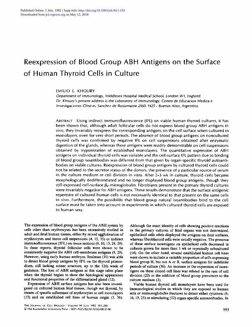
FIGURE 1 2-d-old human thyroid cultures from a blood group O individual, fixed in acetone and stained by indirect IFL with: (a) human serum containing thyroid microsomal autoantibodies, di luted 1:10, showing diffuse intracytoplasmic staining, (b) blood group O normal human serum {RP) at same di lut ion; no IFL staining is present. Original magnification, x 400.
Double-label IFL
For this procedure, viable cultures from blood group A or B thyroids were exposed to a human serum (RP) containing isoantibodies, followed by the FITC ant i-human Fab conjugate. The ceils were then fixed in acetone, and rhodamine- conjugated human immunoglobulins containing thyroid microsomal autoanti- bodies were applied.
Surface IFL Staining of Thyroid Cells in Suspension
180-/d aliquots of the thyroid cell suspension and 20/sl of undiluted serum were placed in 4-ml centrifuge tubes, and the mixture was incubated in ice for 30 min with frequent and gentle shaking. The tubes were then filled with culture medium and centrifuged at 150 g for 5 min. The cell pellet was resuspended in 100 #1 of FITC sheep anti-human Fab conjugate at a 1:50 dilution and incubated for a further 30 min. After washing and centrifugation, the pellet was resuspended in culture medium, and a drop of the cell suspension was placed on a microscope slide, mounted, and examined under fluorescence microscopy. In some experi- ments, thyroid cell monolayers cultured in plastic flasks (Falcon Labware, Oxnard, CA) for 48 h were detached with 0.25% trypsin solution and the cell suspension obtained was then stained by the same procedure.
IFL Staining on Sections of Thyroid Tissue and Thyroid-Cell Pellets
Indirect IFL was performed on unfixed sections of frozen thyroid tissue according to the standard technique (26).
Cell pellets from two blood group A, one blood group B, and one blood group O thyroids, obtained by trypsin digestion of either fresh tissue or 2-d-old established monolayers, were embedded in 5% gelatin and snap frozen according to the method described by Steffelaar et al. (27) but without fixation in paraform- aldehyde. Cryostat sections of these pellets were processed for indirect IFL in the same way.
Complement-mediated Antibody-dependent Cytotoxicity Assay on Thyroid Monolayers
For this purpose, 6-d-old blood group A normal thyroid cultures were incu- bated with fresh, nonheat inactivated, blood group O (RP), or AB (LH) normal serum diluted 1:2 in culture medium for 45, 90, and 180 rain at 37°C in a 5% CO2 incubator. The cover slips were then washed, stained with AO/EB, mounted, and examined by fluorescence microscopy to detect any cytotoxic effect on the ceils still attached. The diluted serum in which the ceils were incubated was centrifuged and the cell pellet was resuspended in the AO/EB solution, mounted on a microscope slide, and examined under UV light.
Determination of "'secretor'" status The secretor status of four blood group A and two blood group B individuals,
whose thyroid ceils were tested in culture, was established by the demonstration of H and the corresponding blood group antigen in saliva by haemagglutination inhibition (24).
E. L
RESULTS
Blood Group Surface Antigens on Thyroid Cells in Culture
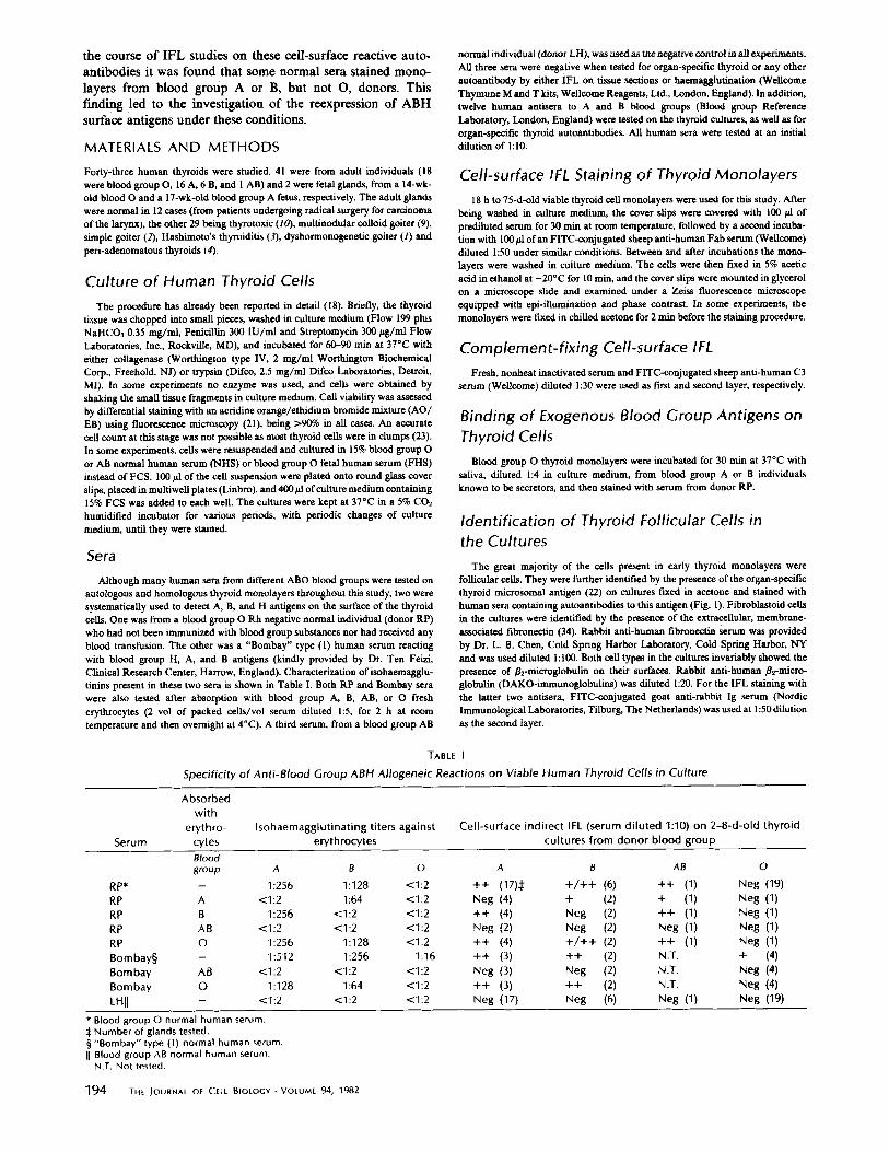
When viable thyroid monolayers from either blood group A, B, or AB individuals were tested by indirect IFL with blood group O RP serum or any other human serum (including 12 commercial anti A or B human antisera) expected to contain isoantibodies to those blood groups, a clear cell-surface staining was observed on the spread thyroid cells in all cases. This consisted of a fine granular pattern covering the whole of the cell membrane. The blood group surface antigens detected in each case invariably corresponded to the genotype of the tissue donor (Table I). Follicular ceils displaying blood group surface antigens were further identified by their counterstaining with rhodamine-conjugated thyroid microsomal antibodies. Al- though the great majority of the thyroid cells were stained, some of them, indistinguishable from the others by either their morphology under phase contrast or the presence of intracy- toplasmic organ-specific microsomal antigen, were completely devoid of blood group surface antigens. Moreover, the density and brightness of the granular staining, though uniform on any particular cell, varied from cell to cell, giving a characteristic "mosaic" pattern on the monolayer and allowing a clear dis- tinction of the cell borders (Fig. 2 a and b). The staining was completely abolished after absorbing the serum with the cor- responding blood group erythrocytes (Table I and Fig. 2 c and d). This cell-surface IFL pattern was clearly distinguishable from the coarser and more uniform granular staining given by sera containing organ-specific thyroid microsomal autoanti- bodies when tested on blood group O normal thyroid cells in culture (18) (Fig. 2 e and f), and also seen by direct IFL on cultured cells obtained from patients with thyroid autoimmune diseases (Khoury et al., unpublished observations). Both organ- specific autoantigens and blood group alloantigens coexisted on the thyroid cell surface during the first week in culture (18).
The cell-surface IFL titer of RP serum on blood group A monolayers was 1:1,000 in most cases; on blood group B cells the titer was somewhat lower. When this serum was used fresh and without prior heat inactivation at 1:I0 dilution, followed by the anti-C3 conjugate, blood group A cultures gave a more exaggerated "mosaic" pattern, with fewer but brighter positive cells. Only occasional thyroid cells showed complement fixing cell surface IFL when blood group B monolayers were tested under the same conditions.
Although only few thyroid cells were obtained without the use of enzymes, they gave an IFL pattern similar to that shown
KHOURY Reexpression of ABH Antigens on Cultured Thyroid Cells 195
FIGURE 2 4-d-old viable thyroid cultures from a blood group A individual. (a), (c), and (e) phase contrast, showing spread thyroid cells. (b) same field as a; indirect IFL with blood group O normal human serum (RP), di luted 1:10, showing a finely granular cell- surface IFL staining which is markedly weaker and less dense on one of the cells. (d) same field as c; indirect IFL with same serum after absorption with blood group A erythrocytes. Only autofluorescent perinuclear granules are visible. (f) same field as e; indirect IFL with a blood group A thyrotoxicosis serum di luted 1:10, showing a coarser granular staining due to organ-specific cell surface autoantibodies. Original magnification, x 650.
by cells dispersed from the glands with either collagenase or trypsin, when stained in culture. Blood group antigens were also detected on the surface of thyroid cells grown in culture medium supplemented with 15% of either blood group AB or O NHS, or blood group O FHS instead of FCS. A clear, though more diffuse, cell-surface-type IFL pattern was ob- tained when the thyroid cultures were fixed in acetone before the staining (Fig. 3).
The cultured thyroid cells displayed blood group surface antigens as soon as they became attached and spread on the cover slip, usually 18-24 h after plating. This was so in all the glands tested except one, in which thyroid cells from a dyshor- monogenetic goiter due to congenital enzymatic deficiency (Pendred's Syndrome) (8) in a blood group A individual were completely negative for this antigen during the first 3 d in culture. In this period, other occasional epithelioid cells in the monolayers (possibly endothelial cells) did show positive stain- ing. Later on, the cultured thyroid cells also expressed blood group antigens in the usual way.
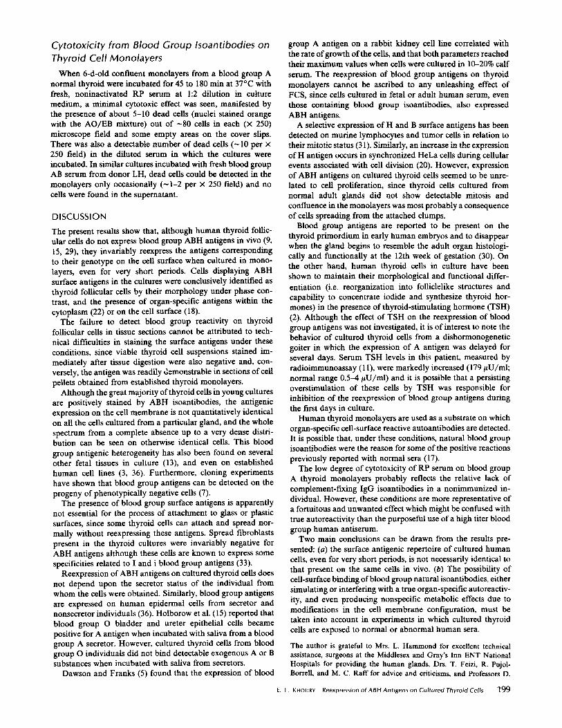
After ~2 wk in culture, thyroid cells from all glands began
to dedifferentiate, their nuclei becoming larger and oval in shape. At this stage they no longer showed A or B surface antigens, and only the remaining differentiated cells were stained by isoantibodies (Fig. 4 a and b), though all cells still displayed p2-microglobulin on their surfaces (Fig, 4 c and d).
Expression of blood group surface antigens did not depend upon the secretor status of the donors, since cultured thyroid cells from three nonsecretor individuals (two from blood group A and one from blood group B) were positively stained by isoantibodies. Similarly, thyroid cells cultured from abnormal glands (thyrotoxicosis, multinodular colloid goiter, Hashi- moto's thyroiditis, etc.) did not differ from normal thyroid cells with respect to the expression of blood group surface antigens.
When blood group O normal thyroid monolayers were in- cubated with normal sera belonging to any blood group, except Bombay, negative staining was obtained in all cases. This was still so when the cells were incubated with saliva from blood group A or B secretor individuals and then stained with RP s e r u m .
When blood group A or B thyroid cultures were stained with
196 THE JOURNAL OF CELL BIOLOGY-VOLUME 94, 1982
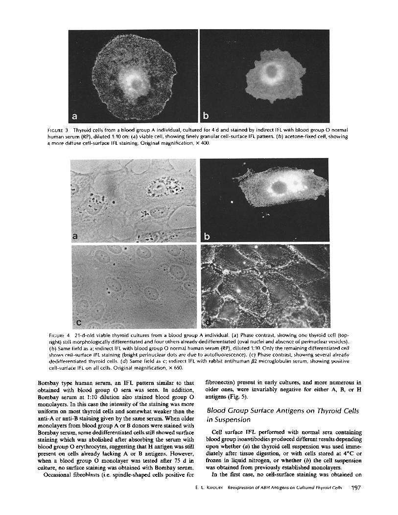
FIGURE 3 Thyroid cells from a blood group A individual, cultured for 4 d and stained by indirect IFL with blood group O normal human serum (RP), di luted 1:10 on: (a) viable cell, showing finely granular cell-surface IFL pattern. (b) acetone-fixed cell, showing a more diffuse cell-surface IFL staining. Original magnification, x 400.
FIGURE 4 21-d-old viable thyroid cultures from a blood group A individual. (a) Phase contrast, showing one thyroid cell (top- right) still morphological ly differentiated and four others already dedifferentiated (oval nuclei and absence of perinuclear vesicles). (b) Same field as a; indirect IFL with blood group O normal human serum (RP), di luted 1:10. Only the remaining differentiated cell shows cell-surface IFL staining (bright perinuclear dots are due to autofluorescence). (c) Phase contrast, showing several already dedifferentiated thyroid cells. (d) Same field as c; indirect IFL with rabbit antihuman/~2 microglobulin serum, showing positive cell-surface IFL on all cells. Original magnification, x 650.
Bombay type human serum, an IFL pattern similar to that obtained with blood group O sera was seen. In addition, Bombay serum at l:10 dilution also stained blood group O monolayers. In this case the intensity of the staining was more uniform on most thyroid cells and somewhat weaker than the anti-A or anti-B staining given by the same serum. When older monolayers from blood group A or B donors were stained with Bombay serum, some dedifferentiated cells still showed surface staining which was abolished after absorbing the serum with blood group O erythrocytes, suggesting that H antigen was still present on cells already lacking A or B antigens. However, when a blood group O monolayer was tested after 75 d in culture, no surface staining was obtained with Bombay serum.
Occasional fibrohlasts (i.e. spindle-shaped cells positive for
fibronectin) present in ear ly cultures, and more numerous in older ones, were invariably negative for either A, B, or H antigens (Fig. 5).
Blood Group Surface Antigens on Thyroid Cells in Suspension
Cell surface IFL performed with normal sera containing blood group isoantibodies produced different results depending upon whether (a) the thyroid cell suspension was used imme- diately after tissue digestion, or with cells stored at 4°C or frozen in liquid nitrogen, or whether (b) the cell suspension was obtained from previously established monolayers.
In the first case, no cell-surface staining was obtained on
E. L. KHOURY Reexpression of ABH Antigens on Cultured Thyroid Cells 197
FIGURE 5 10-d-old viable thyroid cultures from a blood group A individual. (a) Phase contrast, showing fibroblastoid cells. (b) Same field as a; indirect IFL with blood group O normal human serum (RP), di luted 1:10. No IFL staining is present. (c) Phase contrast at lower magnification, showing a bundle of fibroblastoid cells among spread thyroid cells (only their nuclei are visible: arrows). (d) Same field as c; indirect IFL with rabbit antihuman fibronectin serum, showing fibril lar IFL staining only on fibroblastoid cells. Original magnification, a and b x 650. c and d x 2 50.
FIGURE 6 Viable thyroid cell-suspension from a blood group A individual, obtained by trypsinization of a 2-d-old established monolayer. (a) Phase contrast. (b) same field; indirect IFL with blood group O normal human serum (RP) di luted 1:10, showing cell- surface IFL staining. Original magnification, x 650.
TABLE II
Expression of Blood Group A and B Surface Antigens on Adult Thyroid Follicular Cells *
Tissue sections. Viable cell-suspensions after tis-
sue digestion. Cell-pellet sections after tissue
digestion, Viable cell-suspensions from es-
tablished monolayers. Cell-pellet sections from estab-
Positive Positive on differentiated cells. Negative on de-differentiated
cells.
* As detected by indirect irnmunofluorescence
thyroid follicular cells from five different blood group A or AB glands when tested with RP or several other blood group O normal sera. On the other hand, when already established thyroid monolayers were trypsinized 48 h after plating, the cell suspension obtained was positively stained by the same sera (Fig. 6).
IFL on Sections of Thyroid Tissue and Thyroid Cell Pellets
When donor RP serum and 10 of the 12 human anti-A and anti-B antisera were tested on blood group A or B thyroid tissue sections, even undiluted, no staining was observed on thyroid follicular cells and only perifoUicular vascular endo-
198 THt IOURNAL OF CELL BIOLOGY • VOLUM[ 94, 1982
thelium was stained, confLrming previous observations (9, 15, 29). The other two human anti-blood group antisera (one anti- A and one anti-B) stained thyroid follicular cells, irrespective of the blood group of the gland used, when tested undiluted. These two sera were also positive for thyroid microsomal antibodies by haemagglutination. Similarly, donor RP serum did not stain thyroid cells when tested on sections of cell-pellets from blood group A or B glands, obtained/mmediately after tissue digestion. On the other hand, positive rim fluorescence was seen with the same serum when tested on sections of cell- pellets from trypsinized 2-d-old monolayers from a blood group A thyroid (Table II). This positive reaction was abolished when RP serum was tested after absorption with blood group A erythrocytes.
Cytotoxicity from Blood Group lsoantibodies on Thyroid Cell Monolayers
When 6-d-old confluent monolayers from a blood group A normal thyroid were incubated for 45 to 180 rain at 37°C with fresh, noninactivated RP serum at 1:2 dilution in culture medium, a minimal cytotoxic effect was seen, manifested by the presence of about 5-10 dead cells (nuclei stained orange with the AO/EB mixture) out of ~80 cells in each (x 250) microscope field and some empty areas on the cover slips. There was also a detectable number of dead cells (~10 per x 250 field) in the diluted serum in which the cultures were incubated. In similar cultures incubated with fresh blood group AB serum from donor LH, dead cells could be detected in the monolayers only occasionally (~ 1-2 per x 250 field) and no cells were found in the superuatant.
DISCUSSION
The present results show that, although human thyroid follic- ular cells do not express blood group ABH antigens in vivo (9, 15, 29), they invariably reexpress the antigens corresponding to their genotype on the cell surface when cultured in mono- layers, even for very short periods. Cells displaying ABH surface antigens in the cultures were conclusively identified as thyroid follicular cells by their morphology under phase con- trast, and the presence of organ-specific antigens within the cytoplasm (22) or on the cell surface (18).
The failure to detect blood group reactivity on thyroid follicular cells in tissue sections cannot be attributed to tech- nical difficulties in staining the surface antigens under these conditions, since viable thyroid cell suspensions stained im- mediately after tissue digestion were also negative and, con- versely, the antigen was readily demonstrable in sections of cell pellets obtained from established thyroid monolayers.
Although the great majority of thyroid cells in young cultures are positively stained by ABH isoantibodies, the antigenic expression on the cell membrane is not quantitatively identical on all the cells cultured from a particular gland, and the whole spectrum from a complete absence up to a very dense distri- bution can be seen on otherwise identical cells. This blood group antigenic heterogeneity has also been found on several other fetal tissues in culture (13), and even on established human cell lines (3, 36). Furthermore, cloning experiments have shown that blood group antigens can be detected on the progeny of phenotypically negative cells (7).
The presence of blood group surface antigens is apparently not essential for the process of attachment to glass or plastic surfaces, since some thyroid cells can attach and spread nor- mally without reexpressing these antigens. Spread fibroblasts present in the thyroid cultures were invariably negative for ABH antigens although these cells are known to express some specificities related to I and i blood group antigens (33).
Reexpression of ABH antigens on cultured thyroid cells does not depend upon the secretor status of the individual from whom the cells were obtained. Similarly, blood group antigens are expressed on human epidermal ceils from secretor and nonsecretor individuals (36). Holborow et al. (15) reported that blood group O bladder and ureter epithelial ceils became positive for A antigen when incubated with saliva from a blood group A secretor. However, cultured thyroid cells from blood group O individuals did not bind detectable exogenous A or B substances when incubated with saliva from secretors.
Dawson and Franks (5) found that the expression of blood
group A antigen on a rabbit kidney cell line correlated with the rate of growth of the cells, and that both parameters reached their maximum values when cells were cultured in 10-20% calf serum. The reexpression of blood group antigens on thyroid monolayers cannot be ascribed to any unleashing effect of FCS, since cells cultured in fetal or adult human serum, even those containing blood group isoantibodies, also expressed ABH antigens.
A selective expression of H and B surface antigens has been detected on mufine lymphocytes and tumor ceils in relation to their mitotic status (31). Similarly, an increase in the expression of H antigen occurs in synchronized HeLa cells during cellular events associated with cell division (20). However, expression of ABH antigens on cultured thyroid cells seemed to be unre- lated to cell proliferation, since thyroid cells cultured from normal adult glands did not show detectable mitosis and confluence in the monolayers was most probably a consequence of ceils spreading from the attached clumps.
Blood group antigens are reported to be present on the thyroid primordium in early human embryos and to disappear when the gland begins to resemble the adult organ histologi- cally and functionally at the 12th week of gestation (30). On the other hand, human thyroid cells in culture have been shown to maintain their morphological and functional differ- entiation (i.e. reorganization into folliclelike structures and capability to concentrate iodide and synthesize thyroid hor- mones) in the presence of thyroid-stimulating hormone (TSH) (2). Although the effect of TSH on the reexpression of blood group antigens was not investigated, it is of interest to note the behavior of cultured thyroid cells from a dishormonogenetic goiter in which the expression of A antigen was delayed for several days. Serum TSH levels in this patient, measured by radioimmunoassay (11), were markedly increased (179 gU/ml; normal range 0.5-4 #U/ml) and it is possible that a persisting overstimulation of these cells by TSH was responsible for inhibition of the reexpression of blood group antigens during the first days in culture.
Human thyroid monolayers are used as a substrate on which organ-specific cell-surface reactive autoamibodies are detected. It is possible that, under these conditions, natural blood group isoantibodies were the reason for some of the positive reactions previously reported with normal sera (17).
The low degree of cytotoxicity of RP serum on blood group A thyroid monolayers probably reflects the relative lack of complement-fixing IgG isoantibodies in a nonimmunized in- dividual. However, these conditions are more representative of a fortuitous and unwanted effect which might be confused with true autoreactivity than the purposeful use of a high liter blood group human antiserum.
Two main conclusions can be drawn from the results pre- sented: (a) the surface antigenic repertoire of cultured human cells, even for very short periods, is not necessarily identical to that present on the same ceils in vivo. (b) The possibility of cell-surface binding of blood group natural isoantibodies, either simulating or interfering with a true organ-specific autoreactiv- ity, and even producing nonspecific metabolic effects due to modifications in the cell membrane configuration, must be taken into account in experiments in which cultured thyroid cells are exposed to normal or abnormal human sera.
The author is grateful to Mrs. L. H a m m o n d for excellent technical assistance, surgeons at the Middlesex and Gray's Inn ENT National Hospitals for providing the human glands, Drs. T. Feizi, R. Pujol- Borrell, and M. C. Raft for advice and criticisms, and Professors D.
E. L. KHOURY Reexpression of ABH Antigens on Cultured Thyroid Cells 199
D o n i a c h a n d I. M. R o i t t fo r t h e i r p e r m a n e n t g u i d a n c e a n d s u p p o r t ,
M i s s Q. J a y a w a r d e n a p r e p a r e d t h e m a n u s c r i p t .
T h i s w o r k was c a r r i e d o u t w h i l e E. L. K h o u r y was i n rece ip t o f a
W e l l c o m c R e s e a r c h F e l l o w s h i p .
R e c e i v e d f o r pub l i ca t ion 20 F e b r u a r y 1981, a n d in rev i sed f o r m 9
F e b r u a r y 1982.
REFERENCES
1. Bhende, Y. M., C. K. Deshpande, H. M. Bhatia, R. Sangei', R. R. Race, W. T. J. Morgan, and W. M. Watkins. 1952. A "new" blood group character related to the ABO system. Lancet 1:903-904.
2. Bide),, S. P., P. Marsdan, J. Anderson, C. G. McKerron, and H. Berry. 1977. In vitro studies of normal human thyroid cells: responses to thyrotrophin and dibutyryl cyclic AMP. J. Endocrinol. 72:87-96.
3. Chessin, L. N., S. Bramson, W. J. Kuhns, and K. Hirschhorn. 1965. Studies on the A, B, O (H) blood groups on human cells in culture. Blood25:944-953.
4. Coombs, R. R. A., D. Bedford, and L. M. RouiUard. 1956. A and B blood-group antigens on human epidermal ceils demonstrated by mixed agglutination. Lancet 1:461~163.
5. Dawson, A., and Franks D. 1967. Factors affecting the expression of blood group antigen A in cultured cells. Exp. Cell Res. 47:377-385.
6. Forbes, I. J., I. M. Roitt, D. Doniach, and J. L. Solomon. 1962. The thyroid cytntoxic autoantibodies. J. Clin. Invest. 41:996-1006.
7. Franks, D., and A. Dawson. 1966. Variation in the expression of bloud group antigen A in clooal cultures of rabbit ceils. Exp. Cell Res. 42:543-56 I.
8, Fraser. G. R., M. E. Morgans, and W. R. Trotter. 1960. The syndrome of sporadic gnitre and congenital deafness. Quart. J. Mud. 29:279-295.
9, Glyrm, L. E,, and E. J. Holborow, 1959. Distribution of blood-group substances in human tissues. Br. Med, Bull. 15:150-[53,
10. Glynn, L. E.. E. J. Holborow, and G. D. Johnson. 1957. The distribution of blood-group substances in human gastric and duodenal mucosa. Lancet 2:1083-1088.
II. Hall. R, J. Amos, and B. J. Ormston, 1971. Radioimmunoassay of human serum thyrotrophin. Br. Mud. J. 1:582-585.
12. Hudges, R. E., T. C. Evans. J. T. Bradbury, and W. C. KeetteL 1955. The accumulation Of radioactive iodine by human fetal thyroids. J. Clin. Endocrinol. Metab. 15:661-667.
13. H6gnmn, C. F. 1959. Blood group antigens A and B determined by means of mixed agglutination on cultured cells of human fetal kidney, liver, spleen, lung, heart, and skin. Vox sang. 4:319-332.
14. H6gnutn, C. F. 1960. Blood group antigens on human ceils in tissue culture. The effect of prolonged cultivation. Exp. Cell Res. 21:137-143.
15. Holborow, E. 3.. P. C. Brown, L. E. Glynn, M. D. Hawes, G. A. Gresham, T. F. O'Brien. and R. R. A, Coombs. 1960. The distribution of the blood group A antigen in human tissues. Br. J. Exp. PathoL 41:430~37.
16, [twine, W. J. 1960. An investigation of the pathogenesis of Hashimoto's disease. J. Endocrinol. 20:83-90.
17. Jonsson, J., A. Fagraens, and G. Biberfetd. 1968. The mixed haemadsorption test as an aid to the diagnosis of thyroid autoLmmune disease. Clin. Exp. Immunol. 3:287-304.
18. K.houry, E. L., L. J. Hammond, O. F. Bottazzo, and D. Doniach. 1981. Presence of the organ-specific "microsomal" autoantigen on the surface of human thyroid cells in culture: its involvement in complement-mediated cytotoxicity. Clin. Exp. lmmunol. 45:316-328.
19. Kite, J. H., N. R. Rose, K. Kano, and E. Witebsky. 1965. Cytotoxicity of human thyroid autoantibodies. Ann. N. Y. Acad. Sci. 124:626-643.
20. Kuhns, W. J., and S. Bramson. 1968. Variable behaviour of blood group H on He La cell populations synchronized with thymidine. Nature (Long). 219:938-939.
2 I. Lee, S. R., J. Singh, and R. N. Taylor. 1975. Subclasses of T-cells with different sensitivities to cytotoxic antibodies in the presence of anaesthetics. Eur. Y. Immunol. 5:259-262.
22. Nicol, A. G., and J. S. Beck. 1966. PelsLstence of an organ-specific antigen in organ and tissue cultures of hyperplastic human thyroid gland. Nature (Load.). 210:1227-1229.
23. Pekonen, F., and B. D. Weintraub. 1978. Thyrotropin binding to cultured lymphocytes and thyroid cells. Endocrinol. 103:1668-167%
24. Picard, J., D. W. Edward, and T. Feizi. 1978. Changes in the expression of the blood group A, B, H, Le ", and Le b anrigens and the blood group precursor associated 1 (Ma) antigen in giycoprotein-rich extracts of gastric cal~nomas. J. Clin. Lab. Immunol. I:119- 128.
25. Pulvertafl, R. J. V., D. Doniach, I. M. Roitt, and R. V. Hudson. 1959. Cytotoalc effects of Hashimoto serum on human thyroid cells. Lancet 2:214-216.
26. Roitt, 1. M., and D. Doniach. 1969. WHO Manual for Autoimmune Serology, World Health Organization, Geneva.
27. Stefferlaar, J. W., C. B. de Graaff-Reitsma, and T. M. Feltkamp-Vroom. t976. Immune complex detection by immunofluore,u~ence on peripheral blood poiymorphonuclear lea- cocytes. Clir~ Exp. lmmunol. 23:272-278.
28. Szulman, A. E. 1960. The histological distribution of blood group substances A and B in man. J. Exp. Mud. 111:785-800.
29. Szulman, A. E. 1962. The histologicsd distribution of the blood group substances in man as disclosed by immunofluorescence. If. The H antigen and its relation to A and B antigens. J. Exp. Mud. 115:977-996.
30. Szulman, A. E. 1964. The histological distribution of the blood group substances in man as disclosed by immunofluorescence. IlL The A, B, and H antigens in embryos and fetuses from 18 mm in length. J. Exp. Mud. 119:503-516.
31. Thomas, D. B. 1971. Cyclic expression of blood group determinants in murine cells and their relationship to growth control. Nmure (Lon~) 233:317-321.
32. Toccafondi, R. S., S. Atterini, M. A. Medici, C. M. Rotella, A. Tanini, and R. Zonefrati. 1980. Thyroid-stimulating antibody (TS Ab) detected in sera of Graves' disease patients using human thyroid cells cultures. Clin. Exp. Immunol. 40:532-539.
33. Toh, B. H., T. A. Diggle, and S. H. Koli. 1979. li blood group antigens on fibroblast cell surfaces. Clin. lmmunol. ImmunopathoL 12:177-182.
34. Yamada, K. M., and K. Olden. 1978. Pibronectins: adhesive glycoproteins of cell surface and blood. Nature (Load.). 275:179-184.
35. Yunis, E., and J. J. Yunis. 1963. Cell antigens and cell specialization. Ill. On the H antigen receptors on human epidermal cells. Blood. 22:750-756.
36. Yunis, E. J., J. J. Yunis, and K. G. Brand. 1965. Demonstration of isoantigenic receptors on human epidermal cells and established cell lines. BIoo¢L 25:96-104.
2 0 0 THE JOURNAL Or CELL BIOLOGY • VOLUME 94, 1982