Refinement of the Nasal Tip Stephanie Cordes, MD Faculty Advisor: Karen Calhoun, MD The University of Texas Medical Branch Department of Otolaryngology Grand Rounds Presentation February 2000
Transcript
Refinement of the Nasal Tip
Stephanie Cordes, MD
Faculty Advisor: Karen Calhoun, MD
The University of Texas Medical Branch
Department of Otolaryngology
Grand Rounds Presentation
February 2000
Introduction
Most difficult aspect in nasal tip surgery is producing a
predictable outcome.
Nasal tip is approached as a separate part of the rhinoplasty
procedure because of its mobility and animation.
Objective is to create a clearly defined stable, and properly
projecting tip that appears symmetric on frontal and basal
views, triangular on basal view, and that flows and blends
well with the rest of the face.
No single technique for refinement of the nasal tip suffices
for the endless anatomical variations encountered.
Anatomy
Anatomic dome of the nasal tip in reality is
a domal segment whose configuration
varies from concave to smooth to convex.
Alar cartilages can be thought of as three
crura (medial, middle, and lateral), each
composed of two segments with distinct
junction points.
Anatomy of Alar Cartilages
Medial crura are the pillar on
which the nasal tip rests, primary
component of the columella.
Subdivided into: lower footplate
segment and superior columellar
segment (scs).
SCS represents narrow waist of
columella and its length has
correlation with visual length of the
nostril.
Columella-lobular junction marks
the transition from nasal base to
lobule, serves as the breakpoint in
the columella’s double break and is
the basis for the columella-lobular
angle.
Alar Cartilage Anatomy
Middle crus begins at the
columella-lobular junction and
ends at the lateral crus.
Divided into a lobular segment and
a domal segment.
Lobular segment has extreme
variability in width and length.
Domal segment has a distinct
domal notch that corresponds to the
shape of the soft tissue triangle of
the lobule.
Domal junction is the critical
landmark in the refined tip, the tip
defining points fall on the domal
junction line.
Alar Cartilage Anatomy
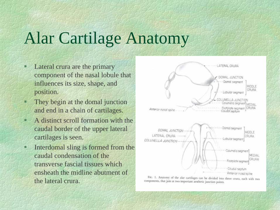
Lateral crura are the primary
component of the nasal lobule that
influences its size, shape, and
position.
They begin at the domal junction
and end in a chain of cartilages.
A distinct scroll formation with the
caudal border of the upper lateral
cartilages is seen.
Interdomal sling is formed from the
caudal condensation of the
transverse fascial tissues which
ensheath the midline abutment of
the lateral crura.
Analysis and Diagnosis
Surgeon's responsibility to balance the patient desires with
what is realistically possible.
Evaluation begins with inspection and palpation of the
patient's nasal skin.
Quality of the skin is an essential indicator of the surgical
outcome, there needs to be enough subcutaneous tissue to
provide adequate cushioning over the nasal skeleton, but
still allow critical definition to the nasal tip.
The size, shape, resilience, and attitude of the alar
cartilages should be assessed by palpation, any asymmetry
should be noted and discussed with the patient.
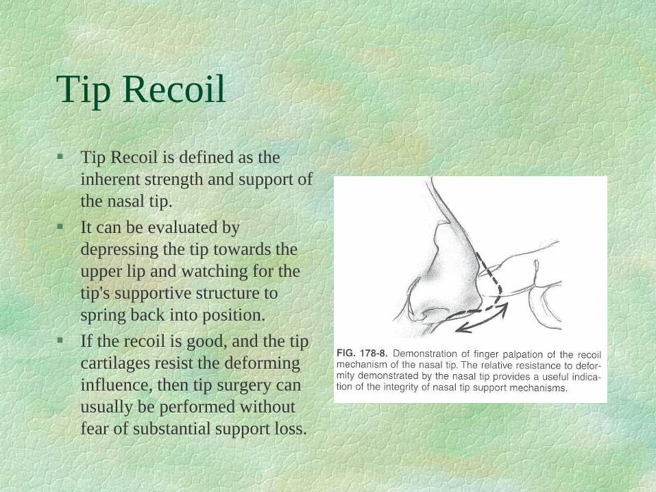
Tip Recoil
Tip Recoil is defined as the
inherent strength and support of
the nasal tip.
It can be evaluated by
depressing the tip towards the
upper lip and watching for the
tip's supportive structure to
spring back into position.
If the recoil is good, and the tip
cartilages resist the deforming
influence, then tip surgery can
usually be performed without
fear of substantial support loss.
Analysis and Diagnosis
Palpate the internal vestibules of the nose for nasal septal twists and
angulations; determine width and length of columella and medial
crura.
Evaluate the size and position of the nasal spine and its related caudal
septal angle.
Surgeon should look at position and inclination of the nasolabial and
nasofrontal angles, the size and shape of the alae, the overall width of
the upper and middle thirds of the nose, and the relationship of the
nose to the rest of the facial features.
Evaluate for facial asymmetries and the relationship of the chin
projection to the nose.
Nasal Tip
It represents the most anterior projecting point on the nose.
Tip projection refers to the posterior to anterior distance
that the tip extends from the facial plane at the alar crease.
Nasal tip rotation is defined as movement of the tip along a
circular arc consisting of a radius centered at the nasolabial
angle that extends to the defining point.
Lower lateral cartilages may be compared to a tripod:
conjoined medial crura form one leg and the lateral crura
represent the other two legs. Shortening or loss of
integrity of any limb changes the spatial position of the
apex (the nasal tip).
Tripod Theory
Preoperative Planning
Standardized photodocumentation is essential.
Realistic expectations and thorough in formed
consent should be discussed.
Any asymmetries should be pointed out to the
patient.
Surgeon should identify what is good and what is
less than ideal about the tip, planning to preserve
the normal, favorable anatomy while correcting
the abnormal anatomy.
Facial Analysis
Surgical Considerations
Surgical Techniques
Ultimate goal is to satisfy the patient’s functional, esthetic,
and psychological expectations for the procedure.
Nasal lobule should be refined, symmetric, and
harmonious with the other nasal features.
Columella should be symmetric and have an appropriate
relationship with the alar margins.
There should be a satisfactory nasal base width and nostrils
of appropriate size and shape.
Loss of tip support and projection in the postoperative
healing period is one of the most common surgical errors
and is usually the result of the sacrifice of tip supports.
Tip Support Mechanisms
Major:
size, shape, and resiliency of the
medial and lateral crura.
wrap-around attachment of the
medial crural footplates to the
caudal septum.
attachment of the caudal margin of
the upper lateral cartilages to the
cephalic margin of the alar
cartilage.
Minor:
dorsal cartilaginous septum,
interdomal ligaments, membranous
septum, nasal spine, surrounding
skin and soft tissues, and alar
sidewalls.
Tip Support
Appropriate tip incisions and approaches should
be planned to preserve as many tip support
mechanisms as possible.
Alar cartilage sculpturing should respect this
principle by conserving the volume and integrity
of the lateral crus and avoiding radical excision
and sacrifice of tip cartilage.
Preferred method is to preserve a majority of the
lateral crus while maintaining a complete,
uninterrupted strip of alar cartilage.
Uninterrupted Cartilage
Technique
Surgical Techniques
Incisions
transcartilaginous
intercartilaginous
marginal
Approaches
delivery of tip
cartilages
non-delivery of tip
cartilages
open approach
Non-Delivery Approach
Good for patients who require
minimal tip cartilage modeling,
have satisfactory preoperative
projection, and minimal interdomal
distance.
Single incision through the
vestibular skin made several mm
cephalic to the caudal margin of the
lower lateral cartilage, vestibular
skin is elevated, resection of few
mm of medial-cephalic portion of
lateral crus.
Mimics nature, disturbs little
normal anatomy, heals predictably
and symmetrically.
Non-Delivery Approach
Delivery Approach
Allows for visual presentation of
the alar cartilages as a bipedicle
chondrocutaneous flap.
Intercartilaginous incision is made,
elevation of skin and soft tissue in
supraperichondrial plane from the
cartilaginous pyramid and septal
angle, marginal incision made at
caudal margin of lower lateral
cartilages, excision of medial
portion of lateral crus leaving at
least 8-10 mm strip.
Vital supports preserved and
healing is predictable.
Delivery Approach
Transdomal Sutures
Transdomal suturing allows
narrowing refinement to the tip
in patients undergoing the
delivery approach.
Strengthen tip support and used
to enhance tip support slightly.
Good for patients with
extremely thin skin, delicate
alar side walls, bulbous
cartilage.
Transdomal Suture
Interrupted Strip Techniques
Used in severe tip deformities and
when more cephalic tip rotation is
indicated.
The complete strip is divided
somewhere along its course and
excessive portions of the medial
and lateral crura are removed.
Asymmetric healing and scarring
are possible anytime the strip is
interrupted, and some tip support is
always sacrificed.
Technique tends to foster cephalic
tip rotation.
Open Approach
Helpful in patients with cleft lip
and nose abnormalities,
asymmetric tips, and
overprojecting tips with variant
anatomy.
More operative edema and
scarring.
Precise direct vision diagnosis and
bimanual surgery.
Soft tissues of the nose are elevated
off the underlying cartilaginous and
bony skeleton, reduction and
augmentation procedures can be
effected precisely with suture
control.
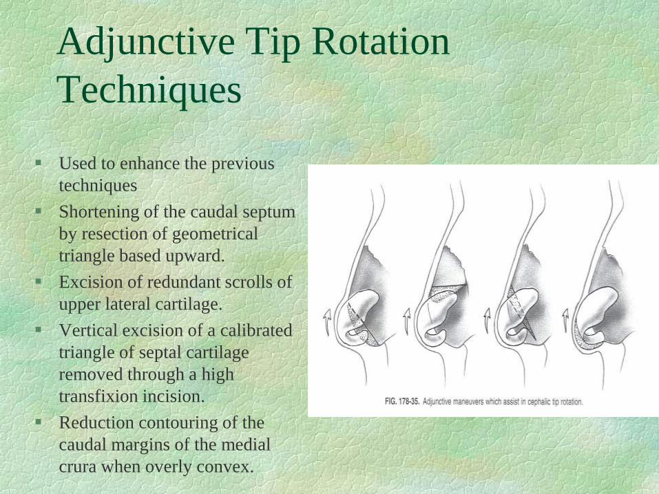
Tip Projection
Complete strip techniques are recommended whenever possible to aid
in maintaining projection. Additional projection may be obtained in a
number of ways.
Cartilage struts positioned below or between the medial crura are
effective in establishing permanent projection.
They should be shaped with a gentle curve to match the anatomy of the
curved columella, but should never extend to the apex of the tip skin.
If medial crural footplates diverge in a widely splayed fashion, further
tip projection can be gained by resection of excessive intercrural soft
tissue and suturing the medial crura together.
Cartilage Struts
Tip Grafts
Autogenous tip grafts can be
used to add height and contour
to the tip of the nose.
Tip grafts may accentuate
favorable tip-defining points
and highlights and can give a
more normal appearance to the
tips with congenital or
postsurgical inadequacies.
Shaped a variety of ways
including triangular,
trapezoidal, or shield-like.
Tip Projection
Goldman technique: complete vertical division of the alar cartilage and
the underlying vestibular skin at the dome. The amount of tip
projection is varied by the location of the cartilage cuts.
Although this is not used much anymore, several surgeons have
devised modifications to this technique that are still used.
Cephalic rotation of the tip may increase projection by advancing the
lateral crura medially and suturing them to lie above the cut ends of the
medial crura.
Transdomal sutures positioned between two complete alar cartilage
strips can create additional projection of the tip.
Overprojected Tip
Aim of procedures is to recess the
tip to a degree that will produce a
desirable profile angle.
Vertical division is made in the
region of the angle and the lateral
crura are advanced and overlapped
on the superior aspect of the medial
crura.
The overlap is usually 2-5 mm and
the crura are resutured to hold them
in this position.
Projection of the lower lateral
cartilages can also be reduced
through a marginal incision.
Tip Rotation
The dynamics of healing play a critical role in tip rotation principles.
Planned degree of rotation depends on: length of nose, face, and upper
lip; facial balance and proportions; patient’s aesthetic desires; and the
surgeon’s aesthetic judgement.
Tip rotation and projection are complementary and interrelated.
Nasal tip rotation results from planned surgical modifications of the
alar cartilages.
There are 6 basic tip rotation techniques: three complete strip and three
interrupted strip techniques.
Complete strip techniques are preferred when the nasal anatomy
permits because projection is preserved, better supported tip, and
asymmetrical healing is less likely. They preserve the normal anatomy
of the nasal tip.
Complete Strip Techniques
Volume reduction of the alar
cartilages results in tissue deficit of
minimal, moderate, or maximal
proportions.
Greater tissue void resulting from
moderate to maximal volume
reduction tends to create
progressively greater degrees of tip
rotation.
Substantial tip rotation depends on
the addition of adjunctive
procedures to achieve cephalic
elevation of the tip complex.
Interrupted Strip Techniques
Break the integrity and spring of the alar cartilages and cephalic
rotation results from the upward scar contracture forces acting on the
alar cartilage segments that are more frail and less well supported.
Caution must be exercised when using interrupted strip techniques in
patients with thin skin or delicate alar cartilages because the loss of
good tip support sets the stage for loss of projection, alar collapse,
notching, pinching, and asymmetry.
Lateral interruption allows for more symmetry and less postoperative
complications because the dividing cut is more lateral and covered
with soft tissue.
Medial interruption techniques are reserved for patients with thicker
skin and supporting structures to minimize the undesirable
consequences of asymmetric healing and even overrotation.