28
Regenerative Biology & Medicine AT THE UNIVERSITY OF NEBRASKA MEDICAL CENTER
Regenerative Biology & Medicineat the University of nebraska Medical center

REGENERATIVE MEDICINE OVERVIEW The invention of new therapies based upon regenerative medicine is likely to be the most important development in 21st century medicine. The most useful definition for regenerative medicine is
“the development of therapies to reconstitute damaged tissues.” There are four primary types of regenerative medicine, listed below from simplest to most complex.
Growth Factor Therapy is the application of growth factors, either systemically or locally, to stimulate existing cells to divide and replace damaged areas of a tissue. Growth factors have been applied with success, experimentally and therapeutically, to the regeneration of bone, skin, cartilage, neurons, and blood vessels. One contemporary strategy of potential interest to the University of Nebraska is attaching specific growth factors to nanoparticles, and then injecting them into regions of tissue damage. In addition to expertise in nanosciences, UNMC has a strong set of researchers with expertise in various aspects of growth factor effects and growth factor receptors, especially applied to regeneration of bone marrow.
Cell Therapy, defined as the application of living cells to restore or enhance the function of tissues and organs. This simple version of regenerative medicine is referred to by the Food and Drug Administration as “minimal manipulation.” Cells used in this type of therapy could be adult or embryonic stem cells, or even fully differentiated adult cells in some cases. Examples would include currently useful therapies such as bone marrow transplantation; and experimental treatments, e.g. injection of bone marrow cells or embryonic stem cells into a vein, or directly into the heart, following an experimental myocardial infarction. This is a strong area at UNMC: a number of UNMC scientists are recognized experts in Cell Therapy, and are capable of utilizing bone marrow cells as well as varieties of embryonic and adult-derived stem cells in these kinds of studies.
Tissue Engineering or Guided Tissue Regeneration is regeneration of biological tissue through the use of cells that are injected into, or are attracted to, natural or fabricated supporting structures that are surgically implanted; or which may be injected. FDA considers this level as “more than minimal manipulation”. Tissue Engineering processes are more elaborate than simple cell therapies and may require complex facilities in addition to the Good Tissue Processing (GTP) facility currently under construction at UNMC. Basic teams may include cell biologists, materials engineers, and other specialists. This level of complexity of regenerative medicine lends itself well to collaborations between UNMC and engineering teams at Peter Kiewit Institute (PKI) and the University of Nebraska-Lincoln. UNMC’s College of Dentistry and orthopedics teams currently are active in development of therapies for guided tissue regeneration. New developments in UNMC Pediatrics and at Children’s Hospital indicate the value of recruitment of biomedical scientists in support of UNMC research and clinical care initiatives combining cardiovascular disease, pediatrics and regenerative medicine.
Synthesized Organs. In this case, new organs are grown in vitro and implanted into experimental models or, ultimately, patients. This area of research is sometimes put in a separate category from the above three types of regenerative medicine. The cells to make the organs may be the patient’s own; adult or embryonic stem cells from a donor; or even cells from a different species. There are a number of examples, with the liver serving as an example an organ with a relatively simple structure that can be grown in vitro, and there are examples of more complex organs that also have been reported as being amenable to in their vitro synthesis, for example, the heart. The University of Nebraska (NU) potential for progress in this area would require the recruitment of teams of physician scientists and engineers.

UNIVERSITY OF NEBRASKA OPPORTUNITIESThere are a number of centers for regenerative medicine in academic health sciences centers in the United States and abroad, upon which the University of Nebraska could model a center. Successful centers range from comprehensive (e.g, experimenting with three or even four of the above subtypes, for example, Pittsburgh or Massachusetts General Hospital) to a more narrow focus upon, for example, adult stem cells (Case Western). Funding for centers is variable and typically includes a mix of private, state and grant/contract support.
NU/UNMC could begin with a scalable model that concentrates upon our strengths in type1, growth factor therapy, and type 2, cell therapy. Initial development of a center would benefit most from recruitment to strengthen these two key areas. We will need to establish a cooperative center that takes advantage of UNMC, UNL and PKI strengths in key disciplines: cell and molecular biology, biochemistry, physiology, nanotechnology, genomics, bioinformatics and drug delivery. In other key areas, e.g. proteomics, we need to continue to grow the research cores at UNMC and UNL and, potentially, carry out some key recruitments.
Ultimately we need to include type 3, “guided regeneration,” since the development and application of tissue scaffolds is one of the most explosive and potentially useful areas in the field. To be fully competitive in guided regeneration research, we’ll need to have well-designed partners among UNMC, UNL and PKI in biomedical and chemical engineering; some recruitment of new talent would be essential. We have good facilities across these campuses that would be attractive to scientists in these areas.
Type 4, synthesized organs, is very expensive. UNMC has little or no activity in this area, and current interpretations of the direction of the field would indicate that 1, 2 and 3 are where the best opportunities are for UNMC, NU and the State of Nebraska.
toM rosenqUist, Ph.d., UNMC ViCe ChaNCellor of researCh
Dr. Rosenquist is a member of the Cardiovascular Differentiation and Development Initial Review Group (study section) at the National Institutes of Health. This group reviews all requests for funding that involve regenerative medicine/stem cell research in the cardiovascular system.

Areas of Research
CANCER page 6
Hamid Band, M.D., Ph.D.
Vimla Band, Ph.D.
Surinder Batra, Ph.D.
Barbara O’Kane, Ph.D.
DRUG DElIVERY AND MATERIAlS MEDICINE page 10
Alexander “Sasha” V. Kabanov Ph.D., Dr.Sc.
EMBRYONIC page 12
Hesham Basma, Ph.D.
David Mercer, M.D., Ph.D.
Angie Rizzino, Ph.D.
HEMATOPOIETIC, & MESENCHYMAl STEM CEllS page 15
Mark Carlson, M.D.
Marcel Devetten, M.D.
John Jackson, Ph.D.
Anne Kessinger, M.D.
Charles Kuszynski, Ph.D.
Graham Sharp, Ph.D.
lUNG page 21
Stephen Rennard, M.D.
NEURAl STEM CEll BIOlOGY AND APPlICATIONS page 22
Iqbal Ahmad, Ph.D.
Sumitra Bhattacharya, Ph.D.
Jialin Zheng, M.D., Ph.D.
STEM CEll EDUCATION page 25
Dave Crouse, Ph.D.
l. Dennis Smith
Contents

James Turpen, Ph.D.iNteriM DireCtorreGeNeratiVe MeDiCiNeProfessorGeNetiCs, Cell BioloGY aND aNatoMY
I am honored to serve as the Interim Director of Regenerative Medicine and appreciate the opportunity to participate in the establishment of this initiative at UNMC.
The Department of Health and Human Services recently published a document in which they said “Regenerative medicine is the next evolution of medical treatments” and represents “the vanguard of 21st century healthcare.”
UNMC has been a world leader in the clinical application of adult stem cells from bone marrow and peripheral blood. I believe we have the experience, resources and talent to capitalize on our expertise and move forward to broaden our portfolio in regenerative medicine by including new and promising areas.
Our initiative in regenerative medicine represents an exciting and challenging step in a field that I have been committed to since my early days as a graduate student. My first rotation was in a laboratory that used nuclear transplantation, now referred to as somatic cell nuclear transfer, to ask fundamental questions about the developmental potential of cancer cells. The technology and manipulations were challenging and the ideas related to understanding how organisms develop and cell populations are programmed were fascinating.
I became, and continue to be, a student of embryology. I entered the stem cell field, concentrating on the development and migration of hematopoietic stem cells in the vertebrate embryo. I have observed the “evolution” of this field from the earliest rigorous demonstration of the existence of pluripotential hematopoietic stem cells to the use of bone marrow and peripheral blood stem cells as important lifesaving therapies. I was inspired by ground breaking work leading to the identification and understanding of the regulation of these stem cells via a multitude of cytokine pathways and the subsequent application of these regulatory molecules as therapeutic agents.
Like many others, I have monitored research into the control of the differentiation of many types of stem cells, including epithelial stem cells, particularly those found in the skin. Learning how to direct these stem cells into hair follicles has led to new practical approaches which hold great promise for the regeneration of hair, a promise of importance for those of us who are follically challenged.
The Department of Health and Human Services projects the U.S. market for regenerative medicine to be on the order of $100 billion and the global potential to exceed $500 billion in the next 20 years. The maturation of the discipline of developmental biology into the area we refer to as regenerative biology and medicine is a testament to the importance of our early investment in basic research leading to our ultimate goal of the betterment of health and treatment of disease.
We now have a realistic and challenging opportunity to make UNMC a player in this emerging field. A key element of success is the recognition that regenerative medicine is a multidisciplinary area that relies on the talents of a broad range of scientists and clinicians. Our initial goal will be to identify and focus on our existing strengths in the broad area of stem cells and establish a working group that will consider a variety of programmatic ideas. It is essential that we identify a niche and establish goals and objectives that will distinguish our program from other centers throughout the world.
We will need to develop a planning document and a business plan to provide a framework for moving forward and use this framework to recruit new talent to UNMC. Our program will need to be cognizant of the political climate, both in Nebraska and nationally, and may require our continued involvement in the legislative process. It will be essential to interact with the general public as advocates for stem cell research and regenerative medicine. Successful programs in regenerative medicine have multiple sources of support from private donors as well as state and national agencies. The ability to take advantage of all possible funding opportunities will be essential to our success.
APPROACH AND VISION:While great strides have been made in reducing breast cancer-associated mortality, more than 40,000 women will lose their battle with breast cancer this year. Resistance to current therapies and recurrence of initially responsive tumors are linked in part to the presence of cancer stem cells in tumors. Understanding the biology of cancer stem cells has become an important area of breast cancer research.
Interactions between Notch ligands and receptors on neighboring cells provide a well-established mechanism to control stem cell fate decisions in higher organisms and Notch receptors or their ligands are aberrantly expressed in breast cancer. A better understanding of Notch signaling pathway in breast cancer is likely to help identify components that may serve as biomarkers of breast cancers and potential targets for development of novel therapeutics towards cancer stem cells.
As Notch signaling pathway components and regulators are well conserved during evolution, we have used the model organism C. elegans to demonstrate the role of microRNA Let-7 downstream of Notch. Based on these findings, we engineered a C. elegans strain in which lethality arising from the loss of Let-7 gene was rescued by the expression of an active Notch allele providing an innovative approach to discover new genes essential for Notch function. Using an RNAi screen, we have identified an evolutionarily-conserved gene as a novel Notch pathway mediator; the expression of this gene is elevated in ER+ breast cancers. Planned studies are designed to investigate if this novel regulator is indeed required for Notch function in normal human mammary epithelial and breast cancer cells and whether its expression is important for Notch-regulated cancer stem cell proliferation. If successful, our basic studies could provide a new biomarker and potential target for therapy of breast cancer. The presence of enzymatic activities in the novel Notch regulator makes it potentially drug-able in order to develop inhibitors of Notch pathway activity that could emerge as cancer stem cell-directed therapeutics.
PUBlICATIONS:Solomon A, Mian Y, Ortega-Cava C, Liu V, Gurumurthy CB, Naramura M, Band V and Band H. Upregulation of the let-7 microRNA with precocious development in lin-12/Notch hypermorphic Caenorhabditis elegans mutants. Dev. Biol. 2008; 316:191-199.
Dimri G, Band H and Band V. Mammary epithelial cell transformation: insights from cell culture and mouse models. Breast Cancer Research 2005; 7:171-179.
FUNDING:National Institutes of Health »
Regulation of normal and cancer cell signaling: cell biology and translation
CANCER
Hamid Band, M.D., Ph.D.Professor, ePPleY iNstitUteDireCtor, CeNter for Breast CaNCer researCh

CANCER
APPROACH AND VISION:Breast cancer remains the second leading cause of cancer-related deaths among women. Recent studies have established that breast cancer is not a single disease but rather a group of diseases, with at least three major subtypes of basal, luminal and ErbB2-positive cancers. The subtype of breast cancer determines the disease-free and overall survival among breast cancer patients. Understanding the origins of various subtypes of breast cancer is directly linked to therapeutic choices and outcomes.
There is increasing evidence that breast cancer originates in and is maintained by a small population of undifferentiated cancer stem cells (CSC) with self-renewal properties while the main mass of the tumor is composed of more differentiated luminal, basal or myoepithelial cell progeny. The origin of CSCs, either from normal stem cells or from intermediate progenitors in which a de novo stem cell program is activated by the oncogenic insults, remains controversial.
A major hurdle in addressing this issue is the lack of immortal human breast stem/progenitor cells can be maintained in vitro and can be deliberately manipulated to ask questions related to origins of breast cancer subtypes. Our laboratory has recently identified, isolated and immortalized normal mammary progenitor/stem cells that include: a non-committed cell type and two phenotypically-committed progenitor cells including a luminal and a myoepithelial cell type.
We are developing methods to induce differentiation, induce tumorigenicity, perform expression profiling and test susceptibility of different therapeutic agents in the three subtypes of progenitor cells we have identified. Given the emerging evidence that stem/progenitor cells are precursors of cancers and multiple subtypes of breast cancer have different survival outcome, our studies are likely to fill a major gap in our understanding of stem cell biology and carry the potential of developing novel therapeutics as well as provide potentially novel markers for diagnostic/prognostic use in breast cancer.
PUBlICATIONS:Dimri G, Band H and Band V. Mammary epithelial cell transformation: insights from cell culture and mouse models. Breast Cancer Res., 2005; 7:171-179.
Band V. In vitro models of early neoplastic transformation of human mammary epithelial cells. Methods Mol. Biol. 2003; 223:237-248.
Dimri GP, Martinez J-L, Jacobs JJL, Keblusek P, Itahana K, van Lohuizen M, Campisi J, Wazer DE and Band V. The Bmi-1 oncogene induces telomerase activity and immortalizes human mammary epithelial cells. Cancer Res., 2002: 62:4736-4745.
Wazer DE, Liu X-L, Chu Q, Gao Q and Band V. Immortalization of distinct human mammary epithelial cell types by human papilloma virus-16 E6 or E7. Proc. Natl. Acad. Sci. USA., 1995; 92:3687-3691.
FUNDING:National Institutes of Health »Department of Defense Breast Cancer Program »
Susceptibility of mammary epithelial cells to transformation: link to stem cells
Vimla Band, Ph.D.Professor aND ViCe Chair of researChDePartMeNt of GeNetiCs, Cell BioloGY aND aNatoMYassoCiate DireCtor, CeNter for Breast CaNCer researChePPleY CaNCer CeNter

APPROACH AND VISION:Cancer is a major public health problem worldwide and more particularly in the developed nations. In the United States, a total of 1,444,920 new cancer cases and 559,650 deaths due to cancers were estimated to occur in 2007. Prostate, breast, pancreatic and ovarian cancers, which are main focus of our group, jointly account for more than 100,000 deaths in the U.S. every year.
A growing body of evidence supports the idea that cancer stem cells are responsible for tumor growth and maintenance, as well as cancer recurrence. Therefore, novel cancer stem cell-targeted therapies could help eradicate cancer. The ideal therapy would be one that is specific for cancer stem cells but spares normal stem cells to avoid unwanted side effects.
Current research in my group is directed to establish the specific biomarkers that distinguish the tumorigenic and metastatic cancer stem/progenitor cells from their differentiated progenies and normal stem/progenitor cells during the progression of locally advanced cancer to metastatic and recurrent disease stages.
We are investigating specific role(s) of various signaling elements (known for maintaining the stem cells self-renewal and multipotent nature) during the malignant transformation. These investigations should also allow us to define the molecular events in stem/progenitor cancer cells responsible for the treatment resistance and establish the beneficial effect of combined targeting of stem cell signaling pathways along with conventional therapies.
PUBlICATIONS:Mimeault M and Batra SK. Recent progress on tissue-resident adult stem cell biology and their therapeutic implications. Stem Cell Reviews, 4(1):27-49, 2008.Mimeault M, Hauke R and Batra SK. Recent advances on the molecular mechanisms involved in the drug resistance of tumorigenic cancer progenitor cell. Clinical Pharmacology and Therapeutics, 83:673-91, 2008.
Mimeault M, Mehta PP, Hauke R and Batra SK. Functions of normal and malignant prostatic stem/progenitor cells in tissue regeneration and cancer progression. Endocrine Reviews, 29:234-52, 2008.
Mimeault M, Hauke R, Mehta PP and Batra SK. Recent advances in cancer stem/progenitor cell research: Therapeutic implications for overcoming resistance to cancers. J. Cell Mol Med, 11, 981-1011, 2007.
Jain M, Kamal N and Batra SK. Engineering antibodies for clinical applications. Mimeault M and Batra SK. Recent advances on the functions of stem cells in tissue regeneration and cancer therapy. Stem Cells 24 2319-2345, 2006.
FUNDING:National Institutes of Health »Department of Defense »
Cancer stem cells: a new generation of markers and targets for cancer diagnosis and therapy
CANCER
Surinder K. Batra, Ph.D. Professor DePartMeNt of BioCheMistrY aND MoleCUlar BioloGY ColleGe of MeDiCiNe

CANCER
Cancer Stem Cells
Barbara O’Kane, MS, Ph.D.assistaNt ProfessorGeNetiCs, Cell BioloGY aND aNatoMYsChool of allieD health - raDiatioN sCieNCes
APPROACH AND VISION:Identification and characterization of the cells responsible for the initiation and maintenance of tumors is essential for the development of more effective therapies for cancer patients. My lab is investigating a rare population of cells identified in breast cancer and lymphoma that are called side population (SP) cells.
SP cells possess many characteristic of normal adult tissue “stem cells” and demonstrate increased tumorigenicity in vivo and in vitro. Stem cells by definition possess the characteristics of longevity, quiescence, self renewal and differentiation, all features that could potentially render them vulnerable to genetic and epigenetic events. The cancer stem cell paradigm provides an explanation for relapses that occur relatively soon or years following treatment.
Our long term goal is to determine if some cancers are the result of mutated tissue stem cells or dysregulated mature cells that retain or acquire stem cell characteristics and investigate their behavior. The ability to separate tumorigenic from non-tumorigenic cells provides an opportunity to generate a phenotypic and molecular profile that could be used to identify potential targets for new and more effective therapies. These targeted therapies may result in more durable responses and better outcomes, particularly in metastatic disease.
PUBlICATIONS:O’Kane Murphy B, Joshi S, Kessinger A, Reed E and Sharp JG. A murine model of bone marrow micrometastases in breast cancer. 2002. Clinical and Experimental Metastasis 19(7):561-569.
O’Kane Murphy BJ, Joshi SS, Kessinger A, Sharp G and Gohr JC. Characterization of a stem cell-like population in breast cancer” Cytotherapy 5:470, 2003.
Hielscher A, O’Kane BJ, Joshi SS and Sharp JG. Mouse and Human Lymphoma Side Poplulation (SP) Cells with Stem Cell-like Characteristics. American Association for Cancer Research The Role of Cancer Stem Cells in the Initiation and Propagation of Tumorigenesis 2008 p B12.
FUNDING:Neuronyx »

APPROACH AND VISION:Our nanomedicine program progressively develops synthetic polymer materials of very small size – 10 to 100 nanometers – that can carry pharmaceutical drugs, proteins, DNA, siRNA and other biologic agents. These functional nanocarriers are designed to safely deliver the biological agents into the cells, to transport agents across biologic barriers, such as the blood brain barrier, and to efficiently release them at the disease site.
We also have developed “molecular beacons,” which can be placed in the same nanocarriers with the biologic agents so that we can not only treat the disease, but also monitor the process of delivery of biological agents and treatment. We collaborate with other UNMC investigators to develop drug delivery and imaging modalities that can be used to treat cancer and neurodegenerative diseases, such as Parkinson’s disease and stroke.
The interaction of synthetic materials with the immune system is another area of our interest. Here, we develop polymers that can interact with immune response cells and facilitate antigen presentation to the immune system so we can achieve, for example, therapeutic vaccination against cancer and other diseases. Gene delivery has been a focus of our research for two decades and we now look for the new applications of our methodologies in vaccine development.
Once this program evolves, we see new opportunities for the biocompatible materials that can interface with cells, especially nanostructured and stimuli responsive cell material constructs that can be used for tissue engineering and other areas of regenerative medicine.
PUBlICATIONS:Kabanov, A.V., Gendelman, H.E. (2007) Nanomedicine in the diagnosis and therapy of neurodegenerative disorders, Progr. Polym. Sci. 32(8-9):1054-82
Sharma, A.K., Zhang, Li., Li, S., Kelly, D.L., Alakhov, V.Yu., Batrakova, E.B., Kabanov, A.V. (2008) Prevention of MDR development in leukemia cells by micelle-forming polymeric surfactant. J. Control. Release, 131(3):220-7.
Gaymalov, Z.Z, Yang, Z, Pisarev, V.M., Alakhov, V.Yu., Kabanov, A.V. (2009) The effect of the nonionic block copolymer pluronic P85 on gene expression in mouse muscle and antigen-presenting cells, Biomaterials, 30 (6): 1232-45
FUNDING:NIH, National Cancer Institute »NIH, Neurodegenerative Disease and Stroke »NIH, National Center of Research Resources »National Science Foundation, »Division of Materials ResearchU.S. Department of Defense »
Designing polymeric materials for biomedical applications
DRUG DElIVERY AND MATERIAlS MEDICINE
Alexander “Sasha” V. Kabanov Ph.D., Dr.Sc.ParKe-DaVis Professor of PharMaCeUtiCal sCieNCes, ColleGe of PharMaCYDireCtor, CeNter for DrUG DeliVerY aND NaNoMeDiCiNe

Researchers and biotech executives foresee the day when the effects of many catastrophic diseases can be reversed. The damaged brains of Alzheimer’s disease patients may be restored. Severed spinal cords may be rejoined. Damaged organs may be rebuilt. Stem cells provide hope that this dream will become a reality.
GeorGeWolff, the BioteCh iNVestor’s BiBle
APPROACH AND VISION:The only definitive treatment of hepatic failure is transplantation of the liver. Liver transplantation, however, is technically complex and dangerous in patients that are most in need. One way to overcome the technical difficulties and risks of liver transplantation is to transplant isolated hepatocytes. Because of the limited supply of organ donors, it is difficult to obtain primary human hepatocytes for research or clinical application.
Extensive literature indicates that transplantation of isolated human hepatocytes potentially could be used as a minimally-invasive treatment for patients with a variety of life-threatening liver disorders. Human hepatocytes have been used to repopulate the livers of immune incompetent mice and facilitating the study of hepatocyte and human hepatitis virus biology.
Embryonic stem (ES) cells are pluripotent and can potentially differentiate into any cell type. The availability of an unlimited supply of ES cell-derived hepatocytes would facilitate development of cell-based therapies for the treatment of liver disease, would facilitate the study of liver diseases, and revolutionize the early stages of the drug discovery process.
In our lab, we have simplified ES cell differentiation strategy to generate cells with many of the defining characteristics of human hepatocytes. We also developed a simple technique for enrichment of the ES-derived hepatocytes based on asialoglycoprotein receptor (ASGPR). Use of this differentiation program would be useful as an alternative source of primary human hepatocytes.
PUBlICATIONS:Basma H, Soto-Gutiérrez A, Yannam GR, Liu L, Ito R, Ellis E, Carson SD, Sato S, Muirhead D, Navarro-Álvarez NR, Mercer DF, Miller JD, Strom SC, Kobayashi N and Fox IJ. Differentiation and Enrichment of Human Hepatocytes Derived from Embryonic Stem Cells (In preparation).
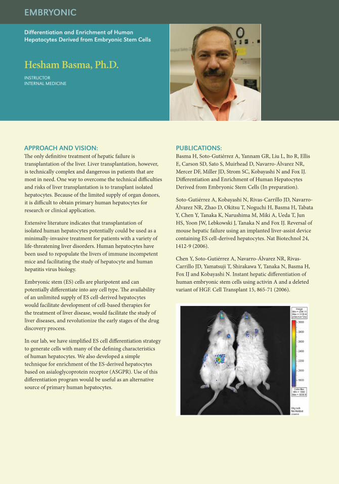
Soto-Gutiérrez A, Kobayashi N, Rivas-Carrillo JD, Navarro-Álvarez NR, Zhao D, Okitsu T, Noguchi H, Basma H, Tabata Y, Chen Y, Tanaka K, Narushima M, Miki A, Ueda T, Jun HS, Yoon JW, Lebkowski J, Tanaka N and Fox IJ. Reversal of mouse hepatic failure using an implanted liver-assist device containing ES cell-derived hepatocytes. Nat Biotechnol 24, 1412-9 (2006).
Chen Y, Soto-Gutiérrez A, Navarro-Álvarez NR, Rivas-Carrillo JD, Yamatsuji T, Shirakawa Y, Tanaka N, Basma H, Fox IJ and Kobayashi N. Instant hepatic differentiation of human embryonic stem cells using activin A and a deleted variant of HGF. Cell Transplant 15, 865-71 (2006).
Differentiation and Enrichment of Human Hepatocytes Derived from Embryonic Stem Cells
EMBRYONIC
Hesham Basma, Ph.D.iNstrUCtor iNterNal MeDiCiNe
EMBRYONIC
Using Human Stem Cell-Derived Hepatocytes for Development of Novel Treatments for Hepatitis C
David Mercer, M.D., Ph.D.assistaNt Professor of sUrGerYliVer/sMall iNtestiNe traNsPlaNt ProGraMDireCtor, iNtestiNal rehaBilitatioN ProGraM
APPROACH AND VISION:Infection with the hepatitis C virus (HCV) is a worldwide epidemic affecting 3% of the US population and an estimated 180 million people worldwide. At UNMC, as in most major transplant centers, HCV is the number one indication for liver transplantation. A major roadblock in the development of HCV antiviral therapies has been the lack of a widely available animal model. In previous work, Dr. Mercer developed the only accepted small animal model for study of HCV, which exploits the unique regenerative properties of liver cells to create a mouse with a partially human liver that is infectable with HCV. Generation of these valuable mice has heretofore been dependent on a steady supply of fresh human hepatocytes for transplantation. In collaboration with Dr. Ira Fox, we will be using a multi-step differentiation process to generate human hepatocytes from both embryonal stem cells (ES cells) and induced pluripotent cells (iPS cells), and use these cells to generate mice with partially human livers. Successful establishment of chimeric human livers in mice from stem cell-derived hepatocytes will be a major advance in the study of human liver function and disease, and allow for accelerated exploration of new drug therapies for HCV.
APPROACH AND VISION:Understanding how gene regulatory networks coordinate the transcription of sets of genes during cell growth and differentiation is one of the biggest challenges facing biomedical scientists. Deciphering the mechanisms that control gene regulatory networks at the molecular level will not only enable us to better understand normal physiology, it will also provide critical insights into aberrant gene regulation in diseases, such as cancer.
A major goal of the work in this laboratory is to understand the fundamental mechanisms and signaling pathways that orchestrate the self-renewal and pluripotency of embryonic stem (ES) cells and induced pluripotent stem (iPS) cells. Currently, we are studying how master regulators, such as Sox2, modulate the transcription of gene regulatory networks that control the self-renewal of stem cells. Recently, we determined that small changes in the levels of Sox2 (<2-fold) trigger the rapid differentiation of ES cells and functions as a molecular rheostat to control the key properties of ES cells.
Moreover, our studies argue strongly that Sox2 levels must be controlled precisely during the generation of iPS cells. Efforts are underway currently to identify Sox2 interacting partner proteins by comprehensive proteomic analyses. We also are examining the roles played by Sox2 in the self-renewal of cancer stem cells and during the reprogramming of somatic cells to a pluripotent stem cell state.
PUBlICATIONS:Boer B, Kopp J, Mallanna S, Desler M, Chakravarthy H, Wilder P, Bernadt C and Rizzino A. Elevating the Levels of Sox2 in Embryonal Carcinoma Cells and Embryonic Stem Cells Inhibits the Expression of Sox2:Oct-3/4 Target Genes. Nucleic Acids Research, 35:1773-1786, 2007.
Rizzino A. A challenge for regenerative medicine: Proper genetic programming, not cellular mimicry. Developmental Dynamics, 36:3199-3207, 2007.
Chakravarthy H, Boer B, Desler M, Mallanna S, McKeithan T and Rizzino A. Identification of DPPA4 and other genes as putative Sox2:Oct-3/4 target genes using a combination of in silico analysis and transcription-based assays. J. Cellular Physiology, 216:651-662, 2008.
Kopp J, Ormsbee B, Desler M and Rizzino A. Small increases in the levels of Sox2 trigger the differentiation of embryonic stem cells. Stem Cells 26:903-911, 2008.
FUNDING:National Institutes of Health »Nebraska Department of Health »Nebraska Research Initiative »UNMC Eppley Cancer Center »
Regulation of Gene Regulatory Networks that Orchestrate Stem Cell Pluripotency
EMBRYONIC
Angie Rizzino, Ph.D. Professor ePPleY iNstitUte for researCh iN CaNCer aND allieD Diseases
HEMATOPOIETIC AND MESENCHYMAl STEM CEllS
Using Mesenchymal Stem Cells for Development of a Skin Replacement Therapy
Mark Carlson, M.D.assoCiate ProfessorDePartMeNt of sUrGerY UNMC aND the oMaha Va MeDiCal CeNter
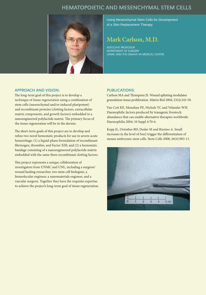
APPROACH AND VISION:The long-term goal of this project is to develop a technique of tissue regeneration using a combination of stem cells (mesenchymal and/or induced pluripotent) and recombinant proteins (clotting factors, extracellular matrix components, and growth factors) embedded in a nanoengineered polylactide matrix. The primary focus of the tissue regeneration will be in the dermis.
The short-term goals of this project are to develop and refine two novel hemostatic products for use in severe acute hemorrhage: (1) a liquid-phase formulation of recombinant fibrinogen, thrombin, and Factor XIII; and (2) a hemostatic bandage consisting of a nanoengineered polylactide matrix embedded with the same three recombinant clotting factors.
This project represents a unique collaboration of investigators from UNMC and UNL, including a surgeon/wound healing researcher, two stem cell biologists, a biomolecular engineer, a nanomaterials engineer, and a vascular surgeon. Together they have the requisite expertise to achieve the project’s long-term goal of tissue regeneration.
PUBlICATIONS:Carlson MA and Thompson JS. Wound splinting modulates granulation tissue proliferation. Matrix Biol 2004; 23(4):243-50.
Van Cott KE, Monahan PE, Nichols TC and Velander WH. Haemophilic factors produced by transgenic livestock: abundance that can enable alternative therapies worldwide. Haemophilia 2004; 10 Suppl 4:70-6.
Kopp JL, Ormsbee BD, Desler M and Rizzino A. Small increases in the level of Sox2 trigger the differentiation of mouse embryonic stem cells. Stem Cells 2008; 26(4):903-11.
APPROACH AND VISION:Mesenchymal Stem Cells (MSC) are a population of multipotent cells that can be isolated (and expanded) from bone marrow, adipose tissue, cord blood and fetal liver, and that have the capacity to proliferate in vitro and to differentiate into several mesenchymal tissues, including bone, cartilage, tendon, muscle, and adipose tissue.
Several studies have shown that human MSC can reverse established severe Graft-versus-Host and Disease (GVHD). To investigate these immune modulating properties of MSC we have studied the effect of MSC on acute GVHD in a mouse model, inducing GVHD by transplanting 5 x 10⁶ C57Bl/6 (H2b) bone marrow cells and 6 x 10⁶ C57Bl/6 splenocytes in radiation ablated DBA/2J (H2d) recipient mice.
This model reliably and reproducibly induces lethal acute GVHD starting around transplant day +10 with a significant improvement in survival if MSC are infused at the time of onset of GVHD. We are hypothesizing that MSC inhibit IL-15 mediated T-cell activation by APC without interfering with regeneration of T regulatory cells. We are using our mouse model to investigate this hypothesis. We are also interested in clinical applications of MSC for treatment of chronic GVHD of the lung (obliterating bronchiolitis) and acute GVHD after solid organ transplantation.
PUBlICATIONS:Kebriaei P, Isola L, Bahceci E, Holland K, Rowley S, McGuirk J, Devetten M, Jansen J, Herzig R, Schuster M and Uberti J: Phase II trial of Prochymal® (ex-vivo cultured adult human mesenchymal stem cells) and corticosteroids as primary treatment for acute graft-versus-host disease (aGVHD). Blood 108: 922a (2006).
Lacy J, Jackson J, Murphy B, Sharp G and Devetten M: Multipotential Mesenchymal Stromal Cells abrogate acute graft-versus-host disease in a murine model. Biol. Blood Marrow Transplant 13: 45 (2007)
FUNDING:UNMC Eppley Cencer Center Cattleman’s Ball Fund »
The Effect of Mesenchymal Stem Cells on Acute Graft-versus-Host Disease in a Mouse Model
HEMATOPOIETIC AND MESENCHYMAl STEM CEllS
Marcel Devetten, M.D.assoCiate Professor of MeDiCiNeDireCtor, heMatoPoietiC Cell traNsPlaNtatioN ProGraM

HEMATOPOIETIC AND MESENCHYMAl STEM CEllS
Bone Marrow Stem Cell Biology
John D. Jackson, Ph.D.assoCiate ProfessorPatholoGY aND MiCroBioloGY
APPROACH AND VISION:Our laboratory is involved in the multi-investigator study of aging effects on stem cells using bone marrow from hip replacement patients. We are particularly interested in the mesenchymal stem cell population. This population has the ability to give rise to osteoblasts, chondrocytes, adipocytes and other cells such as muscle and neural cells.
Aging does not change the number of bone marrow clonogenic stromal stem cells; however, ethanol consumption resulted in a decrease in the number of mesenchymal stem cells potentially through the induction of adipocyte differentiation. As a result of this observation, our laboratory is investigating the effects of ethanol on mesenchymal stem cell growth and differentiation and the subsequent effects of ethanol on hematopoietic stem cell proliferation and differentiation. In addition, we are collaborating with a material physicist, Dr. Fereydoon Namavar, from the Department of Orthopedics.
We are investigating the adhesion, growth and differentiation of mesenchymal stem cells on surfaces of engineered nano-crystal ceramic films produced by ion beam assisted deposition. This technique produces extremely hard and wear-resistant surfaces. Our goal is to produce nano-generated surfaces which will enhance osteo-integration of implanted prosthetic devices.
PUBlICATIONS:Jackson JD, Sharp JG, Haider H, Garvin KL and Namavar F. (2006) Preliminary analysis of attachment, survival and growth of bone marrow stromal cells on nanocrystalline hard ceramic coatings. In: Materials for Scaffolding of Biologically Engineered Systems: Interfaces and Interactions on a Nanoscale, (Ravaglioli A and Krajewski A, eds), Consigloi Nazionale Delle Ricerche, Rome, Italy, pp. 109-118.
Namavar F, Jackson JD, Sharp JG, Mann EE, Bayles K, Cheung BCL, Feschuk CA, Varma S, Haider H and Garvin KL. (2007) Searching for smart durable coatings to promote bone marrow stromal cell growth while preventing biofilm formation. In: Biofilm-Material Interactions-New Tools, Technologies, And Opportunities, (Liberam M, Camesano T, Kreiswirth B, Li P and Richards RG, eds), Material Research Society Symposium Proceedings 954E, Warrendale, PA, 0954-H04 04.
Sharp JG, O’Kane Murphy B, Jackson JD, Brusnahan SK, Kessinger A and Neff JR. (2005) Promises and pitfalls of stem cell therapy for promotion of bone healing. Clinical Orthopaedics and Related Research, (435):52-61.
FUNDING:NIH, National Institute of Aging »
APPROACH AND VISION:Anne Kessinger, M.D., is professor of oncology/hematology and associate director for clinical research at the UNMC Eppley Cancer Center. In 1984, Dr. Kessinger postulated that immature bone marrow stem cells circulating in the bloodstream could be harvested from the blood and used for transplants. Through Dr. Kessinger’s ingenuity, UNMC became a leader in the transplants of peripheral adult stem cells, which has become standard practice in treating lymphoma cancer patients. This treatment has become more effective, and much less painful, than the traditional bone-marrow transplants. In addition to her research with adult stem cells, Dr. Kessinger has published extensively on therapeutic approaches for cancers and solid tumors. She also was the 1997 winner of the University of Nebraska Outstanding Research and Creative Activity Award. She joined UNMC’s faculty in 1972, later serving as chief of oncology/hematology from 1991 to 1999. She has published 194 journal articles, 59 book chapters and 348 abstracts, and has given 110 invited presentations around the world.
Peripheral Adult Stem Cell Transplantation
HEMATOPOIETIC AND MESENCHYMAl STEM CEllS
Anne Kessinger, M.D.ProfessorDePartMeNt of oNColoGY/heMatoloGYassoCiate DireCtor for CliNiCal researCh, ePPleY CaNCer CeNter

HEMATOPOIETIC AND MESENCHYMAl STEM CEllS
UNMC Cell Analysis Core Facility
Charles Kuszynski, Ph.D.assoCiate ProfessorPatholoGY aND MiCroBioloGYDireCtor, Cell aNalYsis faCilitY
APPROACH AND VISION:The focus of my interest in stem cell research is to provide a resource for the identification and isolation of stem cells, particularly of adult tissue origins. Putative stem cells have been demonstrated in numerous tissues such as lung, liver, heart, muscle, bone marrow, and retina as well as tumors.
Careful quantification, classification and isolation of these cells are critical to our understanding of stem cell biology and differentiation into functional tissue components or tumors. The Cell Analysis Facility provides researchers access to the most advanced instrumentation and technology to understand these aspects of stem cells.
Instrumentation available in the facility include state-of-the-art 15 color instruments which allow identification of extremely rare populations of cells and subsequent selection of these rare events for further studies. My role as director of the Cell Analysis Facility is to provide investigators with an expert interface between the technology of flow cytometry and the biology of their cells through direct collaboration and interaction with the investigators and their staff.
PUBlICATIONS:Bhattacharya S, Jackson JD, Das AV, Thoreson WB, Kuszynski C, James J, Joshi S and Ahmad I. Direct identification and enrichment of retinal stem cells/progenitors by Hoechst 33342 dye efflux assay. Invest. Opthalmo. Vis. Sci. 44(6)(Jun): 2764-2773 2002.
Robinson SN, Seina SM, Gohr JC, Kuszynski CA and Sharp, JG. Evidence for a qualitative hierarchy within the Hoechst 33342 >Side Population@ (SP) of Murine Bone Marrow. Bone Marrow Transplantation. 35: 807-818 2005.
Zhang J-L, Cai J, Jackson JD, Kuszynski CA, Walls S, McIvor RS and Fox IJ. Long-term transgene expression and survival of transgene-expressing grafts following lentivirus transduction of bone marrow Side Population (SP) cells. Transplantation 79: 882-888 2005.
FUNDING:Nebraska Research Initiative »NIH Cancer Center Support Grant »NIH, Center for Virology »
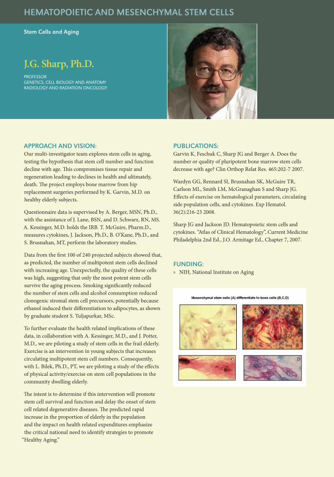
APPROACH AND VISION:Our multi-investigator team explores stem cells in aging, testing the hypothesis that stem cell number and function decline with age. This compromises tissue repair and regeneration leading to declines in health and ultimately, death. The project employs bone marrow from hip replacement surgeries performed by K. Garvin, M.D. on healthy elderly subjects.
Questionnaire data is supervised by A. Berger, MSN, Ph.D., with the assistance of J. Lane, BSN, and D. Schwarz, RN, MS. A. Kessinger, M.D. holds the IRB. T. McGuire, Pharm.D., measures cytokines, J. Jackson, Ph.D., B. O’Kane, Ph.D., and S. Brusnahan, MT, perform the laboratory studies.
Data from the first 100 of 240 projected subjects showed that, as predicted, the number of multipotent stem cells declined with increasing age. Unexpectedly, the quality of these cells was high, suggesting that only the most potent stem cells survive the aging process. Smoking significantly reduced the number of stem cells and alcohol consumption reduced clonogenic stromal stem cell precursors, potentially because ethanol induced their differentiation to adipocytes, as shown by graduate student S. Tuljapurkar, MSc.
To further evaluate the health related implications of these data, in collaboration with A. Kessinger, M.D., and J. Potter, M.D., we are piloting a study of stem cells in the frail elderly. Exercise is an intervention in young subjects that increases circulating multipotent stem cell numbers. Consequently, with L. Bilek, Ph.D., PT, we are piloting a study of the effects of physical activity/exercise on stem cell populations in the community dwelling elderly.
The intent is to determine if this intervention will promote stem cell survival and function and delay the onset of stem cell related degenerative diseases. The predicted rapid increase in the proportion of elderly in the population and the impact on health related expenditures emphasize the critical national need to identify strategies to promote
“Healthy Aging.”
PUBlICATIONS:Garvin K, Feschuk C, Sharp JG and Berger A. Does the number or quality of pluripotent bone marrow stem cells decrease with age? Clin Orthop Relat Res. 465:202-7 2007.
Wardyn GG, Rennard SI, Brusnahan SK, McGuire TR, Carlson ML, Smith LM, McGranaghan S and Sharp JG. Effects of exercise on hematological parameters, circulating side population cells, and cytokines. Exp Hematol. 36(2):216-23 2008.
Sharp JG and Jackson JD. Hematopoietic stem cells and cytokines. “Atlas of Clinical Hematology”, Current Medicine Philadelphia 2nd Ed., J.O. Armitage Ed., Chapter 7, 2007.
FUNDING:NIH, National Institute on Aging »
Stem Cells and Aging
HEMATOPOIETIC AND MESENCHYMAl STEM CEllS
J.G. Sharp, Ph.D.ProfessorGeNetiCs, Cell BioloGY aND aNatoMYraDioloGY aND raDiatioN oNColoGY

lUNG
Stem Cells in lung Disease
Stephen I. Rennard, M.D.ProfessoriNterNal MeDiCiNe
APPROACH AND VISION:Chronic diseases of the lung are among the leading causes of death and disability in the United States. Chronic obstructive pulmonary disease (COPD) is the fourth leading cause of death and is the only major cause of death that is increasing. Asthma, a major cause of morbidity in children and adults, also is increasing in prevalence.
Current treatments are of benefit, but no treatments can cure these disorders. Both asthma and COPD lead to alterations in lung structure that compromise lung function. It is these structural alterations that are refractory to current therapy.
Stem cells likely play a role in these architectural changes, and understanding the mechanisms involved promises to open avenues to prevent the long term consequences of these diseases. Stem cells, which also have the capacity to repair damaged tissue, also hold promise for therapy to restore lost function in those with developed disease.
Our laboratory is attempting to define the cells responsible for lung repair following injury, to determine how the function of these cells is altered in diseases where abnormal lung structure results and to determine mechanisms where the repair capacity of the lung can be manipulated to restore lost function.
PUBlICATIONS:Sugiura H, Liu X, Duan F, Kawasaki S, Togo S, Kamio K, Wang XQ, Mao L, Ahn Y, Ertl RF, Bargar TW, Berro A, Casale TB and Rennard SI. 2007. Cultured lung fibroblasts from ovalbumin-challenged “asthmatic” mice differ functionally from normal. Am J Respir Cell Mol Biol 37(4):424-30.
Togo S, Holz O, Liu X, Sugiura H, Kamio K, Wang X, Kawasaki W, Ahn Y, Fredriksson K, Skold CM, Mueller KC, Branscheid D, Welker L, Watz H, Magnussen H and Rennard SI. 2008. Lung Fibroblast Repair Functions in COPD Patients are Altered by Multiple Mechanisms. Am J Respir Crit Care Med.
Abe S, Boyer C, Liu X, Wen FQ, Kobayashi T, Fang Q, Wang X, Hashimoto M, Sharp JG and Rennard SI. 2004. Cells Derived from the Circulation Contribute to the Repair of Lung Injury. Am J Respir Crit Care Med 170:1158-1163.
Abe S, Lauby G, Boyer C, Rennard SI and Sharp JG. 2003. Transplanted BM and BM side population cells contribute progeny to the lung and liver in irradiated mice. Cytotherapy 5:523-533.
FUNDING:NIH, National Heart Lung and Blood Institute »NIH, National Institute of Environmental Health Sciences »Philip Morris External Research Program »State of Nebraska, Nebraska Research Initiative »University of Nebraska, Larson Endowment »University of Nebraska Medical Center, Dean, College of »Medicine

APPROACH AND VISION: Generation of cellular diversity is central to development, structure and function of the central nervous system (CNS). This process is underpinned by stem cells, multipotential and self-renewing, that generate stage- and site-specifics neurons and glia. Therefore, understanding the biology of stem cells holds the key to mechanisms that regulate neural development and stem cell-based approaches to treat neurodegeneration. Given the complexity and cellular heterogeneity of the CNS, we are studying the regulation of neural stem cells in an accessible and proven model of the CNS, the mammalian retina. There are three major research objectives of our lab: First, to understand the regulation of neural stem cells in terms of their proliferation, state of commitment and differentiation (biology). Second, to utilize information arising from these studies to understand and treat degenerative changes in the retina that causes blindness in devastating diseases such as the age-related macular degeneration, retinitis pigmentosa and glaucoma (applications) and third, to identify alternate sources of neural progenitors (adult and iP stem cells) in order to sustain clinical applications.
PUBlICATIONS:Zhao et al., (2008). Derivation of neurons with functional properties from adult limbal epithelium: implications in autologous cell therapy for photoreceptor degeneration. Stem Cells. 26:939-949.
Das et al., (2006). Neural stem cell properties of Muller glia in the mammalian retina: Regulation by Notch and Wnt signaling. Dev. Biol. 299: 283-302.
Bhattacharya et al., (2007). Maintenance of retinal stem cells by Abcg2 is regulated by Notch signaling. J. Cell. Sci. 120:2652-2662.
Das et al., (2007). SWI/SNF chromatin remodeling ATPase, Brm regulates the differentiation of early retinal stem cells/progenitors by influencing Brn3b expression and Notch signaling. J. Biol. Chem. 282:35187-35201.
Bhattacharya, S., Das, A. V., Mallya, K., Ahmad, I. (2008). CNTF mediated signaling regulates neuronal versus glial differentiation of retinal stem cells/progenitors by concentration-dependent recruitment of MAPK and JAK-STAT pathways in conjunction with Notch signaling. Stem Cells 26:2611-2624.
FUNDING: The Lincy Foundation »The Pearson Foundation »
Neural Stem Cell Biology and Applications
NEURAl STEM CEll BIOlOGY AND APPlICATIONS
Iqbal Ahmad, Ph.D.ProfessoroPhthalMoloGY & VisUal sCieNCes
NEURAl STEM CEll BIOlOGY AND APPlICATIONS
Stem Cells in Retina
Sumitra Bhattacharya, Ph.D.assistaNt ProfessoroPhthalMoloGY aND VisUal sCieNCe
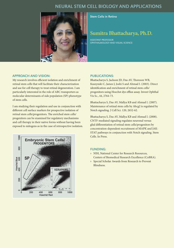
APPROACH AND VISION:My research involves efficient isolation and enrichment of retinal stem cells that will facilitate their characterization and use for cell therapy to treat retinal degeneration. I am particularly interested in the role of ABC transporters as molecular determinants of side population (SP) phenotype of stem cells.
I am studying their regulation and use in conjunction with different cell surface markers for prospective isolation of retinal stem cells/progenitors. The enriched stem cells/progenitors can be examined for regulatory mechanisms and cell therapy in their native forms without having been exposed to mitogens as in the case of retrospective isolation.
PUBlICATIONS:Bhattacharya S, Jackson JD, Das AV, Thoreson WB, Kuszynski C, James J, Joshi S and Ahmad I. (2003). Direct identification and enrichment of retinal stem cells/progenitors using Hoechst dye efflux assay. Invest Ophthal Vis Sc., 44, 2764-73.
Bhattacharya S, Das AV, Mallya KB and Ahmad I. (2007). Maintenance of retinal stem cells by Abcg2 is regulated by Notch signaling. J Cell Sci. 120, 2652-62.
Bhattacharya S, Das AV, Mallya KB and Ahmad I. (2008). CNTF-mediated signaling regulates neuronal versus glial differentiation of retinal stem cells/progenitors by concentration-dependent recruitment of MAPK and JAK-STAT pathways in conjunction with Notch signaling. Stem Cells. In Press.
FUNDING:NIH, National Center for Research Resources, »Centers of Biomedical Research Excellence (CoBRA). Special Scholar Awards from Research to Prevent »Blindness.

APPROACH AND VISION:This laboratory focuses on the influence of inflammation on neurogenesis. Inflammatory factors have varying effects on neural stem/progenitor cell (NPC) proliferation, migration, differentiation, survival and incorporation of newly born neurons into the CNS circuitry during brain disorders.
This laboratory conducts research in this emerging field and examines the potential mechanisms through which inflammation affects neurogenesis during neurological complications. Specifically, we study factors that are released during brain inflammation including the chemokine SDF-1α, cytokines (TNF-α and IL-1β) and the neurotransmitter glutamate. SDF-1α acts on neural progenitor cells (NPCs) via CXCR4 and CXCR7 receptors to recruit NPCs to the site of inflammation and possibly promote the survival, proliferation and/or differentiation of NPC.
On the other hand, SDF-1α is modified by factors released by macrophages, resulting in impairment of normal SDF-1/CXCR4 mediated NPC migration, survival, proliferation and differentiation. Cytokines such as TNF-α and IL-1β, released by activated macrophages/astrocytes, affect NPC proliferation and differentiation during brain complications.
Extracellular glutamate is upregulated during brain inflammation. Glutamate directs the differentiation of NPCs to neurons via the activation of calcium-permeable AMPA receptors containing Q/R-unedited GluR2 subunits. We use in vitro NPC culture systems and in vivo animal model systems to examine the roles and signaling pathways of chemokine, cytokines and glutamate in the regulation of neurogenesis.
PUBlICATIONS:Whitney N, et al. (2008). Calcium-permeable AMPA receptors containing Q/R-unedited GluR2 directs human neural progenitor cells differentiation to neurons. FASEB J. 22:2888-900.
Peng H, et al. (2008). HIV-1-Infected and/or Immune-Activated Macrophage-Secreted TNF-α Affects Human Fetal Cortical Neural Progenitor Cell Proliferation and Differentiation. Glia. 56: 903-16.
Peng H, et al. (2007). Differential expression of CXCL12 and CXCR4 during human fetal neural progenitor cell differentiation. J. Neuroimmunol Pharm 2: 251-258.
Zheng J, Peng H, Rose J and Herek S. (2005). Neurogenesis and its links to brain development, developmental therapeutics and the pathogenesis of neurodegenerative disorders including HIV-1 associated dementia. In The Neurology of AIDS, second edition, P 239-253, Oxford University Press, London.
Peng H, et al. (2004). Stromal cell-derived factor 1 mediated CXCR4 signaling in rat and human cortical neural progenitor cells. J. Neurosci. Res. 76: 35-50.
FUNDING:NIH, National Institute of Neurological Disorders »and Stroke
Brain inflammation, neural stem cell functions and brain disorders
NEURAl STEM CEll BIOlOGY AND APPlICATIONS
Jialin Zheng, M.D.ProfessorPharMaColoGY/exPeriMeNtal NeUrosCieNCe aND PatholoGY/MiCroBioloGY

STEM CEll EDUCATION
Stem Cell Education
David A. Crouse, Ph.D.assoCiate ViCe ChaNCellor for aCaDeMiC affairsexeCUtiVe assoCiate DeaN for GraDUate stUDies
APPROACH AND VISION:My interests in stem cells span nearly 40 years. After a post-doc at the Argonne National Laboratory studying redevelopment of the immune and hematopoietic systems following bone marrow stem cell transplantation, I focused my research on thymic regeneration and redevelopment of T-cell function.
These studies ultimately led to other research on the role of the hematopoietic microenvironment and later, the characterization of circulating (peripheral) stem cells. Most of these studies were supported by the NIH and used animal models but they presented many opportunities for extensive collaboration with clinical colleagues.
Overall, the national emphasis on stem cell biology has grown during this time, particularly over the past 11 years with the publications related to human embryonic stem cells and cloning. During this time, I have taken a lead role in promoting an understanding of the science and ethics that relate to stem cell biology and cloning as well as developing programs and teaching courses related to “Responsible Conduct in Research” and other important career issues for graduate students, post-docs and junior faculty of the UNMC.
Educating the public and our political constituencies about the issues and science related to stem cells has been both rewarding and challenging. Clearly there are major gaps in understanding that make it more challenging to promote public acceptance of some research in this area. The bottom line is often the difference in how individuals or groups consider the embryo and the “beginning of human life.”
Some of these issues have religious origins while others are deeply personal — both of which much be respected. Understanding that, it still is the desire of most scientists to find ways that provide appropriate oversight of stem cell research without completely obstructing the pathway for scientific discovery in this rapidly advancing area. Related to this, UNMC has developed a Stem Cell Biology graduate course (PMM 945) that deals directly with both the basic science that underpins this area and the ethical issues that surround it.

People often ask L. Dennis Smith, Ph.D., what he considers his greatest accomplishment as president of the University of Nebraska. His answer: “Without hesitation, I can say that I tried to foster a culture of excellence in which research and scholarly study are valued.”
President of the University of Nebraska from 1994 to 2004, Dr. Smith was a staunch supporter of stem cell research in 1999, when medical research being conducted at UNMC came under fire. The research, designed to shed light on neurodegenerative diseases such as Alzheimer’s, Parkinson’s, AIDS dementia and multiple sclerosis, relied on fetal tissue. State policymakers and religious leaders warned against using public funds for this research, and a bill was introduced in the legislature to ban the use of such tissue.
In response, Dr. Smith outlined the critical need for the research. The request to cease the research, Dr. Smith wrote, “strikes at the very heart of academic freedom.” The Board of Regents subsequently voted unanimously to support the research. Dr. Smith continues to champion the right of scientists to pursue new knowledge in a responsible manner, and in a climate of academic openness.
For his stand, Dr. Smith was presented with the highly coveted American Association for the Advancement of Science Award for Scientific Freedom and Responsibility in 2002. The award honors scientists and engineers whose exemplary actions, often taken at significant personal cost, have served to foster scientific freedom and responsibility.
Dr. Smith also created the Nebraska Bioethics Advisory Commission, composed of a wide cross section of scientists and lay people, to develop guidelines for the ethical conduct of future biomedical research at the University of Nebraska.
“The ability to undertake legitimate research without external interference, including that from political and religious sectors, is fundamental to the excellence of our postsecondary institutions,” Dr. Smith has said.
After stepping down as president, Dr. Smith briefly returned to the classroom on a part-time basis in Biological Sciences at the University of Nebraska-Lincoln. In the fall of 2008, he taught a class at UNMC on reversibility of the differentiated state and stem cell formation.
Dr. Smith earned his Ph.D. (1964) in experimental embryology, and his bachelor’s degree (1959) in zoology and chemistry from Indiana University. He has published almost 100 research papers and numerous abstracts in such areas as cell biology, developmental biology, biochemistry and molecular biology. He was awarded a Guggenheim Fellowship in 1987. His studies some 30 years ago of cell division in frogs helped lay the groundwork for three researchers who ultimately won a Nobel Prize in medicine for identifying the entire cycle of a cell.
Dr. Smith was on the faculty of Purdue University from 1969 to 1987 and received an honorary Doctor of Science from Purdue in 2000. At Purdue, he headed the department of biological sciences (1980-1987). He was employed as a staff scientist at the Argonne National Laboratory (1964-1969) and at Indiana University (1963-1964). He was an instructor in embryology at the Woods Hole Marine Biology Laboratory during five summer sessions in the 1970s and 1980s.
He has also served as executive vice chancellor of the University of California, Irvine (1990-1994) and was acting chancellor of the Irvine campus from October 1992 to July 1993. Earlier, he had been dean of the School of Biological Sciences at Irvine (1987-1990), where he also served as a faculty member in developmental and cell biology.
Stem Cell Education
STEM CEll EDUCATION
L. Dennis SmithPresiDeNt eMeritUs, UNiVersitY of NeBrasKaProfessor of BioloGiCal sCieNCes, UNiVersitY of NeBrasKa-liNColNChaMPioN of UNMC’s reGeNeratiVe MeDiCiNe aND steM Cell researCh iNitiatiVe