Chapter 10 Regenerative Therapies for Liver Diseases Amar Deep Sharma, Ina Rittelmeyer, Tobias Cantz and Michael Ott Abstract The liver responds to injury or tissue loss by rapid restoration of the original cell mass. The high regenerative capacity is sufficient to restore normal volume and function in most forms of acute liver injury and medical interventions are not required. For the development of regenerative therapies a fundamental understanding of these regenerative principles in the liver is required. In this chapter, we discuss the emerging medical approaches for acute liver failure, chronic liver failure, and hereditary liver diseases, which are based on technolo- gies, such as (stem) cell therapy, tissue engineering, bio-artificial devices or gene therapies. 10.1 Principles of Liver Regeneration The liver comprises about one-fiftieth of the total adult body weight [1], receives approximately 25 % of cardiac output [2] and consists of an exceptional anatomical structure in both biliary system and vasculature. The biliary system, A. D. Sharma Á T. Cantz Junior Research Group ‘‘Stem Cell Biology’’, Cluster of Excellence REBIRTH, Hannover Medical School, Hannover, Germany I. Rittelmeyer Á M. Ott (&) Department of Gastroenterology Hepatology and Endocrinology, Hannover Medical School, Hannover, Germany e-mail: [email protected]I. Rittelmeyer Á M. Ott TWINCORE, Centre for Experimental and Clinical Infection Research, A joint venture between the Hannover Medical School (MHH) and the Helmholtz Centre for Infection Research (HZI), Hannover, Germany H. Baharvand and N. Aghdami (eds.), Regenerative Medicine and Cell Therapy, Stem Cell Biology and Regenerative Medicine, DOI: 10.1007/978-1-62703-098-4_10, Ó Springer Science+Business Media New York 2013 203
Transcript
Chapter 10Regenerative Therapies for Liver Diseases
Amar Deep Sharma, Ina Rittelmeyer, Tobias Cantzand Michael Ott
Abstract The liver responds to injury or tissue loss by rapid restoration of theoriginal cell mass. The high regenerative capacity is sufficient to restore normalvolume and function in most forms of acute liver injury and medical interventionsare not required. For the development of regenerative therapies a fundamentalunderstanding of these regenerative principles in the liver is required. In thischapter, we discuss the emerging medical approaches for acute liver failure,chronic liver failure, and hereditary liver diseases, which are based on technolo-gies, such as (stem) cell therapy, tissue engineering, bio-artificial devices or genetherapies.
10.1 Principles of Liver Regeneration
The liver comprises about one-fiftieth of the total adult body weight [1], receivesapproximately 25 % of cardiac output [2] and consists of an exceptionalanatomical structure in both biliary system and vasculature. The biliary system,
A. D. Sharma � T. CantzJunior Research Group ‘‘Stem Cell Biology’’, Cluster of Excellence REBIRTH,Hannover Medical School, Hannover, Germany
I. Rittelmeyer � M. Ott (&)Department of Gastroenterology Hepatology and Endocrinology, Hannover Medical School,Hannover, Germanye-mail: [email protected]
I. Rittelmeyer � M. OttTWINCORE, Centre for Experimental and Clinical Infection Research, A joint venturebetween the Hannover Medical School (MHH) and the Helmholtz Centre for InfectionResearch (HZI), Hannover, Germany
H. Baharvand and N. Aghdami (eds.), Regenerative Medicine and Cell Therapy,Stem Cell Biology and Regenerative Medicine, DOI: 10.1007/978-1-62703-098-4_10,� Springer Science+Business Media New York 2013
203
an exocrine system in the liver, connects the apical surface of hepatocytes to theintestine through bile canaliculi, which drain into the canals of Hering and finallyinto bile ducts [3]. Two afferent vessels, the hepatic artery and the portal veinprovide the blood supply for the liver. Their terminal branches enter the liversinusoids, which are characterized by fenestrated and discontinuous endothelium[1]. No basement membrane lines the sinusoid, which allows higher permeabilityand direct transfer of particles less than 100 nm from the vessels to the basolateralsurface of the hepatocytes.
The adult liver is a quiescent organ and as few as one out of 3,000 hepatocytesdivides at a given time point to maintain the physiological liver mass. In acuteliver damage or through surgical loss of liver mass, however, cell proliferation canbe extensively accelerated until the tissue mass has been restored [4]. In only 7days up to 75 % surgically removed liver mass can be regenerated in rodents [5].Although the term ‘‘liver regeneration’’ is commonly used, restoration of the livermass after partial hepatectomy is actually a form of compensatory growth of theremaining liver (hyperplasia).
In the regenerative phase after acute liver injury or tissue loss the liver immedi-ately induces more than 100 genes, which are not expressed in normal liver [6, 7]. Theearly changes in gene expression reflect both the entry of hepatocytes into the cellcycle as well as the orchestration of specific adjustments that hepatocytes have tomake, so that they can deliver all essential hepatic functions while going through cellproliferation. The extensive ‘‘reprogramming’’ of hepatic gene expression requiresactivation of multiple signaling pathways involving matrix remodeling proteins,growth factors, cytokines, paracrine signals, and neuroendocrine factors.
Small non-coding RNAs, mainly microRNAs (miRNAs), provide an additionallevel of regulation in liver regeneration. Global loss of miRNAs leads to theimpairment of hepatocyte proliferation at the G1-S stage of cell cycle. In partic-ular, miR-21, one of the upregulated miRNAs in HCCs, has been shown toincrease the proliferation of hepatoma cells by targeting Pten and Btg2. As of now,data are limited and mainly restricted to the initiation phase of liver regeneration.Importantly, in vivo functions of individual miRNAs during liver regenerationhave not yet been identified.
The newborn liver contains mostly diploid hepatocytes, but polyploidizationand binuclearity occur rapidly after birth. In perivenous areas hepatocytes are moreoften polyploid and serve different liver functions when compared to cells of theperiportal region (‘‘metabolic zonation’’) [8, 9]. The gradient of less complex cellswith higher proliferation potential (in vitro) in periportal areas and more maturehepatocytes in perivenous areas has been interpreted as evidence for the existenceof a physiological niche for cell renewal [10] in the periportal region. Recentexperimental studies of hepatocytes with acquired mitochondrial mutations in thecytochrome c oxidase gene have also provided arguments for the periportal regionas the ‘‘regenerative niche’’ in normal liver [11]. The ‘‘streaming liver hypothesis’’postulating that the liver lobule is organized similar to the intestinal crypt andcontains a stem/progenitor cell pool arising form the periportal area, however, hasbeen disputed [12].
204 A. D. Sharma et al.
Regenerative responses and cell types involved differ depending on the severityand chronicity of liver injury. Although it is not debatable that mature hepatocytesand cholangiocytes represent the first and most important resource for tissue repair[13, 14], a liver stem/progenitor cell compartment is likely to be involved in therepair of injured livers.
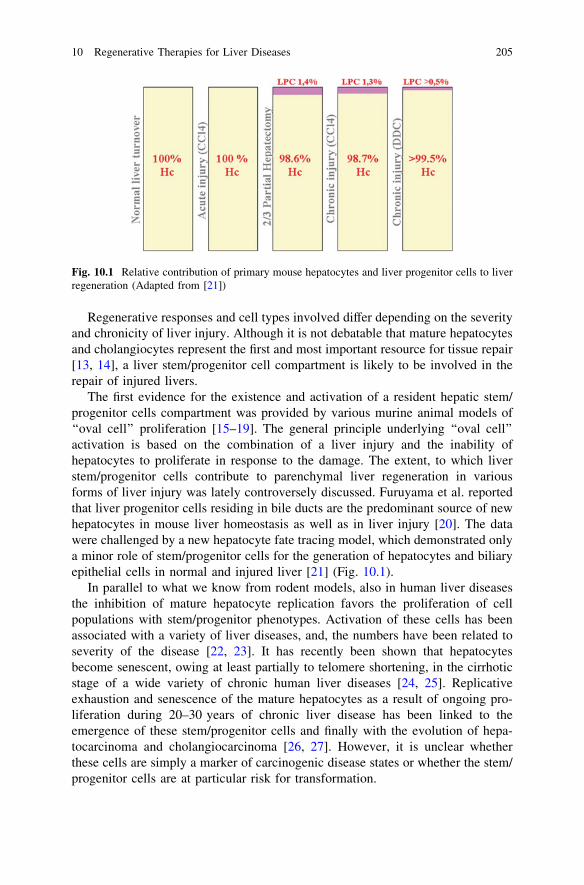
The first evidence for the existence and activation of a resident hepatic stem/progenitor cells compartment was provided by various murine animal models of‘‘oval cell’’ proliferation [15–19]. The general principle underlying ‘‘oval cell’’activation is based on the combination of a liver injury and the inability ofhepatocytes to proliferate in response to the damage. The extent, to which liverstem/progenitor cells contribute to parenchymal liver regeneration in variousforms of liver injury was lately controversely discussed. Furuyama et al. reportedthat liver progenitor cells residing in bile ducts are the predominant source of newhepatocytes in mouse liver homeostasis as well as in liver injury [20]. The datawere challenged by a new hepatocyte fate tracing model, which demonstrated onlya minor role of stem/progenitor cells for the generation of hepatocytes and biliaryepithelial cells in normal and injured liver [21] (Fig. 10.1).
In parallel to what we know from rodent models, also in human liver diseasesthe inhibition of mature hepatocyte replication favors the proliferation of cellpopulations with stem/progenitor phenotypes. Activation of these cells has beenassociated with a variety of liver diseases, and, the numbers have been related toseverity of the disease [22, 23]. It has recently been shown that hepatocytesbecome senescent, owing at least partially to telomere shortening, in the cirrhoticstage of a wide variety of chronic human liver diseases [24, 25]. Replicativeexhaustion and senescence of the mature hepatocytes as a result of ongoing pro-liferation during 20–30 years of chronic liver disease has been linked to theemergence of these stem/progenitor cells and finally with the evolution of hepa-tocarcinoma and cholangiocarcinoma [26, 27]. However, it is unclear whetherthese cells are simply a marker of carcinogenic disease states or whether the stem/progenitor cells are at particular risk for transformation.
Fig. 10.1 Relative contribution of primary mouse hepatocytes and liver progenitor cells to liverregeneration (Adapted from [21])
10 Regenerative Therapies for Liver Diseases 205
10.2 Liver Disease States as Targets for RegenerativeTherapies
The acute and self-limiting liver diseases (e.g. due to acute viral disease, toxins,transient ischemia) normally result in complete regeneration and ‘‘restitutio ad inte-grum’’. More massive injuries may temporarily exhaust the regenerative capacity ofthe liver and result in ‘‘acute liver failure’’, a clinical syndrome, which is characterizedby progressive loss of hepatic function and multiorgan failure. Persistent injuries to theliver also induce regenerative responses but eventually result in scarring and excessdeposition of extracellular matrix components including collagen. Fibrosis and cir-rhosis are the end result of chronic inflammatory reactions induced by a variety ofstimuli including persistent infections, autoimmune reactions, allergic responses,chemical insults, radiation, and tissue injury. Although current treatments for fibroticdiseases, such as idiopathic pulmonary fibrosis, systemic sclerosis, and liver fibrosis/cirrhosis typically target the inflammatory response, there is accumulating evidencethat the mechanisms driving liver fibrogenesis are distinct from those regulatinginflammation. The key cellular mediator of fibrosis is the myofibroblast, which, onceactivated, serves as the primary collagen-producing cell. Myofibroblasts are generatedfrom a variety of sources including resident mesenchymal cells (Ito cells) and circu-lating fibroblast-like cells called fibrocytes that are derived from bone marrow stemcells. Myofibroblasts are activated by paracrine signals derived from lymphocytes tomacrophages, autocrine factors secreted by myofibroblasts, and pathogen-associatedmolecular patterns (PAMPS) produced by pathogenic organisms that interact withpattern recognition receptors (i.e. TLRs) on fibroblasts.
The liver is central to many metabolic activities with hundreds of genesinvolved in their regulation. In recent years, the genetic basis for more than 100liver diseases involving malfunction of the organ has been clarified. Hereditaryliver diseases usually result from point mutations, deletions, or other geneticdefects in single or multiple genes, which are normally expressed in the liver andcan cause acute and chronic liver diseases. The liver also secrets many proteins,which deliver functions for other organ systems, and a state of protein deficiencymay not affect the liver function itself. For most of the hereditary liver diseasesliver organ transplantation cures the disease or the state of protein deficiency andhas become the most important therapeutic approach. Conceptually, many of thesedisorders, for which organ transplantation is effective, can be principally cured bycell- or gene therapies.
10.3 Cells for the Treatment of Liver Diseases
Many of the regenerative technologies generated or envisioned to treat liver dis-eases are based on cellular substrates, which are either transplanted/injected intorecipients or utilized in extracorporeal devices. The primary adult hepatocyte is
206 A. D. Sharma et al.
still the most important cellular resource in clinical situations, in which paren-chymal liver functions need to be reconstituted.
Hepatocytes isolated from pig or human livers as well as immortalized humanhepatocytes have been tested in extracorporeal liver devices. Transplanted humanhepatocytes have been shown to engraft in the recipient liver and to respond togrowth stimuli in vivo [28–30]. Despite a high proliferative capacity of hepato-cytes, which can undergo more than 69 cell doublings or a 7.3 9 1020-foldexpansion [31] in vivo, the proliferation capacity in cell culture is limited.
This lack of in vitro expansion protocols has stimulated the search for alter-native cell sources, which can either expand in cell culture or can be easilyharvested from the body in large quantities. Immortalized hepatocytes derivedfrom adult and fetal tissue are restricted to ex vivo applications and have beenapplied in extracorporal liver devices. Human fetal liver derived hepatoblasts havebeen applied in a small number of patients with acute liver failure [32] andrecently in one patient with hereditary bilirubinemia [33]. These cells are alsobeing tested as a cellular substrate for bioartificial liver devices [34]. Although theisolation of clinical grade stem/progenitor cells from human adult livers has beendescribed, clinical applications were not yet reported.
It has been proposed that (subpopulations of) adult hematopoietic stem cells (HSC),mesenchymal stromal cells (MSC), and cord blood stem cells (CBSC) can transdif-ferentiate into hepatocytes after transplantation, but the efficacy, by which these cellsspontaneously form hepatocytes and liver tissue in animal experiments, still seemsquestionable [35–39]. As an alternative concept, HSC, MSC, and CBSC are beingapplied in patients with chronic liver disease with the therapeutic aim to protect residenthepatocytes from injury, induce liver regeneration, and to initiate tissue remodeling.
High therapeutic expectations have been attributed to embryonic stem (ES) cellsand, more recently, iPS cells. These cells can be maintained in a state of pluripotencyfor long periods of time, grown in large quantities [40–45], and differentiated intovirtually all cell types of the body. ES-like cells have been generated by transfer andsimultaneous expression of four genes and termed induced pluripotent stem (iPS)cells. The direct transcription factor-mediated conversion of fibroblasts into hepaticcells, which could at least temporarily rescue a murine model of metabolic liverfailure, was recently demonstrated by two independent groups [46, 47]. To date, itremains speculative, whether direct ‘‘trans-programming’’ of adult stem cells orfibroblasts into the desired phenotype by forced expression of sets of transcriptionfactors represents an alternative approach and may circumvent the state of pluri-potency, which is associated with teratoma formation in transplanted recipients.
10.3.1 Modes of Therapeutic Activity
Various modes of therapeutic activity have been proposed for transplanted cells.Transplanted primary hepatocytes, fetal hepatoblasts, and adult liver progenitorcells engraft in the recipient liver and function as parenchymal liver cells. In vitro
10 Regenerative Therapies for Liver Diseases 207
hepatic differentiation protocols for stem cells, in particular embryonic and iPScells, aim to generate cell phenotypes compatible with long-term engraft in theliver and hepatocyte functionality.
Several ways to obtain therapeutic activity have been proposed for transplantedunmodified stem cells. For example, the injection of hematopoietic stem cells(HSC) isolated from adult bone marrow or cord blood was shown to generatehepatocytes at therapeutically significant levels in animal models [35–37]. Initialstudies suggested that those extrahepatic stem cells transdifferentiate fromhematopoietic to hepatic lineage in the recipient organ, but more recent work hasdemonstrated fusion of stem cells with hepatocytes as the main mechanism [48]. Inan alternative concept HSC and mesenchymal stromal cells have been injected inanimals and humans with acute and chronic liver disease in order to preventapoptosis, to induce regeneration, or to remodel the recipient liver without forminghepatocytes. Paracrine signaling of the transplanted cells and direct cell–cellcontact have been proposed as main mechanisms in this setting.
10.3.2 Hepatocytes from ES/iPS Cells
Hepatocytes derived from ES cells may serve as an unlimited cell source for invitro and in vivo applications. In order to generate hepatocytes from pluripotentstem cells, various differentiation protocols have been published, which usuallymimic the events occurring during embryonic development of the liver. In onetype of protocol the pluripotent ES cells are differentiated into the hepatocyte stateby initial formation of embryoid bodies, followed by the induction of definitiveendoderm using instructive cytokines such as Activin A. The endoderm cellpopulation can be further instructed to develop toward the hepatocyte lineage byexposure to bone morphogenetic protein (BMP) 4 and fibroblast growth factor(FGF) 2, both important signals from the cardiac mesoderm in early liverembryogenesis [49]. Assessment of the hepatic phenotype is commonly based onhepatocyte-specific gene expression profiles and metabolic activities, such ascytochrome p450 activity, glycogen storage, or urea synthesis, which determinethe efficacy of the differentiation protocol. At the end of a differentiation process itis important to remove contaminating undifferentiated ES cells from the hetero-geneous cell culture to minimize the risk of teratoma formation. This can beachieved by various FACS/MACS sorting techniques or by the transfer of celltype-specific expression of antibiotic resistance genes [50].
Transplantation of ES derived hepatic cells to the liver results in engraftment asboth, mature hepatocytes and bile duct epithelial cells [49, 51]. The level of liverrepopulation obtained with hepatocyte-differentiated ESCs is very low, but can beincreased somewhat when cells are transplanted in a favorable liver environment,which allows positive selection of the transplanted cells. To date, most publishedESC differentiation protocols generate hepatocyte-like cells, but not the fully
208 A. D. Sharma et al.
functional, mature, and transplantable equivalents of hepatocytes that are isolatedfrom adult livers.
The pioneering work of Yamanaka and colleagues has paved the way forgeneration of embryonic stem cell-like cells from almost any postnatal organ, suchas skin, liver, and blood. These cells have been named induced pluripotent stem(iPS) cells. The iPS cells are generated from somatic cells by transduction withviral vectors expressing the stem cell genes oct4, sox2, c-myc, and klf4 [44, 45].The combination of these four transcription factors was identified from initially 24different transcription factors. It was shown that these 4 factors were sufficient toalso induce somatic cell reprogramming in human cells. Subsequently, iPS cellswere generated without viral integration [52] to avoid integrated virus associatedgenotoxicity. iPS cells resemble ES cells as they possess self-renewal capacity, theability to differentiate into cells of ectoderm, mesoderm and endoderm, and ter-atoma formation after transplantation in mice. Similar to mouse ES cells, hepa-tocyte differentiation of mouse and human iPS cells has been documented [53–56].
10.4 Liver Tissue Engineering
Liver tissue engineering is an emerging research field and aims to create functionalliver structures using isolated hepatocytes and/or other cells types. It has beendemonstrated in animal models, that sheets of liver tissue can be grown under therenal capsule or under the skin [57, 58]. Alternatively, hepatic tissues could beengineered ex vivo to produce therapeutic effects allowing this approach tobecome an effective modality for the treatment of acute liver failure. Threedimensional liver bioreactors, which are the main component of cell-based liversupport devices, may be considered as a ‘‘tissue engineering’’ approach. A muchmore complex and not yet achieved task will be the generation of transplantableliver tissue with a functional blood supply and biliary system.
10.5 Therapeutic Concepts in Liver Disease
Regenerative therapies or treatments involving regenerative technologies arecurrently being developed for liver diseases of diverse etiologies. In acute liverfailure syndromes, acute on chronic liver failure and non-function of transplantedlivers the therapeutic approaches aim to substitute liver function (synthesis ofproteins, metabolism, and detoxification) either by extracorporal support devices,transplantation of liver cells, or by engineering and transplantation of functionalliver tissue. In chronic liver diseases conventional drugs, cytokines, stem celltherapy, and gene transfer techniques are being employed to specifically interferewith the inflammatory and profibrotic pathways. For hereditary metabolic liverdisease the experimental and clinical approaches focus on substitution of defective
10 Regenerative Therapies for Liver Diseases 209
genes and proteins by allogeneic transplantation of hepatocytes or by gene therapy.Therapies involving regenerative technologies, such as (stem) cell therapies andgene transfer protocols, which emerge for liver cancer, viral infections, andimmune-mediated liver diseases, are beyond the scope of this book chapter andhave been reviewed elsewhere.
10.5.1 Acute Liver Failure
Acute liver failure (ALF) is a syndrome of diverse etiology, in which patientswithout previously recognized liver disease sustain a liver injury that results inrapid loss of hepatic function. Depending on the etiology and severity of the insult,some patients undergo rapid hepatic regeneration and spontaneously recover.However, nearly half of the patients with ALF require and undergo orthotopic livertransplantation or die. Even with optimal early management many patients withALF develop a cascade of complications often presaged by the systemic inflam-matory response syndrome, which involves failure of nearly every organ system.No satisfactory treatment exists for those patients other than liver transplantation.However, the number of donor livers available is limited and the outcome of livertransplantation for ALF is significantly lower than transplantation for chronic liverdisease. Furthermore, many ALF patients are not placed on the transplant list dueto exclusion criteria, such as sepsis, psychiatric illness, and multi-system organfailure. Specialized treatment algorithms for the intensive care of patients withliver failure and the introduction of antioxidative drug treatments have alreadysignificantly improved the survival of affected patients in the past. Trials ofplasmapheresis and hypothermia from European consortia are near completion anddrugs that facilitate the excretion of ammonia, such as L-ornithine phenylacetate[59], may provide a neuroprotective bridge to orthotopic liver transplantation.
Future therapies for ALF would ideally maintain the patient’s clinical stabilitylong enough to allow liver regeneration to occur, which would obviate the need fororthotopic liver transplantation. Realistically, the goal of such therapies will be toserve as a bridge to orthotopic liver transplantation. Extracorporeal liver supportdevices have been developed to achieve the goal of ‘‘bridging’’ by temporarilysupporting liver detoxification function. Artificial liver support refers to purelymechanical devices including albumin dialysis, while bioartificial liver supportrefers to devices with a cellular component. Artificial systems remove toxins byfiltration or adsorption while bioartificial liver systems perform these functionsalong with biotransformation and synthetic functions of biochemically activehepatocytes.
The molecular adsorbent recirculating system (MARSTM; Gambro, Lund,Sweden) is the most frequently used type of albumin dialysis [60]. The key featureto the function of albumin dialysis is the concentration gradient of low-molecular-weight substrates between the patient’s blood and the 20 % albumin in the sec-ondary circuit. This concentration gradient allows diffusible low-molecular-weight
210 A. D. Sharma et al.
substrates to flow down their gradient over the membrane where they are tran-siently bound by albumin in the secondary circuit [61].The low-molecular-weightsubstrate is then removed from the system by conventional dialysis and hemo-diafiltration within the secondary circuit.
The initial clinical study described a series of 13 patients who underwenttreatment after failure of response to best medical therapy for acute-on-chronichepatic failure. In this series, the overall survival was 69 % and the authors citedthat all patients showed a positive response to therapy [62]. Other encouragingcase reports and small studies eventually led to more widespread use of the system.To date, roughly 7,500 patients have been treated with MARS for various hepaticdiseases, including acute liver failure patients. A meta-analysis assessing the use ofMARS looked at four randomized controlled trials including a total of 67 patientsand two selected nonrandomized trials including 61 patients [63]. Patients hadeither acute or acute-on-chronic liver failure. Primary meta-analysis did not show astatistically significant survival benefit. Recently, the results of a large multicenterrandomized trial of MARS in patients with ALF fulfiling high-urgency liver-transplant criteria in France were presented [64]. The data showed a trend towardbetter surivival in the MARS treatment group, but the difference did not reachsignificance. The transplant-free 6 month survival, however, was significantlyprolonged in those patients treated with at least three sessions of MARS. ‘‘Pro-metheusTM, which employs fractionated plasma separation, is a close variant ofMARS. While MARS is a two-circuit system separated by an albumin imper-meable membrane, Prometheus utilizes a membrane with a 250 kDa cutoffbetween circuits, thereby making the membrane permeable to albumin and hencealbumin-bound toxins. While a large portion of the toxins, which accumulateduring liver failure are water soluble, many are still bound by albumin. Thereforefractionated plasma separation may be advantageous in regard to toxin removal.Other factors that distinguish Prometheus from MARS include the fact that whileMARS is prefilled with 120 g of exogenous human albumin, the patient’sendogenous albumin loads the secondary circuit in Prometheus. Because Prome-theus is loaded with the patient’s albumin, there may be a drop in the patient’salbumin levels during treatment [65, 66].
Most of the clinical data involving Prometheus are either uncontrolled or ret-rospective. A controlled trial, published as an abstract, looking at the effect offractionated plasma separation on hepatic encephalopathy, demonstrated that a 6 htreatment course improved clinical grade and sensory-evoked potentials [67].Multiple case series describe both, acute and acute-on-chronic liver failure patientsbeing treated with Prometheus. Only recently, the results of a controlled ran-domized multicenter clinical trial in 145 patients with acute on chronic liver failurewere reported in abstract form [68]. Survival rates after 1 and 3 month were notsignificantly different in the treated versus the control group. However, patientswith hepatorenal syndrome type I and MELD score of [30 showed a significantsurvival benefit. Currently available data thus illustrate a need for new prospectiverandomized controlled trials to clarify indications and clinical impact of extra-corporeal artificial liver support devices.
10 Regenerative Therapies for Liver Diseases 211
It is unlikely that the complex mechanism, by which the liver ensureshomeostasis, can be replaced by means of nonbiologic detoxification alone. Abioartificial liver, which incorporates hepatocytes from various sources, has thetheoretical advantage of not only providing blood purification through dialysis, butalso providing the hepatocyte-specific functions which are lost with ALF. The firstbiologically based liver assist device to be tested in FDA-approved phase II/IIItrial was HepatAssistTM by Arbios (formerly Circe, Waltham, MA). The deviceemployed a hollow fiber extracorporeal bioreactor loaded with cryopreservedprimary porcine hepatocytes. A randomized, controlled, multicenter phase II/IIIclinical trial was conducted in patients with fulminant/subfulminant liver failureand primary graft nonfunction [69]. The study demonstrated favorable safety, butfailed to demonstrate improved 30-day survival in the overall study population.Although subgroups of the study population showed significant survival benefits,HepatAssist is not yet approved by the FDA. The extracorporeal liver assist device(ELADTM) developed by Vital Therapies (San Diego, CA) utilizes hollow fibercartridges loaded with cells from the C3A human hepatoblastoma cell line. Themost current model also contains a conventional hemodialysis unit. An earlyrandomized controlled trial of 24 patients with acute alcoholic hepatitis demon-strated that therapy with ELAD produced reduced levels of ammonia and bilirubinalong with improvement in hepatic encephalopathy when compared to controls[70]. However, a statistically significant survival advantage was not demonstrated.The modular extracorporeal liver support system (MELSTM; Charité, Berlin,Germany) is a hepatocyte based liver support therapy composed of four inde-pendently functioning hollow fiber capillary cell compartments. A phase I study in2003 including eight patients with ALF demonstrated safety, with all eight patientsbeing successfully bridged to transplantation [71]. Clinical experience with MELShas been limited by the infrequent and unpredictable supply of human hepatocytesand concerns of xenozoonosis involving pig hepatocytes which are prevalent inEurope. The bioartificial liver support system (BLSSTM) by Excorp medical(Minneapolis, MN,U.S.A) is a system that utilizes *100 g of primary porcinehepatocytes in a single hollow fiber cartridge. Venovenous bypass is used tocirculate the patient’s blood through the system. A phase I trial, in which fourpatients were treated with BLSS, demonstrated safety [72]. Currently, a phase II/III study is underway, and results will further define the role of this device. TheAmsterdam medical Center bioartificial liver (AMC-BALTM; AMC, Amsterdam,The Netherlands) uses 100 g of primary porcine hepatocytes bound to a spiral-shaped polyester fabric with integrated hollow fibers. During treatment, the bio-reactor is perfused with the patient’s plasma. A phase I study of the systemexamined seven patients with ALF who underwent multiple treatments with AMC-BAL [73]. Six were successfully bridged to transplantation, and one patientrecovered liver function without transplantation. Improvements were observed inboth clinical and biochemical parameters including a decrease in both bilirubinand ammonia. No adverse events were associated with treatment. While pre-liminary results were encouraging, larger randomized, controlled trials are neededto determine the role of AMC-BAL.
212 A. D. Sharma et al.
In acute liver failure hepatocyte transplantation may act as a bridge to recoveryand regeneration of the injured native liver or alternatively to orthotopic livertransplantation once an organ becomes available. The procedure may also be usedin patients who are not candidates for organ transplantation. A major advantage ofhepatocyte transplantation is the immediate availability of cryopreserved cells.Sufficient cell mass (approximately 10–15 % of liver cell mass) is needed toprovide enough metabolic function [74]. The mass of cells, which can be trans-planted into the liver, is, however, limited. Other options include intrasplenic orintraperitoneal transplantation, which allow a larger volume of cells. The spleenhas been used successfully as injection site in animals [75, 76] and humantransplantation [77]; however, in view of the number of immunologically activecells located in the spleen, rejection or destruction of the non-native cells needsconsideration. Hepatocyte transplantation in patients with ALF has resulted in areduction in ammonia and bilirubin with improvements in hepatic encephalopathyand cardiovascular instability [77, 78]. In the absence of any randomized con-trolled trials, it is difficult to comment on the true efficacy of the intervention.
There are a few studies on liver cell therapy for treatment of acute liver failurein humans with the intention to bridge the patients to orthotopic liver transplan-tation or recovery. Main challenges for future applications are the appropriatetiming of cell transplantation, the restricted uptake capacity of the recipient liver,the availability of cells, and the need for immunosuppression to prevent therejection of the transplanted cells. The latter point may become more importantthan previously considered, since the liver failure gives a high risk for septiccomplications itself, which will be aggravated by immunosuppressive drugs.
Extended liver resections have been associated with significant morbidity andmortality due to hepatic dysfunction or hepatic failure in the postoperative period.Autologous bone marrow stem cell therapies may offer the potential to enhancehepatic regeneration in this setting, perhaps increasing the safety of the procedure.Preclinical models and initial translational studies have suggested that autologousbone marrow stem cell administration can facilitate hepatic regeneration followingboth acute and chronic liver disease [79]. Infusion of HSC in three patients afterextended liver resection demonstrated the therapeutic potential, however, moreand controlled clinical trial data are needed [80].
10.5.2 Chronic Liver Disease and Liver Fibrosis
Chronic injury and inflammation triggers a gradual loss of liver function anddeposition of extracellular matrix components, which leads to fibrosis and finallyto cirrhosis of the liver. Although acute injury does activate mechanisms offibrogenesis, more sustained signals associated with chronic liver diseases lead to afibrogenic response which engages several different cell types. Cirrhosis of theliver as a clinical endpoint of the fibrogenic process is probably an irreversiblecondition and the only long-term therapeutic solution for end-stage chronic liver
10 Regenerative Therapies for Liver Diseases 213
disease today is liver organ transplantation. However, experimental and clinicaldata indicate that earlier events of the perpetuated fibrogenic process in the livercan be stopped or even reversed.
There is now experimental evidence that several endogenous factors/cytokinesplay important roles in regulation of liver fibrogenesis. The use of interferon alpha-2a and -2b is nowadays the main therapeutic strategy for the treatment of chronicviral hepatitis and compensated viral liver cirrhosis [81, 82]. In addition todecreasing viremia in HBV and HCV infections, it also may lead to reduced liverfibrosis.
New therapeutic targets interfering with fibrogenesis are emerging fromtranslational research and have been recently addressed in clinical trials. Inter-feron-gamma1b (IFN-c1b) is a pleiotropic cytokine that displays antifibrotic,antiviral, and antiproliferative activity. Initial studies conducted in patients withHCV-related liver diseases have shown a fibrosis reduction in some of the patients[83]. In particular, patients with elevated interferon-inducible T cell-alpha che-moattractant (ITAC) levels in their blood and, perhaps less advanced diseasestages, may best be suited for IFN-gamma1b based therapy [84].
Interleukin-10 (IL-10) was first described as a cytokine synthesis inhibitoryfactor for T lymphocytes produced from T helper 2 cell clones. In fact various cellpopulations produce IL-10 in the body, including T cell subsets, monocytes,macrophages, and also various other cell types present in organs such as the liver.IL-10 gene polymorphisms are possibly associated with liver disease susceptibilityor severity. Recombinant human IL-10 is currently tested in clinical trials inpatients not responding to standard Peg-IFN a therapy.
PDGF is the most potent mitogen for hepatic stellate cell-derived myofibro-blasts and levels of the growth factor have been shown to increase in liver diseases.Autocrine signaling by PDGF was the first cytokine loop discovered in hepaticstellate cell activation and is among the most potent ones [85]. Hepatic PDGF-aoverexpression using the CRP-gene promoter was accompanied by a significantincrease in hepatic procollagen III mRNA expression as well as TGF-b1 expres-sion. Liver histology showed increased deposition of extracellular matrix intransgenic but not in wildtype mice. These results point to a mechanism of fibrosisinduction by PDGF-a via the TGF-b1 signaling pathway [86]. On the other hand,Dominant-negative soluble PDGF receptor beta is currently investigated as apossible new antifibrogenic target.
TGFb1 remains, however, the classic fibrogenic cytokine. TGF b1 activatesstellate cells via the SMAD proteins pathway and also stimulates collagenexpression in stellate cells through a hydrogen peroxide and C/EBPb -dependentmechanism. There is experimental evidence that hepatocyte-specific overexpres-sion of TGFb1 in transgenic mice increases fibrosis in vivo, and that soluble TGFbreceptor type II treatment inhibits fibrosis in vivo. Also, it has been shown thatadenovirus encoding antisense TGFb mRNA inhibits fibrogenesis in vivo.
More experimental strategies aim to reduce extracellular matrix deposition byoverexpression of MMP’s. Siller-Lopez et al. have used an extrahepatic humanneutrophil collagenase complementary MMP-8 DNA cloned in an adenovirus
214 A. D. Sharma et al.
vector (AdMMP8) as a therapeutic agent in cirrhosis using CCl4 and bile duct–ligated cirrhotic rats models. Liver fibrosis in bile duct–ligated cirrhotic animalswas decreased by 45 % along with reduced hydroxyproline levels in AdMMP8treated animals. Treatment in both models correlated with improvements inascites, functional hepatic tests, and gastric varices indicating diminished portalhypertension in animals injected with AdMMP8 [87].
Alternative treatment concepts aim to protect existing hepatocytes and/or toincrease the hepatocyte mass. Hepatocyte growth factor (HGF), originally iden-tified and cloned as a potent mitogen for hepatocytes [88–91] has mitogenic andmorphogenic activities for a wide variety of cells [92, 93] and also plays anessential role in the development and regeneration of the liver [94]. The proteinhas also been shown to have antiapoptotic activity in hepatocytes [95]. Trans-duction of the HGF gene has suppressed the increase of transforming growthfactor-b1 (TGF-b1), which plays an essential part in the progression of livercirrhosis and inhibited fibrogenesis and hepatocyte apoptosis leading to completeresolution of fibrosis in the cirrhotic liver in a rat model [96].
10.5.2.1 Stem Cell Therapy of Chronic and Acute of Chronic Liver Disease
Although the concept of stem cell therapy for various diseases is principallyaccepted, the practical approach in humans remains difficult. Bone marrow derivedmononucleated cells, hematopoietic stem and progenitor cells, mesenchymal stem(stromal) cells and sinusoidal endothelial cells are currently being investigated.There are several proposed mechanisms by which stem and progenitor cells mightsupport regeneration in targeted organs including the liver: intercellular signalingthrough cell–cell contacts, paracrine signaling (growth factors, cytokines, andhormones) or cell fate change in the target organ.
The concept of stem/progenitor cell infusions exerting a paracrine regenerativeeffect on the liver is gaining support and is backed up by both rodent and humanstudies, although the latter are small and uncontrolled. Endothelial precursor cells(EPC) have been shown in rodent models to promote angiogenesis and the deg-radation of liver scar tissue thereby contributing to liver regeneration [97–99]. Byparticipation in neovascularization and by the expression of multiple growthfactors, transplanted EPCs significantly accelerate liver regeneration. This isachieved by enhancing proliferative activity of hepatocytes leading to improvedsurvival after chemically induced liver injury [97].
Sakaida et al. have demonstrated that transplanted bone marrow cells degradeextracellular matrix in carbon tetrachloride (CCl4)-induced liver fibrosis, with asignificantly improved survival rate in this animal model. Their findings suggestthat transplanted bone marrow cells can degrade collagen fibers and reduce liverfibrosis by strong expression of MMPs, especially MMP-9 [100].
Other groups have raised concerns about the role of certain subtypes of bonemarrow stem cells in chronic liver injury [101]. It has been shown that bonemarrow-derived myofibroblasts significantly contributed to fibrogenesis in a
10 Regenerative Therapies for Liver Diseases 215
chronic liver injury model in mice. They originated predominantly from bonemarrow cells enriched for mesenchymal progenitor cells. These cells were locatedin the region of hepatic scarring and actively expressed collagen. The data suggestthat an axis of recruitment from the bone marrow to the liver does exist in chronicinjury and that the therapeutic application of certain subsets of bone marrow-derived cells may contribute to, rather than resolve scarring of the liver tissue. Thechoice of the transplanted bone marrow cell type might thus be important withregard to supporting liver regeneration or fibrogenesis.
Taken together the infusion of stem cells might provide an array of factorssupporting not only liver regeneration, but also the remodeling of impaired liverarchitecture by interfering with fibrogenesis. Important experimental findings,however, suggest that infused bone marrow cells may also contribute to fibro-genesis [102, 103] giving some cautious notes for the uncritical use of stem cellsfor chronic liver disease outside of controlled clinical trials [100, 104–106].
Several clinical trials already investigated the effect of bone marrow (stem)cells in patients with liver disease. They were mainly uncontrolled, with only smallnumbers of patients enrolled and have provided heterogeneous results. The trialscan be categorized in 4 groups according to the main endpoint and source of cells:[1] effects of granulocyte colony–stimulating factor (G-CSF) mobilized bonemarrow cells in advanced chronic liver disease, [2] effects of infusion of autolo-gous mononuclear cells collected from bone marrow in advanced chronic liverdisease, [3] effects of collection (with or without ex vivo manipulation), andinfusion of mobilized bone marrow cells in advanced chronic liver disease and [4]effects of bone marrow infusions on liver regeneration (after selective portalvenous embolization) prior to extended hepatectomy for liver tumors [107–118].
The trials are quite heterogeneous with regard to the source of stem cells usedand the number of patients included. The following stem cells sources have beenused: bone marrow from iliac crest (50–400 ml), G-CSF mobilization only, G-CSFmobilization followed by leukapheresis and CD-34+ selection and reinfusion. Allbut one trial were non-randomized. The stem cells were administered by peripheralvein infusions (3 studies), by hepatic artery infusions (5 studies), or portal veininfusions (2 studies). The largest study conducted so far by Lyra et al. was also theonly randomized one and included 30 patients.
Eight out of 11 trials have shown a moderate improvement in liver function(albumin, INR, bilirubin, Child-Pugh score, MELD score) and the follow-upperiod has ranged from 2 to 12 months.
In one recent study safety and efficacy of hepatic artery administration ofmobilized autologous and ex vivo expanded adult CD34+ hematopoietic stem cellsin patients with alcoholic cirrhosis (ALC) was assessed [118]. This study reportedone of the largest numbers of CD34 positive stem cells infused in cirrhotic patientsso far. Nine patients with biopsy-proven ALC and abstinence from alcohol for atleast 6 months were included in the study and all patients tolerated the procedurewell, with no treatment-related side effects or toxicities observed. Significantimprovement in liver function was shown by decrease in serum bilirubin levels,
216 A. D. Sharma et al.
serum alanine transaminase, and aspartate transaminase. The Child-Pugh scoreimproved in 7 out of 9 patients and in 5 patients ascites production had declined.
Two studies so far aimed to ameliorate acute on chronic liver disease byadministration of granulocyte—colony stimulating factor (G-CSF) treatment. Incontrast to an earlier study by Campli et al. [119] a more recent study from Indiashowed profound effects on short-term survival, which was associated with amarked increase of CD34 stem cells in the liver of recipients [120].
10.5.3 Hereditary Liver Disease
Liver organ transplantation can be viewed as a form of gene therapy for inheritedliver diseases since the procedure substitutes a defective gene with a normal copyfrom a healthy donor. Animal studies have shown that for most monogenetic liverdiseases partial substitution of a missing or defective protein is able to reverse theclinical phenotype and can result in complete remission of the disease. Thisredundancy opens the possibility to apply minimally invasive therapies such as celland gene therapies to correct an existing gene defect. Although many hurdles stillexist, feasibility has been proven unequivocally in animal models and therapeuticprotocols are now emerging in the clinical arena.
10.5.3.1 Transplantation of Mature (Adult) Hepatocytes
In recent years the interest in liver cell therapy has been increasing continuously,since the demand for whole liver transplantations in human beings far outweighsthe supply [121]. From the clinical point of view, transplantation of hepatocytes orhepatocyte-like cells may represent an alternative to orthotopic liver transplantsfor the correction of genetic disorders resulting in metabolically deficient states.The aim of hepatocyte transplantation in metabolic disease is to partially replacethe missing function without the need to replace the whole organ. Almost 30children and adults who received liver cell therapy for metabolic liver disease arereported in literature [122, 123]. Clinical therapies up to now have been performedby infusing fresh or cryopreserved primary hepatocyte suspensions isolated fromdonated organs. The availability of high quality liver tissue for cell isolation,however, has slowed the widespread application of this therapy. Furthermore, theclinical situation of target patients is rarely immediately life threatening and oftenacceptable conventional therapies are available. Therefore, the potential benefitmust be carefully weighed against any possible complications, such as side effectfrom immunosuppression, hepatocyte embolization of the pulmonary vascularsystem, sepsis or hemodynamic instability.
Objective parameters such as laboratory data (i.e. bile acid, clotting factors,etc.) can be determined to unequivocally assess the efficacy of the treatment. Theresults of hepatocyte transplantation for many metabolic liver diseases have been
10 Regenerative Therapies for Liver Diseases 217
encouraging with demonstrable, although short-term correction of metabolicdeficiency in the majority of cases. Therapeutic benefit has been reported in a girlwith Crigler–Najjar Syndrome Type I, which is a recessively inherited metabolicdisorder characterized by severe unconjugated hyperbilirubinemia [124]. Isolatedhepatocytes were infused through the portal vein and partially corrected plasmabilirubin levels for more than 11 months. Similarly, a 9 year-old boy received7.5 9 109 hepatocytes, infused via the portal vein, which resulted in a decrease inbilirubin level from 530 ± 38 lmol/L (mean ± SD) before to 359 ± 46 lmol/L[125]. Hughes et al. also reported a 40 % reduction in bilirubin levels in a Crigler–Najjar Syndrome Type I patient following transplantation of hepatocytes [126].Although these data demonstrate efficacy and safety, a single course of cellapplication seems not sufficient to correct Crigler–Najjar Syndrome Type Icompletely.
Sustained response was reported in a patient with argininosuccinate lyasedeficiency after repeated hepatocyte transplantation. Engraftment of the trans-planted cells was analyzed in repeated liver biopsies for more than 12 months byfluorescence in situ hybridization for the Y-chromosome and by measurement oftissue enzyme activity [127]. Promising results have also been obtained in a 47-year-old woman suffering from glycogen storage disease type 1a, an inheriteddisorder of glucose metabolism resulting from mutations in the gene encoding thehepatic enzyme glucose-6-phosphatase [128]. 2 9 109 ABO-compatible hepato-cytes were infused into the portal vein. Nine months after cell transplantation, hermetabolic situation had clearly improved. Successful hepatocyte transplantationhas also been achieved in a 4-year-old girl with infantile Refsum disease, an inbornerror of peroxisome metabolism, leading to increased levels of serum bile acidsand the formation of abnormal bile acids [129]. A total of 2 9 109 hepatocytesfrom a male donor were given during eight separate intraportal infusions.Abnormal bile acid production (for instance pipecholic acid) had decreased by40 % after 18 months. Recently, hepatocyte transplantation has been used suc-cessfully to treat inherited factor VII deficiency [130]. Two brothers (aged3 months and 3 years) received infusions of 1.1 and 2.2 9 109 ABO-matchedhepatocytes into the inferior mesenteric vein. Transplantation clearly improved thecoagulation defect and decreased the necessity for exogenous factor VII toapproximately 20 % of that prior to cell therapy. As with the other metabolic liverdiseases, hepatocyte transplantation has been shown to provide a partial correctionof urea cycle defects. Patients showed clinical improvement, reduced ammonialevels, and increased production of urea [131–135].
10.5.3.2 Transplantation of Stem Cells
In the last few years, many reports have suggested that extrahepatic stem cellsparticipate in liver regeneration and may be useful for treating many diseases[136–140]. However, subsequent work by several independent groups has clearlyshown that hepatocyte replacement levels after injection of extrahepatic stem cells
218 A. D. Sharma et al.
or by bone marrow transplantation are low (\0.01 %), unless those bone marrow-derived hepatocytes have a selective growth advantage [141–143]. Furthermore, inmost of the cases, fusion with host hepatocytes rather than transdifferentiation ofextrahepatic cells, has been described as the underlying mechanism [144–147].
So far no convincing evidence has yet been provided in animal models that stemcells including HSC, MSC, iPS, or cells derived from cord blood or the amnion cangenerate therapeutically significant numbers of hepatocytes for the correction ofhereditary metabolic liver diseases. Consequently, no credible data on the use of stemcells in patients with hereditary liver disease have been published.
10.5.3.3 Gene Therapy
The liver is involved in the synthesis of serum proteins, regulation of metabolism,and maintenance of homeostasis and thus provides a variety of opportunities forgene therapeutic corrections. Gene therapy is the treatment of an inherited oracquired disease through the manipulation of a patients’ genetic status or sequencein selected cells by introducing various types of genetic materials, such as virallybound nucleic acids, plasmid DNAs, antisense oligonucleotides, and short inter-ference RNAs. Both viral and non-viral methods have been developed for effectivegene delivery. Currently, only viral vectors have transduction efficacies needed forliver-based gene therapy of inherited metabolic diseases in humans.
Viral Vectors
Viral gene delivery employs replication deficient viruses as a carrier to bring geneticmaterials into cells through their natural infection mechanism. Viral vectors arecreated using molecular biological techniques by which portion of the viral genomeis replaced with a gene of interest. Major drawbacks of viral vectors are their geneticand immunologic toxicities, which are mainly associated with an arbitrary recom-bination with genomic DNA of the target cells and acute immune stimulation,respectively. Because adult humans have already developed immunity against sev-eral types of viruses from which viral vectors are developed, an exposure of the viralvectors to patients often results in strong immunological reaction, and consequentlydisables efficient gene delivery and long-term gene expression.
Viral vectors frequently used in gene therapy studies are derived from retro-virus, adenovirus, and adeno-associated virus. Retroviruses, enveloped RNAviruses with a particle size of approximately 100 nm, only infect dividing cells andare capable of integrating reverse transcribed DNA into the host genome at anunpredictable location [148]. Viral integration has led to leukemia development asrevealed by recent gene therapy trial on X-linked SCID [149]. The requirement ofhepatectomy (*70 %) to stimulate hepatic proliferation is generally considered asa drawback for retrovirus-mediated gene delivery to the liver [150, 151]. Len-tiviruses, a subclass of retroviruses including human immunodeficiency virus, can
10 Regenerative Therapies for Liver Diseases 219
transduce non-dividing as well as dividing cells. The lentivirus preintegrationcomplex is able to pass the intact nuclear membrane, which allows it to integrateinto the host genome without cell division [152]. It was reported that animals canbe repeatedly infected with lentiviral vectors [153].
Adenoviruses are double-stranded DNA viruses with a diameter of approxi-mately 110 nm. Adenoviruses infect both replicating and non-replicating cells,have a relatively large genome, and are unable to integrate into the host genome[154]. A number of serotypes has been used to create adenoviral vectors andemployed in 24.8 % of clinical trials till the end of March, 2008 [155]. Thesevectors exhibit a broad range of liver tropism with serotype 5 as the most com-monly used to date [156]. Adenoviral vector is the first proven gene carrier for thetreatment of cancer [157]. Because this virus is a natural human pathogen, pre-existing immunity against adenovirus can cause severe allergic reaction andinactivation of viral vectors [158]. The current strategy in avoiding these problemsis to use a serotype which the patients have no immunity against [156]. If theimmunogenic drawbacks can be overcome in the future, adenoviral vectors willprobably find a great diversity of clinical applications.
Adeno-associated virus (AAV) belongs to the Parvoviridae virus family and isapproximately 26 nm in diameter without envelope [159]. It requires a helper virusfor replication such as adenovirus. It is non-pathogenic and can infect quiescentcells. AAV is currently classified into 12 serotypes, and the liver is known to be apreferential target especially for AAV-8 [160]. It was reported that this virus caninsert its genome at a defined site on chromosome 19 termed AAVS1 with nearly100 % certainty [161]. The site-directed integration is controlled by viral Repproteins [162], which are often deleted in recombinant AAV vectors in favor ofmore space for the exogenous gene to be packaged into the tiny viral particle.Cotransfection of plasmids coding for Rep protein was reported to restore capa-bility of the site-directed integration and enable a long-term expression of thetransgene without inducing insertional mutagenesis [163]. Results from a numberof animal studies also indicate that AAV is less immunogenic when compared toadenoviruses [164].
Preclinical Evaluation
Feasability of gene therapies has been demonstrated in a wide variety of animalmodels. Long-Evans cinnamon rats are a model of Wilson disease and transfer ofthe ATP7B gene to hepatocytes ameliorates both biochemical and histologicalpathologies [165]. Transgene products released into blood circulation after suc-cessful gene transfer into the hepatocytes corrected pathological manifestationboth inside and/or outside of the liver in glycogen storage diseases (type Ia, [166]Ib [167] and II [167], mucopolysaccharidosis type I [168], IIIB [169] and VII[170], hereditary tyrosinemia type I [171], UDP glucuronyltransferase deficiency(Crigler-Najjar type I) [172], and hemophilia [173–175].
220 A. D. Sharma et al.
A complete and persistent phenotypic correction of phenylketonuria in mice wasreported after hydrodynamic gene delivery of murine phenylalanine hydroxylasecDNA with the help of phiBT1 phage integrase for long-term gene expression [176].Further, the efficacy of adenovirus-mediated in vivo gene therapy for ornithinetranscarbamylase deficiency was reported in mice and non-human primates [177].Hyperlipidemia was not only effectively treated in the respective genetic mousemodels through delivery of apolipoprotein B [178] or E [179] genes but also in wildtype mice treated with a high-fat diet. A reciprocal pathophysiological condition ofhypoalphalipoproteinemia was effectively reversed by adenoviral transduction ofhuman apolipoprotein A-I gene in model mice as well [180].
Liver-Directed Gene Therapy in Humans
Gene therapy has the potential to offer a definitive cure for monogenic diseases byachieving a long-term correction of pathology. Monogenic diseases in the liver aredivided into two groups depending on whether cell damage in the liver is involved ornot. For example, hemophilia, familiar hypercholesterolemia, and phenylketonuriashow systemic manifestations without significant liver cell damage, and have theleast risk for hepatotoxicity in orthotopic gene delivery. In fact phase I/II clinicaltrials for hemophilia B were completed with promising results [181]. Unfortunately,however, the development of inhibitory antibodies against the exogenous factor IXand/or components of viral vectors diminished a persistent phenotypic correction[181, 182]. One possible solution to avoid antibody development against exogenousgene products is gene delivery into the fetal liver to induce tolerance to the exogenousgene products [172, 175, 183] or alternative injection routes [184]. It is important topoint out that significant difference exists between animal studies and human clinicaltrials with respect to immunological reactions [185, 186].
In case of the monogenic liver diseases with substantial hepatocellular damage,gene therapy should not be a primary indication unless gene delivery can becompleted in all hepatocytes in the liver. Successful delivery of human fumaryl-acetoacetate hydrolase gene into hepatocytes protected FAH(-/-) -mice mimickinghereditary tyrosinemia type 1 disease from fulminant liver failure by restoring theenzyme activity [187, 188]. However, hepatocellular damage continued in the restof hepatocytes that had not received the transgene and resulted in the frequentdevelopment of hepatocellular carcinoma. Liver transplantation should be a pri-mary option for the diseases in this category at this moment.
10.6 Future Directions
Regenerative therapies involving various types of cells as well as gene therapiesare currently being investigated in research laboratories around the world and moreand more find the way into therapeutic algorithms in the clinic. Bioartificial liver
10 Regenerative Therapies for Liver Diseases 221
support systems and cell therapies are currently limited by the availability of goodquality hepatocytes. A renewable source of highly metabolically competenthepatocytes will be essential for any successful bioartificial liver system. To dateporcine hepatocytes are most commonly being used with limited acceptance due toongoing concerns of xenozoonosis. Immortalized human hepatocytes have notshown expression of prerequisite hepatocyte function including ammonia detoxi-fication. Other limitations of first-generation bioartificial liver systems, which needto be solved, include excess device complexity, insufficient number of hepatocytesto support a failing liver, early hepatocyte death, and absence or loss of differ-entiated function.
The application of stem cells in liver cell therapies seems to be a promisingapproach for the treatment of liver diseases. However, several issues still have tobe addressed to fulfil this promise. We need to identify, both inside and outside ofthe liver, the stem cell candidates that are able to form mature hepatocytes in vitroand functional liver tissue after transplantation in vivo. The fundamental molecularpathways involved in the differentiation of hepatocytes and cholangiocytes fromstem/progenitor cells, the factors that are responsible for in vitro differentiation ofvarious stem cells into hepatocytes, the mechanisms involved in the fusion of stemcells and hepatocytes and the aspects that can potentially enhance these mecha-nisms need to be studied in more detail. With future progress in stem cell research,the various stem cell sources including hepatic stem/progenitor cells, embryonic,and adult extrahepatic stem cells should provide great opportunities for thetreatment of liver disorders.
Additional work is also needed in the development of an ideal gene deliverysystem. The efficacy of delivery and the level of transgene expression achieved bythe current methods have resulted in phenotypic correction of various hereditaryliver diseases in animal models. The most efficient vehicles for gene delivery to theliver developed so far are viral vectors. Among the viral vectors applicable to livergene delivery, lentiviral vectors appear to have great advantage because of theirability to transduce the liver cells at resting state and generate persistent geneexpression. Gene toxicity by insertional mutagenesis with the transactivation ofpotentially harmful genes and interactions of the host immune system with theviral proteins and the therapeutic product need to be studied in more detail. Activeparticipation of hepatologists in gene therapy research will accelerate the processin turning gene therapy into a common practice for the treatment of variousdiseases through the liver.
In summary, advanced approaches in regenerative hepatology will coverstrategies to improve endogenous liver regeneration, to correct monogenetic liverdiseases by gene therapy, and to support organ function with additional hepaticcells, either in extracorporal devices or as cell transplants. For the latter aspect,improved cell isolation and propagation techniques to utilize cells from donororgans or advanced stem cell-differentiation protocols become of utmost impor-tance to ensure the supply of functional hepatic cells.
222 A. D. Sharma et al.
References
1. Sherlock S, Dooley J (2002) Disease of the liver and biliary system. Blackwell PublishingCompany, Oxford
2. Schiff ER, Sorrell MF, Maddrey WC (2007) Schiff’s diseases of the liver. LippincottWilliams and Wilkins, Philadelphia
3. Burt AD, Portmann BC, Ferrell LD (2007) MacSween’s pathology of the liver. Elsevier,Philadelphia
4. Fausto N, Campbell JS, Riehle KJ (2006) Liver regeneration. Hepatology 43:S45–S535. Michalopoulos GK, DeFrances MC (1997) Liver regeneration. Science 276:60–666. Taub R (1996) Transcriptional control of liver regeneration. FASEB J 10(4):413–4277. Taub R (2004) Liver regeneration: from myth to mechanism. Nat Rev Mol Cell Biol
10:836–8478. Gorla GR, Malhi H, Gupta S (2001) Polyploidy associated with oxidative injury attenuates
proliferative potential of cells. J Cell Sci 114(16):2943–29519. Jungermann K, Kietzmann T (2000) Oxygen: modulator of metabolic zonation and disease
of the liver. Hepatology 31(2):255–26010. Sigal SH, Gupta S, Gebhard DF, Holst P, Neufeld D, Reid LM (1995) Evidence for a
terminal differentiation process in the rat liver. Differentiation 59:35–4211. Fellous TG, Islam S, Tadrous PJ, Elia G, Kocher HM, Bhattacharya S, Mears L, Turnbull
DM, Taylor RW, Greaves LC, Chinnery PF, Taylor G, McDonald SA, Wright NA, AlisonMR (2009) Locating the stem cell niche and tracing hepatocyte lineages in human liver.Hepatology 49(5):1655–1663
12. Bralet MP, Branchereau S, Brechot C, Ferry N (1994) Cell lineage study in the liver usingretroviral mediated gene transfer: evidence against the streaming of hepatocytes in normalliver. Am J Pathol 144:896–905
13. Quante M, Wang TC (2009) Stem cells in gastroenterology and hepatology. Nat RevGastroenterol Hepatol 6(12):724–737
14. Duncan AW, Dorrell C, Grompe M (2009) Stem cells and liver regeneration.Gastroenterology 137(2):466–481
15. Grisham JW, Hartroft WS (1961) Morphologic identification by electron microscopy of‘‘oval’’ cells in experimental hepatic degeneration. Lab Invest 10:317–332
16. Suzuki A, Zheng YW, Kaneko S, Onodera M, Fukao K, Nakauchi H, Taniguchi H (2002)Clonal identification and characterization of self-renewing pluripotent stem cells in thedeveloping liver. J Cell Biol 156:173–184
17. Fausto N (2004) Liver regeneration and repair: hepatocytes, progenitor cells, and stem cells.Hepatology 39:1477–1487
18. Thorgeirsson SS (1996) Hepatic stem cells in liver regeneration. Faseb J 10:1249–125619. Fausto N, Campbell JS (2003) The role of hepatocytes and oval cells in liver regeneration
and repopulation. Mech Dev 120:117–13020. Furuyama K, Kawaguchi Y, Akiyama H, Horiguchi M, Kodama S, Kuhara T, Hosokawa S,
Elbahrawy A, Soeda T, Koizumi M, Masui T, Kawaguchi M, Takaori K, Doi R, Nishi E,Kakinoki R, Deng JM, Behringer RR, Nakamura T, Uemoto S (2003) Continuous cellsupply from a Sox9-expressing progenitor zone in adult liver, exocrine pancreas andintestine. Nat Genet 43(1):34–41
21. Malato Y, Naqvi S, Schürmann N, Ng R, Wang B, Zape J, Kay MA, Grimm D, WillenbringH (2011) Fate tracing of mature hepatocytes in mouse liver homeostasis and regeneration.J Clin 121(12):4850–4860
22. Roskams T, Yang SQ, Koteish A, Durnez A, DeVos R, Huang X, Achten R, Verslype C,Diehl AM (2003) Oxidative stress and oval cell accumulation in mice and humans withalcoholic and nonalcoholic fatty liver disease. Am J Pathol 163:1301–1311
23. Roskams T (2006) Liver stem cells and their implication in hepatocellular andcholangiocarcinoma. Oncogene 25(27):3818–3822
10 Regenerative Therapies for Liver Diseases 223
24. Marshall A, Rushbrook S, Davies SE, Morris LS, Scott IS, Vowler SL, Coleman N,Alexander G (2005) Relation between hepatocyte G1 arrest, impaired hepatic regeneration,and fibrosis in chronic hepatitis C virus infection. Gastroenterology 128:33–42
25. Wiemann SU, Satyanarayana A, Tsahuridu M, Tillmann HL, Zender L, Klempnauer J,Flemming P, Franco S, Blasco MA, Manns MP, Rudolph KL (2002) Hepatocyte telomereshortening and senescence are general markers of human liver cirrhosis. Faseb J 16:935–942
26. Alison MR, Lovell MJ (2005) Liver cancer: the role of stem cells. Cell Prolif 38:407–42127. Mishra L, Banker T, Murray J, Byers S, Thenappan A, He AR, Shetty K, Johnson L, Reddy
EP (2009) Liver stem cells and hepatocellular carcinoma. Hepatology 49(1):318–32928. Dandri M, Burda MR, Török E, Pollok JM, Iwanska A, Sommer G, Rogiers X, Rogler CE,
Gupta S, Will H, Greten H, Petersen J (2001) Repopulation of mouse liver with humanhepatocytes and in vivo infection with hepatitis B virus. Hepatology 33(4):981–988
29. Bissig KD, Le TT, Woods NB, Verma IM (2007) Repopulation of adult and neonatal mice withhuman hepatocytes: a chimeric animal model. Proc Natl Acad Sci USA 104(51):20507–20511
30. Haridass D, Yuan Q, Becker PD, Cantz T, Iken M, Rothe M, Narain N, Bock M, Nörder M,Legrand N, Wedemeyer H, Weijer K, Spits H, Manns MP, Cai J, Deng H, Santo Di JP,Guzman CA, Ott M (2009) Repopulation efficiencies of adult hepatocytes, fetal liverprogenitor cells, and embryonic stem cell-derived hepatic cells in albumin-promoter-enhancer urokinase-type plasminogen activator mice. Am J Pathol 175(4):1483–1492
31. Overturf K (1997) al-Dhalimy, M., Ou, C.N., Finegold, M., and Grompe, M.: Serialtransplantation reveals the stem-cell-like regenerative potential of adult mouse hepatocytes.Am J Pathol 151:1273–1280
32. Habibullah CM, Syed IH, Qamar A, Taher-Uz Z (1994) Human fetal hepatocytetransplantation in patients with fulminant hepatic failure. Transplantation 58(8):951–952
33. Khan AA, Parveen N, Mahaboob VS, Rajendraprasad A, Ravindraprakash HR,Venkateswarlu J, Rao P, Pande G, Narusu ML, Khaja MN, Pramila R, Habeeb A,Habibullah CM (2008) Treatment of Crigler-Najjar Syndrome type 1 by hepatic progenitorcell transplantation: a simple procedure for management of hyperbilirubinemia. TransplantProc 40(4):1148–1150
34. Poyck PP, van Wijk AC, van der Hoeven TV, de Waart DR, Chamuleau RA, van Gulik TM,Oude Elferink RP, Hoekstra R (2008) Evaluation of a new immortalized human fetal livercell line (cBAL111) for application in bioartificial liver. J Hepatol 48(2):266–275
35. Petersen BE, Bowen WC, Patrene KD, Mars WM, Sullivan AK, Murase N, Boggs SS,Greenberger JS, Goff JP (1999) Bone marrow as a potential source of hepatic oval cells.Science 284:1168–1170
36. Alison MR, Poulsom R, Jeffery R, Dhillon AP, Quaglia A, Jacob J, Novelli M, Prentice G,Williamson J, Wright NA (2000) Hepatocytes from non-hepatic adult stem cells. Nature 406:257
37. Newsome PN, Johannessen I, Boyle S, Dalakas E, McAulay KA, Samuel K, Rae F,Forrester L, Turner ML, Hayes PC, Harrison DJ, Bickmore WA, Plevris JN (2003) Humancord blood-derived cells can differentiate into hepatocytes in the mouse liver with noevidence of cellular fusion. Gastroenterology 124:1891–1900
38. Aurich I, Mueller LP, Aurich H, Luetzkendorf J, Tisljar K, Dollinger MM, Schormann W,Walldorf J, Hengstler JG, Fleig WE, Christ B (2007) Functional integration of hepatocytesderived from human mesenchymal stem cells into mouse livers. Gut 56(3):405–415
39. Cantz T, Sharma AD, Jochheim-Richter A, Arseniev L, Klein C, Manns MP, Ott M (2004)Reevaluation of bone marrow-derived cells as a source for hepatocyte regeneration. CellTransplant 13:659–666
40. Evans MJ, Kaufman MH (1981) Establishment in culture of pluripotential cells from mouseembryos. Nature 292(5819):154–156
41. Rathjen J, Rathjen PD (2001) Mouse ES cells: experimental exploitation of pluripotentdifferentiation potential. Curr Opin Genet Dev 11:587–594
43. Boiani M, Scholer HR (2005) Regulatory networks in embryo-derived pluripotent stemcells. Nat Rev Mol Cell Biol 6:872–884
44. Takahashi K, Yamanaka S (2006) Induction of pluripotent stem cells from mouseembryonic and adult fibroblast cultures by defined factors. Cell 126:663–676
45. Takahashi K, Tanabe K, Ohnuki M, Narita M, Ichisaka T, Tomoda K, Yamanaka S (2007)Induction of pluripotent stem cells from adult human fibroblasts by defined factors. Cell131:861–872
46. Sekiya S, Suzuki A (2011) Direct conversion of mouse fibroblasts to hepatocyte-like cellsby defined factors. Nature 475(7356):390–393
47. Huang P, He Z, Ji S, Sun H, Xiang D, Liu C, Hu Y, Wang X, Hui L (2011) Induction offunctional hepatocyte-like cells from mouse fibroblasts by defined factors. Nature475(7356):386–389
48. Wang X, Willenbring H, Akkari Y, Torimaru Y, Foster M, Al-Dhalimy M, Lagasse E,Finegold M, Olson S, Grompe M (2003) Cell fusion is the principal source of bone-marrow-derived hepatocytes. Nature 422:897–901
49. Gouon-Evans V, Boussemart L, Gadue P, Nierhoff D, Koehler CI, Kubo A, Shafritz DA,Keller G (2006) BMP-4 is required for hepatic specification of mouse embryonic stem cell-derived definitive endoderm. Nat Biotechnol 24:1402–1411
50. Drobinskaya I, Linn T, Saric T, Bretzel RG, Bohlen H, Hescheler J, Kolossov E (2008)Scalable selection of hepatocyte- and hepatocyte precursor-like cells from culture ofdifferentiating transgenically modified murine embryonic stem cells. Stem Cells26(9):2245–2256
51. Touboul T, Hannan NR, Corbineau S, Martinez A, Martinet C, Branchereau S, Mainot S,Strick-Marchand H, Pedersen R, Di Santo J, Weber A, Vallier L (2010) Generation offunctional hepatocytes from human embryonic stem cells under chemically definedconditions that recapitulate liver development. Hepatology 51(5):1754–1765
52. Stadtfeld M, Nagaya M, Utikal J, Weir G, Hochedlinger K (2008) Induced pluripotent stemcells generated without viral integration. Science 322:945–949
53. Li W, Wang D, Qin J, Liu C, Zhang Q, Zhang X, Yu X, Lahn BT, Mao FF, Xiang AP(2010) Generation of functional hepatocytes from mouse induced pluripotent stem cells.J Cell Physiol 222:492–501
54. Si-Tayeb K, Noto FK, Nagaoka M, Li J, Battle MA, Duris C, North PE, Dalton S, DuncanSA (2010) Highly efficient generation of human hepatocyte-like cells from inducedpluripotent stem cells. Hepatology 51:297–305
55. Song Z, Cai J, Liu Y, Zhao D, Yong J, Duo S, Song X, Guo Y, Zhao Y, Qin H, Yin X, WuC, Che J, Lu S, Ding M, Deng H (2009) Efficient generation of hepatocyte-like cells fromhuman induced pluripotent stem cells. Cell Res 19:1233–1242
56. Sullivan GJ, Hay DC, Park IH, Fletcher J, Hannoun Z, Payne CM, Dalgetty D, Black JR,Ross JA, Samue lK, Wang G, Daley GQ, Lee JH, Church GM, Forbes SJ, Iredale JP,Wilmut I (2010) Generation of functional human hepatic endoderm from human inducedpluripotent stem cells. Hepatology 51:329–335
57. Ohashi K, Yokoyama T, Yamato M, Kuge H, Kanehiro H, Tsutsumi M, Amanuma T, Iwata H,Yang J, Okano T, Nakajima Y (2007) Engineering functional two- and three-dimensional liversystems in vivo using hepatic tissue sheets. Nat Med 13(7):880–885
58. Ohashi K, Koyama F, Tatsumi K, Shima M, Park F, Nakajima Y, Okano T (2010)Functional life-long maintenance of engineered liver tissue in mice followingtransplantation under the kidney capsule. J Tissue Eng Regen Med 4(2):141–148
59. Jalan R, Wright G, Davies NA, Hodges SJ (2007) L-Ornithine phenylacetate (OP): a noveltreatment for hyperammonemia and hepatic encephalopathy. Med Hypotheses 69:1064–1069
60. Mitzner SR, Stange J, Klammt S, Koball S, Hickstein H, Reisinger EC (2009) Albumindialysis MARS: knowledge from 10 years of clinical investigation. ASAJO J 55(5):498–502
61. Steiner C, Sen S, Stange J, Williams R, Jalan R (2004) Binding of bilirubin andbromosulphthalein to albumin: implications for understanding the pathophysiology of liverfailure and its management. Liver Transpl 10:1531–1538
10 Regenerative Therapies for Liver Diseases 225
62. Stange J, Mitzner S, Risler T (1999) Molecular adsorbent recycling system (MARS):clinical results of a new membrane-based blood purification system for bioartificial liversupport. Artif Organs 23:319–330
63. Khuroo MS, Khuroo MS, Farahat KL (2004) Molecular adsorbent recirculating system foracute and acute-on-chronic liver failure: a meta-analysis. Liver Transpl 10:1099–1106
64. Saliba F, Camus C, Durand F, Mathurin B, Delafosse K, Barange PF, Perrigault P, Revel L,Serfaty M, Belnard A, Letierce P, Ichai D (2009) Samuel: predictive factors of transplantfree survival in patients with fulminant and subfulminant hepatic failure: results from arandomized controlled multicenter trial. J Hepatol 50(1):S89–S90
65. Santoro A, Faenza S, Mancini E (2006) Prometheus system: a technological support in liverfailure. Transplant Proc 38:1078–1082
66. Rifai K, Ernst T, Kretschmer U (2003) Prometheus: a new extracorporeal system for thetreatment of liver failure. J Hepatol 39:984–990
67. Kramer L (2000) Clinical experience with artifical liver support in chronic liver failure withencephalopathy. ASAIO J a211
68. Rifai K, Kribben A, Gerken G (2010) Extracorporeal liver support by fractionated plasmaseparation and absorption (Prometheus�) in patients with acute on chronic liver failure(HELIOS study): a prospective randomized controlled multicenter study. J Hepatol52(Suppl. 1):S3
69. Demetriou AA, Brown RS Jr, Busuttil RW (2004) Prospective, randomized, multicenter,controlled trial of a bioartificial liver in treating acute liver failure. Ann Surg 239:660–667
70. Ellis AJ, Hughes RD, Nicholl D (1999) Temporary extracorporeal liver support for severeacute alcoholic hepatitis using BioLogic-DT. Int J Artif Organs 22:27–34
71. Sauer IM, Kardassis D, Zeillinger K (2003) Clinical extracorporeal hybrid liver support:phase I study with primary porcine liver cells. Xenotransplantation 10:460–469
72. Mazariegos GV, Kramer DJ, Lopez RC (2001) Safety observations in phase I clinicalevaluation of the Excorp Medical bioartificial liver support system after the first fourpatients. ASAIO J 47:471–475
73. Van De Kerkhove MP, Di Florio E, Scuderi V (2002) Phase I clinical trial with the AMC-bioartificial liver. Int J Artif Organs 25:950
74. Asonuma K, Gilbert JC, Stein JE, Takeda T, Vacanti JP (1992) Quantitation of transplantedhepatic mass necessary to cure the Gunn rat model of hyperbilirubinemia. J Pediatr Surg27:298–301
75. Cai J, Ito M, Nagata H, Westerman KA, Lafleur D, Chowdhury JR, Leboulch P, Fox IJ(2002) Treatment of liver failure in rats with end-stage cirrhosis by transplantation ofimmortalized hepatocytes. Hepatology 36:386–394
76. Kobayashi N, Ito M, Nakamura J, Cai J, Gao C, Hammel JM, Fox IJ (2000) Hepatocytetransplantation in rats with decompensated cirrhosis. Hepatology 31:851–857
79. Stutchfield BM, Rashid S, Forbes SJ, Wigmore SJ (2010) Practical barriers to deliveringautologous bone marrow stem cell therapy as an adjunct to liver resection. Stem Cells Dev19(2):155–162
80. am Esch JS 2nd, Knoefel WT, Klein M, Ghodsizad A, Fuerst G, Poll LW, Piechaczek C,Burchardt ER, Feifel N, Stoldt V, Stockschläder M, Stoecklein N, Tustas RY, Eisenberger CF,Peiper M, Häussinger D, Hosch SB (2005) Portal application of autologous CD133+ bonemarrow cells to the liver: a novel concept to support hepatic regeneration. Stem Cells23(4):463–470
81. Manns MP, McHutchison JG, Gordon SC, Rustgi VK, Shiffman M, Reindollar R, Goodman ZD(2001) Pegylated interferon alpha-2b plus ribavirin compared with interferon alpha-2b plusribavirin for initial treatment of chronic hepatitis C: a randomized trial. Lancet 358:958–965
226 A. D. Sharma et al.
82. Fried MW, Shiffman ML, Reddy KR, Smith C, Marinos G, Goncales FL Jr et al (2002)Peginterferon alpha 2a plus ribavirin for chronic hepatitis C virus infection. N Engl J Med347:975–982
83. Muir AJ, Sylvestre PB, Rockey DC (2006) Interferon gamma-1b for the treatment of fibrosisin chronic hepatitis C infection. J Viral Hepat 13(5):322–328
84. Pockros PJ, Jeffers L, Afdhal N, Goodman ZD, Nelson D et al (2007) Final results of adouble-blind, placebo-controlled trial of the antifibrotic efficacy of interferon-gamma1b inchronic hepatitis C patients with advanced fibrosis or cirrhosis. Hepatology 45(3):569–578
85. Borkham-Kamphorst E, Kovalenko E, van Roeyen CR, Gassler N, Bomble M et al (2008)Platelet-derived growth factor isoform expression in carbon tetrachloride-induced chronicliver injury. Lab Invest 88(10):1090–1100
86. Thieringer F, Maass T, Czochra P, Klopcic B, Conrad I et al (2008) Spontaneous hepaticfibrosis in transgenic mice overexpressing PDGF-A. Gene 423(1):23–28
87. Siller–Lopez F, Sandoval A, Salgado S, Salazar A, Bueno M (2004) Treatment with humanmetalloproteinase-8 gene delivery ameliorates experimental rat liver cirrhosis.Gastroenterology 126:1122–1133
88. Nakamura T, Nawa K, Ichihara A (1984) Partial purification and characterization ofhepatocyte growth factor from serum of hepatectomized rats. Biochem Biophys ResCommun 122:1450–1459
89. Russell WE, Mcgowan JA, Bucher NL (1984) Partial characterization of a hepatocytegrowth factor from platelets. J Cell Physiol 119:183–192
90. Nakamura T, Nishizawa T, Hagiya M, Seki T, Shimonishi M, Sugimura A, Tashiro K,Shimizu S (1989) Molecular cloning and expression of human hepatocyte growth factor.Nature 342:440–443
91. Miyazawa K, Tsubouchi H, Naka D, Takahashi K, Okigaki M, Arakaki N, Nakayama H,Hirono S, Sakiyama O, Takahashi K et al (1989) Molecular cloning and sequence analysis ofcDNA for human hepatocyte growth factor. Biochem Biophys Res Commun 163:967–973
92. Boros P, Miller CM (1995) Hepatocyte growth factor: a multifunctional cytokine. Lancet345:293–295
93. Michalopoulos GK, DeFrances MC (1997) Liver regeneration. Science 276:60–6694. Schmidt C, Bladt F, Goedecke S, Brinkmann V, Zschiesche W, Sharpe M, Gherardi E,
Birchmeier C (1995) Scatter factor/hepatocyte growth factor is essential for liverdevelopment. Nature 373:699–702
95. Bardelli A, Longati P, Albero D, Goruppi S, Schneider C, Ponzetto C, Comoglio PM (1996)HGF receptor associates with anti-apoptotic protein BAG-1 and prevents cell death. EMBOJ 15:6205–6212
96. Ueki T, Kaneda Y, Tsutsui H, Nakanishi K, Sawa Y et al (1999) Hepatocyte growth factorgene therapy of liver cirrhosis in rats. Nature Med 5(2):226–230
97. Taniguchi E, Kin M, Torimura T et al (2006) Endothelial progenitor cell transplantationimproves the survival following liver injury in mice. Gastroenterology 130:521–531
98. Nakamura T, Torimura T, Sakamoto M et al (2007) Significance and therapeutic potential ofendothelial progenitor cell transplantation in a cirrhotic liver rat model. Gastroenterology133:91–107
99. Ueno T, Nakamura T, Torimura T et al (2006) Angiogenic cell therapy for hepatic fibrosis.Med Mol Morphol 39:16–21
100. Sakaida I, Terai S, Yamamoto N et al (2004) Transplantation of bone marrow cells reducesCCL4-induced liver fibrosis in mice. Hepatology 40:1304–1311
101. Russo FP, Alison MR, Bigger BW et al (2006) The bone marrow functionally contributes toliver fibrosis. Gastroenterology 130:1807–1821
102. Takezawa R, Watanabe Y, Akaike T (1995) Direct evidence of macrophage differentiationfrom bone marrow cells in the liver: a possible origin of Kupffer cells. J Biochem (Tokyo)118:1175–1183
103. Kisseleva T, Uchinami H, Feirt N et al (2006) Bone marrow-derived fibrocytes participatein pathogenesis of liver fibrosis. J Hepatol 45:429–438
10 Regenerative Therapies for Liver Diseases 227
104. Fang B, Shi M, Liao L et al (2004) Systemic infusion of FLK1(_) mesenchymal stem cellsameliorate carbon tetrachloride-induced liver fibrosis in mice. Transplantation 78:83–88
105. Zhao DC, Lei JX, Chen R et al (2005) Bone marrow derived mesenchymal stem cellsprotect against experimental liver fibrosis in rats. World J Gastroenterol 11:3431–3440
106. Oyagi S, Hirose M, Kojima M et al (2006) Therapeutic effect of transplanting HGF-treatedbone marrow mesenchymal cells into CCl4- injured rats. J Hepatol 44:742–748
107. Houlihan DD, Newsome PN (2008) Critical review of clinical trials of bone marrow stemcells in liver disease. Gastroenterology 135:438–450
108. Gaia S, Smedile A, Omede P et al (2006) Feasibility and safety of G-CSF administration toinduce bone marrow-derived cells mobilization in patients with end stage liver disease.J Hepatol 45:13–19
109. Terai S, Ishikawa T, Omori K et al (2006) Improved liver function in patients with livercirrhosis after autologous bone marrow cell infusion therapy. Stem Cells 24:2292–2298
110. Mohamadnejad M, Namiri M, Bagheri M et al (2007) Phase 1 human trial of autologousbone marrow-hematopoietic stem cell transplantation in patients with decompensatedcirrhosis. World J Gastroenterol 28:3359–3363
111. Mohamadnejad M, Alimoghaddam K, Mohyeddin-Bonab M et al (2007) Phase 1 trial ofautologous bone marrow mesenchymal stem cell transplantation in patients withdecompensated liver cirrhosis. Arch Iran Med 10:459–466
112. Lyra AC, Soares MB, da Silva LF et al (2007) Feasibility and safety of autologous bonemarrow mononuclear cell transplantation in patients with advanced chronic liver disease.World J Gastroenterol 13:1067–1073
113. Lyra AC, Soares MB, da Silva LF et al (2007) A pilot randomised controlled study used toevaluate efficacy of autologous bone marrow mononuclear cells transplantation in patientswith advanced chronic liver disease (abstr). Hepatology 46(Suppl 1):271A
114. Gordon MY, Levicar N, Pai M et al (2006) Characterisation and clinical application ofhuman CD34_ stem/progenitor cell populations mobilized into the blood by granulocytecolony-stimulating factor. Stem Cells 24:1822–1830
115. Levicar N, Pai M, Habib NA et al (2008) Long-term clinical results of autologous infusionof mobilized adult bone marrow derived CD34_ cells in patients with chronic liver disease.Cell Prolif 41:115–125
116. Yannaki E, Anagnostopoulos A, Kapetanos D et al (2006) Lasting amelioration in theclinical course of decompensated alcoholic cirrhosis with boost infusions of mobilizedperipheral blood stem cells. Exp Hematol 34:1583–1587
117. am Esch JS 2nd, Knoefel WT, Klein M et al. (2005) Portal application of autologousCD133+ bone marrow cells to the liver: a novel concept to support hepatic regeneration.Stem Cells 23:463–470
118. Pai M, Zacharoulis D, Milicevic MN, Helmy S, Jiao LR et al (2008) Autologous infusion ofexpanded mobilized adult bone marrow-derived CD34+ cells into patients with alcoholicliver cirrhosis. Am J Gastroenterol 103:1952–1958
119. Di Campli MA, Zocco N, Saulnier A, Grieco G, Rapaccini G, Addolorato C, Rumi A,Santoliquido G, Leone G, Gasbarrini A, Gasbarrini C (2007) Safety and efficacy profile ofG-CSF therapy in patients with acute on chronic liver failure. Dig Liver Dis 39:1071–1076
120. Garg V, Garg H, Khan A, Trehanpati N, Kumar A, Sharma BC, Sakhuja P, Sarin SK (2011)Granulocyte-colony stimulating factor mobilizes CD34+ cells and improves survival ofpatients with acute-on-chronic liver failure. Gastroenterology 142(3):505–551
121. Nussler A, Konig S, Ott M et al (2006) Present status and perspectives of cell-basedtherapies for liver diseases. J Hepatol 45:144–159
123. Fitzpatrick E, Mitry RR, Dhawan A (2009) Human hepatocyte transplantation: state of theart. J Intern Med 266(4):339–357
124. Fox IJ, Chowdhury JR, Kaufman SS et al (1998) Treatment of the Crigler-Najjar syndrometype I with hepatocyte transplantation. N Engl J Med 338:1422–1426
228 A. D. Sharma et al.
125. Ambrosino G, Varotto S, Strom SC, Guariso G, Franchin E, Miotto D et al (2005) Isolatedhepatocyte transplantation for Crigler–Najjar syndrome type 1. Cell Transplant 14:151–157
126. Hughes RD, Mitry RR, Dhawan A (2005) Hepatocyte transplantation for metabolic liverdisease: UK experience. J R Soc Med 98:341–345
127. Stephenne X, Najimi M, Sibille C, Nassogne MC, Smets F, Sokal EM (2006) Sustainedengraftment and tissue enzyme activity after liver cell transplantation for argininosuccinatelyase deficiency. Gastroenterology 130:1317–1323
128. Muraca M, Gerunda G, Neri D, Vilei MT, Granato A, Feltracco P et al (2008) Hepatocytetransplantation as a treatment for glycogen storage disease type 1a. Lancet 359:317–318
129. Sokal EM, Smets F, Bourgois A, Van Maldergem L, Buts JP, Reding R et al (2003)Hepatocyte transplantation in a 4-year-old girl with peroxisomal biogenesis disease:technique, safety, and metabolic follow-up. Transplantation 76:735–738
130. Dhawan A, Mitry RR, Lehec S, Hughes RD, Terry C, Bansal S et al (2004) Hepatocytetransplantation for inherited factor VII deficiency. Transplantation 78:1812–1814
131. Horslen SP, McCowan TC, Goertzen TC et al (2003) Isolated hepatocyte transplantation inan infant with a severe urea cycle disorder. Pediatrics 111:1262–1267
132. Mitry RR, Dhawan A, Hughes RD, Bansal S, Lehec S, Terry C et al (2004) One liver, threerecipients: segment IV from split-liver procedures as a source of hepatocytes for celltransplantation. Transplantation 77:1614–1616
133. Stephenne X, Najimi M, Smets F, Reding R, de Ville de Goyet J, Sokal EM (2005)Cryopreserved liver cell transplantation controls ornithine transcarbamylase deficientpatient while awaiting liver transplantation. Am J Transplant 5:2058–2061
134. Dhawan A, Mitry RR, Hughes RD (2005) Hepatocyte transplantation for liver-basedmetabolic disorders. J Inherit Metab Dis 29:431–435
135. Meyburg J, Das AM, Hoerster F, Lindner M, Kriegbaum H, Engelmann G, Schmidt J, OttM, Pettenazzo A, Luecke T, Bertram H, Hoffmann GF, Burlina A (2009) One liver for fourchildren: first clinical series of liver cell transplantation for severe neonatal urea cycledefects. Transplantation 87(5):636–641
136. Alison MR, Poulsom R, Jeffery R, Dhillon AP, Quaglia A, Jacob J, Novelli M, Prentice G,Williamson J, Wright NA (2000) Hepatocytes from non-hepatic adult stem cells. Nature 406:257
137. Herzog EL, Chai L, Krause DS (2003) Plasticity of marrow-derived stem cells. Blood102:3483–3493
138. Lagasse E, Connors H, Al-Dhalimy M, Reitsma M, Dohse M, Osborne L, Wang X,Finegold M, Weissman IL, Grompe M (2000) Purified hematopoietic stem cells candifferentiate into hepatocytes in vivo. Nat Med 6:1229–1234
139. Petersen BE, Bowen WC, Patrene KD, Mars WM, Sullivan AK, Murase N, Boggs SS,Greenberger JS, Goff JP (1999) Bone marrow as a potential source of hepatic oval cells.Science 284:1168–1170
140. Theise ND, Badve S, Saxena R, Henegariu O, Sell S, Crawford JM, Krause DS (2000)Derivation of hepatocytes from bone marrow cells in mice after radiation-inducedmyeloablation. Hepatology 31:235–240
141. Cantz T, Sharma AD, Jochheim-Richter A, Arseniev L, Klein C, Manns MP, Ott M (2004)Reevaluation of bone marrow-derived cells as a source for hepatocyte regeneration. CellTransplant 13:659–666
142. Kanazawa Y, Verma IM (2003) Little evidence of bone marrow-derived hepatocytes in thereplacement of injured liver. Proc Natl Acad Sci USA 100(Suppl 1):11850–11853
143. Wagers AJ, Sherwood RI, Christensen JL, Weissman IL (2002) Little evidence fordevelopmental plasticity of adult hematopoietic stem cells. Science 297:2256–2259
144. Alvarez-Dolado M, Pardal R, Garcia-Verdugo JM, Fike JR, Lee HO, Pfeffer K, Lois C,Morrison SJ, Alvarez-Buylla A (2003) Fusion of bone-marrow-derived cells with Purkinjeneurons, cardiomyocytes and hepatocytes. Nature 425:968–973
145. Quintana-Bustamante O, Alvarez-Barrientos A, Kofman AV, Fabregat I, Bueren JA, TheiseND, Segovia JC (2006) Hematopoietic mobilization in mice increases the presence of bonemarrow-derived hepatocytes via in vivo cell fusion. Hepatology 43:108–116
10 Regenerative Therapies for Liver Diseases 229
146. Vassilopoulos G, Wang PR, Russell DW (2003) Transplanted bone marrow regeneratesliver by cell fusion. Nature 422:901–904
147. Willenbring H, Bailey AS, Foster M, Akkari Y, Dorrell C, Olson S, Finegold M, FlemingWH, Grompe M (2004) Myelomonocytic cells are sufficient for therapeutic cell fusion inliver. Nat Med 10:744–748
148. Sinn PL, Sauter SL, McCray PB Jr (2005) Gene therapy progress and prospects:development of improved lentiviral and retroviral vectors—design, biosafety, andproduction. Gene Ther 12:1089–1098
149. Bey H, Abina S, von Kalle C et al (2003) A serious adverse event after successful genetherapy for X-linked severe combined immunodeficiency. N Engl J Med 348:255–256
150. Rettinger SD, Ponder KP, Saylors RL, Kennedy SC, Hafenrichter DG, Flye MW (1993) Invivo hepatocyte transduction with retrovirus during in-flow occlusion. J Surg Res 54:418–425
151. Branchereau S, Calise D, Ferry N (1994) Factors influencing retroviral-mediated genetransfer into hepatocytes in vivo. Hum Gene Ther 5:803–808
152. Amado RG, Chen IS (1999) Lentiviral vectors—the promise of gene therapy within reach.Science 285:674–676
153. Kafri T, Blomer U, Peterson DA, Gage FH, Verma IM (1997) Sustained expression of genesdelivered directly into liver and muscle by lentiviral vectors. Nat Genet 17:314–317
154. Ghosh SS, Gopinath P, Ramesh A (2006) Adenoviral vectors: a promising tool for genetherapy. Appl Biochem Biotechnol 133:9–29
155. The Journal of Gene Medicine. The Journal of Gene Medicine Clinical Trial site. Availablefrom URL: http://www.wiley.co.uk/wileychi/genmed/clinical/. 22 Dec 2008
156. Jager L, Ehrhardt A (2007) Emerging adenoviral vectors for stable correction of geneticdisorders. Curr Gene Ther 7:272–283
157. Peng Z (2005) Current status of gendicine in China: recombinant human Ad-p53 agent fortreatment of cancers. Hum Gene Ther 16:1016–1027
158. Marshall E (1999) Gene therapy death prompts review of adenovirus vector. Science286:2244–2245
159. Grieger JC, Samulski RJ (2005) Adeno-associated virus as a gene therapy vector: vectordevelopment, production and clinical applications. Adv Biochem Eng Biotechnol 99:119–145
161. Samulski RJ, Zhu X, Xiao X et al (1991) Targeted integration of adeno-associated virus(AAV) into human chromosome 19. EMBO J 10:3941–3950
162. Young SM Jr, McCarty DM, Degtyareva N, Samulski RJ (2000) Roles of adeno-associatedvirus Rep protein and human chromosome 19 in site-specific recombination. J Virol74:3953–3966
163. Howden SE, Voullaire L, Vadolas J (2007) The transient expression of mRNA coding forRep protein from AAV facilitates targeted plasmid integration. J Gene Med 10:42–50
164. Coura Rdos S, Nardi NB (2007) The state of the art of adeno-associated virus-based vectorsin gene therapy. Virol J 4:99
165. Merle U, Encke J, Tuma S, Volkmann M, Naldini L, Stremmel W (2006) Lentiviral genetransfer ameliorates disease progression in Long-Evans cinnamon rats: an animal model forWilson disease. Scand J Gastroenterol 41:974–982
166. Ghosh A, Allamarvdasht M, Pan CJ et al (2006) Long-term correction of murine glycogenstorage disease type Ia by recombinant adeno-associated virus-1-mediated gene transfer.Gene Ther 13:321–329
167. Yiu WH, Pan CJ, Allamarvdasht M, Kim SY, Chou JY (2007) Glucose-6-phosphatetransporter gene therapy corrects metabolic and myeloid abnormalities in glycogen storagedisease type Ib mice. Gene Ther 14:219–226
168. Kobayashi H, Carbonaro D, Pepper K et al (2005) Neonatal gene therapy of MPS I mice byintravenous injection of a lentiviral vector. Mol Ther 11:776–789
169. Di Natale P, Di Domenico C, Gargiulo N et al (2005) Treatment of the mouse model ofmucopolysaccharidosis type IIIB with lentiviral-NAGLU vector. Biochem J 388:639–646
170. Ponder KP, Melniczek JR, Xu L et al (2002) Therapeutic neonatal hepatic gene therapy inmucopolysaccharidosis VII dogs. Proc Natl Acad Sci USA 99:13102–13107
171. Overturf K, Al Dhalimy M, Tanguay R et al (1996) Hepatocytes corrected by gene therapy areselected in vivo in a murine model of hereditary tyrosinaemia type I. Nat Genet 12:266–273
172. Seppen J, van der Rijt R, Looije N, van Til NP, Lamers WH, Oude Elferink RP (2003)Long-term correction of bilirubin UDP glucuronyltransferase deficiency in rats by in uterolentiviral gene transfer. Mol Ther 8:593–599
173. Miao CH, Thompson AR, Loeb K, Ye X (2001) Long-term and therapeutic-level hepatic geneexpression of human factor IX after naked plasmid transfer in vivo. Mol Ther 3:947–957
174. Herzog RW, Yang EY, Couto LB et al (1999) Long-term correction of canine hemophilia Bby gene transfer of blood coagulation factor IX mediated by adeno-associated viral vector.Nat Med 5:56–63
175. Waddington SN, Nivsarkar MS, Mistry AR et al (2004) Permanent phenotypic correction ofhemophilia B in immunocompetent mice by prenatal gene therapy. Blood 104:2714–2721
176. Chen L, Woo SL (2005) Complete and persistent phenotypic correction of phenylketonuriain mice by site-specific genome integration of murine phenylalanine hydroxylase cDNA.Proc Natl Acad Sci USA 102:15581–15586
177. Raper SE, Wilson JM, Yudkoff M, Robinson MB, Ye X, Batshaw ML (1998) Developingadenoviral-mediated in vivo gene therapy for ornithine transcarbamylase deficiency.J Inherit Metab Dis 21(Suppl 1):119–137
178. Crooke RM, Graham MJ, Lemonidis KM, Whipple CP, Koo S, Perera RJ (2005) Anapolipoprotein B antisense oligonucleotide lowers LDL cholesterol in hyperlipidemic micewithout causing hepatic steatosis. J Lipid Res 46:872–884
179. Kim IH, Jozkowicz A, Piedra PA, Oka K, Chan L (2001) Lifetime correction of geneticdeficiency in mice with a single injection of helper-dependent adenoviral vector. Proc NatlAcad Sci USA 98:13282–13287
180. Oka K, Belalcazar LM, Dieker C et al (2007) Sustained phenotypic correction in a mousemodel of hypoalphalipoproteinemia with a helper-dependent adenovirus vector. Gene Ther14:191–202
181. Manno CS, Pierce GF, Arruda VR et al (2006) Successful transduction of liver inhemophilia by AAV-Factor IX and limitations imposed by the host immune response. NatMed 12:342–347
182. Mingozzi F, High KA (2007) Immune responses to AAV in clinical trials. Curr Gene Ther7:316–324
183. Sabatino DE, Mackenzie TC, Peranteau W et al (2007) Persistent expression of hF.IX Aftertolerance induction by in utero or neonatal administration of AAV-1-F.IX in hemophilia Bmice. Mol Ther 15:1677–1685
184. Tominaga K, Kuriyama S, Yoshiji H et al (2004) Repeated adenoviral administration intothe biliary tract can induce repeated expression of the original gene construct in rat liverswithout immunosuppressive strategies. Gut 53:1167–1173
185. Ye P, Thompson AR, Sarkar R et al (2004) Naked DNA transfer of Factor VIII inducedtransgene-specific, species-independent immune response in hemophilia A mice. Mol Ther10:117–126
186. Gao G, Lu Y, Calcedo R et al (2006) Biology of AAV serotype vectors in liver-directedgene transfer to nonhuman primates. Mol Ther 13:77–87
187. Overturf K, Al Dhalimy M, Ou CN et al (1997) Adenovirus-mediated gene therapy in amouse model of hereditary tyrosinemia type I. Hum Gene Ther 8:513–521
188. Grompe M, Overturf K, al Dhalimy M, Finegold M (1998) Therapeutic trials in the murinemodel of hereditary tyrosinaemia type I: a progress report. J Inherit Metab Dis 21:518–531