Regional wall motion abnormalities and scarring in severe functional ischemic mitral regurgitation: A pilot cardiovascular magnetic resonance imaging study Michael Flynn, MB, FRCSI (C/Th), a Ronan Curtin, MD, b,c Edward R. Nowicki, MD, MS, a Jeevanantham Rajeswaran, MSc, d Scott D. Flamm, MD, b,c Eugene H. Blackstone, MD, a,d and Tomislav Mihaljevic, MD a Objectives: To relate cardiovascular magnetic resonance–derived segmental wall motion and myocardial scar- ring and determine whether they are associated with postoperative mitral regurgitation following coronary artery bypass grafting and annuloplasty for severe functional ischemic mitral regurgitation. Methods: From January 2001 to October 2006, 29 patients with grade 3 þ chronic functional ischemic mitral regurgitation were studied using cardiovascular magnetic resonance. Wall motion abnormality was graded for 17 standard left ventricular myocardial segments (0 ¼ none, 1 þ¼ hypokinesis, 2 þ¼ severe hypokinesis, 3 þ¼ akinesis, 4 þ¼ dyskinesis), as was degree of hyperenhancement (scarring). Postoperative mitral regurgitation was assessed longitudinally by 71 transthoracic echocardiograms. Results: Wall motion abnormalities grade 2 þ were present in most myocardial segments (median 13). Scar > 25% was present in a median of 3 segments, and 44% of those were in the territory of the posterior papillary muscle. Nearly all segments (95%) with> 25% scar had 2 þwall motion abnormality. Although 90% of patients had no mitral regurgitation at hospital discharge, by 6 months, 34% had mitral regurgitation grade 2 þ . There was little association between wall motion abnormality and recurrence of mitral regurgitation (P > .1). Seventy percent of patients with scar > 25% in the posterior papillary muscle region exhibited postoperative mitral regur- gitation of grade 2 þ by 6 months, compared with 15% with score 25% (P ¼ .07). Conclusions: In a pilot study of cardiovascular magnetic resonance imaging in severe functional ischemic mitral regurgitation, severity of posterior papillary muscle region scarring correlated with decreased segmental wall mo- tion and mitral regurgitation early after coronary revascularization and annuloplasty. Routinely assessing scar burden may identify patients for whom annuloplasty alone is insufficient to eliminate mitral regurgitation. Supplemental material is available online. The mechanism for persistent or recurrent regurgitation after coronary artery bypass grafting (CABG) and annuloplasty for severe functional ischemic mitral regurgitation (MR) is unclear. It is likely related to acute hemodynamic changes and continued left ventricular (LV) remodeling. 1-4 This pro- cess represents the reversible and irreversible consequences of coronary artery disease and therefore does not affect all LV myocardial segments uniformly. Preoperative assess- ment of segmental myocardial dysfunction and scarring with cardiac magnetic resonance (CMR) imaging may shed light on the mechanism and identify patients for whom concomitant annuloplasty alone is insufficient to eliminate MR. 5 Therefore, purposes of this pilot CMR clin- ical investigation were to (1) relate regional wall motion abnormalities to degree of myocardial scarring, and (2) determine if either or both play a role in return of MR follow- ing CABG and conventional mitral annuloplasty. PATIENTS AND METHODS Patients From January 2001 to October 2006, 29 patients scheduled for primary CABG and mitral annuloplasty for chronic severe functional ischemic MR at Cleveland Clinic were studied by CMR imaging. All had grade 3 þ or 4 þ MR and a myocardial infarction that had occurred more than 30 days before operation (Table 1). Clinical data were retrieved from the prospective Car- diovascular Information Registry and follow-up echocardiographic findings from the Echocardiography Registry. These registries have been approved for use in research by the Institutional Review Board, with patient consent waived. Regional Wall Motion and Scarring Wall motion abnormalities and myocardial scar severity were assessed within 30 days of operation using CMR for each of 17 standard LV myocar- dial segments (Figure 1). 6 Images were acquired on a 1.5-T Siemens Sonata From the Departments of Thoracic and Cardiovascular Surgery, a Cardiovascular Med- icine, b Radiology, c and Quantitative Health Sciences, d Cleveland Clinic, Cleveland, Ohio. Eugene H. Blackstone was supported in part by the Kenneth Gee and Paula Shaw, PhD, Chair in Heart Research. This paper was presented at the 88th Annual Meeting of the American Association for Thoracic Surgery, San Diego, California, May 10–14, 2008. Received for publication May 8, 2008; revisions received Nov 17, 2008; accepted for publication Dec 25, 2008. Address for reprints: Tomislav Mihaljevic, MD, Department of Thoracic and Cardio- vascular Surgery, Cleveland Clinic, 9500 Euclid Avenue/Desk F24, Cleveland, OH 44195 (E-mail: [email protected]). J Thorac Cardiovasc Surg 2009;137:1063-70 0022-5223/$36.00 Copyright Ó 2009 by The American Association for Thoracic Surgery doi:10.1016/j.jtcvs.2008.12.023 ACQUIRED CARDIOVASCULAR DISEASE The Journal of Thoracic and Cardiovascular Surgery c Volume 137, Number 5 1063 ACD
Transcript
AC
D
Regional wall motion abnormalities and scarring in severe functionalischemic mitral regurgitation: A pilot cardiovascular magneticresonance imaging study
Michael Flynn, MB, FRCSI (C/Th),a Ronan Curtin, MD,b,c Edward R. Nowicki, MD, MS,a Jeevanantham Rajeswaran, MSc,d
Scott D. Flamm, MD,b,c Eugene H. Blackstone, MD,a,d and Tomislav Mihaljevic, MDa
Objectives: To relate cardiovascular magnetic resonance–derived segmental wall motion and myocardial scar-
ring and determine whether they are associated with postoperative mitral regurgitation following coronary artery
bypass grafting and annuloplasty for severe functional ischemic mitral regurgitation.
Methods: From January 2001 to October 2006, 29 patients with grade �3þchronic functional ischemic mitral
regurgitation were studied using cardiovascular magnetic resonance. Wall motion abnormality was graded for
17 standard left ventricular myocardial segments (0 ¼ none, 1þ¼ hypokinesis, 2þ¼ severe hypokinesis, 3þ¼akinesis, 4þ¼ dyskinesis), as was degree of hyperenhancement (scarring). Postoperative mitral regurgitation
was assessed longitudinally by 71 transthoracic echocardiograms.
Results: Wall motion abnormalities grade �2þwere present in most myocardial segments (median 13). Scar
>25% was present in a median of 3 segments, and 44% of those were in the territory of the posterior papillary
muscle. Nearly all segments (95%) with>25% scar had�2þwall motion abnormality. Although 90% of patients
had no mitral regurgitation at hospital discharge, by 6 months, 34% had mitral regurgitation grade �2þ. There
was little association between wall motion abnormality and recurrence of mitral regurgitation (P> .1). Seventy
percent of patients with scar>25% in the posterior papillary muscle region exhibited postoperative mitral regur-
gitation of grade �2þby 6 months, compared with 15% with score �25% (P ¼ .07).
Conclusions: In a pilot study of cardiovascular magnetic resonance imaging in severe functional ischemic mitral
regurgitation, severity of posterior papillary muscle region scarring correlated with decreased segmental wall mo-
tion and mitral regurgitation early after coronary revascularization and annuloplasty. Routinely assessing scar
burden may identify patients for whom annuloplasty alone is insufficient to eliminate mitral regurgitation.
ACQUIRED CARDIOVASCULAR DISEASE
Supplemental material is available online.
The mechanism for persistent or recurrent regurgitation after
coronary artery bypass grafting (CABG) and annuloplasty
for severe functional ischemic mitral regurgitation (MR) is
unclear. It is likely related to acute hemodynamic changes
and continued left ventricular (LV) remodeling.1-4 This pro-
cess represents the reversible and irreversible consequences
of coronary artery disease and therefore does not affect all
From the Departments of Thoracic and Cardiovascular Surgery,a Cardiovascular Med-
icine,b Radiology,c and Quantitative Health Sciences,d Cleveland Clinic, Cleveland,
Ohio.
Eugene H. Blackstone was supported in part by the Kenneth Gee and Paula Shaw,
PhD, Chair in Heart Research.
This paper was presented at the 88th Annual Meeting of the American Association for
Thoracic Surgery, San Diego, California, May 10–14, 2008.
Received for publication May 8, 2008; revisions received Nov 17, 2008; accepted for
publication Dec 25, 2008.
Address for reprints: Tomislav Mihaljevic, MD, Department of Thoracic and Cardio-
ITA, Internal thoracic artery; LAD, left anterior descending coronary artery; LCx, left
circumflex coronary artery; LV, left ventricular; RCA, right coronary artery; SD, stan-
dard deviation.
rgery c May 2009
Flynn et al Acquired Cardiovascular Disease
AC
D
FIGURE 1. Standard myocardial segments for assessing wall motion and scarring. Left, Short-axis depiction of segments; outer ring contains basilar seg-
ments; middle ring, mid-myocardial segments at papillary muscle level; inner ring, apical segments. Middle, Superimposed are territories of the left anterior
descending coronary artery (LAD), right coronary artery (RCA), and left circumflex coronary artery (LCx). Right, Regions of the anterior and posterior pap-
Wall motion abnormalities grade �2þwere present in the
majority of LV segments (median 13 of 17; Figure 2). In the
LAD territory, the median number of segments exhibiting
a wall motion abnormality was 7 of 7, with 6 of 7 being
grade �2þ. Average LAD mean wall motion grade was
2.1� 0.71. In the RCA territory, the median number of seg-
ments exhibiting a wall motion abnormality was 5 of 5, with
all being grade �2þ. Average RCA mean wall motion grade
was 2.3 � 0.65. In the LCx territory, the median number of
segments exhibiting a wall motion abnormality was 5 of 5,
with 3 of 5 being grade �2þ. Average LCx mean wall mo-
tion grade was 1.6 � 0.70. In the region of the anterior pap-
The Journal of Thoracic and Ca
illary muscle, 25 patients (90%) exhibited a wall motion
abnormality, with 14 (48%) grade�2þ; average wall motion
grade was 1.4� 0.78. In the region of the posterior papillary
muscle, 28 patients (97%) exhibited a wall motion abnor-
mality, with 26 (90%) grade �2þ; average of all patients’
mean wall motion grade was 2.3 � 0.83.
Scar>25% was present in a median of 3 of 17 LV seg-
ments. Distribution of scar was most varied in the LAD ter-
ritory, less so in the RCA territory, and least in the LCx
territory (Figure 3). In the region of the anterior papillary
muscle, no patient exhibited scar >25%. In the region of
the posterior papillary muscle, 8 patients (28%) exhibited
scar>25%. In all, 44% of scars>25% were in the region
of the posterior papillary muscle.
FIGURE 2. Distribution of mean wall motion grades overall and in territories of the left anterior descending coronary artery (LAD), right coronary artery
(RCA), left circumflex coronary artery (LCx), and regions of the anterior papillary muscle (Ant. PM) and posterior papillary muscle (Post. PM).
rdiovascular Surgery c Volume 137, Number 5 1065
Acquired Cardiovascular Disease Flynn et al
AC
D
FIGURE 3. Distribution of mean scar scores overall and in territories of the left anterior descending coronary artery (LAD), right coronary artery (RCA), left
circumflex coronary artery (LCx), and regions of the anterior papillary muscle (Ant. PM) and posterior papillary muscle (Post. PM).
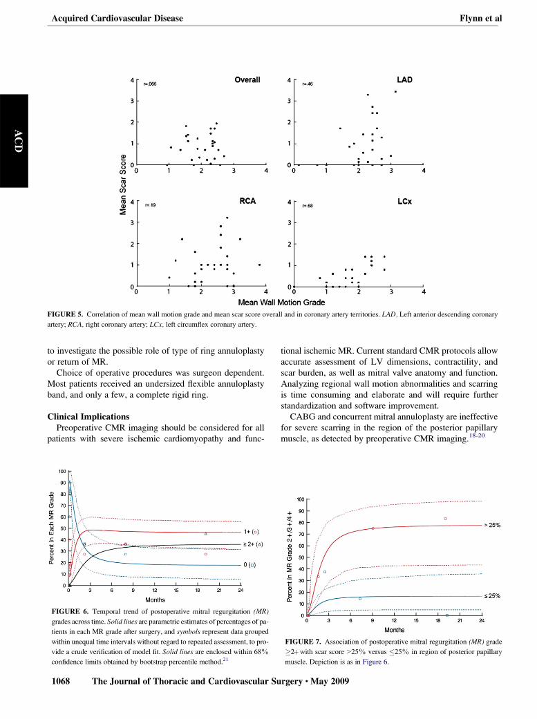
If segmental wall motion was normal, generally no scarring
was detected, as shown for representative segments by bubble
plots (Figure 4). However, when segmental wall motion was
abnormal, extent of scarring was highly variable. This resulted
overall in low correlation between wall motion abnormality
and scar (r¼ .066, Figure 5); however, when examined within
14. Mihaljevic T, Lam BK, Rajeswaran J, Takagaki M, Lauer MS, Gillinov AM, et al.
Impact of mitral valve annuloplasty combined with revascularization in patients with
functional ischemic mitral regurgitation. J Am Coll Cardiol. 2007;49:2191-201.
15. Hundley WG, Morgan TM, Neagle CM, Hamilton CA, Rerkpattanapipat P,
Link KM. Magnetic resonance imaging determination of cardiac prognosis. Cir-
culation. 2002;106:2328-33.
16. Pohl T, Seiler C, Billinger M, Herren E, Wustmann K, Mehta H, et al. Frequency
distribution of collateral flow and factors influencing collateral channel develop-
ment. Functional collateral channel measurement in 450 patients with coronary
artery disease. J Am Coll Cardiol. 2001;38:1872-8.
17. Bolling SF, Pagani FD, Deeb GM, Bach DS. Intermediate-term outcome of mitral recon-
struction in cardiomyopathy. J Thorac Cardiovasc Surg. 1998;115:381-6; discussion 7-8.
18. Kron IL, Green GR, Cope JT. Surgical relocation of the posterior papillary muscle
in chronic ischemic mitral regurgitation. Ann Thorac Surg. 2002;74:600-1.
19. Fukamachi K, Popovic ZB, Inoue M, Doi K, Schenk S, Ootaki Y, et al. Changes in
mitral annular and left ventricular dimensions and left ventricular pressure-vol-
ume relations after off-pump treatment of mitral regurgitation with the Coapsys
device. Eur J Cardiothorac Surg. 2004;25:352-7.
20. Yoon DY, Smedira NG, Nowicki ER, Hoercher K Rajeswaran J, Blackstone EH,
et al. Decision-making in surgical management of ischemic cardiomyopathy
J Thorac Cardiovasc Surg. (in press)
21. Rajeswaran J, Blackstone EH. Interval estimation for individual categories in cu-
mulative logit models. Stat Med. 2007;26:4150-62.
DiscussionDr R. Dion (Genk, Belgium). Dr Flynn, I would like to congrat-
ulate you for the quality of your presentation, and the authors and
The Journal of Thoracic and C
friends from the Cleveland Clinic have to be commended for an
original pilot study trying to elucidate the relation between wall
motion abnormality and scarring and return of MR. CMR was
used, which is certainly very elaborate and time-consuming; there-
fore, one should not underestimate the task of applying CMR and
analyzing it in 29 patients.
The main finding of this study is that extensive scarring, and se-
vere wall motion abnormality to a lesser extent because it is less sig-
nificant in the region of the posterior papillary muscle, correlates
with the return of MR.
My first question concerns the preoperative myocardial infarc-
tion. All 29 patients had a history of myocardial infarction. Could
you specify in which coronary territory? Was it mainly in the region
of the right coronary and the circumflex, as expected, and did it al-
ways correlate with the site of scarring on CMR?
Dr Flynn. Thank you for your kind comments and your good
questions. First, the most predominant area of infarction was in-
ferior, in the right coronary territory. We did not investigate
whether the degree of CMR-assessed scarring correlated with
the presence of myocardial ischemia. We do not have evidence
on that.
Dr Dion. I ask because the LAD mean wall motion was grade
2.1. It was grade 2.3 for the RCA and the posterior papillary mus-
cle, but it was only grade 1.6 for the circumflex and the anterior
papillary muscle. So there was more wall motion abnormality in
the anterior part of the heart than in the territory of the circumflex.
There was also more scar in the LAD than in LCx and in the anterior
papillary muscle territory, which is a bit surprising for me, because
I would have expected more scarring in the territory of the circum-
flex than in the LAD.
However, in the Discussion you state ‘‘the posterolateral wall,
which is predominantly supplied by the LCx, has less collateral cir-
culation in patients with diffuse 3-system coronary artery disease,
making it more prone to scarring, with resulting high correlation be-
tween amount of scarring and regional wall motion abnormalities.’’
But again, the mean scar score is less than 2 in the circumflex ter-
ritory. Can you elaborate on that?
Dr Flynn. Indeed. We feel that the most relevant territory is
possibly that of the right coronary, and to relate this to scarring
or dysfunction in the posterior papillary and the fact that it may
be supplied by 2 territories, as you are aware: if there is disease
or coronary disease related to both of those territories, these are
the patients who are at risk and who possibly have a greater
degree of scarring in the posterior papillary region. And these
are possibly the patients then, as we have demonstrated here,
who have a higher rate of recurrence of ischemic MR after
surgery.
Dr Dion. Thank you. Forty-four percent of the patients had
a scar of more than 25% in the posterior papillary muscle, but
the posterior papillary was attributed 2 segments and the anterior
papillary muscle, only 1 segment. Could it have influenced the
results? Why did you decide that the anterior papillary muscle
would be 1 segment and the posterior papillary muscle 2 seg-
ments?Dr Flynn. Well, we know from science and nature that the pos-
terior papillary is related to 2 coronary territories. We feel that dis-
ease involving both of those coronary territories makes the
posterior papillary more at risk for infarction and a greater determi-
nant of dysfunction.
ardiovascular Surgery c Volume 137, Number 5 1069
Acquired Cardiovascular Disease Flynn et al
AC
D
Dr Dion. Thank you. I am surprised by the relatively high rate of
recurrent MR at 6 months. It is probably explained by the fact that
the choice of the procedure was left at the discretion of the surgeon,
but, on the other hand, it allowed your group to perform this study
with ‘‘only’’ 29 patients. What do you think about the temporal pat-
tern of postoperative MR in 2 phases? I found it very interesting and
very intriguing. Why such an increase in the first 3 months and after
that, stabilization? How do you interpret that?
Dr Flynn. As you are aware, this represents a valvular approach
to a ventricular problem, and this may reflect the fact that annulo-
plasty in these patients, particularly those who got scarring in
that papillary region, may not be the appropriate approach for these
patients. I think to study the overall temporal occurrence of MR was
not within the remit of this study. It wasn’t designed for that pur-
pose; it wasn’t powered for that purpose. So we have not studied
or assessed the temporal degeneration of MR.
Dr. Dion. And finally, I found your conclusion quite severe for
restrictive mitral annuloplasty. You state, ‘‘CABG and concurrent
mitral annuloplasty are ineffective for severe scarring in the region
of the posterior papillary muscle.’’ But even in your setup, 30% of
the patients with severe scarring of the posterior papillary muscle
had no recurrent MR, and 15% with little scarring in the posterior
papillary muscle had recurrent MR. Don’t you think that, besides
the scarring and the wall motion abnormality, you should have
taken into account the LV dilatation? Maybe there is a relation be-
tween the extent of scarring and the LV dilatation, which might ex-
plain that without scarring and with LV dilatation you could have
recurrent MR.
Dr Flynn. I think your comments are well received. I think these
are different ways of looking at very sick patients. These are differ-
ent means of looking at severe LV dysfunction. Yes, indeed, LV
end-diastolic diameter is 1 parameter that can be used. Our mean
LV end-diastolic diameter was 62 mm, but we were unable to study
whether that was a determinant of recurrence of MR or not. You
will remember that our LV ejection fraction mean was 22%.
They were all under 30%. So I think this is perhaps a different
means of looking at a very sick patient group.
Dr Dion. Sure. But if you use CMR, it would probably be inter-
esting as well to also look also at the dimensions: obviously you
plan to extend this type of segmental analysis with CMR, which
is a very time-consuming and elaborate task. And if you were
able to link some of your segmental analysis to global LV dimen-
sion, which is easier to measure, it might simplify your work.
Again, I appreciate very much to review this paper and I congrat-
ulate you for an excellent presentation. I thank the Society for the
privilege of discussing it.
Dr Flynn. Thank you, Professor Dion. Thank you very much.
1070 The Journal of Thoracic and Cardiovascular S
Dr D. Adams (New York). Michael, that was an elegant study
and it is important, and it actually correlates with some of your
previous work from your institution about the importance of via-
ble myocardium in predicting a good result after ischemic repair.
So it is logical that a scar burden would also predict failure, and I
think that is important, particularly in your subgroup, which I
would emphasize for the audience had a very low ejection frac-
tion. So these are difficult patients to make decisions about
whether to operate and what to do. This spectrum of ischemic pa-
tients is very difficult.
My question relates to your specific ring strategy, and I think to
understand any results in restrictive disease, we don’t have data, we
don’t have randomized trials, but I just want to understand, were
they downsized, were they rigid, were they complete, and did
your ring strategy evolve over time? This is a relatively current
study, end point 2001 to 2006, and did that make a difference?
Did you see any patterns you can share with us?
Dr Flynn. First, thank you for your comments and your very
good questions. This was a very small study group. There were 7
patients who had complete rings, 22 who had partial rings. Again,
this study was not designed to compare one ring to another. We
found no difference in the rate of recurrence of MR between the
7 patients with a complete versus the 22 who had a partial ring.
We were unable to assess that.
Your second question regarding the method of downsizing, that
was surgeon-specific. There are different methods used at our insti-
tution depending upon the surgeon.
Dr L. Cohn (Boston, Mass). Excellent data presented beauti-
fully. This is similar to what Dr Dor has been advocating for
some time. Magnetic resonance imaging is something that cardiac
surgeons should really adopt, and I suspect that you agree with
that.
Any suggestions based on these data for more effective surgical
therapy? Based on what you have presented to us, have you and
your colleagues at the Cleveland Clinic decided on a more effective
or more strategic way to treat these patients? And what are your cur-
rent, shall we say, thoughts on recent surgical therapy about this
now, based on the data that you presented to us?
Dr Flynn. Dr Cohn, thank you very much for your kind com-
ments. I think this is a very difficult area and a very difficult patient
group. There are various thoughts and theories. I think the thought
of replacing the mitral valve in this patient group with a tissue valve
is one concept, then ventricular restoration is another possibility.
Again, it depends on the severity of LV dysfunction as to how
one would address the ventricular problems. There are other op-
tions. None of them are ideal. As you are aware, it is a very difficult
problem as to how to approach this.
urgery c May 2009
Flynn et al Acquired Cardiovascular Disease
AC
D
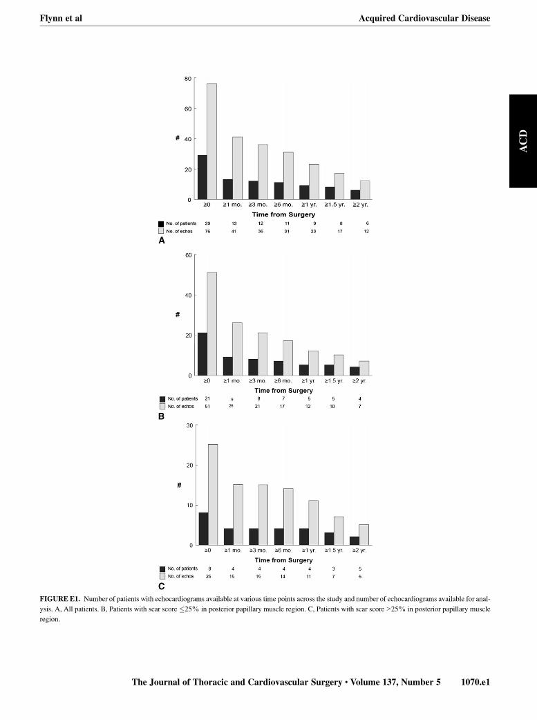
FIGURE E1. Number of patients with echocardiograms available at various time points across the study and number of echocardiograms available for anal-
ysis. A, All patients. B, Patients with scar score �25% in posterior papillary muscle region. C, Patients with scar score>25% in posterior papillary muscle
region.
The Journal of Thoracic and Cardiovascular Surgery c Volume 137, Number 5 1070.e1
Acquired Cardiovascular Disease Flynn et al
AC
D
FIGURE E2. Decomposition of temporal pattern of postoperative mitral regurgitation (MR) in the odds domain.
1070.e2 The Journal of Thoracic and Cardiovascular Surgery c May 2009