Regulation and drug modulation of a voltage-gated sodium channel: Pivotal role of the S4–S5 linker in activation and slow inactivation Jinglei Xiao a,b , Vasyl Bondarenko a , Yali Wang a , Antonio Suma c , Marta Wells a , Qiang Chen a , Tommy Tillman a , Yan Luo b , Buwei Yu b , William P. Dailey d , Roderic Eckenhoff e , Pei Tang a,f,g , Vincenzo Carnevale c , Michael L. Klein c,1 , and Yan Xu a,g,h,i,1 a Department of Anesthesiology and Perioperative Medicine, School of Medicine, University of Pittsburgh, Pittsburgh, PA 15261; b Department of Anesthesiology, Ruijin Hospital, School of Medicine, Shanghai Jiaotong University, Shanghai 200025, China; c Institute for Computational Molecular Science, College of Science and Technology, Temple University, Philadelphia, PA 19122; d Department of Chemistry, University of Pennsylvania, Philadelphia, PA 19104; e Department of Anesthesiology and Critical Care, Perelman School of Medicine, University of Pennsylvania, Philadelphia, PA 19104; f Department of Computational and Systems Biology, School of Medicine, University of Pittsburgh, Pittsburgh, PA 15261; g Department of Pharmacology and Chemical Biology, School of Medicine, University of Pittsburgh, Pittsburgh, PA 15261; h Department of Structural Biology, School of Medicine, University of Pittsburgh, Pittsburgh, PA 15261; and i Department of Physics and Astronomy, University of Pittsburgh, Pittsburgh, PA 15261 Contributed by Michael L Klein, June 1, 2021 (sent for review February 4, 2021; reviewed by Edward J. Bertaccini and Erik Lindahl) Voltage-gated sodium (Na V ) channels control excitable cell func- tions. While structural investigations have revealed conformation details of different functional states, the mechanisms of both activa- tion and slow inactivation remain unclear. Here, we identify residue T140 in the S4–S5 linker of the bacterial voltage-gated sodium chan- nel NaChBac as critical for channel activation and drug effects on inactivation. Mutations at T140 either attenuate activation or render the channel nonfunctional. Propofol, a clinical anesthetic known to inhibit NaChBac by promoting slow inactivation, binds to a pocket between the S4–S5 linker and S6 helix in a conformation-dependent manner. Using 19 F-NMR to quantify site-specific binding by satura- tion transfer differences (STDs), we found strong STDs in inactivated, but not activated, NaChBac. Molecular dynamics simulations show a highly dynamic pocket in the activated conformation, limiting STD buildup. In contrast, drug binding to this pocket promotes and stabi- lizes the inactivated states. Our results provide direct experimental evidence showing distinctly different associations between the S4–S5 linker and S6 helix in activated and inactivated states. Specifically, an exchange occurs between interaction partners T140 and N234 of the same subunit in activation, and T140 and N225 of the domain- swapped subunit in slow inactivation. The drug action on slow inactivation of prokaryotic Na V channels seems to have a mecha- nism similar to the recently proposed “door-wedge” action of the isoleucine-phenylalanine-methionine (IFM) motif on the fast inac- tivation of eukaryotic Na V channels. Elucidating this gating mech- anism points to a possible direction for conformation-dependent drug development. general anesthesia | propofol | NaChBac | NMR | saturation transfer difference V oltage-gated sodium (Na V ) channels control the initiation and propagation of the action potentials in excitable cells, includ- ing most neurons, myocytes, and endocrine cells (1). They convert chemical to electrical signaling by responding to cell membrane depolarization and are responsible for maintaining the normal physiological functions of vital organs such as the heart and brain. Naturally occurring mutations in Na V channels are often associated with pathophysiological conditions (for a recent review, see ref. 2). Dysfunction of Na V channels is related to various diseases, includ- ing epilepsy (3), cardiac arrhythmia (4), myotonias (5), and chronic pain syndromes (6). Understanding how Na V channels function will help uncover their physiological and pathophysiological roles and at the same time facilitate the development of pharmaceuticals to target these channel proteins. The functional cycle of Na V channels involves transitions from resting to activated states by opening a conducting passage for Na + ions to flow into the cells upon membrane depolarization. During prolonged depolarizations, Na V channels undergo fast and slow conformational changes known as inactivation to close the activa- tion gate, leading to functional states that cannot be reactivated or conduct ions (7, 8). Upon membrane repolarization, the channels transition back to the fully closed resting state in a process known as deactivation (9). In eukaryotic cells, the gating process is achieved by a pore-forming α-subunit, which is a single polypeptide chain folded into four homologous but nonidentical repeats in a pseudo- symmetric arrangement. Each repeat is composed of four trans- membrane helices (S1 to S4) as the voltage-sensing module (VSM) and two additional transmembrane helices (S5 and S6) as the pore module (PM). The PMs from the four repeats aggregate around a vestibule having a funnel-shaped ion selectivity filter (SF) near the extracellular entrance, a large water-filled cavity in the middle of the membrane, and an activation gate near the intracellular exit. Significance Voltage-gated sodium channels initiate electric signals in cell communications. The S4–S5 linker between the voltage-sensing and pore modules transmits depolarization signals to trigger channel activation. The mechanisms of this action, however, re- main elusive. By combining biophysical and computational ap- proaches, we identify a critical residue, T140, in the S4–S5 linker of the bacterial sodium channel NaChBac, which plays a pivotal role in channel activation and drug modulation of slow inacti- vation. Specifically, we discovered conformation-dependent drug binding at this site and unveiled a toggling mode of action by T140, which switches interaction partners with different S6 residues to regulate channel activation and slow inactivation. These observations suggest the possibility of conformation- specific drugs targeting the gating machinery of voltage-gated ion channels. Author contributions: P.T., V.C., M.L.K., and Y.X. designed research; J.X., V.B., Y.W., A.S., M.W., Q.C., and T.T. performed research; Y.L., B.Y., W.P.D., R.E., P.T., M.L.K., and Y.X. contributed new reagents/analytic tools; J.X., V.B., A.S., M.W., P.T., V.C., and Y.X. analyzed data; and V.C. and Y.X. wrote the paper. Reviewers: E.J.B., Stanford University School of Medicine; and E.L., Stockholms Universitet. The authors declare no competing interest. This open access article is distributed under Creative Commons Attribution-NonCommercial- NoDerivatives License 4.0 (CC BY-NC-ND). 1 To whom correspondence may be addressed. Email: [email protected] or xu2@ pitt.edu. This article contains supporting information online at https://www.pnas.org/lookup/suppl/ doi:10.1073/pnas.2102285118/-/DCSupplemental. Published July 6, 2021. PNAS 2021 Vol. 118 No. 28 e2102285118 https://doi.org/10.1073/pnas.2102285118 | 1 of 9 BIOPHYSICS AND COMPUTATIONAL BIOLOGY Downloaded by guest on December 5, 2021
Transcript
Regulation and drug modulation of a voltage-gatedsodium channel: Pivotal role of the S4–S5 linker inactivation and slow inactivationJinglei Xiaoa,b
, Vasyl Bondarenkoa, Yali Wanga, Antonio Sumac, Marta Wellsa, Qiang Chena, Tommy Tillmana,
Yan Luob, Buwei Yub
, William P. Daileyd, Roderic Eckenhoffe, Pei Tanga,f,g, Vincenzo Carnevalec,Michael L. Kleinc,1, and Yan Xua,g,h,i,1
aDepartment of Anesthesiology and Perioperative Medicine, School of Medicine, University of Pittsburgh, Pittsburgh, PA 15261; bDepartment ofAnesthesiology, Ruijin Hospital, School of Medicine, Shanghai Jiaotong University, Shanghai 200025, China; cInstitute for Computational Molecular Science,College of Science and Technology, Temple University, Philadelphia, PA 19122; dDepartment of Chemistry, University of Pennsylvania, Philadelphia, PA19104; eDepartment of Anesthesiology and Critical Care, Perelman School of Medicine, University of Pennsylvania, Philadelphia, PA 19104; fDepartment ofComputational and Systems Biology, School of Medicine, University of Pittsburgh, Pittsburgh, PA 15261; gDepartment of Pharmacology and ChemicalBiology, School of Medicine, University of Pittsburgh, Pittsburgh, PA 15261; hDepartment of Structural Biology, School of Medicine, University of Pittsburgh,Pittsburgh, PA 15261; and iDepartment of Physics and Astronomy, University of Pittsburgh, Pittsburgh, PA 15261
Contributed by Michael L Klein, June 1, 2021 (sent for review February 4, 2021; reviewed by Edward J. Bertaccini and Erik Lindahl)
Voltage-gated sodium (NaV) channels control excitable cell func-tions. While structural investigations have revealed conformationdetails of different functional states, the mechanisms of both activa-tion and slow inactivation remain unclear. Here, we identify residueT140 in the S4–S5 linker of the bacterial voltage-gated sodium chan-nel NaChBac as critical for channel activation and drug effects oninactivation. Mutations at T140 either attenuate activation or renderthe channel nonfunctional. Propofol, a clinical anesthetic known toinhibit NaChBac by promoting slow inactivation, binds to a pocketbetween the S4–S5 linker and S6 helix in a conformation-dependentmanner. Using 19F-NMR to quantify site-specific binding by satura-tion transfer differences (STDs), we found strong STDs in inactivated,but not activated, NaChBac. Molecular dynamics simulations show ahighly dynamic pocket in the activated conformation, limiting STDbuildup. In contrast, drug binding to this pocket promotes and stabi-lizes the inactivated states. Our results provide direct experimentalevidence showing distinctly different associations between the S4–S5linker and S6 helix in activated and inactivated states. Specifically, anexchange occurs between interaction partners T140 and N234 ofthe same subunit in activation, and T140 and N225 of the domain-swapped subunit in slow inactivation. The drug action on slowinactivation of prokaryotic NaV channels seems to have a mecha-nism similar to the recently proposed “door-wedge” action of theisoleucine-phenylalanine-methionine (IFM) motif on the fast inac-tivation of eukaryotic NaV channels. Elucidating this gating mech-anism points to a possible direction for conformation-dependentdrug development.
Voltage-gated sodium (NaV) channels control the initiation andpropagation of the action potentials in excitable cells, includ-
ing most neurons, myocytes, and endocrine cells (1). They convertchemical to electrical signaling by responding to cell membranedepolarization and are responsible for maintaining the normalphysiological functions of vital organs such as the heart and brain.Naturally occurring mutations in NaV channels are often associatedwith pathophysiological conditions (for a recent review, see ref. 2).Dysfunction of NaV channels is related to various diseases, includ-ing epilepsy (3), cardiac arrhythmia (4), myotonias (5), and chronicpain syndromes (6). Understanding how NaV channels function willhelp uncover their physiological and pathophysiological roles and atthe same time facilitate the development of pharmaceuticals totarget these channel proteins.The functional cycle of NaV channels involves transitions from
resting to activated states by opening a conducting passage for Na+
ions to flow into the cells upon membrane depolarization. Duringprolonged depolarizations, NaV channels undergo fast and slowconformational changes known as inactivation to close the activa-tion gate, leading to functional states that cannot be reactivated orconduct ions (7, 8). Upon membrane repolarization, the channelstransition back to the fully closed resting state in a process knownas deactivation (9). In eukaryotic cells, the gating process is achievedby a pore-forming α-subunit, which is a single polypeptide chainfolded into four homologous but nonidentical repeats in a pseudo-symmetric arrangement. Each repeat is composed of four trans-membrane helices (S1 to S4) as the voltage-sensing module (VSM)and two additional transmembrane helices (S5 and S6) as the poremodule (PM). The PMs from the four repeats aggregate around avestibule having a funnel-shaped ion selectivity filter (SF) near theextracellular entrance, a large water-filled cavity in the middle ofthe membrane, and an activation gate near the intracellular exit.
Significance
Voltage-gated sodium channels initiate electric signals in cellcommunications. The S4–S5 linker between the voltage-sensingand pore modules transmits depolarization signals to triggerchannel activation. The mechanisms of this action, however, re-main elusive. By combining biophysical and computational ap-proaches, we identify a critical residue, T140, in the S4–S5 linkerof the bacterial sodium channel NaChBac, which plays a pivotalrole in channel activation and drug modulation of slow inacti-vation. Specifically, we discovered conformation-dependent drugbinding at this site and unveiled a toggling mode of action byT140, which switches interaction partners with different S6residues to regulate channel activation and slow inactivation.These observations suggest the possibility of conformation-specific drugs targeting the gating machinery of voltage-gatedion channels.
Author contributions: P.T., V.C., M.L.K., and Y.X. designed research; J.X., V.B., Y.W., A.S.,M.W., Q.C., and T.T. performed research; Y.L., B.Y., W.P.D., R.E., P.T., M.L.K., and Y.X.contributed new reagents/analytic tools; J.X., V.B., A.S., M.W., P.T., V.C., and Y.X. analyzeddata; and V.C. and Y.X. wrote the paper.
Reviewers: E.J.B., Stanford University School of Medicine; and E.L., Stockholms Universitet.
The authors declare no competing interest.
This open access article is distributed under Creative Commons Attribution-NonCommercial-NoDerivatives License 4.0 (CC BY-NC-ND).1To whom correspondence may be addressed. Email: [email protected] or [email protected].
This article contains supporting information online at https://www.pnas.org/lookup/suppl/doi:10.1073/pnas.2102285118/-/DCSupplemental.
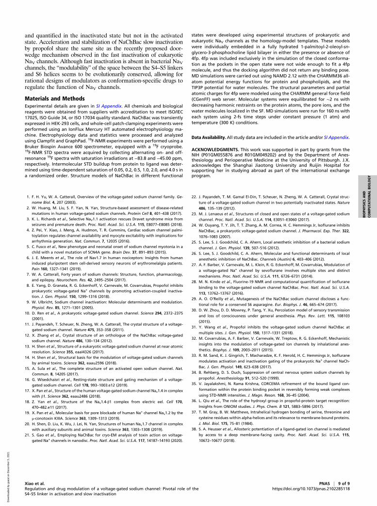
Prokaryotic NaV channels, as exemplified by the ancestral bacterialvoltage-gated sodium channel NaChBac from Bacillus halodurans(10), are composed of four smaller and often identical subunits,each contributing a VSM and PM along with the SF to a tetramericchannel (Fig. 1). In both prokaryotic and eukaryotic NaV channels,each of the VSMs and PMs is connected by a helical segmentcalled the S4–S5 linker (7).Significant advances have been made in recent years in the struc-
tural characterization of prokaryotic and eukaryotic NaV channels(11–21). The first successful crystal structure of a NaV channel atatomic resolution was that from the bacteria Arcobacter butzleri(NavAb) (11), which was captured with the VSM in the activatedconformation and the PM in a nonconducting and presumably pre-open or inactivated conformation. Subsequently, high-resolutionstructures of NavAb (22) and of NavRh from Rickettsiales sp.
HIMB114 (12), which is an ortholog of NaChBac, were solved inputatively inactivated states. Major breakthroughs in cryo-electronmicroscopy (cryo-EM) have led to several high-resolution NaVchannel structures, including NavPaS from American cockroaches(Periplaneta americana) in a nonconducting conformation (13), aswell as NaChBac (21), Nav1.2 (19), and Nav1.7 (20) structures inpresumably inactivated states. Because no crystallization or cryo-EM experiments to date have been attempted at nonzero membranepotentials and the experimental time is often longer than thetimescale of inactivation, capturing NaV channels in either the trueresting or open-channel conformations is technically challenging. Atruncated NavAb structure (23) and a full-length NavMs (fromMagnetococcus marinus) structure (15) were reported to be in pos-sible open conformations. Two cryo-EM structures of Nav1.4 fromhuman and electric eel (17, 18) were captured with the activation
A
B
C
D
E
F
Res
ting
Ope
n (a
ctiv
ated
)C
lose
d (in
activ
ated
)
T140
N225
N234
T140
N225
T140
N225
7Å
3Å
3Å
S1
S2
S3
S4
S5
S6
S1
S2
S3
S4
S5
S6
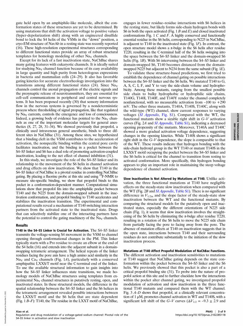
Fig. 1. Structural models of NaChBac showing pivotal position of T140 in the S4–S5 linker relative to S6 and activation gate. (A) Sequence alignment of S4–S5linker among NaV and CaV channels. T140 of NaChBac belongs to the LXXS/T motif (green box) at the C-terminal end of the linker, where possible interactionswith the S6 helix occur. Overview of aligned (B) open (activated) and (C) closed (inactivated) structural models showing different orientations of S6 helices(orange) between S4–S5 linkers (purple) from the same subunits and the S6 helix of the adjacent subunits. Cytosolic views of NaChBac in (D) resting, (E) open,and (F) closed conformations showing the orientations of the S4–S5 linkers (purple) and S6 helices (orange) in the tetrameric channel. Critical residues T140(green), N225 (red), and N234 (blue) and their approximate distances are highlighted in blue. Note the hydrogen bonding between the side chains of T140and N225 in the inactivated state.
2 of 9 | PNAS Xiao et al.https://doi.org/10.1073/pnas.2102285118 Regulation and drug modulation of a voltage-gated sodium channel: Pivotal role of the
gate held open by an amphiphile-like molecule, albeit the con-formation states of these structures are yet to be determined. Byusing mutations that shift the activation voltage to positive values(hyper-depolarization shift) along with an engineered disulfidebond to lock the S4 helix of the VSMs in the “down” position, acryo-EM structure of NavAb in a resting state was recently reported(16). These high-resolution experimental structures correspondingto different functional states provide an array of robust structuraltemplates for homology modeling of other NaV channels.Except for its lack of a fast inactivation state, NaChBac shares
many gating features with eukaryotic channels. It is ideally suitedfor studying NaV channel function because of its easy availabilityin large quantity and high purity from heterologous expressionsin bacteria and mammalian cells (24–29). It also has favorablegating kinetics for accurate electrorheology investigation into thetransitions among different functional states (24). Since NaVchannels control the axonal propagation of the electric signals andthe presynaptic release of neurotransmitters, they are essential forcell–cell communications in peripheral and central nervous sys-tems. It has been proposed recently (30) that sensory informationflow in the nervous systems is governed by a nondeterministicprocess where thresholding of signal propagation, like that regulatedby NaV currents, controls the emergence and loss of consciousness.Indeed, a growing body of evidence has pointed to the NaV chan-nels as one of the important molecular targets for general anes-thetics (8, 27, 28, 31–34). We showed previously that propofol, aclinically used intravenous general anesthetic, binds to three dif-ferent sites in NaChBac (31). Among these sites, we hypothesizedthat a binding cleft in the VSM contributes to the acceleration ofactivation, the nonspecific binding within the central pore cavityfacilitates inactivation, and the binding in a pocket between theS4–S5 linker and S6 has a dual role of promoting activation-coupledinactivation, leading to the net effect of channel inhibition (31).In this study, we investigate the role of the S4–S5 linker and its
relationship to the movement of the S6 helix in channel activationand drug effects on slow inactivation. We show that T140 in theS4–S5 linker of NaChBac is a pivotal residue in controlling NaChBacgating. By placing a fluorine probe at this site and using 19F-NMR tomeasure site-specific binding, we show that propofol binds to thispocket in a conformation-dependent manner. Computational simu-lations show that propofol fits into the amphiphilic pocket betweenT140 and the N225 from the domain-swapped subunit in the inac-tivation conformation, and propofol binding to T140 accelerates andstabilizes the inactivation transition. The experimental and com-putational results reveal a mechanism of T140 switching interactionpartners from the activated state to the inactivated state. Drugsthat can selectively stabilize one of the interacting partners havethe potential to control the gating machinery of the NaV channels.
ResultsT140 in the S4–S5 Linker Is Crucial for Activation. The S4–S5 linkertransmits the voltage-sensing S4 movement in the VSM to channelopening through conformational changes in the PM. This linkertypically starts with a Pro residue to create an elbow at the end ofthe S4 helix (16) and extends into the adjacent subunit in a domain-swapping tetrameric arrangement. The helical repeats of the linkerresidues facing the pore axis have a high amino acid similarity in theNaV and CaV channels (Fig. 1A), particularly with a conservedamphipathic LXXS/T motif near the C-terminal end of the linker.To use the available structural information to gain insights intohow the S4–S5 linker influences state transitions, we made ho-mology models of NaChBac structures using templates from ex-perimental NaV channel structures in putatively resting, open, andinactivated states. In these structural models, the difference in thespatial relationship between the S4–S5 linker and the S6 helices inthe tetrameric arrangement revealed possible interactions betweenthe LXXS/T motif and the S6 helix that are state dependent(Fig. 1 B–F): T140, the Thr residue in the LXXTmotif of NaChBac,
engages in fewer residue–residue interactions with S6 helices inthe resting state, but likely forms side-chain hydrogen bonds withS6 in both the open activated (Fig. 1 B and E) and closed inactivatedconformations Fig. 1 C and F. A highly conserved and functionallyessential residue in the S6 helix, corresponding to N225 in NaChBac,is adjacent to T140 in the inactivated state (Fig. 1F). In contrast, theopen structure model shows a π-bulge in the S6 helix after residueT220, resulting in the C-terminal half of the S6 helix swinging intothe space between the S4–S5 linker and the domain-swapped S6helix (Fig. 1B). With S6 intervening between the S4–S5 linker anddomain-swapped S6, T140 becomes distanced from the domain-swapped N225 but adjacent to N234 from the same subunit (Fig. 1E).To validate these structure-based predictions, we first tried to
establish the dependence of channel gating on possible interactionsbetween the S4–S5 linker and the S6 helix. We mutated T140 to G,A, S, C, I, F, and Y to vary the side-chain volume and hydropho-bicity. Among these mutants, ranging from the smallest possibleside chain to bulky hydrophobic or hydrophilic side chains,T140G, T140I, T140F, and T140Y render the channel completelynonfunctional, with no measurable activation from –100 to +290mV. The other three mutants, T140A, T140S, T140C, along withthe wild-type (WT) channel, show current responses to varyingvoltages (SI Appendix, Fig. S1). Compared with the WT, thefunctional mutants show a sizable right shift in G–V activationcurves (Fig. 2A and SI Appendix, Table S1), indicating a decreasedopening propensity of the mutants. Moreover, T140A and T140Cshowed a more gradual activation voltage dependence, suggestingchanges in the opening kinetics. While T140S shows a significantright shift in the G–V dependence, the slope is comparable to thatof the WT. These results indicate that hydrogen bonding with theside-chain hydroxyl group in the WT T140 or mutant T140S in theLXXS/T motif occupying the pocket between the S4–S5 linker andthe S6 helix is critical for the channel to transition from resting toactivated conformation. More specifically, this hydrogen bondingappears to play an important role in maintaining the steep voltagedependence of channel activation.
Slow Inactivation Is Not Altered by Mutations at T140. Unlike acti-vation, the three functional mutations at T140 have negligibleeffects on the steady-state slow inactivation when compared withtheWT (Fig. 2B and SI Appendix, Table S1). There is no significantdifference in V1/2in and the slope factor of the steady-state slowinactivation between the WT and the functional mutants. Bycomparing the structural models for the putatively open and inac-tivated states, especially the different orientations of N225 sidechain (Fig. 1), it seems that slow inactivation involves the straight-ening of the S6 helix by eliminating the π-bulge after residue T220,resulting in a rotation of the S6 helix to move the N225 side chainfrom partially facing the pore to facing away from the pore. Theabsence of mutation effects at T140 on inactivation suggests that inthe open state, interactions between T140 and their surroundingresidues do not contribute substantially to the initiation of the slowinactivation process.
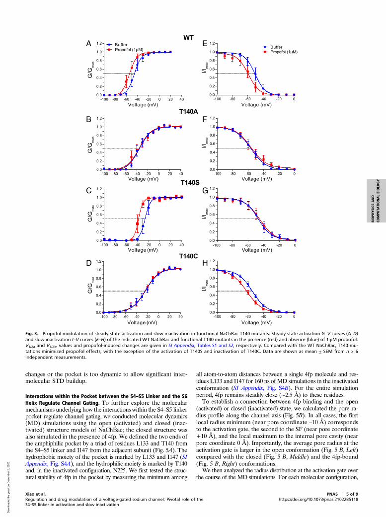
Mutations at T140 Affect Propofol Modulation of NaChBac Functions.The different activation and inactivation sensitivities to mutationsat T140 suggest that NaChBac gating depends on the state con-formation within the pocket between the S4–S5 linker and the S6helix. We previously showed that this pocket is also a part of acritical propofol binding site (31). To probe into the nature of pro-pofol action at this site and to further elucidate how the interactionswithin the pocket alter channel gating, we investigated propofolmodulation of activation and slow inactivation in the three func-tional T140 mutants and compared them with the WT channel.Fig. 3 A–D shows that propofol, at a clinically relevant concentra-tion of 1 μM, promotes channel activation in WT and T140S, with asignificant left shift of the G–V curves (ΔV1/2 = –9.3 ± 2.9 and
Xiao et al. PNAS | 3 of 9Regulation and drug modulation of a voltage-gated sodium channel: Pivotal role of theS4–S5 linker in activation and slow inactivation
–10.0 ± 2.3 mV, respectively; SI Appendix, Table S2). In contrast,the same concentration of propofol produces negligible or smallchanges in the activation of T140A (–0.74 ± 1.6 mV) and T140C(–2.4 ± 0.8 mV). Both Thr and Ser have side chain hydroxylgroups, which appears to be necessary in mediating propofol effectson activation. It should be noted, however, that having a side chainhydroxyl group is not a sufficient condition for the observed propofoleffects: The T140Y mutation, with a bulky hydroxyl-containing hy-drophilic side chain, renders the channel nonfunctional, irrespectiveof the presence or absence of propofol.Propofol effects on steady-state inactivation show a different
pattern: 1 μM propofol promotes steady-state inactivation in WTand T140C but has negligible effects on inactivation of T140A andT140S (Fig. 3 E–H and SI Appendix, Table S2). The removal ofpropofol effects on both activation and slow inactivation by theT140A mutation, which has a smaller side-chain volume than res-idues in the other three functional channels, suggest that 1) stericfitting of propofol in the amphipathic pocket, which would becompromised by this mutation, dominates the observed propofol
actions on NaChBac; and 2) propofol binding at two other previ-ously identified sites [i.e., a cleft in VSM and the central pore (31)],which should be minimally affected by the T140A mutation, playsonly an auxiliary role in modulating NaChBac function. The con-trasting propofol effects on T140S and T140C, whose side chainsare sterically similar, suggest that the propofol’s modes of action onactivation and inactivation are different: Propofol promotes theresting-to-open transition in T140S but not in T140C, whereas itaccelerates the open-to-inactivation transition in T140C but notin T140S.
Propofol Binding to the S4–S5 Linker Is Conformation Dependent. Tounderstand the observed differences in the mutation- and propofol-induced changes in activation and slow inactivation, we furtherinvestigated the conformation-dependent propofol binding in thepocket between the S4–S5 linker and S6 helix by using an addi-tional T220A mutation in both the WT and T140C channels. TheT220A mutation has been shown to abolish slow inactivation inNaChBac (26), which we also confirmed for the double mutantT140C/T220A by electrophysiology (SI Appendix, Fig. S2). It isbelieved that replacing the pore-facing hydrophilic T220 with hy-drophobic Ala residue in the S6 helix stabilizes the π-bulge at F221,prepositioning the S6 helix to assume an activated conformation.This is evidenced by the large left shifts of the activation V1/2 ofT220A relative to the WT (–64.7 vs. –45.7 mV; SI Appendix, TableS1) and of T140C/T220A relative to T140C (–34.6 vs. –25.8 mV).To measure the site-specific propofol binding, we covalently
attached a small, fluorinated probe, 3-bromo-1,1,1-trifluoroacetone(BTFA), to the side chain of T140C in both the T140C and T140C/T220A mutants and measured the intermolecular 19F-NMR satu-ration transfer difference (STD) spectra of 4-fluoro-propofol (4fp),a fluorinated propofol. Intermolecular STD quantifies the cross-relaxation between two nuclei that are in a close and stable contact[<7 Å (35)], hence providing a direct measure of site-specificbinding. We previously showed that 4fp and propofol have nearlyidentical anesthetizing potencies in tadpoles and have similar ef-fects on the activation and slow inactivation of the WT NaChBac(31). In this study, we also confirmed that effects of 4fp and propofolon the T140C mutant were not statistically different (SI Appendix,Fig. S3), validating the use of 4fp as an excellent surrogate forpropofol. Since the NMR experiments were conducted in the ab-sence of a cross-membrane potential (i.e., depolarized), we expectthat the T140C mutant was in an inactivated state under NMRexperimental conditions, whereas the double mutant T140C/T220A,due to mutation-induced abolition of inactivation, was in a pre-openor an activated state. In our experiments, the 19F-labeling efficiencywas 10–50%, which is sufficient for STDmeasurements as unlabeledprotein contributes no NMR signal. The measured cross-relaxationby STD between 4fp and the BTFA label was distinctly different inT140C and T140C/T220A (Fig. 4A). A strong saturation transfer wasobserved from BTFA to 4fp in T140C, with an STDmax of 18.1%and a relatively large 19F intermolecular cross relaxation rate (σ)of ∼0.31 s–1 (Fig. 4B and SI Appendix, Table S3). In the T140C/T220A double mutant, which is devoid of slow inactivation, thesaturation transfer between BTFA and 4fp was greatly dimin-ished, with STDmax at merely ∼2% and the cross-relaxation rateunmeasurable. To confirm that this conformation dependence isspecific to T140, we labeled the nonspecific propofol binding sitein the central cavity at F227C and used the F227C/T220A doublemutant as the corresponding control. The cross-relaxations betweenBTFA and 4fp in F227C and F227C/T220A are essentially identical(Figs. 4 C and D and SI Appendix, Table S3). We conclude that thespecific propofol binding pocket between the S4–S5 linker and S6helix near T140 is conformation dependent: Stable propofol binding,which ensures intermolecular STD buildup, occurs in only theinactivated state, whereas in a channel devoid of slow inactiva-tion, the binding pocket is either removed due to conformation
-100 -80 -60 -40 -20 0 20 400.0
0.2
0.4
0.6
0.8
1.0
1.2
Voltage(mV)
G/G
max
T140S
T140C
WTT140A
-120 -100 -80 -60 -40 -20 00.0
0.2
0.4
0.6
0.8
1.0
1.2
Voltage(mV)
I/IA
B
max
T140S
T140C
WTT140A
Fig. 2. Steady-state activation and slow inactivation in functional NaChBacT140 mutants. (A) The steady-state activation G–V curves of the T140S,T140A, and T140C mutants show right shifts relative to that of the WT. Notea significant decrease in the slopes of the T140A and T140C curves, indicatinga gradual voltage dependence of activation in these mutants. (B) Steady-state inactivation I–V curves of the same T140 mutants compared with theWT. Only T140S shows a slight right shift in V1/2in, but this change is notstatistically significant. The side-chain conformation of T140 is critical for thetransition from the resting to activation state but not for the initiation ofslow inactivation. Data are shown as mean ± SEM from n = 8–10 indepen-dent measurements. The solid lines are best fit to the data using SI Appen-dix, Eq. S2 or Eq. S3, with the best fitting parameters listed in SI Appendix,Table S1.
4 of 9 | PNAS Xiao et al.https://doi.org/10.1073/pnas.2102285118 Regulation and drug modulation of a voltage-gated sodium channel: Pivotal role of the
changes or the pocket is too dynamic to allow significant inter-molecular STD buildup.
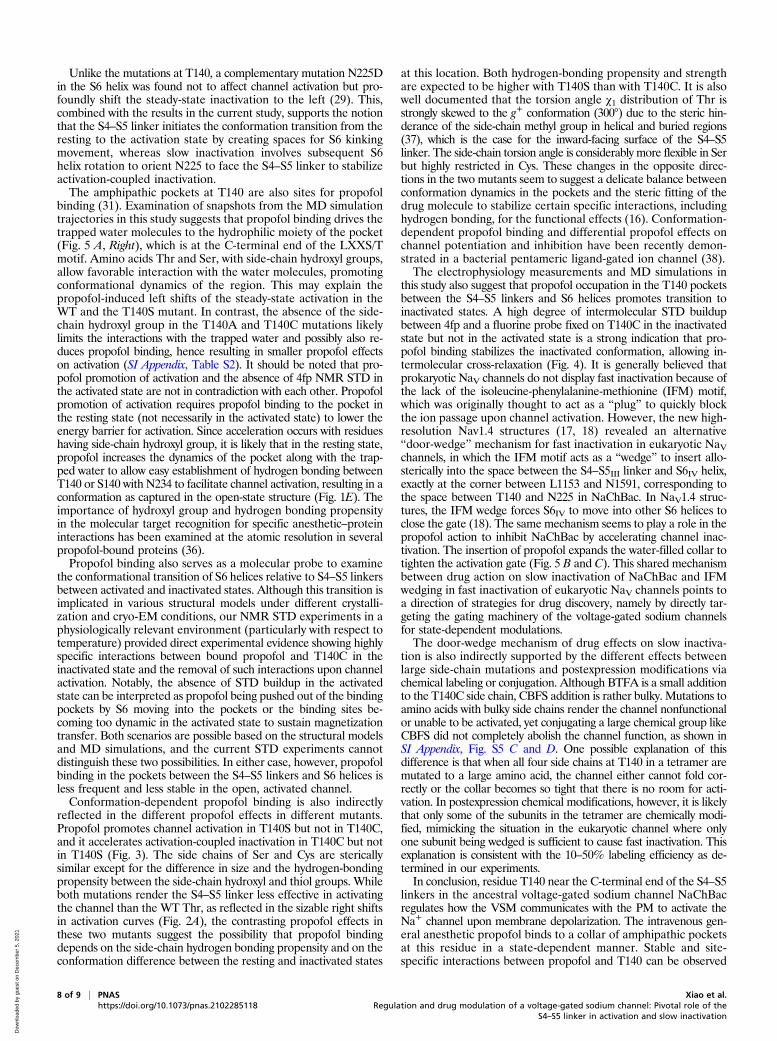
Interactions within the Pocket between the S4–S5 Linker and the S6Helix Regulate Channel Gating. To further explore the molecularmechanisms underlying how the interactions within the S4–S5 linkerpocket regulate channel gating, we conducted molecular dynamics(MD) simulations using the open (activated) and closed (inac-tivated) structure models of NaChBac; the closed structure wasalso simulated in the presence of 4fp. We defined the two ends ofthe amphiphilic pocket by a triad of residues L133 and T140 fromthe S4–S5 linker and I147 from the adjacent subunit (Fig. 5A). Thehydrophobic moiety of the pocket is marked by L133 and I147 (SIAppendix, Fig. S4A), and the hydrophilic moiety is marked by T140and, in the inactivated configuration, N225. We first tested the struc-tural stability of 4fp in the pocket by measuring the minimum among
all atom-to-atom distances between a single 4fp molecule and res-idues L133 and I147 for 160 ns of MD simulations in the inactivatedconformation (SI Appendix, Fig. S4B). For the entire simulationperiod, 4fp remains steadily close (∼2.5 Å) to these residues.To establish a connection between 4fp binding and the open
(activated) or closed (inactivated) state, we calculated the pore ra-dius profile along the channel axis (Fig. 5B). In all cases, the firstlocal radius minimum (near pore coordinate –10 Å) correspondsto the activation gate, the second to the SF (near pore coordinate+10 Å), and the local maximum to the internal pore cavity (nearpore coordinate 0 Å). Importantly, the average pore radius at theactivation gate is larger in the open conformation (Fig. 5 B, Left)compared with the closed (Fig. 5 B, Middle) and the 4fp-bound(Fig. 5 B, Right) conformations.We then analyzed the radius distribution at the activation gate over
the course of the MD simulations. For each molecular configuration,
BufferPropofol (1μM)
T140A
-100 -80 -60 -40 -20 0 20 400.0
0.2
0.4
0.6
0.8
1.0
1.2
Voltage (mV)
T140C
A
B
D
WT
-100 -80 -60 -40 -20 00.0
0.2
0.4
0.6
0.8
1.0
1.2
Voltage (mV)
E
-100 -80 -60 -40 -20 00.0
0.2
0.4
0.6
0.8
1.0
1.2
Voltage (mV)
F
T140S
-100 -80 -60 -40 -20 00.0
0.2
0.4
0.6
0.8
1.0
1.2
Voltage (mV)
G
-100 -80 -60 -40 -20 00.0
0.2
0.4
0.6
0.8
1.0
1.2
Voltage (mV)
H
-100 -80 -60 -40 -20 0 20 400.0
0.2
0.4
0.6
0.8
1.0
1.2
Voltage (mV)
Voltage (mV)
-100 -80 -60 -40 -20 0 20 400.0
0.2
0.4
0.6
0.8
1.0
1.2
Voltage (mV)
BufferPropofol (1μM)
G/G
max
G/G
max
G/G
max
G/G
max
C
I/Im
axI/I
max
I/Im
axI/I
max
-100 -80 -60 -40 -20 0 20 400.0
0.2
0.4
0.6
0.8
1.0
1.2
Fig. 3. Propofol modulation of steady-state activation and slow inactivation in functional NaChBac T140 mutants. Steady-state activation G–V curves (A–D)and slow inactivation I–V curves (E–H) of the indicated WT NaChBac and functional T140 mutants in the presence (red) and absence (blue) of 1 μM propofol.V1/2a and V1/2in values and propofol-induced changes are given in SI Appendix, Tables S1 and S2, respectively. Compared with the WT NaChBac, T140 mu-tations minimized propofol effects, with the exception of the activation of T140S and inactivation of T140C. Data are shown as mean ± SEM from n > 6independent measurements.
Xiao et al. PNAS | 5 of 9Regulation and drug modulation of a voltage-gated sodium channel: Pivotal role of theS4–S5 linker in activation and slow inactivation
we defined the radius of the activation gate as the spatial averageover the pore coordinates between –15 and –6 Å (Fig. 5B). Asexpected, the activated and inactivated configurations show arelatively large (∼2.5-Å) and small (∼1-Å) gate radius, respectively(Fig. 5C). Importantly, wider pores are hydrated and allow perme-ation of water molecules, while the narrow ones do not (Fig. 5D).Interestingly, the 4fp-bound conformation shows the same averagevalue of the gate as the closed channel but with significantly smallerfluctuations (a narrower distribution width). Hence, the probabilityof observing an instantaneous configuration with a wide pore isgreatly decreased in the 4fp-bound configurations. These resultsindicate that the binding of 4fp to the pocket between the S4–S5linker and the S6 helix “locks” the channel in the closed (inacti-vated) conformation by reducing fluctuations of the activation gate.We further set out to investigate the mechanistic link between
drug binding and pore radius. We noticed that, in the apo config-urations, the 4fp binding site is often occupied by water molecules(Fig. 5A). The number of occupying water molecules varies asthe binding pocket undergoes structural fluctuations. Since thefour binding pockets in the tetrameric NaChBac form a collar sur-rounding the pore near the activation gate, we hypothesized thatfluctuations that increase the binding pocket volume would tightenthe collar and decrease the pore radius at the activation gate, or vice
versa. To test this, we measured the number of water moleculeswithin 6 Å from the reference residues L133 and I147, which markthe distal end of the pocket from T140, for each frame in thesimulations. Fig. 5E shows the two-dimensional probability distri-butions as a function of the pore radius and the number of watermolecules in the pocket. In the open conformation (Fig. 5 E,Upper), a peak distribution of wider pore radii (∼2.5 Å) is as-sociated with fewer numbers of water molecules (∼15) in the pocket,whereas in the closed conformation (Fig. 5E, Lower), the probabilitydistribution is associated with a narrower gate radius of ∼ 1 Å and∼25 water molecules in the pocket. We interpret this anticorrelationbetween the radius and the number of water molecules in the pocketas supporting evidence that as the channel fluctuates toward theclosed (inactivated) conformation, the size of the pocket increases.Indeed, for the closed conformation (Fig. 5 E, Lower), only onepopulation is present in the probability distribution, with a gate ra-dius of ∼1 Å and ∼25 water molecules in the pocket.To experimentally verify that modifying the size of the binding
pocket can impact inactivation, we covalently conjugated8-(chloromercuri)-2-dibenzofuransulfonic acid (CBFS) to thecysteine thiol group to partially occupy the hydrophilic end of thepocket at T140C (Fig. 5 A, Right). In the Cys-free WT control, inwhich no conjugation occurred, CBFS produced negligible ef-fects on both activation and inactivation (SI Appendix, Fig. S5 Aand B), and propofol again shifted the activation and inactivationcurves in the hyperpolarization direction. In T140C, in whichCBFS occupied the hydrophilic moiety of the pocket, the activationcurve exhibits a slight left shift relative to the already right-shiftedT140C activation, similar to the minimal propofol effects observedin the unconjugated T140C mutant. Like propofol binding, theinactivation curve also left-shifted significantly due to covalentCBFS conjugation. Interestingly, because the pocket geometry ismodified by CBFS, a small modulation by propofol is now mea-surable in the activation of the CBFS-conjugated T140C mutant(SI Appendix, Fig. S5C). More importantly, the addition of propofolin the presence of CBFS can further stabilize the pocket, leading toan additional left shift of the steady-state inactivation (SI Appendix,Fig. S5D). The effects of propofol on V1/2a and V1/2in with andwithout CBFS conjugation are summarized in SI Appendix, Table S4.
DiscussionIn this study, we demonstrated the dependence of NaChBac gatingon residue T140 in the S4–S5 linker. Mutating this residue pro-foundly changed NaChBac’s steady-state activation, and severalamino acids with side chains too large or too small compared withThr, irrespective of hydrophobicity, rendered the channel non-functional. In mutations that retained channel function, the volt-age dependence of activation was shifted to the right relative tothe WT channel while slow inactivation was unchanged. By ana-lyzing the cryo-EM structure of NaChBac in an inactivated state(21) and homology models of NaChBac in the resting and puta-tively open states, it is clear that T140 is situated at a pivotal lo-cation where its side chains in the tetrameric channel pinch anamphipathic collar surrounding the crossing of the S6 helices atthe level of the activation gate. The collar consists of the inward-facing residues of the S4–S5 linker within four buried pockets ofthe tetramer, and these pockets create spaces between the S4–S5linkers and the adjacent S6 helices for iris-like S6 movement upongating (23). The outward movement of S4 helices in response tomembrane depolarization has been proposed to cause conforma-tional change in the S4–S5 linker to loosen the collar (16), allowingS6 movement. The MD simulations in this study show that thepockets are partially water-filled, and the number of trappedwater molecules is inversely correlated with the averaged radiusof the pore at the activation gate (Fig. 5E). While our simulationsdo not sample full transitions between the open and closed poreconformations and we could not directly observe an associationbetween opening/closing of the pore gate and wetting/dewetting of
A C
DB
Fig. 4. Conformation-dependent 4-fluoropropofol binding to T140. (A)Stack plots of 19F-NMR saturation transfer difference (STD) spectra of 4fp atthe indicated saturation times from a 19F probe labeled at T140C in aninactivated state and in the T140C/T220A double mutant devoid of slowinactivation. (B) Plot of normalized STD from A as a function of saturationtime. Sizable cross-relaxation between 4fp and T140 is quantifiable only inthe inactivated state and negligible in the double-mutant channel that isunable to inactivate. (C and D) Corresponding control experiments with a 19Fprobe labeled at the nonspecific 4fp binding site near F227C and theinactivation-disabled double mutant F227C/T220A. In B and D, solid lines arebest fit to the data using SI Appendix, Eq. S5. Error bars are derived from thesignal-to-noise ratios of the STD spectra. Fitting parameters are listed inSI Appendix, Table S3.
6 of 9 | PNAS Xiao et al.https://doi.org/10.1073/pnas.2102285118 Regulation and drug modulation of a voltage-gated sodium channel: Pivotal role of the
the pockets, it is conceivable that tightening or loosening of thecollar is coupled to the compression of the pockets. The hydro-philic side chain of T140, with its hydrogen-bonding propensity forthe trapped water molecules in the amphipathic pockets, plays acritical role both in facilitating the loosening of the collar to allowthe iris-like movement of S6 into the spaces and in establishingspecific interactions with S6 in a conformation-dependent manner.Specifically, the T140 side chain toggles between two possible
interaction partners in the S6 helices upon gating: In the activatedstate, T140 of each S4–S5 linker interacts with N234 from the S6helix of the same subunit, whereas in the inactivated states, T140releases N234 from the same subunit and establishes a hydrogenbond with N225 from the domain-swapped S6 helix. The contrastingmutation effects on activation and slow inactivation underscorethe conformational distinctions of the pockets in different functionalstates.
4-fluoropropofolOpen ClosedA
L133 L133 L133
I147 I147 I1474-fluoropropofol
Open
Closed
Narrowpore
Widepore
10
-20
20
30
Å
2.2 Å1.9 Å-10
0
Wide Narrow
B
C D E
5 10 15 20 25 30 350.5
1.0
1.5
2.0
2.5
3.0
Total number of waters in pockets
Por
e ra
dius
[Å]
4-fluoropropofolOpen Closed
T140 T140 T140
Fig. 5. Mechanistic association between the drug-binding pocket at T140 and channel activation state. (A) Representative snapshots from MD simulations ofNaChBac channels in open (activated, Left), closed (inactivated, Middle), and 4fp-bound (Right) states. The amphipathic drug-binding pocket is surrounded byL133 and T140 in the S4–S5 linker and L147 from the adjacent subunit. The pocket is water-filled in the closed and 4fp-bound states but has a greatly reducedvolume in the activated state. (B) The pore radius profiles, averaged from all MD simulation frames, along the channel axis in the open (Left), closed (Middle),and 4fp-bound (Right) structures. The activation gate is located from z = –15 Å to z = –6 Å. The lower bound corresponds to the pore entrance and is lined bythe four symmetry-related residues I231. At lower values of z, the pore lining helix S6 is solvent exposed and, in the open structure, partially unfolded andhighly fluctuating. (C) The radius distribution functions are calculated from each instantaneous channel configuration in the MD simulations and averagedfrom z = –15 Å to z = –6 Å for open (red), closed (green), and 4fp-bound (blue) structures. Note that 4fp binding locks the channel in a tightly closedconfiguration with a narrower radius distribution width than the apo activated and inactivated states. (D) Pore water profiles reveal that a wider pore (Left)allows passage of water, and the narrow pore (Right) does not. (E) Two-dimensional probability distribution of the number of water molecules in the T140pocket and the pore radius at the activation gate. The darker colors correspond to a higher probability. The red and blue tones refer to the closed and openstate simulations, respectively.
Xiao et al. PNAS | 7 of 9Regulation and drug modulation of a voltage-gated sodium channel: Pivotal role of theS4–S5 linker in activation and slow inactivation
Unlike the mutations at T140, a complementary mutation N225Din the S6 helix was found not to affect channel activation but pro-foundly shift the steady-state inactivation to the left (29). This,combined with the results in the current study, supports the notionthat the S4–S5 linker initiates the conformation transition from theresting to the activation state by creating spaces for S6 kinkingmovement, whereas slow inactivation involves subsequent S6helix rotation to orient N225 to face the S4–S5 linker to stabilizeactivation-coupled inactivation.The amphipathic pockets at T140 are also sites for propofol
binding (31). Examination of snapshots from the MD simulationtrajectories in this study suggests that propofol binding drives thetrapped water molecules to the hydrophilic moiety of the pocket(Fig. 5 A, Right), which is at the C-terminal end of the LXXS/Tmotif. Amino acids Thr and Ser, with side-chain hydroxyl groups,allow favorable interaction with the water molecules, promotingconformational dynamics of the region. This may explain thepropofol-induced left shifts of the steady-state activation in theWT and the T140S mutant. In contrast, the absence of the side-chain hydroxyl group in the T140A and T140C mutations likelylimits the interactions with the trapped water and possibly also re-duces propofol binding, hence resulting in smaller propofol effectson activation (SI Appendix, Table S2). It should be noted that pro-pofol promotion of activation and the absence of 4fp NMR STD inthe activated state are not in contradiction with each other. Propofolpromotion of activation requires propofol binding to the pocket inthe resting state (not necessarily in the activated state) to lower theenergy barrier for activation. Since acceleration occurs with residueshaving side-chain hydroxyl group, it is likely that in the resting state,propofol increases the dynamics of the pocket along with the trap-ped water to allow easy establishment of hydrogen bonding betweenT140 or S140 with N234 to facilitate channel activation, resulting in aconformation as captured in the open-state structure (Fig. 1E). Theimportance of hydroxyl group and hydrogen bonding propensityin the molecular target recognition for specific anesthetic–proteininteractions has been examined at the atomic resolution in severalpropofol-bound proteins (36).Propofol binding also serves as a molecular probe to examine
the conformational transition of S6 helices relative to S4–S5 linkersbetween activated and inactivated states. Although this transition isimplicated in various structural models under different crystalli-zation and cryo-EM conditions, our NMR STD experiments in aphysiologically relevant environment (particularly with respect totemperature) provided direct experimental evidence showing highlyspecific interactions between bound propofol and T140C in theinactivated state and the removal of such interactions upon channelactivation. Notably, the absence of STD buildup in the activatedstate can be interpreted as propofol being pushed out of the bindingpockets by S6 moving into the pockets or the binding sites be-coming too dynamic in the activated state to sustain magnetizationtransfer. Both scenarios are possible based on the structural modelsand MD simulations, and the current STD experiments cannotdistinguish these two possibilities. In either case, however, propofolbinding in the pockets between the S4–S5 linkers and S6 helices isless frequent and less stable in the open, activated channel.Conformation-dependent propofol binding is also indirectly
reflected in the different propofol effects in different mutants.Propofol promotes channel activation in T140S but not in T140C,and it accelerates activation-coupled inactivation in T140C but notin T140S (Fig. 3). The side chains of Ser and Cys are stericallysimilar except for the difference in size and the hydrogen-bondingpropensity between the side-chain hydroxyl and thiol groups. Whileboth mutations render the S4–S5 linker less effective in activatingthe channel than the WT Thr, as reflected in the sizable right shiftsin activation curves (Fig. 2A), the contrasting propofol effects inthese two mutants suggest the possibility that propofol bindingdepends on the side-chain hydrogen bonding propensity and on theconformation difference between the resting and inactivated states
at this location. Both hydrogen-bonding propensity and strengthare expected to be higher with T140S than with T140C. It is alsowell documented that the torsion angle χ1 distribution of Thr isstrongly skewed to the g+ conformation (300°) due to the steric hin-derance of the side-chain methyl group in helical and buried regions(37), which is the case for the inward-facing surface of the S4–S5linker. The side-chain torsion angle is considerably more flexible in Serbut highly restricted in Cys. These changes in the opposite direc-tions in the two mutants seem to suggest a delicate balance betweenconformation dynamics in the pockets and the steric fitting of thedrug molecule to stabilize certain specific interactions, includinghydrogen bonding, for the functional effects (16). Conformation-dependent propofol binding and differential propofol effects onchannel potentiation and inhibition have been recently demon-strated in a bacterial pentameric ligand-gated ion channel (38).The electrophysiology measurements and MD simulations in
this study also suggest that propofol occupation in the T140 pocketsbetween the S4–S5 linkers and S6 helices promotes transition toinactivated states. A high degree of intermolecular STD buildupbetween 4fp and a fluorine probe fixed on T140C in the inactivatedstate but not in the activated state is a strong indication that pro-pofol binding stabilizes the inactivated conformation, allowing in-termolecular cross-relaxation (Fig. 4). It is generally believed thatprokaryotic NaV channels do not display fast inactivation because ofthe lack of the isoleucine-phenylalanine-methionine (IFM) motif,which was originally thought to act as a “plug” to quickly blockthe ion passage upon channel activation. However, the new high-resolution Nav1.4 structures (17, 18) revealed an alternative“door-wedge” mechanism for fast inactivation in eukaryotic NaVchannels, in which the IFM motif acts as a “wedge” to insert allo-sterically into the space between the S4–S5III linker and S6IV helix,exactly at the corner between L1153 and N1591, corresponding tothe space between T140 and N225 in NaChBac. In NaV1.4 struc-tures, the IFM wedge forces S6IV to move into other S6 helices toclose the gate (18). The same mechanism seems to play a role in thepropofol action to inhibit NaChBac by accelerating channel inac-tivation. The insertion of propofol expands the water-filled collar totighten the activation gate (Fig. 5 B and C). This shared mechanismbetween drug action on slow inactivation of NaChBac and IFMwedging in fast inactivation of eukaryotic NaV channels points toa direction of strategies for drug discovery, namely by directly tar-geting the gating machinery of the voltage-gated sodium channelsfor state-dependent modulations.The door-wedge mechanism of drug effects on slow inactiva-
tion is also indirectly supported by the different effects betweenlarge side-chain mutations and postexpression modifications viachemical labeling or conjugation. Although BTFA is a small additionto the T140C side chain, CBFS addition is rather bulky. Mutations toamino acids with bulky side chains render the channel nonfunctionalor unable to be activated, yet conjugating a large chemical group likeCBFS did not completely abolish the channel function, as shown inSI Appendix, Fig. S5 C and D. One possible explanation of thisdifference is that when all four side chains at T140 in a tetramer aremutated to a large amino acid, the channel either cannot fold cor-rectly or the collar becomes so tight that there is no room for acti-vation. In postexpression chemical modifications, however, it is likelythat only some of the subunits in the tetramer are chemically modi-fied, mimicking the situation in the eukaryotic channel where onlyone subunit being wedged is sufficient to cause fast inactivation. Thisexplanation is consistent with the 10–50% labeling efficiency as de-termined in our experiments.In conclusion, residue T140 near the C-terminal end of the S4–S5
linkers in the ancestral voltage-gated sodium channel NaChBacregulates how the VSM communicates with the PM to activate theNa+ channel upon membrane depolarization. The intravenous gen-eral anesthetic propofol binds to a collar of amphipathic pocketsat this residue in a state-dependent manner. Stable and site-specific interactions between propofol and T140 can be observed
8 of 9 | PNAS Xiao et al.https://doi.org/10.1073/pnas.2102285118 Regulation and drug modulation of a voltage-gated sodium channel: Pivotal role of the
and quantified in the inactivated state but not in the activatedstate. Acceleration and stabilization of NaChBac slow inactivationby propofol share the same site as the recently proposed door-wedge mechanism observed in the fast inactivation of eukaryoticNaV channels. Although fast inactivation is absent in bacterial NaVchannels, the “modulability” of the space between the S4–S5 linkersand S6 helices seems to be evolutionarily conserved, allowing forrational designs of modulators as conformation-specific drugs toregulate the function of NaV channels.
Materials and MethodsExperimental details are given in SI Appendix. All chemicals and biologicalreagents were obtained from suppliers with accreditation to meet ISO/IEC17025, ISO Guide 34, or ISO 17034 quality standard. NaChBac was transientlyexpressed in HEK-293 cells, and whole-cell patch-clamping experiments wereperformed using an IonFlux Mercury HT automated electrophysiology ma-chine. Electrophysiology data and statistics were processed and analyzedusing Clampfit and GraphPad. 19F-NMR experiments were performed using aBruker Biospin Avance 600 spectrometer, equipped with a 19F cryoprobe.19F-NMR STD spectra were acquired by collecting alternating on- and off-resonance 19F spectra with saturation irradiations at −83.8 and −45.00 ppm,respectively. Intermolecular STD buildup from protein to ligand was deter-mined using time-dependent saturation of 0.05, 0.2, 0.5, 1.0, 2.0, and 4.0 s ina randomized order. Structure models of NaChBac in different functional
states were developed using experimental structures of prokaryotic andeukaryotic NaV channels as the homology-model templates. These modelswere individually embedded in a fully hydrated 1-palmitoyl-2-oleoyl-sn-glycero-3-phosphocholine lipid bilayer in either the presence or absence of4fp. 4fp was included exclusively in the simulation of the closed conforma-tion as the pockets in the open state were not wide enough to fit a 4fpmolecule, and thus the docking algorithm did not return any binding pose.MD simulations were carried out using NAMD 2.12 with the CHARMM36 all-atom potential energy functions for protein and phospholipids, and theTIP3P potential for water molecules. The structural parameters and partialatomic charges for 4fp were modeled using the CHARMM general force field(CGenFF) web server. Molecular systems were equilibrated for ∼2 ns withdecreasing harmonic restraints on the protein atoms, the pore ions, and thewater molecules localized in the SF. MD simulations were run for 160 ns witheach system using 2-fs time steps under constant pressure (1 atm) andtemperature (300 K) conditions.
Data Availability.All study data are included in the article and/or SI Appendix.
ACKNOWLEDGMENTS. This work was supported in part by grants from theNIH (P01GM055876 and R01GM049202) and by the Department of Anes-thesiology and Perioperative Medicine at the University of Pittsburgh. J.X.acknowledges the Shanghai Jiaotong University and Ruijin Hospital forsupporting her in studying abroad as part of the international exchangeprogram.
1. F. H. Yu, W. A. Catterall, Overview of the voltage-gated sodium channel family. Ge-
nome Biol. 4, 207 (2003).
2. W. Huang, M. Liu, S. F. Yan, N. Yan, Structure-based assessment of disease-related
mutations in human voltage-gated sodium channels. Protein Cell 8, 401–438 (2017).
3. K. L. Richards et al., Selective NaV1.1 activation rescues Dravet syndrome mice from
seizures and premature death. Proc. Natl. Acad. Sci. U.S.A. 115, E8077–E8085 (2018).
4. Z. Pei, Y. Xiao, J. Meng, A. Hudmon, T. R. Cummins, Cardiac sodium channel palmi-
toylation regulates channel availability and myocyte excitability with implications for
17. X. Pan et al., Structure of the human voltage-gated sodium channel Nav1.4 in complex
with β1. Science 362, eaau2486 (2018).
18. Z. Yan et al., Structure of the Nav1.4-β1 complex from electric eel. Cell 170,
470–482.e11 (2017).
19. X. Pan et al., Molecular basis for pore blockade of human Na+ channel Nav1.2 by the
μ-conotoxin KIIIA. Science 363, 1309–1313 (2019).
20. H. Shen, D. Liu, K. Wu, J. Lei, N. Yan, Structures of human Nav1.7 channel in complex
with auxiliary subunits and animal toxins. Science 363, 1303–1308 (2019).
21. S. Gao et al., Employing NaChBac for cryo-EM analysis of toxin action on voltage-
gated Na+ channels in nanodisc. Proc. Natl. Acad. Sci. U.S.A. 117, 14187–14193 (2020).
22. J. Payandeh, T. M. Gamal El-Din, T. Scheuer, N. Zheng, W. A. Catterall, Crystal struc-
ture of a voltage-gated sodium channel in two potentially inactivated states. Nature
486, 135–139 (2012).
23. M. J. Lenaeus et al., Structures of closed and open states of a voltage-gated sodium
channel. Proc. Natl. Acad. Sci. U.S.A. 114, E3051–E3060 (2017).
24. W. Ouyang, T. Y. Jih, T. T. Zhang, A. M. Correa, H. C. Hemmings Jr, Isoflurane inhibits
NaChBac, a prokaryotic voltage-gated sodium channel. J. Pharmacol. Exp. Ther. 322,
1076–1083 (2007).
25. S. Lee, S. J. Goodchild, C. A. Ahern, Local anesthetic inhibition of a bacterial sodium
channel. J. Gen. Physiol. 139, 507–516 (2012).
26. S. Lee, S. J. Goodchild, C. A. Ahern, Molecular and functional determinants of local
anesthetic inhibition of NaChBac. Channels (Austin) 6, 403–406 (2012).
27. A. F. Barber, V. Carnevale, M. L. Klein, R. G. Eckenhoff, M. Covarrubias, Modulation of
a voltage-gated Na+ channel by sevoflurane involves multiple sites and distinct
mechanisms. Proc. Natl. Acad. Sci. U.S.A. 111, 6726–6731 (2014).
28. M. N. Kinde et al., Fluorine-19 NMR and computational quantification of isoflurane
binding to the voltage-gated sodium channel NaChBac. Proc. Natl. Acad. Sci. U.S.A.
113, 13762–13767 (2016).
29. A. O. O’Reilly et al., Mutagenesis of the NaChBac sodium channel discloses a func-
tional role for a conserved S6 asparagine. Eur. Biophys. J. 46, 665–674 (2017).
30. D. W. Zhou, D. D. Mowrey, P. Tang, Y. Xu, Percolation model of sensory transmission
and loss of consciousness under general anesthesia. Phys. Rev. Lett. 115, 108103
(2015).
31. Y. Wang et al., Propofol inhibits the voltage-gated sodium channel NaChBac at
multiple sites. J. Gen. Physiol. 150, 1317–1331 (2018).
32. M. Covarrubias, A. F. Barber, V. Carnevale, W. Treptow, R. G. Eckenhoff, Mechanistic
insights into the modulation of voltage-gated ion channels by inhalational anes-
thetics. Biophys. J. 109, 2003–2011 (2015).
33. R. M. Sand, K. J. Gingrich, T. Macharadze, K. F. Herold, H. C. Hemmings Jr, Isoflurane
modulates activation and inactivation gating of the prokaryotic Na+ channel NaCh-
Bac. J. Gen. Physiol. 149, 623–638 (2017).
34. B. Rehberg, D. S. Duch, Suppression of central nervous system sodium channels by
propofol. Anesthesiology 91, 512–520 (1999).
35. V. Jayalakshmi, N. Rama Krishna, CORCEMA refinement of the bound ligand con-
formation within the protein binding pocket in reversibly forming weak complexes
using STD-NMR intensities. J. Magn. Reson. 168, 36–45 (2004).
36. L. Qiu et al., The role of the hydroxyl group in propofol-protein target recognition:
Insights from ONIOM studies. J. Phys. Chem. B 121, 5883–5896 (2017).
37. T. M. Gray, B. W. Matthews, Intrahelical hydrogen bonding of serine, threonine and
cysteine residues within alpha-helices and its relevance to membrane-bound proteins.
J. Mol. Biol. 175, 75–81 (1984).
38. S. A. Heusser et al., Allosteric potentiation of a ligand-gated ion channel is mediated
by access to a deep membrane-facing cavity. Proc. Natl. Acad. Sci. U.S.A. 115,
10672–10677 (2018).
Xiao et al. PNAS | 9 of 9Regulation and drug modulation of a voltage-gated sodium channel: Pivotal role of theS4–S5 linker in activation and slow inactivation