Volume 14, Issue 2 93 REGULATION OF CELL PROLIFERATION BY EPIDERMAL GROWTH FACTOR Authors: Joseph Schlessinger Alain B. Schreiber Andrea Levi Irit Lax Towia Libermann Yosef Yarden The Weizmann Institute of Science Rehovot, Israel Referee: Harvey Henchman University of California School of Medicine Los Angeles. Calirornia I. INTRODUCTION In the last decade it became clear that the proliferation of cells in vivo and in vitro is controlled by several hormones and growth factors which are present in serum and in other tissue fluids. Since most of the growth factors are present in tissue fluids at very low concentrations, it is difficult to purify them in quantities which allow the characterization of their properties. Nevertheless, several growth factors have now been isolated and purified; the better characterized growth factors are nerve growth factor (NGF), epidermal growth factor (EGF), fibroblast growth factors (FGF), and platelet derived growth factor (PDGF).‘-4 Other growth factors which play an important role in the proliferation and differentiation of the hematopoietic system (e.g., colony stimulating factor (CSF) and macrophage-granulocyte inducer (MGI)5’6 or of the immune system (e.g., interleukins, T-cell growth factors and others)”* are not yet characterized to a similar extent. Interest in growth factors was greatly stimulated by the finding that transformed cells generally have lower serum requirements than their normal counterparts. This led many investigators to the idea that study of the growth factors of normal and transformed cells in vitro might shed light on the control of normal and malignant growth in vivo. Since then several studies have shown that certain transformed cells produce growth factors which bind to membrane receptors and therefore reduce the binding of the exogenous growth factors to their membrane receptors. Todaro et have shown that cells transformed by the RNA murine or feline sarcoma viruses rapidly lose their ability to bind EGF; whereas cells transformed by the DNA tumor virus, polyoma, and SV40, or infected with nontransforming RNA tumor viruses have normal levels of functional EGF receptors.’ Subsequently it was shown that the virally transformed cells produce an “EGF-like” substance which competes with the authentic EGF for EGF receptors.” Therefore, this factor was called sarcoma growth factor (SGF). Todaro et al. postulated that the transformed cells which produce the “EGF-like” factor would be permanently stimulated to grow and will need less of exogenous growth factors for their proliferation. Similar growth factors termed “transforming growth factors” (TGF) were found in the conditioned medium of various cultured transformed cells,”-’4 in fetal calf serum” and in mouse embryos.’6 These TGFs have the following properties: they interact with EGF receptors and are strong mitogens which induce the overgrowth of cells in monolayer cultures. Moreover, they induce the morphologic transformation of normal cells and Critical Reviews in Biochemistry and Molecular Biology Downloaded from informahealthcare.com by University Of Massachusetts on 10/14/12 For personal use only.
Transcript
Volume 14, Issue 2 93
REGULATION O F CELL PROLIFERATION BY EPIDERMAL GROWTH FACTOR
Authors: Joseph Schlessinger Alain B. Schreiber Andrea Levi Irit Lax Towia Libermann Yosef Yarden The Weizmann Institute of Science Rehovot, Israel
Referee: Harvey Henchman University of California
School of Medicine Los Angeles. Calirornia
I. INTRODUCTION
In the last decade it became clear that the proliferation of cells in vivo and in vitro is controlled by several hormones and growth factors which are present in serum and in other tissue fluids. Since most of the growth factors are present in tissue fluids at very low concentrations, it is difficult to purify them in quantities which allow the characterization of their properties. Nevertheless, several growth factors have now been isolated and purified; the better characterized growth factors are nerve growth factor (NGF), epidermal growth factor (EGF), fibroblast growth factors (FGF), and platelet derived growth factor (PDGF).‘-4 Other growth factors which play a n important role in the proliferation and differentiation of the hematopoietic system (e.g., colony stimulating factor (CSF) and macrophage-granulocyte inducer (MGI)5’6 o r of the immune system (e.g., interleukins, T-cell growth factors and others)”* are not yet characterized to a similar extent.
Interest in growth factors was greatly stimulated by the finding that transformed cells generally have lower serum requirements than their normal counterparts. This led many investigators to the idea that study of the growth factors of normal and transformed cells in vitro might shed light on the control of normal and malignant growth in vivo. Since then several studies have shown that certain transformed cells produce growth factors which bind to membrane receptors and therefore reduce the binding of the exogenous growth factors to their membrane receptors. Todaro et have shown that cells transformed by the RNA murine or feline sarcoma viruses rapidly lose their ability to bind EGF; whereas cells transformed by the DNA tumor virus, polyoma, and SV40, or infected with nontransforming RNA tumor viruses have normal levels of functional EGF receptors.’ Subsequently it was shown that the virally transformed cells produce an “EGF-like” substance which competes with the authentic E G F for E G F receptors.” Therefore, this factor was called sarcoma growth factor (SGF). Todaro et al. postulated that the transformed cells which produce the “EGF-like” factor would be permanently stimulated to grow and will need less of exogenous growth factors for their proliferation. Similar growth factors termed “transforming growth factors” (TGF) were found in the conditioned medium of various cultured transformed cells,”-’4 in fetal calf serum” and in mouse embryos.’6 These TGFs have the following properties: they interact with EGF receptors and are strong mitogens which induce the overgrowth of cells in monolayer cultures. Moreover, they induce the morphologic transformation of normal cells and
Cri
tical
Rev
iew
s in
Bio
chem
istr
y an
d M
olec
ular
Bio
logy
Dow
nloa
ded
from
info
rmah
ealth
care
.com
by
Uni
vers
ity O
f M
assa
chus
etts
on
10/1
4/12
For
pers
onal
use
onl
y.
94 CRC Critical Reviews in Biochemistry
anchorage independent growth: a property in cell culture that correlates best with tumorigenicity in vivo.” However, the TGFs do not cross-react with anti-EGF antibodies suggesting structural differences between EGF and the TGFs. It is not yet known whether all the effects of TGF are mediated via EGF receptor.
In this review we summarized studies which explore the molecular mechanism of the action of EGF. Our goal is to analyze various questions of mechanism concerning the action and regulation of EGF-receptor complexes leading to the repertoire of biological responses mediated by the growth factor. We would like to bring to the attention of the reader two recent reviews by Gospodarowicz“ and Carpenter and Cohen,lg which summarize important aspects of the biological effects, action, and regulation of EGF.
Mouse EGF is a single chain polypeptide of 53 amino acid residues (MW 6045). It is isolated from the submaxillary glands of adult male mice where it is found at levels of approximately 0.5% of the dry weight p r ~ t e i n . ’ ” ~ Human EGF (urogastrone) is isolated from human urine.2072’ The amino acid sequence of human-EGF urogastrone is very similar to the sequence of mouse EGF, although it has a smaller molecular weight (5400 daltons). Both mouse and human EGF bind to the same receptor sites on mouse, human, rat, and chick cells.’,’8 The biological activity of mouse and human EGF is identical.
Various tissues respond to EGF. EGF stimulates the proliferation of the corneal epithelium and the epidermis. In skin, EGF induces the growth of epidermal and epithelial cells leading to an increase in the thickness of the epidermis accompanied by a decrease in fat. Other tissues which respond to EGF include the liver, lung, and kidney. EGF also stimulates the growth of organ cultures from mammary gland epithelial tissue. Another response of EGF is the inhibition of the secretion of gastric acid which is mediated by various types of stimuli.20 EGF has also some tumor promoting activity, as it enhances the carcinogenicity of 3-methylcholanthene and potentiates the tumorigenicity of Kirsten Sarcoma
EGF stimulates the proliferation of various cultured cells from many different species.’ In addition to its mitogenic response, EGF induces various early and delayed responses. Early response’s include stimulation of ion and nutrient t r a n ~ p o r t ’ ~ ’ ~ , * ~ enhancement of the phosphorylation of endogenous membrane proteins,*’ induction of specific changes in the organization of the cytoskeleton,z6 and changes in cell m ~ r p h o l o g y . ~ ~ Delayed responses include the activation of the enzyme ornithine decarboxylase” and enhancement of the biosynthesis of fibronectin2’ and keratin.30 Like other growth factors, EGF induces a variety of cellular responses, the so called “pleiotropic response”. The relationship between the early and delayed responses mediated by EGF is not known.
Maximal stimulation of DNA synthesis is achieved at partial occupancy of EGF receptors. Occupation of not more than 25% of the available receptors on the cell surface gives rise to maximal stimulation of DNA synthesis. The induction of DNA synthesis by EGF starts approximately 15 hr after the exposure to the growth factor and reaches maximal value after about 24 hr.’,’9331 Several studies have shown that EGF must be continuously present in the medium for about 5 to 8 hr in order to initiate at least a partial enhancement of DNA synthesis. However, removal of EGF from the medium after 14 hr does not affect DNA synthesis measured at 24 hr after the addition of EGF. After 15 hr exposure to the hormone the cells become “committed” for DNA synthesis (for review see Reference 1).
11. A COMMON PATHWAY FOR THE REGULATION OF RECEPTORS FOR POLYPEPTIDE HORMONES
Cells which are exposed to increased concentrations of EGF or other hormones such as insulin or nerve growth factor (NGF) gradually lose a substantial fraction of their
Cri
tical
Rev
iew
s in
Bio
chem
istr
y an
d M
olec
ular
Bio
logy
Dow
nloa
ded
from
info
rmah
ealth
care
.com
by
Uni
vers
ity O
f M
assa
chus
etts
on
10/1
4/12
For
pers
onal
use
onl
y.
Volume 14, Issue 2 95
receptors for the respective h o r r n o n e ~ . ’ ~ ~ , ’ ~ ~ ~ ~ - ~ ~ This phenomenon called “down regulation” depends on hormone concentration, time, and temperature. When the cells are returned to medium free of hormone, the number of receptors returns t o normal within 8 to 16 hr. Hormone induced receptor loss is usually accompanied by the appear- ance of degradative products of the cell associated hormone. Carpenter and C ~ h e n ’ ~ followed the fate of ‘”I-EGF after its binding to cell surface receptors. They have shown that the binding of ’”I-EGF at 37OC is followed by rapid internalization, degradation in lysosomes, and release of ‘251-iodotyrosine from the cells. At 4°C the radiolabeled hormone remains associated with the cell surface. Moreover, the degradation of the internalized I-EGF is blocked by chloroquine, ammonium ions, local anesthetics, and metabolic inhibitor^.'^ The biological role of E G F internalization and degradation is not known. Aharonov et al. demon~t ra t ed ’~ that the initial internalization of E G F and receptor down regulation are not sufficient for E G F mitogenesis. They suggested that the major role of the down regulation of E G F receptors is t o adjust the cell’s sensitivity to EG F.
The fate of EGF after binding to cell surface receptors was investigated in several laboratories by different techniques. Fluorescence microscopy and fluorescence photobleaching recovery (FPR) were used to trace the distribution and measure the mobility of fluorescent conjugate of E G F on living ~ e l l s . ’ ~ - ~ ~ Electron microscopy was used to follow the fate of ferritin EGF39 and colloidal gold avidin EGF4’ and electron microscope autoradiography was used to visualize the internalization of 12sI-EGF.4’
The picture which emerges from these studies indicates that E G F becomes internalized by a process called receptor mediated endocytosis. EGF binds to diffusely d i s t r i b ~ t e d ~ ~ - ~ ~ laterally mobile3* membrane receptors which rapidly c l ~ s t e r , ~ ~ - ~ ’ in a temperature dependent process, and become endocytosed and degraded by lysosomal enzyme^.^'
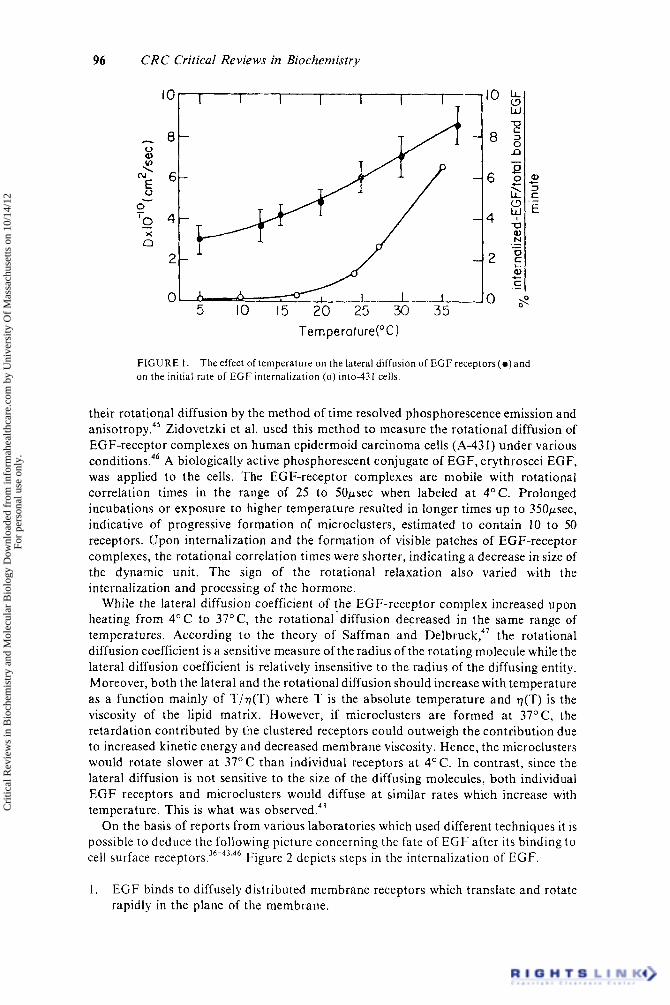
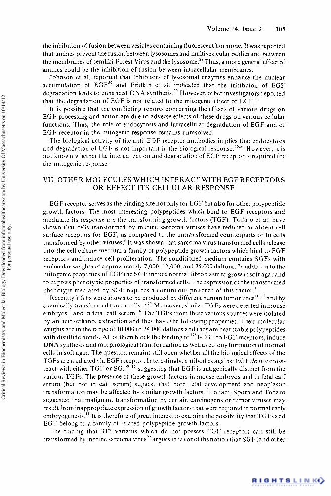
Receptor clustering and receptor mediated endocytosis are temperature-dependent processes which are completely inhibited a t 4’ C.36-42 If receptor clustering and subsequent receptor mediated endocytosis occur at specific sites on the cell surface (i.e., coated pit areas), its inhibition at low temperature could be due to decreased lateral diffusion of the occupied E G F receptors. T o test these possibilities Hillman and Schlessinger measured the lateral diffusion of rhodamine labeled EGF (R-EGF) bound to E G F receptors on human epidermoid carcinoma cells as a function of temperature. The value of D increased gradually from D = 3 X lo-’’ cm2/sec at 4OC up to D = 8.5 X lo-’’ cm2/sec at 37°C.43 No phase transition was observed in this temperature range as previously reported for the lateral diffusion of various proteins incorporated in artificial lipid b i l a y e r ~ . ~ ~ An activation energy of 6 kcal/mol was calculated for the temperature dependence of the lateral diffusion of EGF-receptor c0mplexes;4~ a value similar t o the activation energies measured for membrane proteins incorporated in artificial bilayers. Hillman and Schlessinger compared the temperature dependence of the lateral diffusion of EGF receptor complexes to the temperature dependence of the onset of patch formation and rate of internalization of ’’’I-EGF into A-431 cells.43 Figure 1 depicts the effect of temperature on the lateral diffusion of EGF-receptor complexes and on the rate of internalization of I2’I-EGFinto these cells. Interestingly, a t 15OC the internalization of E G F is very slow, while the lateral diffusion D is only half of the D value at 37” C.43 From the calculation of the collision frequency of the occupied EGF receptors with coated regions using the measured values of D at 4” C and at 37O C, it was concluded that lateral diffusion is not the rate limiting step for either endocytosis or patching.43 Moreover, since the internalization of radiolabeled E G F receptors precedes the formation of “visible patches” it was suggested that the bright patches observed by light microscopy are endocytic vesicles formed by the coalescence of several vesicles, each containing microclusters of EGF-receptor c ~ m p l e x e s . ~ * ’ ~ ’
A new approach to study the dynamic properties of membrane receptors is to measure
Cri
tical
Rev
iew
s in
Bio
chem
istr
y an
d M
olec
ular
Bio
logy
Dow
nloa
ded
from
info
rmah
ealth
care
.com
by
Uni
vers
ity O
f M
assa
chus
etts
on
10/1
4/12
For
pers
onal
use
onl
y.
96 CRC Critical Reviews in Biochemistry
I I I I
d zz , , 0
5 10 15 20 25 30 35 Temperature("C1
0)
3 c +
.- E
?
FIGURE 1 . on the initial rate of E G F internalization ( 0 ) into-431 cells.
The effect of temperature on the lateral diffusion of EGF receptors(o)and
their rotational diffusion by the method of time resolved phosphorescence emission and a n i ~ o t r o p y . ~ ~ Zidovetzki et a]. used this method to measure the rotational diffusion of EGF-receptor complexes on human epidermoid carcinoma cells (A-43 1) under various conditions.46 A biologically active phosphorescent conjugate of EGF, erythroscei EGF, was applied to the cells. The EGF-receptor complexes are mobile with rotational correlation times in the range of 25 to 50psec when labeled a t 4°C. Prolonged incubations or exposure to higher temperature resulted in longer times up to 350psec, indicative of progressive formation of microclusters, estimated to contain 10 to 50 receptors. Upon internalization and the formation of visible patches of EGF-receptor complexes, the rotational correlation times were shorter, indicating a decrease in size of the dynamic unit. The sign of the rotational relaxation also varied with the internalization and processing of the hormone.
While the lateral diffusion coefficient of the EGF-receptor complex increased upon heating from 4 ° C to 37"C, the rotational-diffusion decreased in the same range of temperatures. According to the theory of Saffman and D e l b r ~ c k , ~ ' the rotational diffusion coefficient is a sensitive measure of the radius of the rotating molecule while the lateral diffusion coefficient is relatively insensitive to the radius of the diffusing entity. Moreover, both the lateral and the rotational diffusion should increase with temperature as a function mainly of T/v(T) where T is the absolute temperature and r](T) is the viscosity of the lipid matrix. However, if microclusters are formed at 37"C, the retardation contributed by the clustered receptors could outweigh the contribution due to increased kinetic energy and decreased membrane viscosity. Hence, the microclusters would rotate slower a t 37OC than individual receptors at 4°C. In contrast, since the lateral diffusion is not sensitive to the size of the diffusing molecules, both individual E G F receptors and microclusters would diffuse at similar rates which increase with temperature. This is what was observed.43
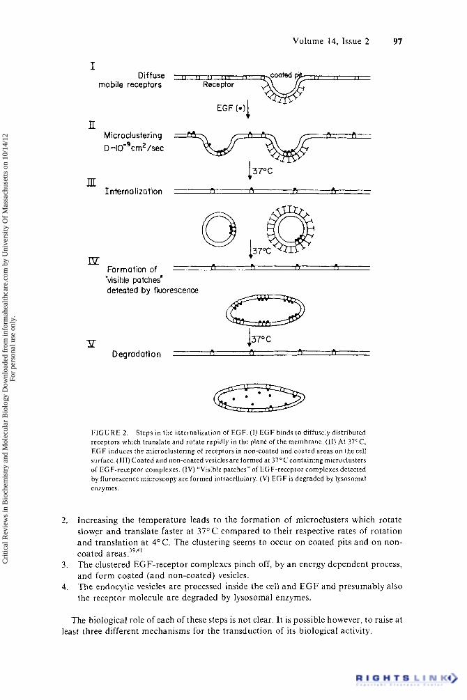
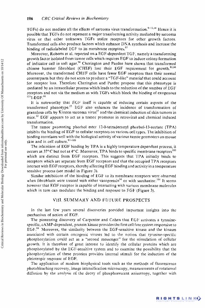
On the basis of reports from various laboratories which used different techniques it is possible to deduce the following picture concerning the fate of E G F after its binding to cell surface receptor^.^^-^^.^^ Figure 2 depicts steps in the internalization of EGF.
1. EGF binds to diffusely distributed membrane receptors which translate and rotate rapidly in the plane of the membrane.
Cri
tical
Rev
iew
s in
Bio
chem
istr
y an
d M
olec
ular
Bio
logy
Dow
nloa
ded
from
info
rmah
ealth
care
.com
by
Uni
vers
ity O
f M
assa
chus
etts
on
10/1
4/12
For
pers
onal
use
onl
y.
Volume 14, Issue 2 97
c > < I ( 1 I 1 1 r ,
I Diffuse
mobile receptors Receptor
EGF (-11
lI Microclustering -+ /=ztY=Bb D -IO-'cm2 /sec
137°C m J l L - . Interno liza t ion i 1 I 1 , I 1
Ip Formation of - Qk70ca L 1 [ I I 1 1 1 I
"visible patches' deteated by fluorescence
P D egrad a t i o n ( 1 ( 1 < 1 I 1
F I G U R E 2. Steps in the internalization of EGF. ( I ) E G F binds t o diffusely distributed receptors which translate and rotate rapidly in the plane of the membrane. (11) At 37'C, E G F induces t h e microclustering of receptors in non-coated and coated areas on the cell surface. (111) Coated and non-coated vesicles are formed a t 37°C containing microclusters of EGF-receptor complexes. (IV) "Visible patches" of EGF-receptor complexes detected by fluroescence microscopy a re formed intracellulary. (V) E G F is degraded by lysosomal enzymes.
Increasing the temperature leads to the formation of microclusters which rotate slower and translate faster at 37" C compared to their respective rates of rotation and translation a t 4°C. The clustering seems to occur on coated pits and on non- coated areas .39'4 ' The clustered EGF-receptor complexes pinch off, by an energy dependent process, and form coated (and non-coated) vesicles. The endocytic vesicles are processed inside the cell and E G F and presumably also the receptor molecule are degraded by Iysosomal enzymes.
The biological role of each of these steps is not clear. It is possible however, to raise at least three different mechanisms for the transduction of its biological activity.
Cri
tical
Rev
iew
s in
Bio
chem
istr
y an
d M
olec
ular
Bio
logy
Dow
nloa
ded
from
info
rmah
ealth
care
.com
by
Uni
vers
ity O
f M
assa
chus
etts
on
10/1
4/12
For
pers
onal
use
onl
y.
98 CRC Critical Reviews in Biochemistry
1. E G F acts at the level of the cell surface to generate directly or indirectly via a “second messenger” (e.g., phosphorylation of intracellular proteins, concentration of ions and metabolites), a signal that triggers both rapid and delayed responses. Alternatively, one or more of the rapid responses may produce the signal for the long-term effects. In this model internalization does not participate in the signalling process, it is rather related to the desensitization of the cell t o the growth factor. E G F generates a signal a t the level of the plasma membrane for the rapid responses, and the internalized hormone or its fragments produce an independent signal by interaction with putative intracellular sites for the long-term effects. E G F binding to cell surface receptors creates the appropriate perturbation in the receptor molecule which by itself is the active species.
2.
3.
Obviously it is possible to suggest additional mechanisms. Nevertheless, these three alternatives set the stage for systematic investigation of the mechanism of E G F action. In the next section we discuss various questions concerning the mechanism of E G F response which shed light on the three alternative mechanism proposed below.
111. A POSSIBLE ROLE FOR THE MICROAGGREGATION O F EGF RECEPTORS
E G F has a single methionyl at position 21. Cleavage with cyanogen bromide results in the formation of two polypeptide chains connected by the three disulphide bonds of the molecule.48349 CNBr-cleaved EGF (CNBr-EGF) retains approximately 10% of the binding activity of EGF but is virtually devoid of mitogenic activity in vitro4s,so,s’ and in viva.'' It is not known yet whether the non-mitogenic property of CNBr-EGF is due to the cleavage at the methionyl residue per se or due to further changes induced in the E G F molecule during the reaction with cyanogen bromide.
In contrast to EGF, CNBr-EGF does not induce the clustering of the EGF-receptor complexes. However, the addition of bivalent anti-EGF antibodies restores both patch formation and the mitogenic activity of this analogue.48 The mitogenic activity is not restored by adding monovalent Fab’ fragments of the antibodies. The concentration of bivalent antibodies required for full restoration of DNA synthesis is far below the concentration at which visible patches of R-EGFcan be seen. Thus, it was concluded that microaggregation of perhaps only a few receptors rather than the formation of visible patches may be the relevant mechanism for the induction of the mitogenic activity of
Two additional experiments demonstrate the significance of receptor clustering, First, concentrations of native EGF far below those that normally show activity can elicit dramatic responses when bivalent anti-EGF antibodies are added. Monovalent antibody fragments cannot enhance DNA synthesis. Second, cells that a re relatively refractory to EGF, despite unimpaired binding, can be markedly sensitized and respond to the hormone after cross-linking with anti-EGF an t ibod ie~ .~ ’ Thus it was proposed that receptor aggregation potentiates the biological response of EGF.33.4R*52 It was previously argued that ligand induced receptor clustering could cause a n efficient amplification of biological signals across the plasma membrane of various ligand-receptor systems.52
An alternative interpretation of the effect of anti-EGF antibodies on the response of E G F is that the bivalent anti-EGF antibodies enhance the apparent affinity of EGF towards its receptors rather than enhance receptor clustering. The binding constant (K) of EGF to E G F receptor equals ken/ kolf where:
~ ~ ~ . 3 3 , 4 8 , 5 2
k o n ECF + R e c e p t o r e E G F - Receptor
kotr
Cri
tical
Rev
iew
s in
Bio
chem
istr
y an
d M
olec
ular
Bio
logy
Dow
nloa
ded
from
info
rmah
ealth
care
.com
by
Uni
vers
ity O
f M
assa
chus
etts
on
10/1
4/12
For
pers
onal
use
onl
y.
Volume 14, Issue 2 99
Anti-EGF antibodies are able to increase the binding constant K by either increasing k,, or by decreasing k,rr (or both). Since the cells are exposed to EGF in the presence of the antibodies it is impossible to rule out a direct effect on the ken. Moreover, the effect of anti-EGF antibodies on the k,fr was not studied in detail. Therefore it is required to study the effect of anti-EGF antibodies on the binding parameters of E G F in order to understand the antibody-induced potentiation of the biological response to EGF.
IV. EGF RECEPTOR AND EGF INDUCED-PROTEIN PHOSPHORYLATION
The membrane receptor of EGF plays a key role in the transduction of the signals mediated by EGF leading to cell proliferation. The EGF receptor interacts with transforming growth factors (TGF) leading to anchorage-independent growth.’-I6 Tumor promoters such as phorbol esters53 induce a decrease in the affinity of E G F towards its receptors. Recent studies indicate that E G F receptor could also interact with receptors for vasopressin on 3T3 cells.54 Schreiber et al. have recently reported on the generation of a monoclonal antibody against EGF re~ep to r .~ ’ This antibody blocks the binding of radiolabeled E G F to E G F receptors on 3T3 cells, human fibroblasts, and A-43 1 Interestingly it also induces “EGF-like” activity: the phosphorylation of membrane proteins, activation of ornithine decarboxylase, and the stimulation of DNA synthesis and cell p r~ l i f e ra t ion . ” ,~~ These results support the notion that EGF receptor rather than E G F itself is the active moiety and that the role of the hormone is t o perturb the receptor which then stimulates the pleiotropic responses mediated by EGF.
E G F receptor is a membrane glycoprotein containing sialic acid residues. E G F binds to the EGF receptor with a n apparent dissociation constant of 2 X lo-’’ M. Most cells which are responsive to EGF bear 40,000 to 100,000 EGF receptors per cell. A cell line with an unusually high number of E G F receptors which is commonly used for the investigation of E G F receptors is the human epidermoid carcinoma A-43 1 which bears approximately 2 x lo6 receptors per
Various approaches have been used to characterize and isolate the receptor for EGF. Cross-linking reagents have been used to produce covalent EGF-receptor complexes of approximately 190,000 daltons from 3T3 fibroblasts5* and 100,000 daltons from liver membrane^.'^ Linsley et al. have shown that a fraction of ‘251-EGFforms a direct-linkage complex with EGF receptors.60 This “covalent association” of E G F receptor appears t o be due to an oxidative effect on E G F by chloramine T.61 Similar direct linkages were reported for ‘251-insulin and ’2’I-thrombin with their respective receptors.62363 Anti-EGF antibodies were used for the immunoprecipitation of ”’I-EGF directly linked receptor complex. The immunoprecipitated receptor appeared as a 160,000 to 140,000 daltons proteins.64 EGF-affinity chromatography of E G F receptor from membranes prepared from A-431 cells indicated that EGF receptors is a protein of 170,000 d a l t o n ~ . ~ ~
Although limited progress has been made in purifying the E G F receptor by solubilization of membranes, much more progress was made in an attempt to obtain a cell-free system which is responsive to EGF. Carpenter and Cohen discovered that EGF induces a rapid, cyclic nucleotide-independent and tyrosine-specific phosphorylation of a number of membrane proteins when added to purified plasma membranes from human epidermoid carcinoma cells (A-43 1). Several proteins of molecular weights correspond- ing t o 170,000, 150,000, 80,000, and 22,000 are p h ~ s p h o r y l a t e d . ~ ~ - ~ ’ The major phosphorylated proteins with molecular weight of 170,000 to 150,000 daltons were identified as EGF r e ~ e p t o r . ~ ~ - ~ ~ Both ATP and GTP can act as the phosphate donor of the phosphorylation reaction and either Mn+2 or Mgt2 is required for optimal activity of the protein kinase.68 The kinase activity is copurified with EGF-binding activity by EGF af f in i ty-~hromatography.~~ This finding together with the fact that the phosphorylation
Cri
tical
Rev
iew
s in
Bio
chem
istr
y an
d M
olec
ular
Bio
logy
Dow
nloa
ded
from
info
rmah
ealth
care
.com
by
Uni
vers
ity O
f M
assa
chus
etts
on
10/1
4/12
For
pers
onal
use
onl
y.
100 CRC Critical Reviews in Biochemistry
l igand
toplasm
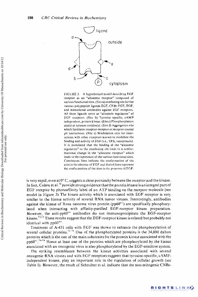
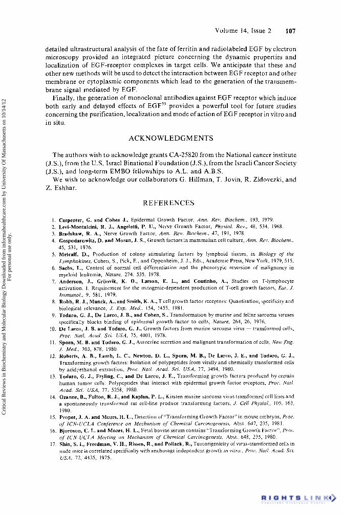
F I G U R E 3. A hypothetical model describing E G F receptor as a n "allosteric receptor" composed of various functional sites. (Site a ) combiningsite for the various polypeptide ligands EGF, CNBr-EGF, T G F , and monoclonal antibodies against E G F receptors. All these ligands serve a s "allosteric regulators" of E G F receptors. (Site b) Tyrosine specific, C A M P independent, protein kinase. (Site c) Phosphorylation site(s) a t tyrosine residue(s). (Si ted) Aggregation site which facilitates receptor-receptor or receptor-coated pit interactions. (Site e) Modulation sites for inter- actions with other receptors known to modulate the binding and activity of E G F (i.e., TPA, vasopressin). It is postulated that the binding of the "allosteric regulators" to the combining site leads to a confor- mational change in the "allosteric receptor" which leads to the expression of the various functional sites. Continuous lines indicate the conformation of the sites in the absence of E G F and dotted lines represent the conformation of the sites in the presence of E G F .
is very rapid, even at 0" C, suggests a close proximity between the receptor and the kinase. In fact, Cohen et ak7' provide strong evidence that the protein kinase is a n integral part of EGF receptor by photoaffinity label of an ATP binding on the receptor molecule (see model in Figure 3) The kinase activity which is associated with EGF receptor is very similar to the kinase activity of several RNA tumor viruses. Interestingly, antibodies against the kinase of Rous sarcoma virus protein (pp60"') are specifically phosphory- lated when interacting with affinity-purified EGF-receptor kinase preparation. However, the anti-pp60"'" antibodies d o not immunoprecipitate the EGF-receptor k i n a ~ e . ~ ' , ~ ~ These results suggest that the EGF-receptor kinase is related but probably not identical with pp6OarC.
Treatment of A-431 cells with E G F was shown to enhance the phosphorylation of several cellular protein^.^'-^^ One of the phosphorylated proteins is the 34,000 dalton proteins which is the one of the main substrates for the protein kinase associated with the pp60src.73-74 Hence at least one of the proteins which are phosphorylated by the kinase associated with an oncogenic virus is also phosphorylated by the EGF-sensitive system.
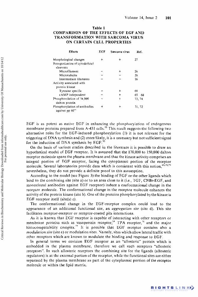
The striking resemblance between the kinase activities associated with several oncogenic RNA viruses and with E G F receptors suggests that tyrosine-specific, CAMP- independent kinase, play an important role in the regulation of cellular growth (see Table 1). However, the result of Schreiber et al. indicate that the non-mitogenic CNBr-
Cri
tical
Rev
iew
s in
Bio
chem
istr
y an
d M
olec
ular
Bio
logy
Dow
nloa
ded
from
info
rmah
ealth
care
.com
by
Uni
vers
ity O
f M
assa
chus
etts
on
10/1
4/12
For
pers
onal
use
onl
y.
Volume 14, Issue 2 101
Table 1 COMPARISON OF THE EFFECTS OF EGF A N D TRANSFORMATION WITH SARCOMA VIRUS
ON CERTAIN CELL PROPERTIES
Effects
Morphological changes Reorganization of cptoskeletal
elements Microfilament Microtubules Intermediate filaments
Activity associated with protein kinase
Tyrosine specific C A M P independent
Phosphorylation of 34,000
Phosphorylation of antibodies dalton protein
against pp 60”‘
EGF
t
t -
-
+ t t
+
Sarcoma virus
+ Ref.
27
26 26 26
69
73, 74
71. 72
65-68
EGF is as potent as native EGF in enhancing the phosphorylation of endogenous membrane proteins prepared from A-43 I cells.50 This result suggests the following two alternative roles for the EGF-induced phosphorylation (1 ) it is not relevant for the triggering of D N A synthesis and (2) more likely, it is a necessary but not sufficient signal for the induction of DNA synthesis by EGF.”
On the basis of various studies described in the literature it is possible to draw an hypothetical model of EGF receptor. It is assumed that the 170,000 to 150,000 dalton receptor molecule spans the plasma membrane and that the kinase activity comprises an integral portion of EGF receptor, facing the cytoplasmic portion of the receptor molecule. Several laboratories provide data which is consistent with this n ~ t i o n , ~ ~ ’ ~ ’ ” ~ nevertheless, they do not provide a definite proof to this assumption.
According to the model (see Figure 3) the binding of EGF o r the other ligands which bind to the combining site (site a) or to an area close to it (i.e., TGF, CNBr-EGF, and monoclonal antibodies against EGF receptor) induce a conformational change in the receptor molecule. The conformational change in the receptor molecule enhances the activity of the protein kinase (site b). One of the proteins phosphorylated by the kinaseis EGF receptor itself (site(s) c).
The conformational change in the EGF-receptor complex could lead to the appearance of an additional functional site, an aggregation site (site d). This site facilitates receptor-receptor or receptor-coated pits interactions. As it is known that EGF receptor is capable of interacting with other receptors or
membrane proteins such as vasopressin r e ~ e p t o r , ’ ~ TPA receptor,’6 and the major histocompatability complex.” It is possible that EGF receptor contains also a modulation site (site e) or modulation sites. Namely, sites which allow lateral traffic with other receptors which are known to modulate the binding and response to EGF.
In general terms we envision E G F receptor as an “allosteric” protein which is embedded in the plasma membrane, therefore we call such receptors “allosteric receptors”. In such allosteric receptors the combining site for the ligands (allosteric regulators) is at the external portion of the receptor, while the functional sites are either separated by the plasma membrane as part of the cytoplasmic portion of the receptor molecule or within the lipid matrix.
Cri
tical
Rev
iew
s in
Bio
chem
istr
y an
d M
olec
ular
Bio
logy
Dow
nloa
ded
from
info
rmah
ealth
care
.com
by
Uni
vers
ity O
f M
assa
chus
etts
on
10/1
4/12
For
pers
onal
use
onl
y.
102 CRC Critical Reviews in Biochemistry
Table 2 BIOCHEMICAL EFFECTS OF THE MONOCLONAL
ANTIBODIES AGAINST EGF RECEPTOR
Concentration of Concentration of 2GZ-IgM EGF
50% inhibition of binding of 20nM 2n M
Enhancement of phosphorylation 50nM 20nM
Induction of changes in cell >50nM > lOnM
Activation of the enzyme lOnM 2n M
Maximal induction of DNA synthesis lOnM 2nM Stimulation of cell proliferation 20nM 2nM
EGF (2nM)
of membrane proteins
cell morphology
ornithine decarboxylase
Thus, EGF and other ligands which bind to the combining site act as allosteric regulators inducing a conformational change in the allosteric receptor which lead to the expression and activation of new functional sites.
V. MONOCLONAL ANTIBODIES AGAINST EGF RECEPTOR INDUCE “EGF-LIKE” ACTIVITY IN RESPONSIVE CELLS
A powerful tool for the isolation and investigation of membrane receptors is to use specific antibodies directed against the membrane receptors. .Haigler and Carpenter prepared antibodies directed against the membrane or human epidermoid carcinoma cells A-431.” They reported that these antibodies inhibit the binding of EGFto cellsand membrane bearing EGF receptors. Hence, these antibodies inhibit EGF binding, and bioactivity and do not possess any intrinsic biological activity.
Schreiber et al. reported on the generation of monoclonal antibodies against EGF receptors.55 They raised antibodies against A-43 1 cells and selected monoclonal antibodies against EGF receptors. One fast growing clone that secreted IgM antibodies (denoted 2G2) capable of inhibiting the binding of EGF to membrane receptors, was grown in culture in large quantities and in the form of ascites in mice.
The properties of this antibody are as follows.
1. 2G2 antibody binds to cells bearing EGF receptors in proportion to the amount of EGF receptors on these cells. 2G2 antibody inhibits specifically the binding of radiolabeled EGF to human, rat, and mouse cells and to membranes prepared from these cells. It does not bind to cells which do not bear EGF receptors. 2G2 antibody induces various early and delayed responses which are mediated by EGF. This antibody induces the enhancement of phosphorilation of endogenous membrane proteins, changes in the morphology of A-431 cells, activation of the enzyme ornithine decarboxylase, stimulation of DNA synthesis in responsive cells, enhancement of the proliferation of cells bearing EGF receptors. Thus 2G2 antibody acts as a competitive agonist of EGF inducing both the early and the delayed effects mediated by the growth factor (see summary in Table 2).
2.
In contrast to the potency of 2G2-IgM in inhibiting the binding of radiolabeled EGF to its membrane receptors, an excess of EGF could not displace the bound 2G2 from the membrane receptor.55959 However, monovalent Fab’ fragments of 2G2 inhibit the binding
Cri
tical
Rev
iew
s in
Bio
chem
istr
y an
d M
olec
ular
Bio
logy
Dow
nloa
ded
from
info
rmah
ealth
care
.com
by
Uni
vers
ity O
f M
assa
chus
etts
on
10/1
4/12
For
pers
onal
use
onl
y.
Volume 14, Issue 2 103
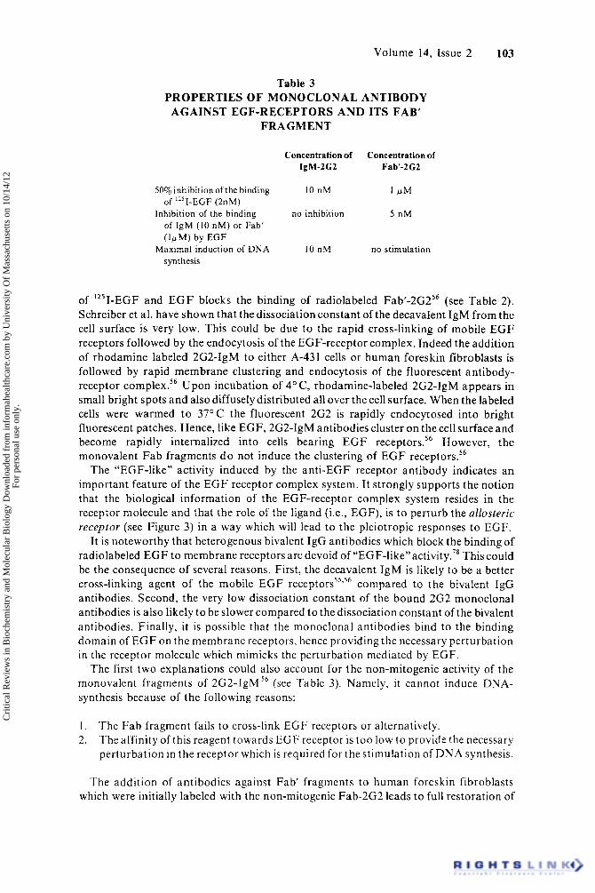
Table 3 PROPERTIES OF MONOCLONAL ANTIBODY
FRAGMENT AGAINST EGF-RECEPTORS A N D ITS FAB’
Concentration of Concentration of IgM-ZGZ Fab’-2G2
50% inhibition of the binding 10 nM 1 PM of ”’I-EGF (2nM)
of IgM (10 nM) or Fab’ (1pM) by EGF
synthesis
Inhibition of the binding no inhibition 5 nM
Maximal induction of DNA 10 nM no stimulation
of ‘251-EGF and E G F blocks the binding of radiolabeled Fab’-2G256 (see Table 2). Schreiber et al. have shown that the dissociation constant of the decavalent IgM from the cell surface is very low. This could be due to the rapid cross-linking of mobile E G F receptors followed by the endocytosis of the EGF-receptor complex. Indeed the addition of rhodamine labeled 2G2-IgM to either A-431 cells or human foreskin fibroblasts is followed by rapid membrane clustering and endocytosis of the fluorescent antibody- receptor complex.56 Upon incubation of 4’ C, rhodamine-labeled 2G2-lgM appears in small bright spots and also diffusely distributed all over the cell surface. When the labeled cells were warmed t o 37OC the fluorescent 2G2 is rapidly endocytosed into bright fluorescent patches. Hence, like EGF, 2G2-IgM antibodies cluster on the cell surface and become rapidly internalized into cells bearing E G F receptor^.'^ However, the monovalent Fab fragments d o not induce the clustering of E G F receptor^.^^
The “EGF-like” activity induced by the anti-EGF receptor antibody indicates an important feature of the E G F receptor complex system. It strongly supports the notion that the biological information of the EGF-receptor complex system resides in the receptor molecule and that the role of the ligand (i.e., EGF), is t o perturb the allosteric receptor (see Figure 3) in a way which will lead to the pleiotropic responses to EGF.
It is noteworthy that heterogenous bivalent IgG antibodies which block the binding of radiolabeled E G F to membrane receptors are devoid of “EGF- l ike”a~ t iv i ty .~~ This could be the consequence of several reasons. First, the decavalent IgM is likely to be a better cross-linking agent of the mobile E G F compared to the bivalent IgG antibodies. Second, the very low dissociation constant of the bound 2G2 monoclonal antibodies is also likely to be slower compared to the dissociation constant of the bivalent antibodies. Finally, it is possible that the monoclonal antibodies bind to the binding domain of E G F on the membrane receptors, hence providing the necessary perturbation in the receptor molecule which mimicks the perturbation mediated by EGF.
The first two explanations could also account for the non-mitogenic activity of the monovalent fragments of 2G2-lgM56 (see Table 3). Namely, i t cannot induce DNA- synthesis because of the following reasons:
1 . 2.
The Fab fragment fails to cross-link EGF receptors or alternatively. The affinity of this reagent towards EGF receptor is too low to provide the necessary perturbation in the receptor which is required for the stimulation of DNA synthesis.
The addition of antibodies against Fab’ fragments to human foreskin fibroblasts which were initially labeled with the non-mitogenic Fab-2G2 leads to full restoration of
Cri
tical
Rev
iew
s in
Bio
chem
istr
y an
d M
olec
ular
Bio
logy
Dow
nloa
ded
from
info
rmah
ealth
care
.com
by
Uni
vers
ity O
f M
assa
chus
etts
on
10/1
4/12
For
pers
onal
use
onl
y.
104 CRC Critical Reviews in Biochemistry
DNA synthesis. This experiment seems to suggest that receptor clustering is required for the restoration of DNA synthesis. However, it is also possible that the anti-Fab antibody enhances the affinity of the Fab’ fragment towards EGF receptors.56
VI. POSSIBLE RELATIONSHIP BETWEEN EGF INTERNALIZATION AND ITS MITOGENIC EFFECT
Several studies support the idea that intracellular degradation of EGF or E G F receptors is necessary for the mitogenic response to EGF. Fox and Das have shown that at low concentration of EGF added to 3T3 fibroblasts the rate of receptor internalization and degradation correlates well with its mitogenic a~t ivi ty . ’~ They have suggested that the proteolytically cleaved EGF receptor may be involved in the activation of DNA ~ n y t h e s i s . ~ ~ It is not clear yet whether the proteolytic fragments of EGF receptor are related to the EGF-induced protein activator of DNA synthesis which was isolated from cytoplasmic extracts of 3T3 cells treated with EGF.80
One approach to investigate the biological role of E G F internalization and degradation is to use drugs which inhibit specific stages in the processing of EGF- receptor complexes and then to examine their effect on the biological response mediated by EGF.
Various amines block the degradation of the endocytosed E G F without affecting the binding of the hormone to its surface receptors.34 Several reports presented data indicating that primary amines inhibit the formation of visible patches on the cell surface of fluorescently labeled EGF, a2-macroglobulin and 3,3’,5-triiodo-L-thyronine and also their subsequent internalization into the cells.81.82 Hence these drugs seem to provide a tool for investigating the biological role of hormone clustering and internalization. However, amines at doses which inhibit receptor clustering d o not inhibit, and even potentiate EGF stimulated DNA synthesis by 3T3 cells.83 This suggests that receptor clustering and internalization are related to the removal of the hormone from the cell surface rather than to the mechanism of tim mu la ti on.^^ Recently Haigler et al. reported that amines block the endocytosis of a2-macroglobulin but do not block the endocytosis of E G F into 3T3 cells.84 Previous studies have shown that amines did not inhibit the endocytosis EGF into human epidermoid carcinoma cells ( A 4 3 1) and fibroblast^,^' and that the main action of amines is inhibition of hormone d e g r a d a t i ~ n . ~ ’ ~ ~ ~ ~ ~ ~ ~ ~ ~
Since various primary amines and inhibitors of the enzyme transglutaminase block the formation of visible patches of rhodamine az-macroglobulin and rhodamine E G F it was postulated that this enzyme cross-links covalently the receptors and putative proteins in the coated pits by forming t-(y-glutaminyl) lysine cro~s-bridges.~’ The conflicting reports concerning the effects of amines on EGF processing and activity led several laboratories to perform a comprehensive investigation of the effects of amines on the fate of EGF after the binding to cultured ~ e l l s . ~ ~ , ~ ~ ~ ~ ~ ~ ~ ~ Current studies from several laboratories provide the following view on the effect of amines on EGF processing and action. Amines do not affect the rate and the extent of internalization of E G F into 3T3 cells. Studies with fluorescent conjugates of E G F show that amines slow the formation of visible patches of E G F on 3T3 cells.42 Fluorescence photobleaching recovery (FPR) and video intensification microscopy cannot detect the formation of microclusters of E G F on the cell surface. However, Haigler et al. have shown by electron microscopy that ferritin E G F forms microclusters composed of 2 to 6 molecules a t 37O C and that amines did not block this process.” Such microclusters are presumably mobile and therefore capable of coalescing into patches on the cell surface. However, several studies suggest that microclusters of EGF-receptor complexes are internalized and that the “visible patches” are in fact endocytic vesicle^.^^,^^^^^ Therefore the effect of amines could be interpreted as
Cri
tical
Rev
iew
s in
Bio
chem
istr
y an
d M
olec
ular
Bio
logy
Dow
nloa
ded
from
info
rmah
ealth
care
.com
by
Uni
vers
ity O
f M
assa
chus
etts
on
10/1
4/12
For
pers
onal
use
onl
y.
Volume 14, Issue 2 105
the inhibition of fusion between vesicles containing fluorescent hormone. It was reported that amines prevent the fusion between lysosomes and multivesicular bodies and between the membranes of semliki Forest Virus and the lysosome.88 Thus, a more general effect of amines could be the inhibition of fusion between intracellular membranes.
Johnson et al. reported that inhibitors of lysosomal enzymes enhance the nuclear accumulation of EGFS9 and Fridkin et al. indicated that the inhibition of E G F degradation leads to enhanced DNA synthesis." However, other investigators reported that the degradation of E G F is not related t o the mitogenic effect of EGF."
It is possible that the conflicting reports concerning the effects of various drugs on E G F processing and action are due to adverse effects of these drugs on various cellular functions. Thus, the role of endocytosis and intracellular degradation of E G F and of E G F receptor in the mitogenic response remains unresolved.
The biological activity of the anti-EGF receptor antibodies implies that endocytosis and degradation of E G F is not important in the biological r e s p ~ n s e . ' ~ ' ~ ~ However, it is not known whether the internalization and degradation of EGF receptor is required for the mitogenic response.
VII. OTHER MOLECULES WHICH INTERACT WITH EGF RECEPTORS OR EFFECT ITS CELLULAR RESPONSE
E G F receptor serves as the binding site not only for EGF but also for other polypeptide growth factors. The most interesting polypeptides which bind to E G F receptors and modulate its response are the transforming growth factors (TGF). Todaro et af. have shown that cells transformed by murine sarcoma viruses have reduced or absent cell surface receptors for EGF, as compared to the untransformed counterparts or to cells transformed by other v i r ~ s e s . ~ It was shown that sarcoma virus transformed cells release into the cell culture medium a family of polypeptide growth factors which bind to E G F receptors and induce cell proliferation. The conditioned medium contains SGFs with molecular weights of approximately 7,000, 12,000, and 25,000 daltons. In addition to the mitogenic properties of E G F the S G F induce normal fibroblasts to grow in soft agar and to express phenotypic properties of transformed cells. The expression of the transformed phenotype mediated by S G F requires a continuous presence of this factor."
Recently TGFs were shown to be produced by different human tumor lines"-'3 and by chemically transformed tumor cells.""3 Moreover, similar TGFs were detected in mouse embryos" and in fetal calf serum.16 The TGFs from these various sources were isolated by a n acid/ethanol extraction and they have the following properties. Their molecular weights are in the range of 10,000 to 24,000 daltons and they are heat stable polypeptides with disulfide bonds. All of them block the binding of "'I-EGF to E G F receptors, induce DNA synthesis and morphological transformation as well as colony formation of normal cells in soft agar. The question remains still open whether all the biological effects of the TGFs are mediated via EGF receptor. Interestingly, antibodies against EGF do not cross- react with either T G F or SGF9-I6 suggesting that EGF is antigenically distinct from the various TGFs. The presence of these growth factors in mouse embryos and in fetal calf serum (but not in calf serum) suggest that both fetal development and neoplastic transformation may be affected by similar growth factors." I n fact, Sporn and Todaro suggested that malignant transformation by certain carcinogens or tumor viruses may result from inappropriate expression of growth factors that were required in normal early embryogenesis." It is therefore of great interest to examine the possibility that TGFs and EGF belong to a family of related polypeptide growth factors.
The finding that 3T3 variants which do not possess E G F receptors can still be transformed by murine sarcoma virusg2 argues in favor ofthe notion that SGF(and other
Cri
tical
Rev
iew
s in
Bio
chem
istr
y an
d M
olec
ular
Bio
logy
Dow
nloa
ded
from
info
rmah
ealth
care
.com
by
Uni
vers
ity O
f M
assa
chus
etts
on
10/1
4/12
For
pers
onal
use
onl
y.
106 CRC Critical Reviews in Biochemistry
TGFs) d o not mediate all the effects of sarcoma virus t r a n s f ~ r m a t i o n . ~ - ” ” ~ Hence it is possible that TGFs d o not represent a major transforming activity mediated by sarcoma virus or that other unknown TGFs utilize receptors for other growth factors. Transformed cells also produce factors which enhance DNA synthesis and increase the binding of radiolabeled E G F to its membrane receptors.93
Moreover, Roberts et al. reported on a EGF-dependent TGF, namely a transforming growth factor isolated from tumor cells which requires E G F to induce colony formation of indicator cell in soft agar.94 Cherington and Pardee have shown that transformed Chinese hamster fibroblast (CHEF) lose their E G F requirement for Moreover, the transformed C H E F cells have fewer E G F receptors than their normal counterparts but they d o not seem to produce a “TGF-like” material that could account for receptor loss. Therefore Cherington and Pardee propose that this phenotype is mediated by an intracellular process which leads to the reduction of the number of E G F receptors and not via the medium as with TGFs which block the binding of exogenous ‘251-EGF.95
It is noteworthy that E G F itself is capable of inducing certain aspects of the transformed p h e n ~ t y p e . ~ ’ EGF also enhances the incidence of transformation of granulosa cells by Kirsten sarcoma virus23 and the chemical induction of skin tumors in mice.** EGF appears to act as a tumor promoter in retroviral and chemical induced transformation.
The tumor promoting phorbol ester 12-0-tetradecanyl phorbol-13-acetate (TPA) inhibits the binding of E G F to cellular receptors on various cell types. The inhibition of binding correlates well with the biological activity of various tumor promoters on mouse skin and in cell c u l t ~ r e . ~ ’ - ’ ~ ~
The inhibition of E G F binding by TPA is a highly temperature dependent process, it occurs at 37°C but not a t 4°C. Moreover, TPA binds to specific membrane receptors”’ which are distinct from EGF receptors. This suggests that TPA initially binds to receptors which are separate from E G F receptors and that the occupied TPA receptors interact with E G F receptors, thereby affecting EGF binding and activity in a temperature sensitive process (see model in Figure 3).
Similar inhibitions of the binding of EGF to its membrane receptors were observed when fibroblasts were treated with either vasopressins4 or with saccharine.”’ It seems however that E G F receptor is capable of interacting with various membrane molecules which in turn can modulate the binding and response to E G F (Figure 3).
VIII. SUMMARY AND FUTURE PROSPECTS
In the last few years several discoveries provided important insights into the mechanism of action of EGF.
The pioneering discovery of Carpenter and Cohen that E G F activates a tyrosine- specific, CAMP-dependent, protein kinase provides the first cell free system responsive to EGF.66 Moreover, the similarity between the EGF-sensitive kinase and the kinases associated with certain oncogenic viruses led to the notion that tyrosine-specific phosphorylation could act as a “second messenger” for the stimulation of cellular growth. It is therefore of great interest to identify the cellular proteins which are phosphorylated by the EGF-sensitive system and to examine the possibility that the phosphorylation of these proteins provides internal stimuli for the induction of the pleiotropic response of EGF.
The application of modern biophysical tools such as the methods of fluorescence photobleaching recovery, image intensification microscopy, measurements of rotational diffusion by the analysis of the decay of phosphorescent anisotropy, together with
Cri
tical
Rev
iew
s in
Bio
chem
istr
y an
d M
olec
ular
Bio
logy
Dow
nloa
ded
from
info
rmah
ealth
care
.com
by
Uni
vers
ity O
f M
assa
chus
etts
on
10/1
4/12
For
pers
onal
use
onl
y.
Volume 14, Issue 2 107
detailed ultrastructural analysis of the fate of ferritin and radiolabeled E G F by electron microscopy provided an integrated picture concerning the dynamic properties and localization of EGF-receptor complexes in target cells. We anticipate that these and other new methods will be used to detect the interaction between E G F receptor and other membrane or cytoplasmic components which lead to the generation of the transmem- brane signal mediated by EGF.
Finally, the generation of monoclonal antibodies against E G F receptor which induce both early and delayed effects of EGF” provides a powerful tool for future studies concerning the purification, localization and mode of action of E G F receptor in vitro and in situ.
ACKNOWLEDGMENTS
The authors wish t o acknowledge grants CA-25820 from the National cancer institute (J.S.), from the U.S. Israel Binational Foundation (J.S.), from the Israeli Cancer Society (J.S.), and long-term EMBO fellowships to A.L. and A.B.S.
We wish to acknowledge our collaborators G . Hillman, T. Jovin, R. Zidovezki, and 2. Eshhar.
REFERENCES
I . Carpenter, G. and Cohen J., Epidermal Growth Factor, Ann. Rev. Biochem., 193, 1979. 2. Levi-Montalcini, R. J., Angeletti, P. U., Nerve Growth Factor, Physiol. Rev., 48, 534, 1968. 3. Bradshaw, R. A., Nerve Growth Factor, Ann. Rev. Biochern., 47, 191, 1978. 4. Gospodarowitz, D . and Moran, J. S., Growth factors in mammalian cell culture, Ann. Rev. Biochem.,
45, 531, 1976. 5. Metcalf, D . , Production of colony stimulating factors by lymphoid tissues, in Biology of the
Lymphokines, Cohen, S., Pick, E., and Oppenheim, J. J., Eds., Academic Press, New York, 1979,515. 6. Sachs, L., Control of normal cell differentiation and the phenotypic reversion of malignancy in
myeloid leukemia, Nature. 274, 535, 1978. 7. Anderson, J., Gronvik, K. O., Larson, E. L., and Coutinho, A., Studies on T-lymphocyte
activation. 1. Requirement for the mitogenic-dependent production of T-cell growth factors, Eur. J. Immunol., 9, 581, 1979.
8. Robb, R. J. , Munck, A., and Smith, K . A. , Tcell growth factor receptors: Quantitation, specificity and biological relevance, J. Exp. Med., 154, 1455, 1981.
9. Todaro, G . J., De Larco, J. B., and Cohen, S., Transformation by murine and feline sarcoma viruses specifically blocks binding of epidermal growth factor to cells, Nature, 264, 26, 1976.
10. D e Larco, J. B. and Todaro, C. J., Growth factors from murine sarcoma virus - transformed cells, Proc. Natl. Acad. Sci. USA, 75, 4001, 1978.
1 1 . Sporn, M. B. and Todaro, G . J., Autocrine secretion and malignant transformation of cells, New Eng. J. Med., 303, 878, 1980.
12. Roberts, A. B., Lamb, L. C., Newton, D . L., Sporn, M . B., De Larco, J. E., and Todaro, G . J., Transforming growth factors: Isolation of polypeptides from virally and chemically transformed cells by acid/ethanol extraction, Proc. Narl. Acad. Sci. USA, 77, 3494, 1980.
13. Todaro, G . J., Fryling, C., and De Larco, J. E., Transforming growth factors produced by certain human tumor cells: Polypeptides that interact with epidermal growth factor receptors, Proc. Natl. Acad. Sci. USA, 77, 5258, 1980.
14. Ozanne, B., Fulton, R. J. , and Kaplan, P. L., Kirsten murine sarcoma virus transformed cell lines and a spontaneously transformed rat cell-line produce transforming factors, J . Cell Physiol., 105, 163, 1980.
15. Proper, J. A. and Mozes, H. L., Detection of “Transforming Growth Factor”in mouse embryos, Proc. of ICN- UCLA Conference on Mechanism of Chemical Carcinogenesis, Abst. 647, 235, 1981.
16. Bjoroson, C. L. and Mozes, H . L., Fetal bovine serum contains “Transforming Growth Factor”, Pror. of ICN-UCLA Meeting on Mechanism of Chemical Carcinogenesis, Abst. 648, 235, 1980.
17. Shin, S. I., Freedman, V. H., Rissen, R., and Pollack, R., Tumorigenicity of virus-transformed cells in nude mice is correlated specifically with anchorage independent growth in virro., Proc. Narl. Acad. Sci. USA, 72, 4435, 1975.
Cri
tical
Rev
iew
s in
Bio
chem
istr
y an
d M
olec
ular
Bio
logy
Dow
nloa
ded
from
info
rmah
ealth
care
.com
by
Uni
vers
ity O
f M
assa
chus
etts
on
10/1
4/12
For
pers
onal
use
onl
y.
108 CRC Critical Reviews in Biochemistry
18. Gospodarowicz, D., Epidermal and nerve growth factors in mammalian development, Ann. Rev. Physiol., 43, 251, 1981.
19. Carpenter, G. and Cohen, S., EGF: Receptor interaction and stimulation of cell growth, in Receptor and Recognition (Series B), Vol. 13, Lefkowitz, R . L., Ed., Chapman and Hall, 41.
20. Gregory, H., Isolation and structure of urogastrone and its relationship to epidermal growth factor, Nature, 257, 325, 1975.
21. Cohen, S. and Carpenter, G., Human epidermal growth factor: Isolation and chemical and biological properties, Proc. Nail. Acad. Sci. USA, 72, 1317, 1975.
22. Rose, S. P., Stahn, R., Passovoy, D. S., and Herschman, H., Epidermal growth factor enhancement of skin tumor induction in mice. Experientia, 32, 913, 1976.
23. Harrison, J. and Auersperg, N., Epidermal growth factor enhances viral transformation of granulosa cells, Science, 213, 218, 1981.
24. Hollenberg, M. D. and Cuatrecasas, P., Epidermal growth factor: Receptors in human fibroblasts and modulation of action by cholera toxin, Proc. Natl. Acad. Sci. USA, 70, 2964, 1973.
25. Carpenter, G., King, L. Jr., and Cohen, S., Rapid enhancement of protein phosphorylation in A-431 cell membrane preparations by epidermal growth factor, J . Biol. Chem., 254, 4884, 1979.
26. Schlessinger, J. and Geiger, B., Epidermal Growth Factor induces redistribution of actin and a-actinin in human epidermal carcinoma cells, E-rp. Cell Res., 134, 273, 1981.
27. Chinkers, M., McKanna, T. J. A., and Cohen, S., Rapid induction of morphological changes in human carcinoma cells A-431 by epidermal growth factor, J. Cell Biol., 83, 260, 1979.
28. Stasbury, M. and Cohen, S., The stimulation of ornithine decarboxylase activity in tests of the neonatal mouse, Biochem. Biophys. Acln, 261, 177, 1972.
29. Chen, L. B., Gudor, R. C., Sun, T. T., Chen, A. B., Mosesson, M. W., Control of a cell surface major glycoprotein by epidermal growth factor, Science, 197, 776, 1977.
30. Rheinwald, J. G. and Green, H., Epidermal growth factor and the multiplication of cultured human epidermal keratinocytes, Nature: 265, 421, 1977.
31. Hollenberg, M. D., Receptors for insulin and epidermal growth factor: relation to synthesis of D N A in cultured rabbit lens epithelium, Arch. Biochern. Bzophys., 171, 371, 1975.
32. Kahn, C. R., Neville, D. M., and Roth, J., Insulin-receptor interactions in the obese hyperglycenic mouse - a model of insulin resistance. J . Biol. Chem., 248, 244, 1973.
33. Schlessinger, J., The mechanism and role of hormone-induced clustering of membrane receptors, Trends in Biochern. Sci., 5, 210, 1980.
34. Carpenter, G. and Cohen, S., '251-labeled human epidermal growth factor, J. Cell Biol., 71, 159, 1976. 35. Aharonov, A., Pruss, R . M., and Herschman, H. R., Epidermal growth factor. Relationship between
receptor regulation and mitogenesis in 3T3 cells, J . Biol. Cbem., 253, 3970, 1978. 36. Schlessinger, J., Shechter, Y., Willingham, M. C., and Pastan, I., Direct visualization of binding,
aggregation and internalization of insulin and epidermal growth factor on living fibroblastic cells, Proc. Natl. Acad. Sci. USA 75, 2659, 1978.
37. Haigler, H. T., Ash, J . F., Singer, S. J., and Cohen, S., Visualization by fluorescence of the binding and internalization of epidermal growth factor in human carcinoma cells A-431, Proc. Natl. Acad. Sci. USA, 75, 3317, 1978.
38. Schlessinger, J., Shechter, Y., Cuatrecasas, P., Willingham, M. C., and Pastan, I., Quantitative determination of the lateral diffusion coefficients of the hormone-receptor complexes of insulin and epidermal growth factor on the plasma membrane of cultured fibroblasts, Proc. Nail. Acad. Sci. USA, 75, 5353, 1978.
39. Haigler, H. T., McKanna, J. A., and Cohen, S., Direct visualization of the binding and internalization of a ferritin conjugate of epidermal growth factor in a human carcinoma cells A-431, J . Cell Biol., 81, 382, 1979.
40. Hopkins, C. R. and Boothroyd, B., Early events in the binding of epidermal growth factor to surface receptors on ovarian granulosa cells, Eur. J. Cell Biol., 24, 259, 1981.
41. Gorden, P., Carpentier, J., Cohen, S., and Orci, L., Epidermal growth factor: Morphological demonstration of binding internalization and Iysosomal association in a human fibroblasts, Proc. Natl. Acad. Sci. USA, 75, 5025, 1978.
42. Yarden, Y., Gabbay, M., and Schlessinger, J., Primary amines d o not prevent the endocytosis of epidermal growth factor into 3T3 fibroblasts, Biochern. Biophys. Acta, 674, 188, 1981.
43. Hillman G. and Schlessinger, J., The lateral diffusion of epidermal growth factor complexed to its surface receptors does not account for the thermal sensitivity of patch formation and endocytosis, Biochemislrv, 21, 1667, 1982.
44. Vaz, W. L. C., Kapitza, H. G., Stumpel, J., Sackmann, E., and Jovin, T. M., Translational mobility of glycophorin in bilayer membranes of dimyristoyl-phosphatidylcholine, Biochemisfry. 20, 1392, 1981.
45. Austin, R. H., Chan, S. S., and Jovin, T. M., Rotational diffusion of cell surface components by time- resolved phosphorescence anisotropy, Proc. Nail. Acad. Sci. USA, 76, 5650, 1979.
Cri
tical
Rev
iew
s in
Bio
chem
istr
y an
d M
olec
ular
Bio
logy
Dow
nloa
ded
from
info
rmah
ealth
care
.com
by
Uni
vers
ity O
f M
assa
chus
etts
on
10/1
4/12
For
pers
onal
use
onl
y.
Volume 14, Issue 2 109
46. Zidovetzki, R., Yarden, Y., Schlessinger, J., Jovin, T. M., Rotational diffusion of epidermal growth factor complexed to cell surface receptors reflects the rapid microaggregation and endocytosis of occupied receptors, Proc. Natl. Acad. Sci. USA, 78, 6981, 1981.
47. Saffman, P. G. and Delbruck, M. Brownian motion in biological membranes, Proc. Natl. Acad. Sci. USA, 72, 3111, 1975.
48. Shechter, Y., Hernaez, L., Schlessinger, J., and Cuatrecases, P., Local aggregation of hormone- receptor complexes is required for activation by epidermal growth factor, Nafure, 278, 835, 1978.
49. Holladay, L. A., Savage, C. R. Jr., Cohen, S., and Puett, D., Conformation and unfolding thermodynamics of epidermal growth factor and derivatives, Biochemistry, 15, 2624, 1976.
50. Schreiber, A. B., Yarden, Y., and Schlessinger, J., A non-mitogenic analogue of epidermal growth factor enhances the phosphorylation of endogenous membrane proteins, Biochem. Biophys. Res. Commun., 101, 517, 1981.
5 I . Yarden, Y., Schreiber, A. B., and Schlessinger, J., A non-mitogenic analogue of E G F induces the early responses mediated by EGF, J. Cell Biol., 92, 687, 1982.
52. Schlessinger, J., Receptor aggregation as a mechanism for transmembrane signalling models for hormone action, in “Physical Chemical Aspecfs of Cell Surface Events in Cellular Regulation, De Lisi, C. and Blumenthal, R., Eds., Eisevier Press, N.Y., 1979, 89.
53. Lee, L. S. and Weinstein, B., Epidermal growth factor, like phorbol esters, induces plasminogen activator in Hela cells, Nature, 274, 696, 1978.
54. Rosengurt, E., Brown, K. D., and Pettican, Vasopressin inhibition of epidermal growth factor binding to cultured mouse cells, J. Biol. Chem., 256, 716, 1981.
55. Schreiber, A. B., Lax, I., Yarden, Y., Eshhar, Z., and Schlessinger, J., Monoclonal antibodies against the receptor for epidermal growth factor induces early and delayed effects of epidermal growth factor, Proc. Natl. Acad. USA, 78, 7535, 1981.
56. Schreiber, A. B., Liberrnan, T. A., Lax, I., Yarden, Y., and Schlessinger, J., Biological role of EGF- receptor clustering. Investigation with monoclonal antibodies against EGF-receptor, J. Biol. Chem., in press.
57. Fabricant, Xi. N., De Larco, J. E., and Todaro, G. J., Nerve growth factor receptors on human melanoma cells in culture, Proc. Natl. Acad. Sci. U S A , 74, 565, 1977.
58. Das, M. and Fox, C. F., Molecular mechanism of mitogen action: processing of receptor induced by epidermal growth factor, Proc. Natl. Acad. USA, 75, 2644, 1978.
59. Hollenberg, M. D., Epidermal growth factor-urogastrone: a polypeptide acquiring hormonal status, Vitamins Hormones, 37, 69, 1979.
60. Linsley, P. S., Blifeld, C., Wrarn, W., and Fox, C. F., Direct linkage of epidermal growth factor to its receptor, Nature, 278, 745, 1979.
61. Buhrow, S. A., Cohen, S., and Staros, J . V., Affinity labeling of the protein kinase associated with the EGF receptor in membrane residues, J. B id . Chem., 257, 4019, 1982.
62. Saviolokis, G . A., Harrison, L. C., and Roth, J., The binding of 1251-insulin to specific receptors in IM-9 human lymphocytes. Detection of radioactivity covalently linked to receptors, J. Biol. Chem., 256, 4924, 1981.
63. Baker, J . B., Simmer, R. L., Glenn, K. C., and Cunningham, D. D., Thrombin and epidermal growth factor become linked to cell surface receptors during mitogenic stimulation, Nature, 278, 743, 1979.
64. Linsley, P. S. and Fox, C. F., Direct linkage of EGF to its receptor. Characterization and biological relevance, J. Supramol. Structure, 14, 441, 1980.
65. Cohen, S., Carpenter, G., and King, L., Jr., Epidermal growth factor - receptor-protein kinase interactions. Co-purification of receptor and epidermal growth factor-enhanced phosphorylation activity, J. Biol. Chern., 255, 4834, 1979.
66. Carpenter, G., King, L. Jr., and Cohen, S., Epidermal growth factor stimulates phosphorylation in membrane preparation in vitro, Nature, 276, 409, 1978.
67. Carpenter, G., King, L. Jr., and Cohen, S., Rapid enhancement of proteins phosphorylation in A-43 I cell membrane preparations by epidermal growth factor, J. Biol. Chern., 254, 4884, 1979.
68. King, L., Jr., Carpenter, G., and Cohen, S., Characterization by electrophoresis of epidermal growth factor stimulated phosphorylation using A-431 membranes, Biochemistry, 19, 1524, 1980.
69. Ushiro, H. and Cohen, S., Identification of phosphotyrosine as a product of epidermal growth factor- activated protein kinase in A-431 cell membranes, J . Biol. Cbem., 255, 8363, 1980.
70. Cornens, P. G., Simmer, R. L., and Baker, J . B., Direct Linkage of 12’I-EGF to all surface receptors: A usefut artifact of chloramine -- T treatment, J . Biol. Chern.. 257, 42 (1982).
71. Chinkers, M. and Cohen, S., Purified EGF receptor-kinase interacts specifically with antibodies to Rous sarcoma virus transforming protein, Nature, 290, 519, 1981.
72. Kudlow, J . E., Buss, J. E., and Gill, G. N., Anti-pp60”‘ antibodies are substrates for EGF-stimulated protein kinase, h’arure, 290, 519, 1981.
Cri
tical
Rev
iew
s in
Bio
chem
istr
y an
d M
olec
ular
Bio
logy
Dow
nloa
ded
from
info
rmah
ealth
care
.com
by
Uni
vers
ity O
f M
assa
chus
etts
on
10/1
4/12
For
pers
onal
use
onl
y.
110 CRC Critical Reviews in Biochemistry
73. Hunter, T. and Cooper, J. A., Epidermal growth factor induces rapid tyrosine phosphorylation of proteins in A-431 human tumor cells. Cell. 24, 741, 1981.
74. Erikson, E., Shealy, P. J., and Erikson, R. L., Evidence that viral transforming gene products and epidermal growth factor stimulate phosphorylation of the same cellular protein with same specificity, J. Biol. Chem., 256, 11281, 1981.
75. Linsley, P. S., Das, M., and Fox, C. F., Affinity labeling of hormone receptors and other ligand binding proteins, in Membrane Receptors: Methods f o r Purification and Characterization, (Recept0.n and recognition, series B), Vol. 11, Jacobs, S. and Cuatrecasas, P., Chapman and Hall, London, 1981, 89.
76. Lee, L. S. and Weinstein, I. B., Mechanism of tumor promoter inhibition of cellular binding of epidermal growth factor, Proc. Narf. Acad. Sci. USA, 76, 5168, 1979.
77. Schreiber, A. B., Edidin, M., and Schlessinger, J., Interaction of major histocompatibility antigens and the EGF receptor, Cell, in press.
78. Haigler, H. T. and Carpenter, G., Production and characterization of antibodies blocking epidermal growth factor receptor interactions. Biochem. Biophys. Acta, 598, 314, 1980.
79. Das, M. and Fox, C. F., Molecular mechanism of mitogen action: Processing of receptor induced by epidermal growth factor, Proc. Natl. Acad. Sci. USA, 75, 2644, 1978.
80. Das M., Mitogenic hormone-induced intracellular message: Assay and partial characterization of an activator of DNA replication induced by epidermal growth factor, Proc. Natl. Acad. Sci. USA, 77, 1 12, 1980.
81. Maxfield, F. R., Willingham, M. C., Davies, P. J. A., and Pastan, I., Amines inhibit the clustering of a2-macroglobulin and EGF on the fibroblast cell surface, Nature, 277, 661, 1979.
82. Cheng, S.-Y., Maxfield, F. R., Robbins, J., Willingham, M. C., and Pastan, I. , Receptor-mediated uptake of 3,3',5-triiodo-L thyronine by cultured fibroblasts, Proc. Natl. Acad. Sci. USA, 77, 3425, 1980.
83. Maxfield, F. R., Davies, P. J. A., Klempner, L., Willingham, M. C., and Pastan, I., Epidermal growth factor stimulation of DNA synthesis is potentiated by compounds that inhibit its clustering in coated pits, Proc. Narl. Acad. Sci. USA, 76, 5731, 1979.
84. Haigler, H. T., Willingham, M. C., and Pastan, I., Inhibitors of '2J1-epidermal growth factor internalization, Biochem. Biophys. Res. Commun., 94, 630, 1980.
85. McKanna, J. A., Haigler, H. T., and Cohen, J., Hormone receptor topology and dynamics: Morphological analysis using ferritin-labeled epidermal growth factor, Proc. Natl. Acad. Sci. USA. 76, 5689, 1979.
86. King, A. C., Hernaez-Davis, L., and Cuatrecasas, P., Lysomotropic amines cause intracellular accumulation of receptors for epidermal growth factor, Proc. Natl. Acad. Sci. USA, 77, 3283, 1980.
87. Davies, P. J. A., Davies, D. R., Levitzki, A., Maxfield, F. R., Milhaud, D., Willingham, M. C., and Patan, I., Transglutaminase is essential in receptor-mediated endocytosis of a2-macroglobulin and polypeptide hormones, Nature, 283, 162, 1980.
88. Helenius, A., Kartenbeck, J., Simons, K., and Fries, E., The entry of Semliki Forest Virus into BHK-21 cells, J . Cell Biol., 84, 404, 1980.
89. Johnson, L. K., Baxter, J. D., Vlodavsky, Y., and Gospodarowicz, D., Epidermal growth factor and expression of specific genes: Effect on cultured rat pituitary cells are dissociable from the mitogenic response, Proc. Natl. Acad. Sci. USA, 77, 394, 1980.
90. Friedkin, M., Legg, A., and Rozengurt, E., Antitubulin agents enhance the stimulation of DNA synthesis by polypeptide growth factors in 3T3 mouse fibroblasts, Proc. Natl. Acad. Sci. USA. 76, 3909, 1979.
91. Savion, N., Vlodavsky, I., and Gospodarowicz, D., Role of degradation process in mitogenic effect of epidermal growth factor, Proc. Natl. Acad. Sci. LISA, 77, 1466, 1980.
92. Pruss, R. M., Herschman, H. R., and Klement, V., 3T3 variants lacking receptors for epidermal growth factor are susceptible to transformation by Kirsten sarcoma virus, Nature, 274, 1978.
93. Gunivan, P. and Ladda, R. L., Decrease in epidermal growth factor receptor levels and production of material enhancing epidermal growth factor binding accompany the temperature-dependent changes from normal to transformed phenotype, Proc. Natl. Acad. Sci. USA. 76, 3377, 1979.
94. Roberts, A. B., Anzano, M. A., Lamb, L. C., Smith, J. M., and Sporn, M. B., New class of transforming growth factors potentiated by epidermal growth factor: Isolation from non-neoplastic tissues, Proc. Natl. Acad. Sci. USA, 98, 5339, 1981.
95. Cherington, P. V. and Pardee, A. B., On the basis for loss of the EGF growth requirement by transformed cells, in Cold Harbor Conference on Hormones and Cell Culture in press.
96. Cherington, P. V., Smith, B. I.., and Pardee, A., Loss of epidermal growth factor requirement and malignant transformation, Proc. Narl. Acad. Sci. USA, 76, 3937, 1979.
97. Shoyab, M., De-Larco, J. E., and Todaro, J., Biologically active phorbol esters specifically alter affinity of epidermal growth factor membrane receptors, Nature, 279, 387, 1979.
Cri
tical
Rev
iew
s in
Bio
chem
istr
y an
d M
olec
ular
Bio
logy
Dow
nloa
ded
from
info
rmah
ealth
care
.com
by
Uni
vers
ity O
f M
assa
chus
etts
on
10/1
4/12
For
pers
onal
use
onl
y.
Volume 14, Issue 2 111
98. Brown, K. D., Dicker, P., and Rozengurt, E., Inhibition of epidermal growth factor binding to surface receptors by tumor promoters, Biochem. Biophys. Res. Commun., 86, 1037, 1979.
99. Weinstein, I. B., Mufson, R . A., Lee, L. S., Fisher, P. B., Laskin, J. , Horwitz, A. D. , and Ivanovic, V., Membrane and other biochemical effects of the phorbol esters and their relevance to tumor promotion, in Carcinogenesis: Fundamenlal Mechanisms and Environmental Effecis Pullman, B., Ts’o, P. 0. P., and Gelboin, H . , Eds., Reidel, 1980, 543.
100. Virendra, S. and Thomas, J . S., Specific binding of phorbol ester tumor promoters to intact primary epidermal cells from Sencar mice, Proc. Nail. Acad. Sci. USA, 78, 2549, 1981.
101. Lee, L. S., Saccharin and cyclamate inhibit binding of epidermal growth factor, Proc. Nart. Acad. Sci. USA, 78, 1042, 1981.