30
| Date post: | 15-Oct-2014 |
| Category: |
Documents |
| Upload: | tripat-singh |
| View: | 542 times |
| Download: | 10 times |


Normocardia: defined as normal heart rate
Range - 60 - 100 beats / min. average 70/min
Bradycardia: defined as decrease in heart rate to less than 60 beats / min-in athletes.
Tachycardia: defined as increase in heart rate to
more than 100 beats / min. in emotion, exercise, and fever.

Normal sinus rhythm refers to the heartbeat which originates from the SA node.
Physiological variations or Factors Physiological variations or Factors AffectingAffecting
Heart Rate:Heart Rate:1.Age: As age increases HR decreases. In
very old age HR is more than young adult.2.Sex: HR is higher in females than males.
3. Surface area: HR 1 Surface area
When S.A increases, the HR decreases.

4. Muscular Exercise increases HR.5. Sleep decreases the HR.
6.Respiration: Effect of respiration on H.R is called
sinus arrhythmia. During inspiration the HR increases and
during expiration HR decreases.7. Effect of body temperature on HR.
Fever. Every 10F rise in body temperature increase HR by 10 beats /min.
Fall in body temperature decrease HR.

Effect of posture on HRHR increases in the standing posture.HR is lowest in supine posture.This is due to the
activation of the baroreceptor (Sino.aortic) reflexes.
Effect of BP on HR:Marey’s law states that in a normal individual theHR varies inversely as the BP. that is HR 1
BPwhen BP increases the HR decreases, when BPdecreases the HR increases. Exception to Mareys law is muscular exercise whereboth BP and HR increase.

Regulation of Heart rate HR is one of the important factors which
determine CO and BP.
So it determines the amount of blood flow to the tissue.
Eg.: HR increases during exercise to increase the blood flow to the working muscle.
HR decreases during sleep or at rest because the demand for blood flow is less.
Regulation of heart rate is therefore one of the homeostatic mechanisms.
Mechanisms Regulating Heart Rate:Role of Cardiac Nerves a. Sympatheticb. Parasympathetic Role of Cardiac CentersRole of cardiovascular reflexes in
regulation of Heart ratea. Baroreceptor reflexes. b. Chemoreceptor Reflexes. Other Factors a. Role of cerebral and limbic cortex. b. Role of Hormones.

Role of Cardiac Nerves on HRa. Cardiac Sympathetic Nerves.
The pre.ganglionic fibers arise from the lateral horn cells of T1 – T5 spinal segments
and end in the superior, middle and inferior cervical
ganglia.
The post ganglionic fibers arise from the ganglia and run in the
superior, middle and inferior cardiac nerves to supply SAN, AVN, atria and the ventricles.

Effect of sympathetic stimulationThis results in release
of neurotransmitter nor- epinephrine at the sympathetic post ganglionic nerve endings. This acts on B1 adrenergic receptors on the heart
to produce.1.Increase in HR - + ve Chronotropic effect
2.Increase in force of contraction - + ve inotropic effect.
3.Increase in excitability - +ve bathmotropic effect.4.Increased conductivity through the conducting
tissue. + ve dromotropic effect
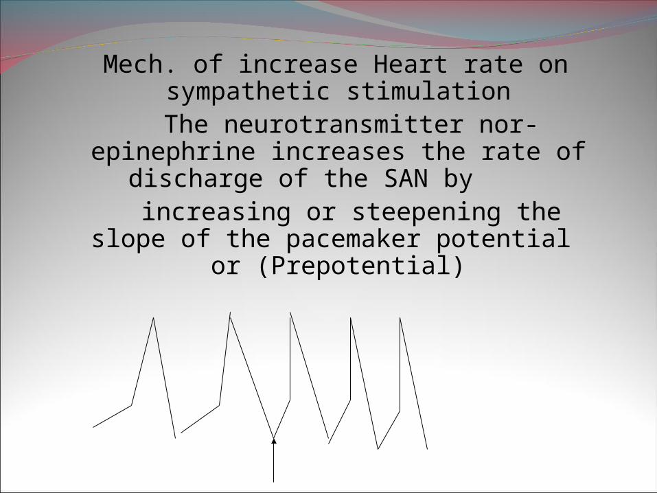
Mech. of increase Heart rate on
sympathetic stimulation The neurotransmitter nor-epinephrine
increases the rate of discharge of the SAN by
increasing or steepening the slope of the pacemaker potential or (Prepotential)

Cardiac Parasympathetic Nerve is the Vagus Nerve:
The preganglionic cardiac vagal fibers originate from three vagal nucleii in the
medulla1. Dorsal motor nucleus of the vagus
2. Nucleus of tractus solitarius 3. Nucleus Ambiguus
Preganglionic vagal fibers run
with the sympathetic nerves to end in parasympathetic ganglion cells in the
wall of the atria, SAN, AVN.
HR decreases during vagal stimulation due to release of the neurotransmitter
Acetylcholinefrom the parasympathetic post ganglionic endings.
ACh decreases the conduction rate in SA node & AVnode by decreasing the slope of prepotential
Postganglionic vagal fibers are short and supply both
atria. No vagal nerve supply to ventricles.
The Right vagus supplies the sino atrial node (SAN) and
the Left vagus supplies the A V Node.

In humans if a parasympatholytic (blocking) drug like atropine is given the HR rises from 70/min to 150 – 180 /min, due tounopposed action of sympathetic nerves. At rest there is a vagal inhibitory tone that keeps the resting HR at 70/min.
If both sympathetic and parasympathetic nerves areblocked, then the HR is about 100/min.
The resting HR is due to a balance between the sympathetic and parasympathetic nerve stimulation.

Causes of cardiac arrest due to vagal stimulation.
Blow on eyeball Blow on eyeball Traction on abdominal viscera Traction on abdominal viscera Effects of parasympathetic (vagal N)
stimulation1.Decrease in HR – vely chronotropic effect.2.Decrease in force of contraction – vely
inotropic effect3.Decrease excitability of heart – vely
Bathmotropic effect4.Decrease conductivity of heart – vely
dromotropic
Moderate vagal stimulation leads to slowing ofHR. Intense vagal stimulation leads to cardiac arrest .
Definition of Vagal Tone:
The tonic discharge of inhibitory impulses in thevagus nerve at rest is known as vagal tone.
Experimental evidence for vagal tone. If both the vagi are cut in an experimental animal
theHR is seen to go up.

II. Role of cardiac centers and cardiovascular reflexes on HR regulation.
The cardiac centers are present in the Medulla or Brain stem. They are
1. Vasomotor Center – cell bodies are present in RVLM that is Rostral ventrolateral medulla.
2. Cardio Inhibitory center – present in the medulla. CIC consists of the three vagal
Nuclei.3. Nucleus Ambiguus (NA)
4. Nucleus of the tractus solitarius (NTS)5. Dorsal Motor Nucleus of vagus.
Efferent nerve from CIC to heart is the vagus Nerve:
CARDIO VASCULAR reflexes
that control vagal tone and Heart rate are1.Baroreceptor reflexes are important
for regulation of BP and HR.Sites are
1. Sino Aortic Baroreceptor reflex (Most Imp)
2. Atrial stretch receptor reflex3. Left ventricular stretch receptor reflex.4. Receptors present in pulmonary blood
vessels.5. Bainbridge reflex.
Role of atrial stretch receptors in control of HR and vagal tone:
Atrial stretch receptors are of 2 types. Type A receptors discharge during atrial
systole and
Type B Receptors which are stimulated by atrial distension due to increased venous
return.
Stimulation causes reflex vasodilation resulting in
hypotension and Tachycardia.
c. Bainbridge Reflex: Bainbridge first described this in 1915.
He observed that rapid infusion of saline in anesthetized animals produces
an increase in heart rate if the intial heart rate is low.
The receptors may be the tachycardia producing Atrial Type B receptors.
Bainbridge reflex is a true reflex as it is abolished by bilateral vagotomy.
It may be a protective reflex to prevent over stretching of atrium.
Physiological significance not known.

Sino aortic baroreceptor reflex.Receptor: Stretch receptors located in the
carotid sinus and aortic arch.Stimulus : increase BP causes stretch of the
baroreceptorAfferent Nerve: from carotid sinus is the sinus nerve branch of glossopharyngeal N. From the aortic arch impulses are carried in the vagus
nerves to the
Center: CIC in the nucleus of the tractus solitarius
Efferent N for S.A reflex is the vagus Nerve. Effector organ is the HEART

Mechanism of Baroreceptor reflex when BP is high
Increase blood pressure (standing to supine)
Increase stretch of carotid sinus
Increase impulse traffic in sinus nerve
Increase stimulation of the cardio inhibitory center
Increased vagal stimulation or Increase in vagal
tone
Decrease Heart Rate

Mechanism of Baroreceptor reflex when the BP is decreased:
Supine to standing the BP is decreased
Decrease stretch of carotid sinus
Decrease impulse traffic in sinus nerve
Decrease stimulation of cardio inhibitory center
Decrease impulses in vagal nerve
Decrease vagal tone
Increase in Heart Rate

Role of left ventricular receptors on HR
Distension of L.V in experimental animals causes hypotension and bradycardia.
Stretch of L.V
Increase impulses in baroreceptor afferents
Stimulate CIC or dorsal M.N of vagus
Bradycardia (increase vagal tone)

II. Chemoreceptor reflexes.important for regulation of respiration, BP & HR.They are
a. Peripheral chemoreceptors present in the carotid body and aortic body.
b. Coronary chemoreflex or Bezold Jarisch reflex.
c. Pulmonary chemoreflex.
d. Cushing Ischemic reflex

The peripheral chemoreceptorsLocation: the carotid body (at the bifurcation of common carotid Art) and aortic body in the arch of
aorta. Stimuli are hypoxia, hypercapnia and acidosis.
Afferent nerve is Glossopharyngeal from Carotid
body and vagus N from the aortic body.
Center:It stimulates the vasomotor center to produce an increase in BP and decrease
HR(Bradycardia).However hypoxia releases catecholamines from
adrenal medulla, which produces Tachycardia (increase HR) and increase in BP.

Coronary Chemoreflex or Bezold Jarisch reflex:
Injection of serotonin,& veratridine into the
left coronary artery which supplies the left ventricle
produces apnea followed by rapid breath and reflex hypotension and Bradycardia.Clinical implication: In myocardial infarction we get hypotension and bradycardia. It is thought that chemical
substances liberated from infarcted tissue stimulate nerve fiber endings, which are the receptors. Afferent nerve is the vagus nerve
Pulmonary chemoreflex.
Injection of serotonin, capsaicin, veratridine
into the pulmonary artery produces apnea followed by rapid
breathing, hypotension and bradycardia. This is mediated by vagal fibers present
close to the lung capillaries.

Cushings Ischemic Reflex:Patient with increase in intracranial pressure
Decrease blood supply to vasomotor center
Local hypoxia and hypercapnia
Stimulation of vasomotor center
Vasoconstriction and increase blood pressure
Increase discharge from baroreceptors
Increased stimulation of CIC
Increase in vagal tone and Bradycardia

Effect of painful stimuli: Stimulation of C fibers causes tachycardia Stimulation of A delta fibers causes
Bradycardia Effect of emotional stimuli on HR Emotions affect HR by acting via higher
centers that is the limbic Cortex and hypothalamus They act on the Cardio inhibitory Center. Anger, excitation increase HR, Fear and grief decrease HR.
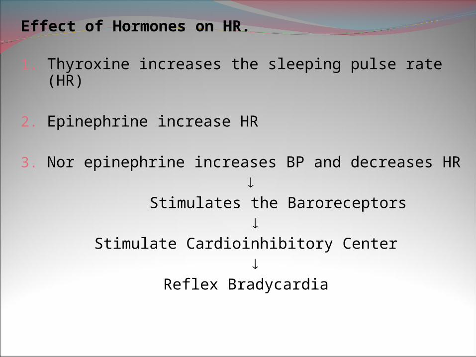
Effect of Hormones on HR. 1. Thyroxine increases the sleeping pulse rate (HR) 2. Epinephrine increase HR 3. Nor epinephrine increases BP and decreases HR
Stimulates the Baroreceptors
Stimulate Cardioinhibitory Center
Reflex Bradycardia


