REGULATION OF POSTNATAL NEUROGENESIS AND BRAIN ANGIOGENESIS BY THYROID HORMONE Liqun Zhang Center for Brain Repair and Rehabilitation Department of Clinical Neuroscience and Rehabilitation Institute of Neuroscience and Physiology at Sahlgrenska Academy University of Gothenburg Sweden 2009
Transcript
REGULATION OF POSTNATAL NEUROGENESIS AND BRAIN ANGIOGENESIS BY THYROID
HORMONE
Liqun Zhang
Center for Brain Repair and Rehabilitation
Department of Clinical Neuroscience and Rehabilitation
Institute of Neuroscience and Physiology at Sahlgrenska Academy
University of Gothenburg
Sweden
2009
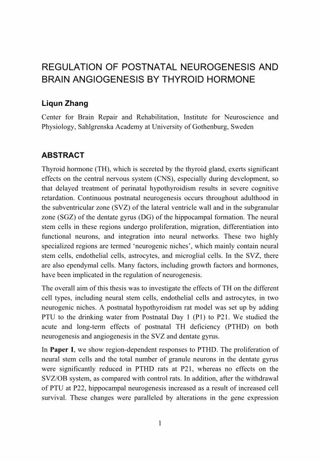
Cover illustration: Sagittal section of rat brain and thyroid gland showing the: dentate gyrus (dark blue), subventricular zone of the lateral ventricle wall (light blue), thyroid gland (orange) and blood vessel (red). Illustration by Nina Hellström.
ISBN 978-91-633-5666-7
1
REGULATION OF POSTNATAL NEUROGENESIS AND BRAIN ANGIOGENESIS BY THYROID HORMONE
Liqun Zhang Center for Brain Repair and Rehabilitation, Institute for Neuroscience and Physiology, Sahlgrenska Academy at University of Gothenburg, Sweden
ABSTRACTThyroid hormone (TH), which is secreted by the thyroid gland, exerts significant effects on the central nervous system (CNS), especially during development, so that delayed treatment of perinatal hypothyroidism results in severe cognitive retardation. Continuous postnatal neurogenesis occurs throughout adulthood in the subventricular zone (SVZ) of the lateral ventricle wall and in the subgranular zone (SGZ) of the dentate gyrus (DG) of the hippocampal formation. The neural stem cells in these regions undergo proliferation, migration, differentiation into functional neurons, and integration into neural networks. These two highly specialized regions are termed ‘neurogenic niches’, which mainly contain neural stem cells, endothelial cells, astrocytes, and microglial cells. In the SVZ, there are also ependymal cells. Many factors, including growth factors and hormones, have been implicated in the regulation of neurogenesis.
The overall aim of this thesis was to investigate the effects of TH on the different cell types, including neural stem cells, endothelial cells and astrocytes, in two neurogenic niches. A postnatal hypothyroidism rat model was set up by adding PTU to the drinking water from Postnatal Day 1 (P1) to P21. We studied the acute and long-term effects of postnatal TH deficiency (PTHD) on both neurogenesis and angiogenesis in the SVZ and dentate gyrus.
In Paper I, we show region-dependent responses to PTHD. The proliferation of neural stem cells and the total number of granule neurons in the dentate gyrus were significantly reduced in PTHD rats at P21, whereas no effects on the SVZ/OB system, as compared with control rats. In addition, after the withdrawal of PTU at P22, hippocampal neurogenesis increased as a result of increased cell survival. These changes were paralleled by alterations in the gene expression
2
patterns of growth factors and apoptotic factors, i.e., Fgf2, Ngf, Wnt3a, Vegfa and Bcl2, at both P21 and P90.
In Paper II, we describe a reduction in angiogenesis at P21 due to PTHD, as evidenced by reductions in the complexity and density of the microvessels, both in the neocortex and dentate gyrus. However, these defects were fully recovered by P90, following PTU withdrawal at P22. In the neocortex, these changes were paralleled by altered levels of VEGFA and FGF2. Furthermore, we report that the physiologic plasma concentration of TH promotes proliferation and tube-like structure formation, and inhibits the death of brain-derived endothelial (RBE4) cells in vitro.
In Paper III, we investigate the region-specific contribution of astrocytes to the activities of neural stem cells after T3 (50 nM) treatment. Conditioned medium (CM) was collected from cultures of SVZ and hippocampal astrocytes after T3 (50 nM) treatment, and was added to the cultures of neural stem cells (NSCs) from the corresponding brain regions. The CM from T3-treated hippocampal astrocyte cultures promoted hippocampal NSCs survival by increasing the proliferation and decreasing the cell death, whereas the CM from T3-treated SVZ astrocyte cultures did not have similar effects on the activities of the SVZ NSCs. Interestingly, the migration of neuroblast from both SVZ and hippocampus was significantly increased after culturing with CM after T3-treatment from corresponding regions. Furthermore, the astrocytes after T3-treatment from these two regions displayed different expression patterns for the Bdnf, Noggin, Wnt3a, Pedf, and Thrb genes, which are implicated in the regulation of neurogenesis.
In summary, this thesis demonstrates the hyper-susceptibility of hippocampal neurogenesis to PTHD including NSCs and astrocytes, and provides evidence of a strong linkage between brain angiogenesis and thyroid hormone levels.
Sköldkörteln, thyroidea, är en körtel som sitter på halsen strax under struphuvudet. Sköldkörtelns hormon är bland annat viktiga för normal tillväxt under uppväxten. Brist på sköldkörtelhormon (hypothyreos) under tidiga barndomsår, samt fördröjd behandling, kan leda till kognitiv utvecklingsstörning, eftersom sköldkörtelhormonerna också har en betydande inverkan på det centrala nervsystemet. I vuxen ålder förekommer det nybildning av nervceller (neurogenes) i två regioner i hjärnan: i subventrikulärzonen (SVZ) i den laterala ventrikelväggen och subgranulärzonen (SGZ) i hippocampus. För att nervcellsnybildningen ska vara möjlig krävs en speciell miljö, den så kallade neurogena nischen. Stamceller, astrocyter och endotelceller är viktiga celltyper i denna miljö. Stamcellerna genomgår tillväxt, migration samt differentiering till funktionella nervceller och integreras i neurala nätverk.
Syftet med avhandlingen var att förstå de akuta och/eller långsiktiga effekterna av postnatal sköldkörtelhormonbrist på neuronala stam/progenitorceller, astrocyter och endotelcellerna i de två neurogena regionerna.
Vi observerade en skillnad mellan de två neurogena områdena i deras svar på akut sköldkörtelhormonbrist. Tillväxten av neuronala stam/progenitorceller och det totala antalet nervceller i hippocampus minskade avsevärt, men ingen effekt sågs i luktbulben. Nervcellsnybildningen i hippocampus återhämtade sig dock efter att normala sköldkörtelhormonnivåer hade återställts, detta genom ökad cellöverlevnad.
Astrocyter, isolerade från SVZ och hippocampus, har visat sig olika uttrycksmönster vid sköldkörtelhormonbehandling in vitro. Hippocampala astrocyter stimulerade av sköldkörtelhormon främjade tillväxt och neuronal differentiering av hippocampala stamceller, men inte astrocyter isolerade från SVZ. Dessutom konstaterade vi att sköldkörtelhormonbrist in vivo, orsakade minskad nybildning av blodkärl (angiogenes) i både hippocampus och hjärnbarken i unga individer, vid vuxen ålder återgick angiogenesen till normala nivåer. Vi har även visat att fysiologiska koncentrationer av sköldkörtelhormon in vitro, främjar spridning, angiogenes och hämmar celldöd hos endotelceller.
Sammanfattningsvis visar denna avhandling att nervcellsnybildningen i hippocampus är mer mottaglig för postnatal hypothyreos, samt att en stark
4
koppling existerar mellan hjärnans angiogenes och dess sköldkörtelhormon-nivåer.
5
LIST OF ORIGINAL PAPERS
The thesis is based on the following papers:
Paper I: Liqun Zhang, Klas Blomgren, H. Georg Kuhn, Christi M. Cooper-Kuhn
Effect of postnatal thyroid hormone deficiency on neurogenesis in the juvenile and adult rat
Neurobiology of Disease (2009) 34: 366-374
Paper II: Liqun Zhang, Christi M. Cooper-Kuhn, Ulf Nannmark, Klas Blomgren, H. Georg Kuhn
In vivo and in vitro effects of thyroid hormone on brain angiogenesis
Accepted by the Journal of Cerebral Blood Flow and Metabolism (2009)
Paper III: Liqun Zhang, Klas Blomgren, H.Georg Kuhn
Astrocytes stimulated by thyroid hormone promote hippocampal neural stem cell growth and neuronal differentiation
Submitted
6
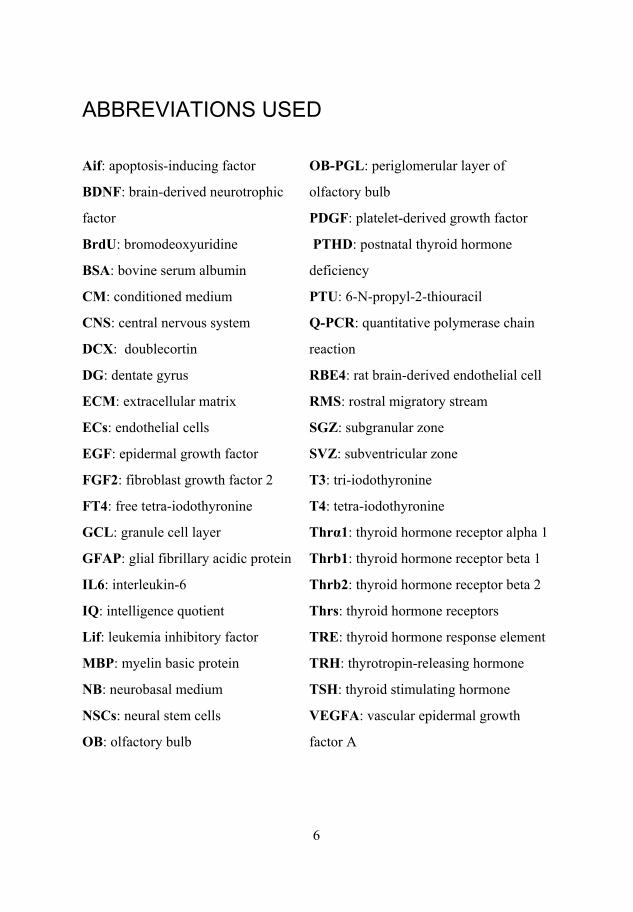
ABBREVIATIONS USED
Aif: apoptosis-inducing factor
BDNF: brain-derived neurotrophic
factor
BrdU: bromodeoxyuridine
BSA: bovine serum albumin
OB-PGL: periglomerular layer of
olfactory bulb
PDGF: platelet-derived growth factor
PTHD: postnatal thyroid hormone
deficiency
CM: conditioned medium PTU: 6-N-propyl-2-thiouracil
CNS: central nervous system
DCX: doublecortin
Q-PCR: quantitative polymerase chain
reaction
DG: dentate gyrus RBE4: rat brain-derived endothelial cell
RESULTS AND DISCUSSION ........................................................................... 27
PTU-induced PTHD results in decreased brain volume and neurogenesis of the dentate gyrus at P21 .............................................................................. 27
Following PTU withdrawal at P22, PTHD rats demonstrate increased cell survival and neuronal commitment in the hippocampus at P90 ................. 28
PTHD has no impact on the volume or neurogenesis of the subventricular zone-olfactory bulb system ......................................................................... 29
Compensatory mechanism of hippocampal neurogenesis after PTU withdrawal at P22 ....................................................................................... 29
PTHD reduces angiogenesis in the dentate gyrus and neocortex at P21 .... 30
Decreased brain angiogenesis caused by PTHD is recovered at P90 ......... 31
Compensatory mechanism of neocortex angiogenesis after PTU withdrawal at P22 ....................................................................................... 31
Stimulatory effects of TH on RBE4 cells in vitro ...................................... 32
Astrocyte isolated from hippocampus and SVZ response differently to T3 treatment ...................................................................................................... 33
CONCLUSION AND OUTLOOK ...................................................................... 35
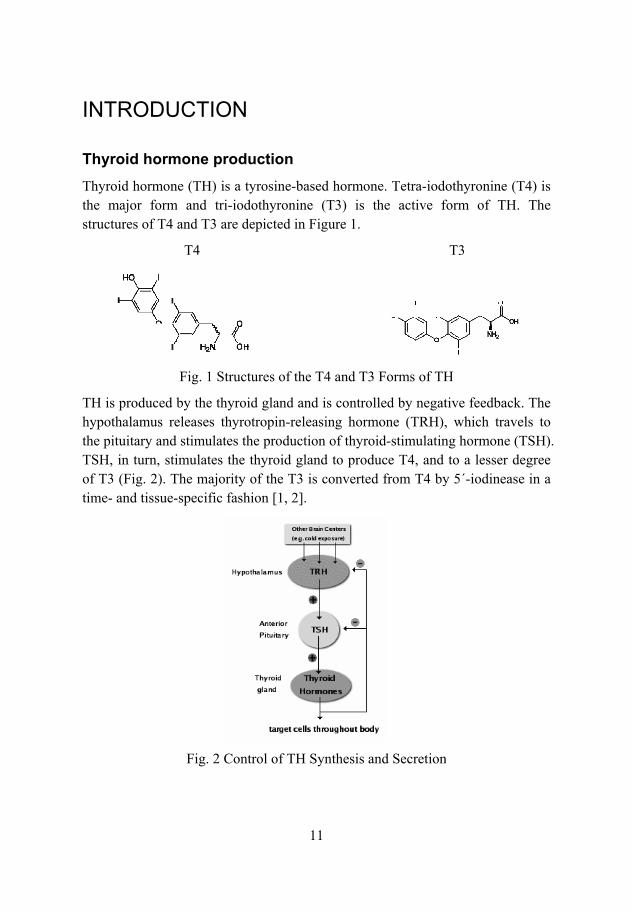
TH receptors and inhibitor The actions of TH are mediated through binding to the TH receptors (Thrs). The major forms of Thrs are: TH receptor 1 (Thra1), TH receptor 1 (Thrb1) and TH receptor 2 (Thrb2). The expression patterns of the Thrs are region- and developmental state-dependent. For example, Thra1 is predominantly expressed in the hippocampus, whereas Thrb is predominantly expressed in the neocortex [3, 4]. Thrs are members of nuclear receptor family and bind both TH and the TH response element (TRE) located in the promoters of target genes. The genomic actions of TH are mediated through the TRE, while the non-genomic actions of TH are mediated through binding beyond TRE [5]. A potent TH antagonist both in vivo and in vitro is NH-3, which binds TH directly, interacts with TRE and inhibits TRE-responsive genes [6].
Adult NeurogenesisNew neurons are generated throughout life through multiple processes, such as neural stem cell (NSC) proliferation, migration, survival/death, differentiation, and functional maturation into neurons [7]. NSCs are found in two distinct regions of the brain: 1) the subgranular zone (SGZ) of the dentate gyrus (DG); and 2) the subventricular zone (SVZ) of the later ventricle wall [8]. Bromodeoxyuridine (BrdU) is widely used to track the newly generated cells from the neurogenic regions. BrdU, which is a thymidine analogue, is incorporated into the DNA strands of dividing cells during the S-phase of the cell cycle and it can be detected by immunocytochemistry [9]. The following factors are known to regulate ongoing neurogenesis: trophic factors, such as brain-derived neurotrophic factor (BDNF) and fibroblast growth factor 2 (FGF2) [10, 11]; hormones, such as estrogen and adrenal steroids [12, 13]; neurotransmitters, such as acetylcholine and serotonin [14, 15]; and extracellular matrix (ECM) components, such as fibronectin and laminin.
Neurogenesis in the adult hippocampus Hippocampal NSCs located in the SGZ proliferate and give rise to neuroblasts, and then migrate to the granule cell layer, in which they send out large dendritic processes. However, they become neurons only after extension of their axons along the mossy fibers to the CA3 region. In the adult hippocampus, approximately 9,000 new cells are generated daily [16]. However, most of these new cells do not survive for more than 2 weeks [17]. Those cells that survive for longer periods differentiate into neurons and become involved in the short-term
13
spatial learning and memory processes [18]. Under physiologic conditions, exercise stimulates hippocampal neurogenesis [19], and the enriched environment promotes the survival of newly generated granule cells [20] and even in the aging brain [21]. Under pathologic conditions, epilepsy [21] and irradiation [22] modulate the mobility of hippocampal stem/progenitor cells.
Neurogenesis in the adult olfactory system The NSCs in the SVZ proliferate locally, migrate along the rostral migratory stream (RMS), and differentiate into either periglomerular neurons or granule cells in the olfactory bulb (OB) [23-25]. Approximately 30,000 new cells are generated daily in the adult mouse OB [8]. The number of new cells is stringently regulated by sensory activity [26]. Recently, it has been demonstrated that neural stem/progenitor cells in the SVZ are heterogeneous and specify the neuronal fate commitment based on the original position of the cells in the SVZ [27].
Neurogenic niches The microenvironments of the NSCs in the SVZ and SGZ are termed neurogenic niches. In addition to NSCs, the niches include astrocytes, endothelial cells, microglial cells, mature neurons, extracellular matrix and so on. In the SVZ, the neurogenic niche also includes ependymal cells. In the SVZ neurogenic niche, primary progenitor cells (type-B, GFAP /S100 /Nestin /Sox2 ) give rise to transient proliferating cells (type-C, Mash1 ) cells, which develop into migrating neuroblasts (type-A, PSA-NCAM ) [28-30]. The SGZ neurogenic niche includes radial glia-like astrocytes (GFAP /Sox2 /Nestin ), which give rise to transient proliferating progenitor cells, which differentiate into DCX neuroblasts, and subsequently into granule neurons in the dentate gyrus [31]. The differences in the subtypes of neural stem cells between these two regions indicate the existence of region-specific neurogenic niches in the adult brain. Furthermore, the interactions that occur between the different cell types are of crucial importance for neurogenesis in each region. In the SGZ of the DG, a dense cluster of dividing cells is located close to the microvessels, which indicates that neurogenesis occurs within an angiogenic niche [32]. Consequently, soluble factors secreted from endothelial cells can promote neuronal differentiation from NSCs [33]. Astrocytes isolated from the hippocampus, but not those from the spinal cord, have been shown to promote neurogenesis from NSCs in a co-culturing system [34]. Although non-neurogenic neural stem/progenitor cells
14
collected from the spinal cord can differentiate into neurons only after transplantation to the neurogenic region of the adult brain [35, 36].
Neural stem cell culturingNeural stem cell (NSCs) isolated from the SVZ or SGZ of the adult mammalian brain can differentiate into neurons, astrocytes and oligodendrocytes when cultured in serum-free medium without growth factors [37]. Furthermore, NSCs can proliferate when cultured in adequate concentrations (20 ng/ml) of growth factors, e.g., FGF2 and EGF [38]. Currently, two NSCs culture systems are used [39, 40]: 1) monolayer culturing on a plastic surface that is coated with fibronectin or laminin; and 2) suspension “free-floating” neurosphere culturing. The monolayer culturing allows better access to the environmental stimuli, whereas neurosphere culturing results in improved cell expansion and survival [41].
AstrocytesAstrocytes, which are the major cell type of the CNS, are characterized by staining for glial fibrillary acidic protein (GFAP), which appears as “stars” [42]; these cells reveal very fine processes when filled with dye [43, 44]. Astrocytes represent a heterogeneous cell population, and mRNA array studies with astrocytes identified different astrocyte-specific gene expression profiles of astrocytes isolated from the different brain regions [45]. In addition to their traditional roles in neuronal support and neurotransmitter re-uptake, astrocytes produce numerous peptide factors, such as interleukin 6 (IL6), which are implicated in the regulation of neurogenesis [46]. Furthermore, astrocytes isolated from the hippocampus, in contrast to those isolated from non-neurogenic regions, such as the spinal cord, can support neuronal differentiation [34].
Angiogenesis in the brain Brain angiogenesis is the process by which new blood vessels are formed from pre-existing microvessels. The angiogenic mechanism is influenced by both physiologic and pathologic conditions [47, 48]. New blood vessel formation is stimulated by the release of proteases from “active” endothelial cells (ECs), which in turn leads to degradation of the basal membrane. The ECs then migrate to the interstitial space, proliferate, form the lumen of the new vessel, generate a new basal membrane, and recruit pericytes. Many factors contribute to
15
angiogenesis, e.g., the members of the vascular epidermal growth factor (VEGF) family [49], FGF2 [50], and platelet-derived growth factor (PDGF) [51].
TH and diseaseDiseases related to TH have been identified. Hyperthyroidism is caused by the overproduction of TH, and the major cause in humans is Graves’ disease, which involves autoimmune-related over-functioning of the thyroid gland, whereas hypothyroidism is caused by the reduced production of TH. In the peripheral system, TH controls energy metabolism and the basal metabolic rate. In the CNS, TH controls each step of neurogenesis and TH receptors are predominantly expressed in neurons, although are also present in oligodendrocytes [52], astrocytes [53], and microglial cells [54] in vitro. The degree of neurologic damage depends on the magnitude and duration of the TH reduction, as well as the developmental stage in which the reduced level of TH occurs. If not corrected shortly after birth, both hyperthyroidism and hypothyroidism lead to irreversible brain damage [55]. The adult-onset hypothyroidism is not so sever as in the development stage, whereas depression, mental fatigue and reduced memory function are the most significant symptoms.
In the developing brain, TH deficiency leads to various neuronal abnormalities in the CNS and cretinism is the severe congenital hypothyroidism in infants. It is manifested by significant long-term defects, such as visual-spatial impairment, language problems, and a lower intelligence quotient (IQ) level in later childhood [56, 57]. These abnormalities include irregular neuronal proliferation [58], delayed migration [59], decreased dendritic densities, and impaired synaptic transmission [60] from specific brain regions. During the maturation of the cerebral cortex, TH deficiency results in a poorly defined cortical layer due to altered migration and inter-hemisphere connections [61, 62]. This may be due to changes in the ECM proteins and dramatic morphologic changes in the cerebellum [63]. In addition, TH promotes in a dose-dependent manner neuronal survival, by preventing the apoptosis of newly formed granule neurons through the action of BCL2 [64].
Astrocytes play a central role in the metabolism of TH, by transporting T4 across the blood-brain barrier into the neural tissues [65, 66] and by converting the non-active T4 form to the active T3 form. TH regulates astrocyte morphology and promotes astrocyte differentiation and maturation [67, 68]. Interestingly, neonatal cerebellar astrocytes increase their proliferation without morphologic differentiation in response to TH [68]. This differential response to TH indicates
16
that TH-mediated effects are dependent upon the stage of astrocyte development. Furthermore, TH regulates ECM components [69, 70], such as laminin and fibronectin, as well as the secretion of growth factors [71], such as FGF2 [70] and EGF [72]. Moreover, TH increases the number and differentiation of microglial cells [54].
TH plays an important role in oligodendrocyte function. In vivo, postnatal TH deficiency leads to significantly decreased myelination, which is correlated with the cerebral concentrations of T3 [73]. Culturing of dissociated brain cells without T3 results in the diminished synthesis of myelin-associated glycolipids, and this reduction can be reversed by adding T3 to the medium [74].
17
AIMS OF THE STUDIES
The overall aim of this thesis was to investigate the responses of different cell types in the neurogenic niches to thyroid hormone.
Specific aims To investigate the acute and long-term effects of PTHD on neurogenesis in two adult neurogenic regions.
To investigate the acute and long-term effects of PTHD on brain angiogenesis and the effect of TH on RBE4 cells in vitro.
To investigate the effects of astrocyte-secreted soluble factors after T3-treatment, on the activities of NSCs from the corresponding adult neurogenic regions.
18
METHODS
Ethical approvals and animals (Papers I, II and III) All animal experiments were performed in accordance with the ethics guidelines of the University of Gothenburg, and were approved by the Gothenburg Committee of the Swedish Agency for Animal Welfare (permits 421-2004 and 26-2008). Wistar rats (Charles River, Sulzfeld, Germany) were used in all the animal experiments.
Pregnant rats (Papers I and II) were housed singly in cages until they gave birth, after which the litters were culled to 8 pups per cage. The litters were randomly divided into control and hypothyroid groups. To establish the hypothyroidism rat model, 6-N-propyl-2-thiouracil (PTU; Sigma-Aldrich, St. Louis, MO, USA) was added to the drinking water (0.1% w/v). The drinking water was replaced daily with fresh PTU solution. Animals underwent PTU treatment from birth until postnatal day 10 (P10) or 21 (P21), and were subsequently sacrificed. For the long-term survival group, PTU treatment was terminated at P22. The pups were weaned at P25, separated according to gender, and housed in groups of 3-4 animals until P90. The P21 rats were used for the isolation of cortical microvessels (Paper II).
Cultures of primary astrocytes (Paper III) were prepared from P1 rats, and cultures of neural stem cells (Paper III) were established from P21 rats.
Immunohistochemistry and immunofluorescence staining (Papers I, II, and III) For the tissue analyses, rats were deeply anesthetized with isofluorane and perfused with 0.9% NaCl, followed by 4% paraformaldehyde. The brains were removed, post-fixed in paraformaldehyde, and then transferred to a 30% sucrose solution. The brains were sagitally cut into 40 m thick sections and stored at 4°C until further analysis. For the in vitro analyses (Papers II and III), cells were fixed with Histofix (Histo Lab AB, Gothenburg, Sweden) and permeabilized with Triton X-100 before staining.
For immunohistochemical staining, the following primary antibodies were used: purified monoclonal mouse anti- -tubulin III (1:200 dilution; Covance, Denver, PA, USA); mouse anti-thyroid hormone receptor beta 1 (THRB1; 1:500; Santa Cruz Biotechnology, Santa Cruz, CA, USA); mouse anti-RECA (1:300; AbD
19
Serotec, Dusseldorf, Germany); mouse anti-NeuN (1:200; Chemicon/Millipore, Billerica, MA, USA); rat anti-BrdU (1:250; BioSite, Täby, Sweden); polyclonal rabbit anti-GFAP (1:200; Dako, Glostrup, Denmark); rabbit anti-thyroid hormone receptor alpha 1 (THRA1; 1:500; Santa Cruz Biotechnology); rabbit anti-phosphor-histone H3 (pHH3; 1:200; Upstate Biotechnology, New York, NY, USA); rabbit anti-tyrosine hydroxylase (1:500; Chemicon); rabbit anti-Prox1 (1:4000; Chemicon); goat anti-DCX (1:250; Chemicon,); goat anti-thyroid hormone receptor beta 2 (THRB2; Santa Cruz Biotechnology); and sheep anti-S100 (1:500; Swant, Bellinzona, Switzerland). For immunoperoxidase detection, the following biotinylated secondary antibodies were used: donkey anti-mouse IgG; donkey anti-rat IgG; donkey anti-rabbit IgG; and donkey anti-goat IgG (all at 1:1000; Jackson ImmunoResearch, West Grove, PA, USA). For fluorescence detection, the following secondary antibodies were used: Alexa 488-conjugated donkey anti-mouse IgG; Alexa 555-conjugated donkey anti-rat IgG; Alexa 555-conjugated donkey anti-rabbit IgG; Alexa 555-conjugated donkey anti-goat IgG; and Alexa 647-conjugated donkey anti-sheep IgG (all at 1:1000; Jackson ImmunoResearch).
PCR and Q-PCR analyses (Papers I, II, and III) For tissue collection, each animal was deeply anesthetized with isofluorane and euthanized by decapitation. The brain was removed and the midline was separated. A portion of the neocortex was removed, to access the lateral ventricle wall. A strip of tissue, which contained both ependymal and sub-ependymal tissues, was pinched off. Hippocampal tissue was collected by removing the entire hippocampal formation, including the DG and CA regions. The dissected tissues were placed in RNA-later (Qiagen, Hilden, Germany), stored at 4°C for 24 hrs, and then transferred to -20°C until total RNA extraction.
For microvessel isolation, the cortex was removed, followed by homogenization and centrifugation at 1,000 × g for 10 min. The collected pellet was suspended in 17.5% dextran, and centrifuged at 4,400 × g for 15 min. The pellet was suspended in 1% bovine serum albumin (BSA), and passed through 100 m and 20 m nylon meshes; the isolated microvessels were stored at -80°C until further total RNA extraction.
Total RNA was extracted using the RNeasy Mini kit (Qiagen), and DNase I (RNase-free, Qiagen) was used to remove genomic DNA. cDNA was synthesized from 2 g of total RNA at 37°C in a 50 l reaction mixture that contained RNase inhibitor, random primers, dNTPs, and M-MLV reverse
20
transcriptase (all from Promega, Madison, WI, USA). For the self-designed primers, the following sequences were used: for Thra1, forward 5 -cccccatctcttctctcctt-3 and reverse 5 -aagttcatttgtcgccctgt-3 ; for Thrb1, forward 5 -tcgctgtagacttggtgtgg-3 and reverse 5 -ctctttggacggagaactgg-3 ; and for Thrb2, 5 -ggtggttattcatcccctctc-3 and reverse 5 -tggcacgcagtagttcattt-3 . For Q-PCR, thermal cycling was initiated at 95°C for 5 min activation, followed by 40 cycles at 95°C for 10 sec, and at 60°C for 30 sec. The primers (Qiagen) for the Aif (QT00180943), Bad (QT00190407), Bax (QT01081752), Bcl2 (QT00184863), Bclxl (QT01081346), Bdnf (QT00375998), Bmp4 (QT00180474), Egf (QT00183449), Fgf2 (QT00189035), Flk (QT00408352), Flt1 (QT00183498), Gadph (QT00199633), Gdnf (QT01084132), Lif (QT00189147), Ngf (QT00506884), Noggin (QT00379337), Pedf (QT00187243), Shh (QT00184912), Thra (QT01169791), Thrb (QT00193690), Vegfa (QT00198954), and Wnt3a(QT00472794) genes were used.
Electron microscopy (Paper II) Animals were anesthetized with isofluorane and euthanized by decapitation. The neocortex was removed and fixed in Karnowsky solution, followed by post-fixation in OsO4 and potassium ferrocyanide. The tissues were treated with uranyl acetate and embedded in agar resin. Sections were examined by electron microscopy, following contrast staining with lead citrate and uranyl acetate.
Assays for plasma T3 and free T4 (Papers I and II) Animals were sacrificed with isofluorane. Blood was collected from the heart and centrifuged at 3000 × g for 20 min to separate out the plasma. The plasma was collected and stored at -20°C. The concentrations of plasma T3 and free tetra-iodothyronine (FT4) were determined using a radioimmunoassay kit (MP Biomedicals; Orangeburg, NY, USA).
Cell counting, microvessel assessments and volume measurements (Papers I and II) The total numbers of cells and microvessels, branching points, and diameters of the microvessels were estimated using a stereology system (StereoInvestigator; MicroBrightField, Williston, VT, USA), which provides a consistent quantitative estimate using unbiased random sampling. In principle, cells or objects of interest located within the counting frames are counted. Two of four counting frame borders were predetermined to be exclusion planes. This counting
21
principle was also applied in the Z-axis. Every 12th DG, SVZ, and cortex, and every 6th OB section were analyzed. The total volume was determined by tracing the area, and the sum of all the areas multiplied by the section thickness and serial section factor (e.g., 12 for the 1:12 series).
The total number of granule cells in the DG was counted in 50 × 50- m counting frames, which were placed in a 250 × 250- m counting grid. The total number of BrdU cells in the granule cell layer of the OB (OB-GCL) was counted in 100 × 100- m counting frames placed in a 500 × 500- m grid. The total numbers of microvessels and branching points, and the average diameters of the microvessels were estimated in 50 × 50- m counting frames placed in a 250 × 250- m counting grid. The numbers of BrdU cells in the DG and in the periglomerular layer of the OB (OB-PGL) were counted.
Quantification of VEGFA and FGF2 levels in neocortex (Paper II) Neocortex tissues were dissected out as described above and stored at -80°C until further analysis. To measure the protein concentrations, the tissues were homogenized in a solution of protease inhibitors (Roche, Basel, Switzerland), phosphatase inhibitor (Sigma-Aldrich), and sodium orthovanadate (Sigma-Aldrich). The supernatant was collected after centrifugation at 3,000 × g for 10 min at 4 C. To obtain the cytosolic fraction, the supernatant was centrifuged at 15,000 × g for 30 min at 4 C. Aliquot of 30 g of total protein were used in the VEGFA and FGF2 assays described below.
Quantification of VEGFA and FGF2 levels in cell culture supernatant (Papers II and III) Culture medium was centrifuged at 1,500 × g for 10 min at 4°C, to remove particles, and then diluted 10-fold with calibrator diluent RD57, as recommended by the manufacturer. The amounts of VEGFA and FGF2 in the culture medium were measured using the mouse VEGFA and human FGF2 immunoassays (R&D Systems, Minneapolis, MN, USA), respectively, which have cross-reactivity of 98% to rat VEGFA and 96% to rat FGF2, respectively.
RBE4 cell culturing (Paper II) For the culturing of rat brain-derived endothelial (RBE4) cells, the culture plates or flasks were coated with 100 g/ml of type I collagen (Roche Applied Science, Indianapolis, IN, USA) before use. The RBE4 cells were maintained in 1:1 (v/v)
MEM/Ham’s F12 medium (Invitrogen, Carlsbad, CA, USA) that contained 10%
22
FBS, 30 mg/ml Geneticin, 1 ng/ml FGF2, and penicillin/streptomycin solution (all from Invitrogen). The RBE4 cells were passaged before they reached confluence, and the medium was replaced every other day. To remove TH from the culture medium, charcoal-stripped FBS (Gentaur, Brussels, Belgium) was used in place of FBS. T3 (10 nM; Sigma-Aldrich) or T4 (100 nM; Sigma-Aldrich) was added to the charcoal-stripped culture medium to investigate the effect of TH on the activities of RBE4 cells [75, 76]. The pan-TH receptor inhibitor NH-3 was used at a concentration of 10 M [6].
Primary astrocyte culturing (Paper III) Primary astrocyte cultures were prepared from P1 rats. Both the hippocampus and SVZ were dissected out [77, 78]. The tissues were mechanically passed through an 80 m nylon mesh into MEM (Invitrogen), which was supplemented with (v/v) FBS Gold (PAA Laboratories GmbH, Pasching, Austria), 1% penicillin-streptomycin (Invitrogen), 1.6 × amino acids (Invitrogen), 3.2 × vitamins (Invitrogen), 1.6 mM L-glutamine (Invitrogen), 7.15 mM glucose, and 48.5 mM NaHCO3. The medium was changed after 3 days of culture and thereafter three times per week. Astrocytes were used after 14 or 17 days in culture when a confluent layer had formed.
Primary neural stem cell culturing (Paper III) Primary NSCs was prepared from the P21 rat hippocampus and SVZ. The tissues were mechanically dissociated by incubation in warm 0.01% Papain (Worthington, Lakewood, NJ, USA)/0.1% Dispase II (Sigma-Aldrich)/0.01% DNase I (Worthington). The dissociated cells were grown in Neurobasal medium (NB; Invitrogen) that was supplemented with B27 (Invitrogen), GlutaMAX™ (Invitrogen), 100 g/ml penicillin-streptomycin (Invitrogen), 2 g/ml heparin (Sigma-Aldrich), 20 ng/ml FGF2 (PeproTech, Rocky Hill, NJ, USA), and 20 ng/ml EGF (Invitrogen). The growth medium was renewed every second day. Primary cultures were passaged by incubation with TrypLE Express (Invitrogen). The number of viable cells was determined by Trypan blue exclusion before use. Cells between the 3rd and 9th passages were used in the assays.
Adhesion assay for RBE4 cells (Paper II) RBE4 cells were tested for adhesion in 96-well plates that were pre-coated with collagen [79].The wells were blocked with BSA, and loaded with 1 x 10⁵ cells per well in 250 l of charcoal-stripped medium with or without TH (10 nM T3 or
23
100 nM T4). The plates were incubated for 2 hrs or 4 hrs before gentle washing. The total numbers of attached cells were measured with the CyQUANT NF Cell Proliferation Assay Kit (Invitrogen).
NSCs proliferation assay (Paper III) NSCs proliferation was determined using the BrdU incorporation assay. The 96-well plates were loaded with 800 NSCs per well in 150 l of NB/B27 that was supplemented with growth factor FGF2 and EFG, and 150 l of conditioned medium (CM) from astrocyte culturing from the corresponding region. For the control wells, astrocytes-derived CM from untreated astrocytes was supplemented with the same amount of residual T3 detected in the CM after T3 treatment. BrdU (10 M) was added to the NSCs for 2 hrs. The incorporated BrdU was quantified using a BrdU labeling and detection ELISA kit (Roche Diagnostics, Indianapolis, IN, USA).
Migration assay for RBE4 cells (Paper II) RBE4 cell migration was measured using an 8 m pore size Transwell system (Corning Life Sciences, Lowell, MA, USA). The upper chamber was coated with collagen before use, and then loaded with 1 × 10⁵ cells in 100 l of charcoal-stripped RBE4 culture medium. The lower chamber was filled with 600 l of charcoal-stripped culture medium with or without TH (10 nM T3 or 100 nM T4). The Transwell apparatus was incubated at 37°C for 4 hrs, after which the cells in the upper chamber were gently washed away. The total number of migrated cells in the bottom chamber was quantified under a microscope in ten random fields at 20 × magnification.
Migration assay for neurosphere (Paper III) Neurosphere migration in response to astrocytes-derived CM was determined by measuring the area of migrating neuroblasts embedded in the Matrigel (BD Biosciences, San Jose, CA, USA). Neurospheres of similar size from the hippocampus and SVZ were used in this assay. Neurosphere cells in 250 l of NB/B27 medium without growth factors were mixed with 750 l of Matrigel. Then, 60 l of the mixture was added to each well of a 96-well plate and incubated at 37°C for 30 min to allow gel formation. Thereafter, culture medium, which consisted of 50% NB/B27 without growth factors and 50% astrocytes-derived CM after T3-treatment, was added. For control wells, 50% CM collected from untreated astrocytes was supplemented with the same amount of residual
24
T3 detected in the CM after T3 treatment. This combinatorial medium was renewed daily for 3 days. The wells were fixed with Histofix (Histo Lab). Images were taken and analyzed using the ImageJ software (available at http://rsb.info.nih.gov; developed by Wayne and Rasband, National Institutes of Health, Bethesda, MD) and the NeuronJ plug-in (available at http://www.imagescience.org/meijering/software/neuronj; developed by Erik Meijering, Swiss Federal Institute of Technology, Zürich, Switzerland).
Tubulogenesis assay for RBE4 cells (Paper II) The tubulogenesis of RBE4 cells in response to TH was assessed by the total length of the tubes formed in the Matrigel. In total, 8.75 × 10⁴ cells were mixed with 500 l of Matrigel, and 50 l aliquots of this mixture were loaded into the wells of a 96-well plate and incubated at 37°C for 30 min to allow gel formation. Then, 250 l of RBE4 cell culture medium was added and the plate was incubated at 37°C for 48 hrs. After washing, the cell medium was replaced with charcoal-stripped RBE4 cell culture medium with or without TH (10 nM T3 or 100 nM T4). The medium was replaced daily for 4 days. Images were captured, and the total length of the capillary-like network that formed in the Matrigel was measured using the ImageJ software and the NeuronJ plug-in.
Expansion assay for RBE4 cells (Paper II)RBE4 cell expansion was determined in a collagen-coated 96-well plate. Thus, 5,000 RBE4 cells in 250 l of RBE4 medium were added to each well and incubated at 37°C for 4 hrs to allow cell adhesion. After gentle washing, 250 l of charcoal-stripped RBE4 medium was added to each well with or without TH (10 nM T3 or 100 nM T4). The medium was replaced daily for 4 days. The total cell number was determined using the CyQUANT NF Cell Proliferation Assay kit (Invitrogen).
Expansion assay for NSCs (Paper III) NSCs expansion was determined in a 96-well plate that was coated with polyornithine (10 g/ml; Sigma-Aldrich) and fibronectin (1 g/ml; Sigma-Aldrich). Thus, 400 cells were added to each well of a 96-well plate in 300 l NB/B27 medium that was supplemented with factors (heparin, FGF2, and EGF). The NSCs were incubated at 37°C for 36 hrs, to allow attachment. Thereafter, the medium was switched to 50% NB/B27 supplemented with the above factors and 50% CM collected from T3-treated astrocytes. As a control, the astrocytes-
25
derived CM from untreated astrocytes was supplemented with the same amount of residual T3 detected in the CM collected after T3 treatment. The medium was renewed every 2 days for a period of 8 days. On Day 8, 10 l of Premix WST-1 were added to each well, followed by incubation for 4 hrs. Cell expansion was determined using a cell proliferation assay (Premix WST-1; TaKaRa Bio).
Cell death assays for RBE4 cells and NSCs (Papers II and III) RBE4 cell death was determined in collagen-coated 24-well plates. Thus, 5,000 cells were added to each well and incubated at 37°C for 24 hrs. After washing, the medium was replaced with charcoal-stripped RBE4 culture medium with or without TH (10 nM T3 or 100 nM T4). Medium replacement was performed daily for 4 days.
The NSCs death assay was determined in 24-well plates that were coated with polyornithine (10 g/ml; Sigma-Aldrich) and fibronectin (1 g/ml; Sigma-Aldrich). A total of 2.78 × 10⁵ NSCs was cultured for 36 hrs in NB/B27 that was supplemented with growth factors, to allow cell attachment. After gentle washing, the medium was switched to medium that contained 50% NB/B27 and 50% CM collected from T3-treated astrocytes. For the control wells, the CM collected from untreated astrocytes was supplemented with the same amount of residual T3 detected in the CM after T3 treatment. The medium was renewed daily for 3 days.
The attached cells were detached using Trypsin (Invitrogen) and divided into two parts. One part was used for the measurement of cytoplasmic histone-associated DNA fragments using the Cell Death Detection ELISA kit (Roche), while the remaining part was used for total cell measurement using the Premix WST-1 cell proliferation kit (TakaRa Bio, Otsu, Japan). Cell death was calculated as the total amount of DNA fragmentation with correction for the total cell number in the corresponding well.
NSCs differentiation assay (Paper III) NSCs differentiation assay was performed in an 8-well chamber slide (Daigger, Vernon Hills, IL, USA) that was coated with polyornithine and laminin before use. In total, 5 × 10³ NSCs were loaded into each chamber and cultured in medium that contained 50% NB/B27 culture medium without growth factors and 50% CM from T3-treated cultures of astrocytes derived from the corresponding region of the brain. For the control wells, CM collected from untreated astrocytes was supplemented with the same amount of residual T3 detected in the CM after
26
T3 treatment. This combinatory medium was renewed every 3 days for 12 days. The following primary antibodies were used in the assays: for neurons, purified monoclonal mouse anti- tubulin III (1:200; Covance); and for astrocytes, polyclonal rabbit anti-GFAP (1:200; Dako). The following secondary antibodies were used: Alexa 488-conjugated donkey anti-mouse IgG; and Alexa 555-conjugated donkey anti-rabbit IgG (both at 1:1000; Jackson ImmunoResearch). ProLong Gold with DAPI (Invitrogen) was used to label the nuclei.
27
RESULTS AND DISCUSSION
Paper I: Neurogenesis
PTU-induced PTHD results in decreased brain volume and neurogenesis of the dentate gyrus at P21 In the postnatal rat brain, important developmental processes, such as the production of granule cells in the dentate gyrus, olfactory bulb, and cerebellum, are ongoing [80, 81]. During this period, cell proliferation plays a key role in neurogenesis. To study the effects of postnatal thyroid hormone deficiency (PTHD) on brain development, rats were treated with PTU, which is a reversible blocker of TH synthesis, from birth until P21.
To investigate the acute effects of PTHD on neurogenesis in the hippocampus, rats were sacrificed at P22. The rats that were treated postnatally with PTU showed significantly smaller volumes for the dentate gyrus and each subregion, as compared to control rats. This reduction in volume may be attributed to either a decrease in cell number and/or an increased cell density of granular cells in the dentate gyrus. According to the previous study, it is possible that continued thyroid hormone deficiency into adulthood results in a further decrease of the volume in the GCL of the dentate gyrus [82].
We determined the total number of granule cells in the dentate gyrus. Granule cells are mainly produced postnatally, which corresponds to the experimental hypothyroid period. Using Prox1, which a marker of dentate gyrus granule cells, we observed a significant decrease in the total number of neurons at P21 due to PTHD. We also detected a significantly lower total number of dividing cells that were positive for phosphor-histone H3 (pHH3), which is a marker of cells in the M-phase of the cell cycle. This is in accordance with a similar study using a primary inherited hypothyroid model, which demonstrated decreased proliferation in the dentate gyrus of juvenile mice [83]. Since the continued neurogenesis in the dentate gyrus is functionally important for short-term spatial memory and involved in the brain cognition [84], our findings are in accordance to the cognitive deficits and learning impairment observed in patients [85, 86]. We conclude that PTHD has a detrimental effect on postnatal hippocampal neurogenesis.
28
Since the brains of the PTHD rats were smaller, we also examined the cell densities, to determine whether the number of neural stem/progenitor cells was normal relative to the smaller size of the hippocampus. There was no difference in the densities of the pHH3 cells in the subgranular zone of the dentate gyrus in PTHD rats, as compared to the control at P21. This indicates that although there is a reduction in the number of cells, the pool of proliferating cells in the dentate gyrus is active relative to the smaller size of the dentate gyrus, and that neural stem cell activity is not completely exhausted even after 3 weeks of PTHD. Other studies have suggested that the neural stem cell pool is not permanently depleted by cell death, but rather reduced in size due to cell cycle arrest during TH deficiency [87]. We hypothesize that these findings have implications for the structural and functional recovery in hippocampus after PTU was withdrawal at P22.
Following PTU withdrawal at P22, PTHD rats demonstrate increased cell survival and neuronal commitment in the hippocampus at P90 New neurons continue to be generated in the adult rodent hippocampus and SVZ [88] even in the elder human brain [89]. To study the long-term effect of PTHD on adult neurogenesis, PTU-treatment was terminated at P22, and the animals were sacrificed at P90. To address the long-term effect of PTHD at P90, we investigated several aspects of adult hippocampal NSCs, i.e., cell proliferation, migration, differentiation, and survival/integration. We observed no differences in PTHD rats in terms of the density or number of pHH3 cells in the SGZ of the dentate gyrus, as compared with control rats at P90. We used DCX, which is a marker of neuroblasts, to estimate the number of neuronal committed progenitor cells. We also observed no difference in PTHD rats on the density of DCX cells as compared with control rats. To study the effects on survival and differentiation of the newly generated cells, we administered BrdU at P60, and analyzed the brains at P90. Although there was no change in the number of BrdU+ cells, a significantly increased cell density of BrdU cells was noted in the PTHD rats, as compared with control rats at P90. Furthermore, the cell fate determination of surviving cells was estimated by triple fluorescence with the neuronal marker Prox1 and glial marker S100 , together with BrdU. The PTHD rats exhibited an increased density of new neurons (BrdU /Prox1 ), but no difference of new astrocytes (BrdU /S100 ) in the GCL of the dentate gyrus, as compared with control rats. These results suggest that, in the smaller dentate
29
gyrus of PTHD rats, more cells survive, and possibly indicating a compensatory mechanism for PTHD. And the more new neurons added in the hippocampus might contribute the recovery of cognitive deficit.
PTHD has no effect on the volume or neurogenesis in subventricular zone and olfactory bulb system To test whether the effects of PTHD are specific for the dentate gyrus, we investigated the SVZ/OB system. Surprisingly, we observed no differences in the volumes of SVZ and OB at either P21 or P90 in PTHD rats, as compared with control rats. In addition, no difference in the density of pHH3 cells at either P21 or P90. Furthermore, no difference in the cell density of BrdU cells either in OB-GCL or OB-Glom regions in PTHD rats at P90. Therefore we conclude that PTHD has no effect on SVZ/OB neurogenesis and neural stem cells in the SVZ/OB is unaffected by PTHD. It indicates that the two neurogenic niches are different when response to PTHD. These results are in contrast to some previous studies, hypothyroid wildtype and transthyretin null mice demonstrated a decreased proliferation of NSCs in the SVZ [90]. And adult-onset hypothyroid rats demonstrated the reduced production of NSCs and neuronal migration in the SVZ/OB system [91]. This discrepancy could due to strain difference, quantification methods, the period during which PTU was delivered, or various region- and developmental stage-specific expression of the thyroid hormone receptors [92]. And it is also possible that NSCs in the SVZ posses better compensatory mechanisms than NSCs in the hippocampus.
Compensatory mechanism of hippocampal neurogenesis after PTU withdrawal at P22 In the hippocampus, we observed the increased cell survival and neuronal differentiation in PTHD rats, as compared with control rats at P90, although no difference in the proliferating cells at either P21 or P90 rats. Since cell survival reflects a balance between cell generation and cell death, we investigated the mRNA levels of genes that regulate neurogenesis and apoptotic cell death. In quantitative PCR of hippocampal tissue, we observed the decreased the mRNA levels of Ngf, Vegfa, Fgf2, and Wnt3a in PTHD rats, as compared with control rats at P21, whereas no difference was found at P90. Since previous studies demonstrated that FGF2 regulate adult neurogenesis through increased neuronal differentiation in SVZ/OB system [93], NGF acts only on the survival of neuronal progenitor cells [94], VEGFA can increase neurogenesis in both
30
hippocampus and SVZ/OB system [95]and Wnt signaling pathway is associated with neuronal specification in the dentate gyrus [96], it indicates a compensatory mechanism in the mRNA level in the PTHD rats after PTU withdrawal.
Cell death of the newly generated cells is the major factor that involved in the regulation of adult neurogenesis [18, 97]. In our study, the mRNA levels of the anti-apoptotic gene Bcl2 were decreased and the pro-apoptotic gene Bax was increased at P21 and no difference at P90 in PTHD rats, as compared with control rats. Furthermore, we detected no change in the mRNA level for the apoptosis-inducing factor Aif in PTHD rats at P21, but this level was decreased at P90, as compared with control rats. Although the changes of mRNA levels do not necessarily reflect changes in the corresponding protein levels, it indicates that apoptotic cell death is affected by PTHD.
Taken together, these results suggest that once TH levels return to normal, intracellular conditions that favor cell survival is induced and try to compensate the deficits of PTHD. Our results are in accordance to previous studies, such as cholinergic and cytochrome oxidase activity, dentate gyrus neuronal fiber layers and hippocampal functional metabolic activity were recovered when the levels of TH are normal [98-100].
Paper II: Angiogenesis
PTHD reduces angiogenesis in the dentate gyrus and neocortex at P21 Angiogenesis, which involves the formation by endothelial cells of new blood vessels from pre-existing microvessels, is influenced by physiologic and pathologic conditions, such as stroke and ischemia [47]. Brain microvessels are specialized, since they are part of the blood-brain barrier, which allows only certain molecules to enter the neural parenchyma. Since an adequate blood supply is crucial for brain development and function, we investigated the effects of PTHD on microvessels. We used RECA, an endothelial cell marker, to examine angiogenesis in the dentate gyrus and neocortex. We determined microvessel density by counting the total number of microvessels in systematically placed counting frames. In addition, we determined the diameters of the microvessels and microvessel complexity by quantifying the vessel branching points. We observed similar effect of PTHD in both the neocortex and dentate gyrus. PTHD resulted in significant reduced microvessel density and
31
complexity, and larger diameter at P21, as compared with control rats. These changes were considered to be a consequence of pathologic condition of PTHD, rather than a developmental retardation of the brain vascular system, because no significant changes of these parameters were observed during the postnatal period from previous study [101]. The reduced capillary complexity and density might explain the dramatically decreased cerebral blood flow in PTU-treated rats [102] and changes in the blood-brain barrier nutrient transport in iodine-deficient animals [103]. Furthermore, we used electron microscopy to investigate the ultrastructure of brain capillary at P21. And no changes were observed in terms of endothelial cell ultrastructure, pericyte morphology, basal membrane configuration or astrocyte endfeet morphology. The EM results demonstrated that the enlargement of vessel diameter did not result in any structural alterations to the vessel wall, which indicates the recovery possibility of the brain microvessels when the levels of TH reach to normal. In addition, reduced angiogenesis may disrupt nutrient and growth factor supply, which would have negative effects on neurogenesis and neuronal development and consequently contribute to impaired brain function.
Decreased brain angiogenesis caused by PTHD is recovered at P90Following the withdrawal of PTU at P22, the brain microvessels of the rats were investigated at P90, at which time-point the TH levels had returned to the control levels. The impairment of brain angiogenesis was fully recovered to the level seen in the control group, with respect to microvessel density, complexity and diameter. These results indicate that the effects of PTHD on brain microvessels are transient.
Compensatory mechanism of neocortex angiogenesis after PTU withdrawal at P22 Many factors contribute to the promotion of angiogenesis in the CNS, such as VEGFA specially act on endothelial cells (ECs), whereas FGF2 activates a broad range of target cells, including ECs. We measured the levels of VEGFA and FGF2 in the neocortex. PTHD resulted in significantly reduced levels of VEGFA and FGF2 in the neocortex at P21, whereas no difference of them at P90. This result is accordance to previous study in a chick chorioallantoic membrane model, TH exhibited FGF2-dependent pro-angiogenic effects [75] and patients who have lower VEGF blood level due to short-term TH deficiency, can be
32
recovered by TH treatment [104]. In addition, we also observed decreased levels of mRNA for Fgf2, Vegfa, and the VEGF receptors Flt1 and Flk1 in PTHD rats at P21, while no differences of them were noted at P90, as compared with control rats. This result is in accordance to previous study that demonstrated substantial coronary angiogenesis was coincided with the upregulated Fgf2 expression [105]. We conclude that the compensatory mechanism exist after PTU withdrawal and can fully recover the deficits of PTHD on brain angiogenesis.
Stimulatory effects of TH on RBE4 cells in vitroThe central element of angiogenesis is the endothelial cell. Therefore, we investigated the effect of TH in the range of physiological levels on the activities of the RBE4 in vitro. We observed no effect of TH on RBE4 cell adhesion or migration. However, TH treatment resulted in reduced apoptotic cell death, increased cell expansion of RBE4 cells and the formation of tube-like structures. This indicates a stimulatory effect of TH on RBE4 cells in vitro. Furthermore, TH increased the levels of Vegfa and Fgf2 mRNA in RBE4 cells, and promotes the secretion of VEGFA and FGF2 from RBE4 cells. It indicates an autocrine regulation of TH on RBE4 cells. However, we cannot exclude the fact that higher levels may have different effects in our system. And previous study demonstrated that T3 (1 M) exhibited no effects on tubulogenesis with human foreskin ECs [106] and T3 ( 1 nM) reduced cell adhesion, but promoted migration in human mesenchymal stem cells [107].
Paper III: Astrocyte-Stem Cell Interactions
T3-stimulated astrocytes promote hippocampal NSCs cell growth and neuronal differentiationAstrocytes represent a major component of the neurogenic niche and are in intimate contact with neural stem cells. Previous studies have demonstrated that, in the intact and injured brain, astrocytes secrete growth and inflammatory factors, thereby regulating neuronal survival and regeneration [108, 109]. We hypothesized that astrocytes are important mediators of the effects that TH has on stem cell activity. To explore this hypothesis, we culture astrocytes from both neurogenic regions, i.e., the hippocampus and SVZ. Astrocyte-conditioned culture medium was collected after incubation with or without T3 (50 nM).
33
NSCs were then cultured with conditioned medium from the corresponding region. When hippocampal NSCs were cultured with conditioned medium derived from T3-treated astrocyte cultures, the rates of proliferation, survival, neuronal differentiation, and neuroblast migration were increased, while the rate of cell death was reduced. We conclude that hippocampal NSCs are dependent on astrocytes when response to T3 and astrocyte-derived factors may play a significant role in mediating the effects of TH.
T3-stimulated astrocytes promote SVZ neuroblast migrationSince we observed different in vivo responses of the adult SVZ neurogenesis to PTHD (Paper I), we decided to investigate the effects of NSCs isolated from SVZ cultured in SVZ astrocyte-derived conditioned medium after T3 (50 nM) treatment. The conditioned medium from T3-treated astrocytes had no effect on cell proliferation, survival, cell death or neuronal differentiation, although increased neuroblast migration was observed. These results confirmed the vulnerability of hippocampal NSCs niche to TH deficiency in vivo. This region-specific response of NSCs raises questions as to whether this effect is due to some factor(s) in the astrocyte-derived conditioned medium, or whether this difference can be attributed to the NSCs themselves.
Astrocyte isolated from hippocampus and SVZ response differently to T3 treatment Previous studies have demonstrated that astrocytes are heterogeneous population and have different gene expression profiles isolated from different brain region. We performed quantitative PCR analysis on astrocytes harvested from two regions of the brain after T3-treatment. The expression levels of the Bdnf, Noggin, Lif, Wnt3a and TH receptor alpha (Thra) genes were up-regulated in the hippocampal astrocytes but not in the SVZ astrocytes after T3-treatment, as compared with none-treated groups. In contrast the levels of Pedf mRNA were increased exclusively in the SVZ astrocytes. Vegfa and TH receptor beta (Thrb) were up-regulated in the astrocytes from both the hippocampus and SVZ. Furthermore, we observed increased levels of VEGFA in the supernatants of the astrocytes cultures from both the hippocampus and SVZ after T3-treatment. Therefore we conclude that astrocytes in hippocampus and SVZ responded differently with T3-treatment. These results indicate that hippocampal astrocytes can provide significant trophic support for NSCs, which is dependent upon the presence of T3. However, our results do not exclude the possibility that the
34
different response of NSCs to CM is due to the intrinsic properties of NSCs, since previous studies also demonstrated that SVZ and hippocampal NSCs follow distinct development lineage [110-112].
Differential responses of hippocampal and subventricular NSCs have been observed under various physiologic and pathologic conditions. For example, although Ascl1 gene overexpression occurs in the NSCs of both adult neurogenic regions, only the hippocampal NSCs can redirect themselves from neuronal to oligodendrocytic differentiation, albeit not in the SVZ/OB system [21]. In addition, running and enriched environment increase hippocampal neurogenesis, whereas have no effect in the SVZ/OB system [113]. Under the lesion condition, only progenitor cells from the SVZ migrate over a long distance to the lesion site [114]. In addition, the NSCs in the SVZ seem to be more resilient than their hippocampal counterparts, since SVZ/OB neurogenesis can recover from irradiation (1 × 6 Gy), whereas hippocampal neurogenesis does not recover from this dose of irradiation [22].
35
CONCLUSION AND OUTLOOK
CONCLUSIONBased on the data presented in this thesis, we conclude that NSCs in the hippocampus are more vulnerable than that in the SVZ/OB system to PTHD. Continued adult hippocampal neurogenesis can partially compensates for the PTHD although a significantly less-developed dentate gyrus remains. Hippocampal astrocytes provide trophic support for the dentate gyrus neurogenesis process in a T3-dependent manner. We first detected that PTHD was associated with extensive defects in brain angiogenesis, which suggests that the response to TH involves interactions between several cell types. Although angiogenesis was fully recovered at P90, the insufficient supply of nutrients during a key developmental phase for the hippocampus may have a significant impact on the development of functional deficits.
OUTLOOKIn our third study, we show that the hippocampal astrocytes-derived CM promote hippocampal NSCs growth and neuronal differentiation after T3-treatment, whereas this was not the case for SVZ astrocytes. Although we observed differences between the astrocytes of the two neurogenic niches after T3-treatment, we cannot exclude the possibility that the different responses of the NSCs might due to the difference between the NSCs themselves when response to TH. It will be very interesting to investigate the response of NSCs derived from the two neurogenic niches to T3 stimulation in vitro. This might improve our understanding of the behaviors of NSCs from two distinct neurogenic niches under both physiological and pathologic conditions.
Previous studies have revealed structural and functional defects in patients who have missed the critical postnatal window for TH replacement therapy. Our study demonstrates a reduction of approximately 20% in the number of hippocampal granule neurons for PTHD rats. Upon diagnosis, perinatal hypothyroidism is clinically treated by TH replacement therapy, which is effective in restoring normal levels of TH. However, there is currently no therapy to reverse the structural deficits caused by it, which will subsequently hinder the proper cognitive development of the patient. It is very important to develop a therapy that will improve the life quality for them. With respect to hippocampal formation, voluntary running stimulates cell proliferation and neurogenesis [115]
36
and has positive effects on learning and LTP [19], while in the cortex, voluntary running stimulates the proliferation of astrocytes and microglial cells [116]. In addition, physical exercise induces the production of neurotrophic factors [117], which are important for promoting angiogenesis, neurogenesis, and brain plasticity. Another study demonstrated that prolonged exercise induced angiogenesis and increased cerebral blood volume in primary motor cortex of rat [118]. Furthermore, physical exercise-induced increase of cerebral blood volume in the dentate gyrus was correlated with postmortem measurement of neurogenesis by MRI study in human [119]. In future experimental series, it is planned to investigate the effects of long-term voluntary running on the structural and functional recovery of PTHD animals in order to improve neuronal rehabilitative therapy, thereby enhancing the quality of life for patients with severe postnatal hypothyroidism.
37
ACKNOWLEDGMENTS
The work described in this thesis was performed at the Center for Brain Repair and Rehabilitation (CBR) at the Institute of Neuroscience and Physiology of Göteborg University. I would like to express my sincere gratitude to all the people who helped me over the several years of this work. Special thanks go to:
Georg Kuhn, my main supervisor. Thank you so much for accepting me into your research group, for giving me the opportunity to set up many experiments independently, and for listening to me and understanding me. You are an expert in our field, and your thoughts and idea elevate my work to a much higher level. Thanks also for your exceptional English writing support. Thanks for everything!!!
Klas Blomgren, my co-supervisor. Thank you for providing financial support when I was registered as a PhD student. I sincerely appreciate your original idea for my thesis and our good collaboration. I am looking forward to future collaborations with you!
In memoriam: Peter Eriksson, Thank you for introducing me to Georg and giving me the opportunity to work in the field of neuroscience. I remember the first day that we met in the Department of Clinic Neuroscience. I miss you!
Birgit Linder, Ann-Marie, and Rita Grandér, Thank you for keeping track of all the lab supplies. I always get excellent assistance from all of you whenever I set up a new experiment. You are the best lab assistants!
Christi Kuhn, Thank you for correcting my manuscript. Thank you for the big hug when I told you I was pregnant. Thank you for the parties in your house. I wish you the best!
Nina Hellström, It is a great pleasure to get to know you and share many things with you about work, life, and culture. I learned a lot from you about being an independent woman, and I am very happy for the changes. Thanks for everything and I wish you the best life!
Olle Linderg, You are a very nice and kind office-mate. Thank for correcting my Swedish writing in the thesis. I wish you the best!
Åsa Persson, Thank you for the nice dinner in your apartment. You are such wonderful company. I am glad to see you back after your trip to Asia. I wish you good luck in your work!
38
Martina Boström, I enjoy listening to you talking about life. Thank you for those relaxing moments. I wish you the best!
Guilia Zanni, It is very nice to know you. Thanks for your caring and the warm “welcome to Italy”. Keep in touch! I will let you know if I get chance to visit Italia!
Axel Jansson, I really miss the time when you were here. Thanks for showing me this computer stuff!
Marie Nilsson, Thank you for the very fruitful collaboration, and I am looking forward to the next one!
Andrew Naylor, the gentleman who says “lady first” in the corridor. Thank you for bringing me the “running idea” and good scientific suggestions!
Tomas S Scanlan: Thank you for providing the thyroid hormone receptor antagonist NH-3 for the second paper!
Ulf Nannmark: Thank you for the collaboration on the EM work!
Johan Rung, Thanks for the help of my failed behavior experiment and the friendly chat!
Linda Paulson, Thank you for the party in your apartment and the visit to Oslo!
Johanna Zandén, Thanks for your warm wishes when Sherry was born and for the good organization in the lab!
Niklas Karlsson, Thank you for the help with intellicage - we learnt a better way to solve the problems. I will always be by your side whenever you need help!!
Maurice Curitis, Thank you for the many things you told me when we made the trip together to Oslo!
Tomoyo Ochiishi, It is a real pleasure to get to know you. Thanks for the very friendly chat and scientific suggestions!
Yoshiaki Sato, Thanks for the friendly chat and the very warm and thoughtful Christmas card!
Changlian Zhu and your students, Thanks for the nice working atmosphere and company over many weekends and late evenings in the empty CBR!
Milos Pekny and Marcela Pekna, Thanks for the exciting seminars!
Lizhen Li, Thanks for the friendship and chat in my apartment!
39
Michelle Porritt, Thanks for the many “Great questions” and wonderful chat!
Henrik Landgren, Thanks for the many “Great questions” and suggestions regarding RNAi transfection!
Jonas Faijerson, Thanks for the encouragement and suggestions for cell work!
Thanks for all the colleagues in the CBR or 4th floor: Ali Reza Kazemi, Amed Osman, Anders Ståhlbergg, Angelica Kloberg, Anke Brederlau, Anna Wolf, Cecilia Bull, Charlotta Lindwall, Daniel Andersson, Fredrik Blomstrand, Gunnel Nordström, Helene Andersson, Ina Nodin, Jenny Nyberg, Karin Gustafsson, Karin Hultman, Karolina Roughton, Lina Bunketorp Käll, Lind Hou, Marie Kalm, Mathilda Zettersttröm Axell, Maryam Faiz, Michael Nilsson, Mila Komitova, Pete Smith, Sachiyo Misumi, Ulrika Wilhelmsson, Åsa Widerstrand and so on.
To my friends,
Dajin, I want to tell you how wonderful you are. Whatever happens, you are someone I can always count on - without fail. I appreciate your gracious heart that always makes me smile!
Haiyan, I miss the time when we studied, exercised, and worked together in Beijing! Thanks for the great help, especially when my mom was sick in Beijing!
Meihua, my most humorous friend, I am so glad to have you as a friend in my life. Whatever happens, you are someone who makes me laugh again. It is really great that we shared the abroad experience and you are the great Mom!
Yan Jiang, my most smart friend, I miss the time when we studied TOFEL and GRE together. I admire your strength to persevere to get what you want in science. You are an excellent lady!
Huipeng Chen, Qingming Wang, Jizhong Chen, and Guochang Fan, Thanks for the opportunity and support to go down the road of science!
Aunt Zhou’s family, Xuejing’s family and other school friends, Thanks for the friendship beyond age. For all the great times we spent together cooking, dancing, singing, writing, computer work, paper folding, fun time in the red summer house, beach and so on. All this make me feel that Sweden is another home!
40
YingFei and Lizhong, Thanks for the many great dinners in your house, and the karaoke, dancing and relaxing chats and joking! It means a lot to me, especially when I am far away from China.
Lina, Wong, and Ryan, Thanks for introducing me to a new world, and for the great time with me when I was alone. Thanks for your patience in improving my English and love you Ryan!
Johan and Anna, Thank you for improving my English and Swedish! It was kind of you to introduce and explain Swedish culture and history to me, and it is very interesting to hear your opinions of China. Thanks for the New Year’s Eve and wonderful dinners.
Ping, Ingemar, and Oskar, Thank you for the nice dinners, deep chats, cozy home and great help when I really need! Love you Oskar!
Katarina and Brent, I miss having you around and the time we spent together in the forest! I am so lucky to meet such sweet people as you here.
Qiqi, Thanks for your caring ways, especially to Sherry! I really consider you as my little sister.
Babala, It is really amazing that we met in Sweden for a complicated reason. I miss the time we went out in the Nature and lots of fun in the corridor!
Rui Li, Ye Tian, and Emma, Thanks for the wonderful New Year’s dinner! It has been such a pleasure to get to know you and chat whenever we meet. Love you Emma!
Jiang Jia and Yu Cao, Thanks for the wonderful Christmas dinner! It was really lots of fun to spend so many Saturdays together.
Ning and Niklas, Thanks for the many calls and suggestions from you when Sherry was born, and for the great times we spent together!
Xixi, Bent, Robin, and Kim, Thanks for your friendship and the many fun times together with the kids. Love you Robin and Kim!
Fang Zhang and Jing, Thanks for your help when I first arrived in Sweden!
Xiang Zou, Thanks for your warm heart and help when I first arrived in Sweden!
To my families,
My parents, I feel so lucky for growing under your protection when I was young. You give me everything. Without you, I would not be here in Sweden getting my
41
PhD! I will do my best to be a better person and a good scientist. I will always love you!
Mingzi, Zhongchun, and Franklin, Thank you for helping me to know the beautiful country – Sweden. I know that between sisters the words “Thank you” is not enough. Your endless support and encouragement for my life and work are very important to me! Love you Franklin!
Xintian, Zhengjian, and Alison, Glad to see that my youngest sister became a Mom! You are all sweethearts, caring about every member of our big family. Your suggestion and unconditional support for my life and work are very important to me! Love you Alison!
My husband, I have learned a lot from you, and you have helped me a lot in both work and life. Your understanding and support are very important to me. THANKS!!!
Sherry, my loving daughter, your smile, hug, and kiss give me strength in life. You mean the world to me! I wish you happiness every day!
42
43
REFERENCES
1. Yen, P.M., Physiological and molecular basis of thyroid hormone action. Physiol Rev, 2001. 81(3): p. 1097-142.
2. Cheng, S.Y., Multiple mechanisms for regulation of the transcriptional activity of thyroid hormone receptors. Rev Endocr Metab Disord, 2000. 1(1-2): p. 9-18.
3. Hodin, R.A., M.A. Lazar, and W.W. Chin, Differential and tissue-specific regulation of the multiple rat c-erbA messenger RNA species by thyroid hormone. J Clin Invest, 1990. 85(1): p. 101-5.
4. Guadano-Ferraz, A., et al., Lack of thyroid hormone receptor alpha1 is associated with selective alterations in behavior and hippocampal circuits. Mol Psychiatry, 2003. 8(1): p. 30-8.
5. Hiroi, Y., et al., Rapid nongenomic actions of thyroid hormone. Proc Natl Acad Sci U S A, 2006. 103(38): p. 14104-9.
6. Lim, W., et al., A thyroid hormone antagonist that inhibits thyroid hormone action in vivo. J Biol Chem, 2002. 277(38): p. 35664-70.
7. Nowakowski, R.S., et al., Population dynamics during cell proliferation and neuronogenesis in the developing murine neocortex. Results Probl Cell Differ, 2002. 39: p. 1-25.
8. Lois, C. and A. Alvarez-Buylla, Long-distance neuronal migration in the adult mammalian brain. Science, 1994. 264(5162): p. 1145-8.
9. Miller, M.W. and R.S. Nowakowski, Use of bromodeoxyuridine-immunohistochemistry to examine the proliferation, migration and time of origin of cells in the central nervous system. Brain Res, 1988. 457(1): p. 44-52.
10. Gage, F.H., et al., Survival and differentiation of adult neuronal progenitor cells transplanted to the adult brain. Proc Natl Acad Sci U S A, 1995. 92(25): p. 11879-83.
11. Palmer, T.D., J. Ray, and F.H. Gage, FGF-2-responsive neuronal progenitors reside in proliferative and quiescent regions of the adult rodent brain. Mol Cell Neurosci, 1995. 6(5): p. 474-86.
12. Brannvall, K., L. Korhonen, and D. Lindholm, Estrogen-receptor-dependent regulation of neural stem cell proliferation and differentiation. Mol Cell Neurosci, 2002. 21(3): p. 512-20.
44
13. Cameron, H.A. and E. Gould, Adult neurogenesis is regulated by adrenal steroids in the dentate gyrus. Neuroscience, 1994. 61(2): p. 203-9.
14. Cooper-Kuhn, C.M., J. Winkler, and H.G. Kuhn, Decreased neurogenesis after cholinergic forebrain lesion in the adult rat. J Neurosci Res, 2004. 77(2): p. 155-65.
15. Banasr, M., et al., Serotonin mediates oestrogen stimulation of cell proliferation in the adult dentate gyrus. Eur J Neurosci, 2001. 14(9): p. 1417-24.
16. Cameron, H.A. and R.D. McKay, Adult neurogenesis produces a large pool of new granule cells in the dentate gyrus. J Comp Neurol, 2001. 435(4): p. 406-17.
17. Gould, E., et al., Learning enhances adult neurogenesis in the hippocampal formation. Nat Neurosci, 1999. 2(3): p. 260-5.
18. Dayer, A.G., et al., Short-term and long-term survival of new neurons in the rat dentate gyrus. J Comp Neurol, 2003. 460(4): p. 563-72.
19. van Praag, H., et al., Running enhances neurogenesis, learning, and long-term potentiation in mice. Proc Natl Acad Sci U S A, 1999. 96(23): p. 13427-31.
20. Kempermann, G., H.G. Kuhn, and F.H. Gage, More hippocampal neurons in adult mice living in an enriched environment. Nature, 1997. 386(6624): p. 493-5.
21. Jessberger, S. and F.H. Gage, Stem-cell-associated structural and functional plasticity in the aging hippocampus. Psychol Aging, 2008. 23(4): p. 684-91.
22. Hellstrom, N.A., et al., Differential recovery of neural stem cells in the subventricular zone and dentate gyrus after ionizing radiation. Stem Cells, 2009. 27(3): p. 634-41.
23. Gall, C.M., et al., Evidence for coexistence of GABA and dopamine in neurons of the rat olfactory bulb. J Comp Neurol, 1987. 266(3): p. 307-18.
24. Kosaka, T., et al., Differential effect of functional olfactory deprivation on the GABAergic and catecholaminergic traits in the rat main olfactory bulb. Brain Res, 1987. 413(1): p. 197-203.
25. Belluzzi, O., et al., Electrophysiological differentiation of new neurons in the olfactory bulb. J Neurosci, 2003. 23(32): p. 10411-8.
26. Corotto, F.S., J.R. Henegar, and J.A. Maruniak, Odor deprivation leads to reduced neurogenesis and reduced neuronal survival in the olfactory bulb of the adult mouse. Neuroscience, 1994. 61(4): p. 739-44.
45
27. Merkle, F.T., Z. Mirzadeh, and A. Alvarez-Buylla, Mosaic organization of neural stem cells in the adult brain. Science, 2007. 317(5836): p. 381-4.
28. Doetsch, F., J.M. Garcia-Verdugo, and A. Alvarez-Buylla, Cellularcomposition and three-dimensional organization of the subventricular germinal zone in the adult mammalian brain. J Neurosci, 1997. 17(13): p. 5046-61.
29. Merkle, F.T. and A. Alvarez-Buylla, Neural stem cells in mammalian development. Curr Opin Cell Biol, 2006. 18(6): p. 704-9.
30. Curtis, M.A., et al., Human neuroblasts migrate to the olfactory bulb via a lateral ventricular extension. Science, 2007. 315(5816): p. 1243-9.
31. Seri, B., et al., Cell types, lineage, and architecture of the germinal zone in the adult dentate gyrus. J Comp Neurol, 2004. 478(4): p. 359-78.
32. Palmer, T.D., A.R. Willhoite, and F.H. Gage, Vascular niche for adult hippocampal neurogenesis. J Comp Neurol, 2000. 425(4): p. 479-94.
33. Shen, Q., et al., Endothelial cells stimulate self-renewal and expand neurogenesis of neural stem cells. Science, 2004. 304(5675): p. 1338-40.
34. Song, H., C.F. Stevens, and F.H. Gage. Astroglia induce neurogenesis from adult neural stem cells. Nature 2002 May 2 [cited 417 6884]; 2002/05/03:[39-44]. Available from: http://www.ncbi.nlm.nih.gov/entrez/query.fcgi?cmd=Retrieve&db=PubMed&dopt=Citation&list_uids=11986659.
35. Shihabuddin, L.S., et al., Adult spinal cord stem cells generate neurons after transplantation in the adult dentate gyrus. J Neurosci, 2000. 20(23): p. 8727-35.
37. Gage, F.H., et al., Multipotent progenitor cells in the adult dentate gyrus. J Neurobiol, 1998. 36(2): p. 249-66.
38. Palmer, T.D., J. Takahashi, and F.H. Gage, The adult rat hippocampus contains primordial neural stem cells. Mol Cell Neurosci, 1997. 8(6): p. 389-404.
39. Gottlieb, D.I., Large-scale sources of neural stem cells. Annu Rev Neurosci, 2002. 25: p. 381-407.
40. Reynolds, B.A. and S. Weiss, Generation of neurons and astrocytes from isolated cells of the adult mammalian central nervous system. Science, 1992. 255(5052): p. 1707-10.
46
41. Svendsen, C.N., et al., Restricted growth potential of rat neural precursors as compared to mouse. Brain Res Dev Brain Res, 1997. 99(2): p. 253-8.
42. Eng, L.F., R.S. Ghirnikar, and Y.L. Lee, Glial fibrillary acidic protein: GFAP-thirty-one years (1969-2000). Neurochem Res, 2000. 25(9-10): p. 1439-51.
43. Bushong, E.A., et al., Protoplasmic astrocytes in CA1 stratum radiatum occupy separate anatomical domains. J Neurosci, 2002. 22(1): p. 183-92.
44. Wilhelmsson, U., et al., Absence of glial fibrillary acidic protein and vimentin prevents hypertrophy of astrocytic processes and improves post-traumatic regeneration. J Neurosci, 2004. 24(21): p. 5016-21.
45. Bachoo, R.M., et al., Molecular diversity of astrocytes with implications for neurological disorders. Proc Natl Acad Sci U S A, 2004. 101(22): p. 8384-9.
46. Barkho, B.Z., et al., Identification of astrocyte-expressed factors that modulate neural stem/progenitor cell differentiation. Stem Cells Dev, 2006. 15(3): p. 407-21.
47. Harrigan, M.R., Angiogenic factors in the central nervous system. Neurosurgery, 2003. 53(3): p. 639-60; discussion 660-1.
48. Plate, K.H., Mechanisms of angiogenesis in the brain. J Neuropathol Exp Neurol, 1999. 58(4): p. 313-20.
49. Namiecinska, M., K. Marciniak, and J.Z. Nowak, [VEGF as an angiogenic, neurotrophic, and neuroprotective factor]. Postepy Hig Med Dosw (Online), 2005. 59: p. 573-83.
50. Kawamata, T., E.K. Speliotes, and S.P. Finklestein, The role of polypeptide growth factors in recovery from stroke. Adv Neurol, 1997. 73: p. 377-82.
51. Cao, Y., R. Cao, and E.M. Hedlund, R Regulation of tumor angiogenesis and metastasis by FGF and PDGF signaling pathways. J Mol Med, 2008. 86(7): p. 785-9.
52. Sarlieve, L.L., A. Rodriguez-Pena, and K. Langley, Expression of thyroid hormone receptor isoforms in the oligodendrocyte lineage. Neurochem Res, 2004. 29(5): p. 903-22.
53. Leonard, J.L., et al., Differential expression of thyroid hormone receptor isoforms in neurons and astroglial cells. Endocrinology, 1994. 135(2): p. 548-55.
54. Lima, F.R., et al., Regulation of microglial development: a novel role for thyroid hormone. J Neurosci, 2001. 21(6): p. 2028-38.
47
55. Wong, C.C. and M.S. Leung, Effects of neonatal hypothyroidism on the expressions of growth cone proteins and axon guidance molecules related genes in the hippocampus. Mol Cell Endocrinol, 2001. 184(1-2): p. 143-50.
56. Fisher, D.A., et al., Screening for congenital hypothyroidism: results of screening one million North American infants. J Pediatr, 1979. 94(5): p. 700-5.
57. Porterfield, S.P. and C.E. Hendrich, The role of thyroid hormones in prenatal and neonatal neurological development--current perspectives. Endocr Rev, 1993. 14(1): p. 94-106.
58. Thompson, P., Jr., et al., Iodothyronine levels in human cerebrospinal fluid. J Clin Endocrinol Metab, 1982. 54(3): p. 653-5.
59. Potter, B.J., et al., Retarded fetal brain development resulting from severe dietary iodine deficiency in sheep. Neuropathol Appl Neurobiol, 1982. 8(4): p. 303-13.
60. Rosman, N.P., et al., The effect of thyroid deficiency on myelination of brain. A morphological and biochemical study. Neurology, 1972. 22(1): p. 99-106.
61. Berbel, P., et al., Organization of auditory callosal connections in hypothyroid adult rats. Eur J Neurosci, 1993. 5(11): p. 1465-78.
62. Berbel, P., et al., Role of thyroid hormones in the maturation of interhemispheric connections in rats. Behav Brain Res, 1994. 64(1-2): p. 9-14.
63. Koibuchi, N., et al., Current perspectives on the role of thyroid hormone in growth and development of cerebellum. Cerebellum, 2003. 2(4): p. 279-89.
64. Muller, Y., et al., Thyroid hormone promotes BCL-2 expression and prevents apoptosis of early differentiating cerebellar granule neurons. Int J Dev Neurosci, 1995. 13(8): p. 871-85.
65. Bernal, J., Iodine and brain development. Biofactors, 1999. 10(2-3): p. 271-6.
66. Dratman, M.B. and J.T. Gordon, Thyroid hormones as neurotransmitters. Thyroid, 1996. 6(6): p. 639-47.
67. Gavaret, J.M., et al., Thyroid hormone action: induction of morphological changes and protein secretion in astroglial cell cultures. Brain Res Dev Brain Res, 1991. 58(1): p. 43-9.
48
68. Trentin, A.G. and V. Moura Neto, T3 affects cerebellar astrocyte proliferation, GFAP and fibronectin organization. Neuroreport, 1995. 6(2): p. 293-6.
69. Trentin, A.G., D. Rosenthal, and V. Maura Neto, Thyroid hormone and conditioned medium effects on astroglial cells from hypothyroid and normal rat brain: factor secretion, cell differentiation, and proliferation. J Neurosci Res, 1995. 41(3): p. 409-17.
70. Trentin, A.G., et al., Thyroid hormone modulates the extracellular matrix organization and expression in cerebellar astrocyte: effects on astrocyte adhesion. Glia, 2003. 42(4): p. 359-69.
71. Martinez, R. and F.C. Gomes, Proliferation of cerebellar neurons induced by astrocytes treated with thyroid hormone is mediated by a cooperation between cell contact and soluble factors and involves the epidermal growth factor-protein kinase a pathway. J Neurosci Res, 2005. 80(3): p. 341-9.
72. Martinez, R. and F.C. Gomes, Neuritogenesis induced by thyroid hormone-treated astrocytes is mediated by epidermal growth factor/mitogen-activated protein kinase-phosphatidylinositol 3-kinase pathways and involves modulation of extracellular matrix proteins. J Biol Chem, 2002. 277(51): p. 49311-8.
73. Martinez-Galan, J.R., et al., Myelin basic protein immunoreactivity in the internal capsule of neonates from rats on a low iodine intake or on methylmercaptoimidazole (MMI). Brain Res Dev Brain Res, 1997. 101(1-2): p. 249-56.
74. Bhat, N.R., et al., Investigations on myelination in vitro. Regulation by thyroid hormone in cultures of dissociated brain cells from embryonic mice. J Biol Chem, 1979. 254(19): p. 9342-4.
75. Davis, F.B., et al., Proangiogenic action of thyroid hormone is fibroblast growth factor-dependent and is initiated at the cell surface. Circ Res, 2004. 94(11): p. 1500-6.
76. Kansara, M.S., et al., Physiological concentrations of insulin and T3 stimulate 3T3-L1 adipocyte acyl-CoA synthetase gene transcription. Am J Physiol, 1996. 270(5 Pt 1): p. E873-81.
77. Hansson, E., et al., Cellular composition of primary cultures from cerebral cortex, striatum, hippocampus, brainstem and cerebellum. Brain Res, 1984. 300(1): p. 9-18.
78. Marshall, G.P., 2nd, et al., Subventricular zone microglia possess a unique capacity for massive in vitro expansion. Glia, 2008. 56(16): p. 1799-808.
49
79. Rigot, V., et al., Integrin ligation and PKC activation are required for migration of colon carcinoma cells. J Cell Sci, 1998. 111 ( Pt 20): p. 3119-27.
80. Battie, C.N., Postnatal development of [3H]yohimbine binding in the olfactory bulb of male and female rats. Brain Res, 1986. 395(1): p. 129-32.
81. Gould, E. and H.A. Cameron, Regulation of neuronal birth, migration and death in the rat dentate gyrus. Dev Neurosci, 1996. 18(1-2): p. 22-35.
82. Madeira, M.D., et al. Effects of hypothyroidism upon the granular layer of the dentate gyrus in male and female adult rats: a morphometric study. in J Comp Neurol. 1991.
83. Uchida, K., et al., Impaired neurogenesis in the growth-retarded mouse is reversed by T3 treatment. Neuroreport, 2005. 16(2): p. 103-6.
84. Drapeau, E., et al., Spatial memory performances of aged rats in the water maze predict levels of hippocampal neurogenesis. Proc Natl Acad Sci U S A, 2003. 100(24): p. 14385-90.
85. Sawin, S., et al., Development of cholinergic neurons in rat brain regions: dose-dependent effects of propylthiouracil-induced hypothyroidism. Neurotoxicol Teratol, 1998. 20(6): p. 627-35.
86. Sutan-Assin, M., Developmental and behavioral disorders in children with congenital hypothyroidism. Paediatr Indones, 1990. 30(3-4): p. 125-32.
87. Lauder, J.M., The effects of early hypo- and hyperthyroidism on the development of rat cerebellar cortex. III. Kinetics of cell proliferation in the external granular layer. Brain Res, 1977. 126(1): p. 31-51.
88. Kuhn, H.G., H. Dickinson-Anson, and F.H. Gage, Neurogenesis in the dentate gyrus of the adult rat: age-related decrease of neuronal progenitor proliferation. J Neurosci, 1996. 16(6): p. 2027-33.
89. Eriksson, P.S., et al., Neurogenesis in the adult human hippocampus. Nat Med, 1998. 4(11): p. 1313-7.
90. Richardson, S.J., et al., Cell division and apoptosis in the adult neural stem cell niche are differentially affected in transthyretin null mice. Neurosci Lett, 2007. 421(3): p. 234-8.
91. Lemkine, G.F., et al., Adult neural stem cell cycling in vivo requires thyroid hormone and its alpha receptor. Faseb J, 2005. 19(7): p. 863-5.
92. Bradley, D.J., H.C. Towle, and W.S. Young, 3rd, Spatial and temporal expression of alpha- and beta-thyroid hormone receptor mRNAs, including the beta 2-subtype, in the developing mammalian nervous system. J Neurosci, 1992. 12(6): p. 2288-302.
50
93. Kuhn, H.G., et al., Epidermal growth factor and fibroblast growth factor-2 have different effects on neural progenitors in the adult rat brain. J Neurosci, 1997. 17(15): p. 5820-9.
94. Frielingsdorf, H., et al., Nerve growth factor promotes survival of new neurons in the adult hippocampus. Neurobiol Dis, 2006.
95. Schänzer, A., et al., Direct stimulation of adult neural stem cells in vitro and neurogenesis in vivo by vascular endothelial growth factor. Brain Pathol, 2004. 14(3): p. 237-48.
96. Lie, D.C., et al., Wnt signalling regulates adult hippocampal neurogenesis. Nature, 2005. 437(7063): p. 1370-5.
97. Biebl, M., et al., Analysis of neurogenesis and programmed cell death reveals a self-renewing capacity in the adult rat brain. Neurosci Lett, 2000. 291(1): p. 17-20.
98. Farahvar, A., et al., Marked recovery of functional metabolic activity and laminar volumes in the rat hippocampus and dentate gyrus following postnatal hypothyroid growth retardation: a quantitative cytochrome oxidase study. Exp Neurol, 2007. 204(2): p. 556-68.
99. Farahvar, A. and E. Meisami, Novel two-dimensional morphometric maps and quantitative analysis reveal marked growth and structural recovery of the rat hippocampal regions from early hypothyroid retardation. Exp Neurol, 2007. 204(2): p. 541-55.
100. Patel, A.J., et al., Effect of thyroid deficiency on postnatal cell formation in the rat brain: a biochemical investigation. Brain Res, 1976. 104(1): p. 33-48.
101. Michaloudi, H., et al., Developmental changes in the vascular network of the rat visual areas 17, 18 and 18a. Brain Res, 2006. 1103(1): p. 1-12.
102. Lefauconnier, J.M., P. Lacombe, and G. Bernard, Cerebral blood flow and blood-brain influx of some neutral amino acids in control and hypothyroid 16-day-old rats. J Cereb Blood Flow Metab, 1985. 5(2): p. 318-26.
103. Sunitha, Y., P. Udaykumar, and M. Raghunath, Changes in blood-brain barrier nutrient transport in the offspring of iodine-deficient rats and their preventability. Neurochem Res, 1997. 22(7): p. 785-90.
104. Dedecjus, M., et al., Influence of L-thyroxine administration on poor-platelet plasma VEGF concentrations in patients with induced short-term hypothyroidism, monitored for thyroid carcinoma. Endocr J, 2007. 54(1): p. 63-9.
51
105. Tomanek, R.J. and T.L. Busch, Coordinated capillary and myocardial growth in response to thyroxine treatment. Anat Rec, 1998. 251(1): p. 44-9.
106. Lansink, M., et al., Effect of steroid hormones and retinoids on the formation of capillary-like tubular structures of human microvascular endothelial cells in fibrin matrices is related to urokinase expression. Blood, 1998. 92(3): p. 927-38.
107. Benvenuti, S., et al., Thyroid hormones promote cell differentiation and up-regulate the expression of the seladin-1 gene in in vitro models of human neuronal precursors. J Endocrinol, 2008. 197(2): p. 437-46.
108. Ridet, J.L., et al., Reactive astrocytes: cellular and molecular cues to biological function. Trends Neurosci, 1997. 20(12): p. 570-7.
109. Chen, Y. and R.A. Swanson, Astrocytes and brain injury. J Cereb Blood Flow Metab, 2003. 23(2): p. 137-49.
110. Yu, R.K. and M. Yanagisawa, Glycosignaling in neural stem cells: involvement of glycoconjugates in signal transduction modulating the neural stem cell fate. J Neurochem, 2007. 103 Suppl 1: p. 39-46.
111. Ellis, P., et al., SOX2, a persistent marker for multipotential neural stem cells derived from embryonic stem cells, the embryo or the adult. Dev Neurosci, 2004. 26(2-4): p. 148-65.
112. Corti, S., et al., Isolation and characterization of murine neural stem/progenitor cells based on Prominin-1 expression. Exp Neurol, 2007. 205(2): p. 547-62.
113. Brown, J., et al., Enriched environment and physical activity stimulate hippocampal but not olfactory bulb neurogenesis. Eur J Neurosci, 2003. 17(10): p. 2042-6.
114. Goings, G.E., V. Sahni, and F.G. Szele, Migration patterns of subventricular zone cells in adult mice change after cerebral cortex injury. Brain Res, 2004. 996(2): p. 213-26.
115. van Praag, H., G. Kempermann, and F.H. Gage, Running increases cell proliferation and neurogenesis in the adult mouse dentate gyrus. Nat Neurosci, 1999. 2(3): p. 266-70.
116. Ehninger, D. and G. Kempermann, Regional effects of wheel running and environmental enrichment on cell genesis and microglia proliferation in the adult murine neocortex. Cereb Cortex, 2003. 13(8): p. 845-51.
117. Cotman, C.W. and N.C. Berchtold, Exercise: a behavioral intervention to enhance brain health and plasticity. Trends Neurosci, 2002. 25(6): p. 295-301.
52
118. Swain, R.A., et al., Prolonged exercise induces angiogenesis and increases cerebral blood volume in primary motor cortex of the rat. Neuroscience, 2003. 117(4): p. 1037-46.
119. Pereira, A.C., et al., An in vivo correlate of exercise-induced neurogenesis in the adult dentate gyrus. Proc Natl Acad Sci U S A, 2007. 104(13): p. 5638-43.