101 Dra. Andrea Jobe Executive Director and Director of Speech Programs / RSF-EARTHSPEAK REHABILITATION IN PATIENTS WITH CLEFT PALATE SPEECH Three Major Factors in the Rehabilitation of Cleft Palate Speech The rehabilitation of cleft palate speech ideally requires an integrated, team approach incorporating the professional efforts of: • plastic and reconstructive surgery • speech and language pathology • dentististry and orthodontia The impact a cleft palate has upon speech production cannot be overstated. A number of the major organs of speech have interrupted function due to this deformity. Even with surgical correction, speech may not proceed normally without therapeutic help. If surgical correction of the cleft lip and/or palate is done before 1 year of age, there is a good likelihood that speech development will be normal. However, if such correction occurs after 1 year of age or the age of speech onset, a significant number of children may still require speech therapy in order to overcome their incorrect method of sound production. Even with children who have had cleft lip and palate repair before the onset of speech, as many as 25% of them may have the need for some speech therapy Historically, such correction has been problematic in developing nations. There are many reasons for this: economic, geographical and availability of speech therapy services. The conventional methods of speech therapy require that an individual be Cleft Lip & Palate Manual L E C T U R E S
Transcript
101
Dra. Andrea JobeExecutive Director and Director of Speech Programs / RSF-EARTHSPEAK
REHABILITATION IN PATIENTSWITH CLEFT PALATE SPEECH
Three Major Factors in the Rehabilitation of Cleft PalateSpeech
The rehabilitation of cleft palate speech ideally requires an integrated, team approachincorporating the professional efforts of:
• plastic and reconstructive surgery • speech and language pathology• dentististry and orthodontia
The impact a cleft palate has upon speech production cannot be overstated. Anumber of the major organs of speech have interrupted function due to thisdeformity. Even with surgical correction, speech may not proceed normally withouttherapeutic help.
If surgical correction of the cleft lip and/or palate is done before 1 year of age, thereis a good likelihood that speech development will be normal. However, if suchcorrection occurs after 1 year of age or the age of speech onset, a significant numberof children may still require speech therapy in order to overcome their incorrectmethod of sound production. Even with children who have had cleft lip and palaterepair before the onset of speech, as many as 25% of them may have the need forsome speech therapy
Historically, such correction has been problematic in developing nations. There aremany reasons for this: economic, geographical and availability of speech therapyservices. The conventional methods of speech therapy require that an individual be
C l e f t L i p & Pa l a t e M a n u a l
L
E
C
T
U
R
E
S
1270 manual ok 19/1/07 15:37 Página 101
102
brought to the professional’s office 2 to 3 times a week for individual or group lessonsthat can last up to 1 hour. The fee can be high for such service where it is available.Poor teeth alignment or missing teeth due to complete cleft lip and palate can alsocontribute to the speech problem
For the past 18 years, a method to provide speech correction for cleft palate speechto patients who have no access to speech therapy has been sought by the individualsof RSF-EARTHSPEAK. Exploration of ways that dental and orthodontic assistancecan be brought to these individuals is also being done.
Over the years a method called Corrective Babbling has been developed. Thismethod is delivered through educating parents and caregivers to become the speechteachers their children need. This bypasses the geographical, economic andavailability barriers that currently exist. Corrective Babbling is a scientifically basedapproach that uses what we know about the organs of speech, speech soundproduction, developmental stages of speech learning, speech teaching and how theproblems of cleft lip and palate challenge normal speech development.
How Speech is effected by a Cleft Palate
There are no special organs devoted to speech in mankind. Organs that are used forrespiration and eating are adapted for speech use.
Shortly after birth, children begin to use the organs of respiration and eating to learnthe speech sounds of their native language. This process begins at birth with the firstcry, proceeds through stages of “cooing” and proceeds on to babbling.
Crying begins the development of oral and nasal airflow distinction. Cooing formsthe motor basis for learning the vowel sounds and babbling teaches the consonantsand vowel combinations that later form words. This development proceeds andintensifies until the child is about 1 year of age. At this time, the stored sound-motorpatterns will then be further refined and employed in word development as expressivelanguage begins.
The developing child first learns the sounds of his native language by observingsounds that he sees, feels and hears. He is constantly bathed in an environment ofthese sounds as his mother and others “talk” to him
This cycle, external reinforcement and storage is what permits us to habituate soundsand make them readily available to use when we begin speaking.
L
E
C
T
U
R
E
S
Cirujanos
MundiPlástikos
1270 manual ok 19/1/07 15:37 Página 102
103
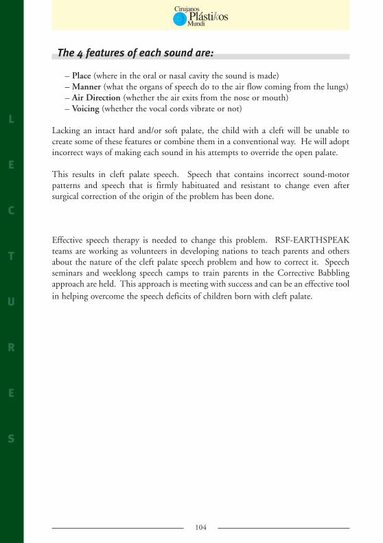
Child— sees a “cat” and says cat
With or without a cleft palate this process occurs normally in all children and isessential to speech development
Children with unrepaired cleft palates during this vital speech learning time willengage in this cycle as well. However, these children are handicapped by the inabilityto feel the sounds they see and hear or to reproduce them with intact organs ofspeech. This in turn leads to the habituated storage of incorrect sound motorpatterns for each speech sound.
Each sound has 4 characteristic features that make it different from each other sound.These features are created by using the organs of speech in different combinations.Each sound has its own sound-motor pattern just like a musical note.
C l e f t L i p & Pa l a t e M a n u a l
L
E
C
T
U
R
E
S
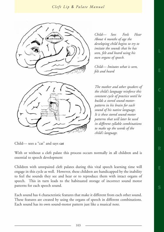
Child— Sees Feels HearAbout 4 months of age thedeveloping child begins to try toimitate the sounds that he hasseen, felt and heard using hisown organs of speech.
Child— Imitates what is seen,felt and heard
The mother and other speakers ofthe child’s language reinforce thisconstant cycle of practice until hebuilds a stored sound-motor-pattern in his brain for eachsound of his native language.It is these stored sound-motorpatterns that will later be usedin different syllable combinationsto make up the words of thechild’s language.
1270 manual ok 19/1/07 15:37 Página 103
104
The 4 features of each sound are:
– Place (where in the oral or nasal cavity the sound is made)– Manner (what the organs of speech do to the air flow coming from the lungs)– Air Direction (whether the air exits from the nose or mouth)– Voicing (whether the vocal cords vibrate or not)
Lacking an intact hard and/or soft palate, the child with a cleft will be unable tocreate some of these features or combine them in a conventional way. He will adoptincorrect ways of making each sound in his attempts to override the open palate.
This results in cleft palate speech. Speech that contains incorrect sound-motorpatterns and speech that is firmly habituated and resistant to change even aftersurgical correction of the origin of the problem has been done.
Effective speech therapy is needed to change this problem. RSF-EARTHSPEAKteams are working as volunteers in developing nations to teach parents and othersabout the nature of the cleft palate speech problem and how to correct it. Speechseminars and weeklong speech camps to train parents in the Corrective Babblingapproach are held. This approach is meeting with success and can be an effective toolin helping overcome the speech deficits of children born with cleft palate.
L
E
C
T
U
R
E
S
Cirujanos
MundiPlástikos
1270 manual ok 19/1/07 15:37 Página 104
105
Dr. Debra JohnsonChief of Surgery for the Sutter Community Hospitals,and Co-Director of the Sutter Cleft Lip and Palate Panel.Member of the Board of Directors for Interplast, Inc.
THE GENETICS OF CLEFT LIP AND PALATE
Clefts occur in approximately 1:700 births.1:750 births in Caucasians1:500 births in Asians1:1200 in Africans
Clefts can occur as cleft lip alone, cleft lip/palate, or cleft palate aloneCleft lip alone: 21% of cleftsCleft lip/palate together: 46% of cleftsCleft palate alone: 33% of cleftsBilateral cleft lip is associated with a cleft palate in 86% of cases
Clefts occur in boys more commonly than in girlsRatio of cleft lip: left 6:right 3: bilateral 1
Causes of clefting:For most, no single factor can be identified as the cause.Isolated clefts are those that have no other birth anomaly.Syndromic clefts are those associated with other birth disorders.
Clefts are a feature of over 300 Syndromes, and most are rare.More common syndromes: Pierre-Robin sequence, Crouzon,
Apert, Pfeiffer, Van der Woude, Treacher Collins,Velocardiofacial
Syndromal clefts make up 15% of cleft lip +/- palate50% of isolated cleft palate75% of VPI are syndromic
Isolated clefts: caused by an interaction between an individual’s genes and certainenvironmental factors (often impossible to identify). Phenytoin, Accutane, alcohol,tobacco, folic acid and pyridoxine deficiencies have been associated with clefting.Increasing parental age, especially an older father, is associated. About 35% of cleftshave a positive family history.
C l e f t L i p & Pa l a t e M a n u a l
L
E
C
T
U
R
E
S
1270 manual ok 19/1/07 15:37 Página 105
106
Human Genome Project: identifying genes related to clefting. Genes related to theproduction of tissue growth factor alpha, and fibroblast growth factor receptors haveshown an association with clefts. Genetic testing may soon allow us to predict those families that may have a higher riskof cleft babies, although, as Dr. Millard says, “There is little chance that the moleculargeneticist will arrive in an operating room with a cleft repair gene attached to aretrovirus anytime soon.”
What are the risks that another family member will be bornwith a cleft?
For parents of one cleft child: 2-5% risk of a second child with a cleftIf additional family members have clefts: 10-12% risk
For a person born with a cleft: 2-5% risk of having a cleft childIf additional family members have clefts: 10-12% risk
For siblings of a person with a cleft: 1% riskIf additional family members have clefts: 5-6% risk
If a syndrome is involved: risk can be as high as 50%. These patients should have a genetic evaluation.
Cleft lip and/or palate implies a risk of recurrence that ranges fromincomplete CL alone to bilateral CL and P.
Cleft palate alone implies a risk for cleft palate only; these familiesare not at risk for cleft lip.
Genetic Evaluation:
Obtain a detailed family history, a medical history, and physical examination of the cleftpatient, and laboratory testing.
1. Verify that the cleft is “isolated” and not part of a syndrome.2. Evaluate whether other relatives have similar conditions, andhow closely related
they are. The greater the number of relatives known to have clefts, and the closertheir biologic relation, the greater the risk of recurrence.
3. Consider the type and severity of the cleft. Clefts tend to be consistent withinfamilies, although severity can vary.
4. Testing: chromosomal testing is a syndrome is suspected; radiographs; moleculartesting is available for some specific conditions; photographs.
5. Counseling: discussion of treatment necessary; risk of recurrence; discussion ofstrategies for future prenatal diagnosis.
L
E
C
T
U
R
E
S
Cirujanos
MundiPlástikos
1270 manual ok 19/1/07 15:37 Página 106
107
Donald R. Laub, M.D., FACS Adjunct Clinical Associate Professor of Surgery,Stanford Medical School Founder, Interplast
Welcome
You are reading a manual on helping the cleft patient. This work is the most nobleand most important and most effective work of mankind. You don’t require to becongratulated. Because you are—
Professional person
A professional person; each one of you is a professional person, that is a person whohas devoted his or her life to a skill and a science, and you practice that skill for thegood of the other person. This act produces a very comfortable feeling—immediate gratification if you will— when you have helped someone with a severedeformity and significantly changed their fortunes for the rest of their lives.
Psychic Income
This is the Psychic Income. A professional person does the work for the good of theother person, which is in contrast to the non-professional person who performs hisskill for the good of himself primarily. For example, the infamous CEOs of largecorporations.
This Psychic Income is so powerful that it possibly produces a chemical affecting thebrain that addicts us to do this type of work. Psychic Income is the motor which runsall Plastic Surgery Voluntary Foundations, and it is the motor for CPM (CirujanosPlastikos Mundi), and it is what makes the world go around as far as the PlasticSurgery organizations designed to help the developing world are concerned.
For example, the founders of most plastic surgery helping foundations and most ofthe foundations devoted to orthodontia, speech therapy, ENT, dentistry, to help thecleft child, both the founders and the members, relate a similar personal experienceon the occasion of taking care of the first child they have had the privilege of helpingwith their skill. Many or most of these people relate that at the moment of receiving
C l e f t L i p & Pa l a t e M a n u a l
L
E
C
T
U
R
E
S
1270 manual ok 19/1/07 15:37 Página 107
108
this special psychic income that it caused a change in their value system to be morehumanitarian. In a way it was the child that helped the professional, to change theirlife to be more humanitarian.
It is as if the “tail is now wagging the dog”, the tail being the Psychic Income and theDog being the Physician and the paramedical person, who receives impetus to do thistype of work or to form to a professional foundation. Here the patient becomes theone who produces the result in the surgeon. This phenomenon of the Doctorneeding the patient reverses or erases our preoperative mind set that the surgeon isthe almighty one and the patient is entirely a recipient of the surgeon’s skill andbenevolence.
We are all on a horizontal relationship: doctor + patient both, not a verticalrelationship
The team of a foundation must also consist of businessmen, accountants, lawyers andfundraisers; and all of these are vital to our life.
In this instance, people with these skills are also professionals because they areworking for the other person and they derive the same Psychic Income as thephysicians do, because they are acting as surrogate surgeons or surrogate medicalprofessionals. After all, it is because of these surrogate physician professionals that allof this is made possible.
When these two types of skills work together, we form a new way of practicingmedicine. It is in these foundations where there is a true teamwork, where medicineand business truly work together for the patients benefit and for each other’sbetterment of their individual specialty.
A true combination of business and medicine is produced by the commonality ofhumanitarianism.
In this case, one + one equals three: it is obvious that medicine and business workingtogether is a very powerful unit.
All of the elements in this process evolved at many places simultaneously. Myexperience is for example at Stanford where, as at the other places, it was recognizedthat the needs of the students and the residents in training would be better solvedin the setting of these programs. The acquisition of the psychomotor or surgicalskills by trainees in these programs was excellent. This method of teaching issecond to no other method. The residents who were there for the reason of learningthe clinical or surgical parts of this process were delighted with what was going on;
L
E
C
T
U
R
E
S
Cirujanos
MundiPlástikos
1270 manual ok 19/1/07 15:37 Página 108
109
they were learning what is or was the very epitome of the field: the cleft lip repairand the cleft palate repair, and the nasal reconstruction for the cleft: for them thiswas the highest achievement toward their goal of learning surgery.
Peak Experience
As they learned skills, they were sort of in a state of ecstasy because this constitutes apeak experience for the trainee.
Value Change
A Peak Experience occurs at birth, when we are married, or are divorced, or graduate,or receive an award—or perform our first cleft lip repair. At the time of a peakexperience, our value system can change. For example, at the conclusion of thislandmark operation for them, they might say, “This was the best experience I haveever had in my life; I want to do this for the rest of my life.” At that moment in thetraining stage, the imprinting is apt to be much more permanent, meaningful, long-lasting, and appropriate, than if these values of humanitarianism are transferred oracquired at a post-residency level. This process easily occurs for all practicingphysicians and surgeons at any point in their careers; and it happened to me in earlyresidency, and I assume it occurs for you at every cleft surgery. The imprinting is bestdone at the time in our careers set aside for learning “to the max”, which is residencyor fellowship training.
When the trainee states, “I want to do this for the rest of my life,” she or he hasindicated that an attitude has been transferred to that “student” as well as a skill.Because the operation causing such psychic income was on a child where no fee wascharged, this was a humanitarian act, so in this way the attitude of helping others wasalso transferred and imprinted. And I have heard, on 40 occasions, those words—atthe end of the 1st cleft lip repair (repeat), “This was the best experience I have hadin my life, I want to do this for the rest of my life,” a value change had occurred, andthe skill and the social attitude became part of that person’s life from that time of thepeak experience on.
In Dr. Richard Jobe’s survey of the practicing surgeons who had undergone theseexperiences, as a resident, plus or minus 35% had continued practicing this work,and had retained this value.
C l e f t L i p & Pa l a t e M a n u a l
L
E
C
T
U
R
E
S
1270 manual ok 19/1/07 15:37 Página 109
110
Interdisciplinary contact is necessary
The patient changed the surgeon in a profound way; the “tail wagged the dog”; agood outcome resulted.
Now turning to another aspect of our professional life, let me discuss the danger ofa few in a University who achieve great excellence in a small field, and achievepower and notoriety within their system. The tendency to pout and cry may occurif each of your objectives is not immediately met by the Dean or the ruling class oryour peers. The tendency is to say, “I quit, and I will form my own institute inorder that I can practice excellence.” What happens in this case is that interdisciplinary contact is eliminated. Thecross-fertilization from different methods of thinking and from different relatedfields is eliminated. It’s probably more difficult to form multi-disciplinary effortswith equal emphasis given to all of the “ancillary” fields when your practice iswith an institute without pluralism, even though the institute is seemingly theideal setting.
The advancements in that particular field may then wither.
The multidisciplinary approach has helped develop certain fields of surgery:
MULTIDISCIPLINARY INITIATIVESTHAT HAVE WORKED:
• Plastic Surgery • Cleft Surgery and Humanitarian Surgery • Cosmetic Surgery • Maxillofacial Surgery (both cosmetic and reconstructive). • Surgery for Craniofacial anomalies • Hand Surgery • Skin Physiology and aesthetic improvement based in this science • Microsurgery itself • Joint replacement Surgery • Neurosurgery for Epilepsy • Microsurgery including tissue tolerance and limb transplantation. • Bariatric Microsurgery • Ophthalmic surgery for retinal disease and for nearsighted patients. • Oncologic Surgery
L
E
C
T
U
R
E
S
Cirujanos
MundiPlástikos
1270 manual ok 19/1/07 15:37 Página 110
111
AREAS THAT HAVE NOT BENEFITED AS MUCHFROM THE MULTIDISCIPLINARY INITIATIVES:
• Breast Surgery • Parts of Oncologic Surgery
The theory behind Residents on Trips
And in the future, the equilibrium between oral surgery and ENT and plastic surgerywill require the skill and diplomacy that only occurs in a University environment anddoes not occur in an institute. Hijo de tigre nacio pintado. The nourishment of our young, our own progeny in eachof our “narrow” fields is important, and is on a par with the importance of themultidisciplinary effort.
No doubt about it, the Teaching of the Residents in developed countries is as vital asteaching in other lands, because if the young (students and residents andimpressionable youngsters) are not nurtured and induced to gain the excitement andpleasure of Psychic Income, the field will not reproduce itself. Withholding thisprocess is similar to practicing birth control on your own professional discipline.
Advances in development of our fields
If the professionals do not teach in this way, they are sterilizing themselves. Theyprevent reproduction their own kind.
Furthermore, medicine is not a static science, new operations and new advances areoccurring at regular intervals.
The inclusion of our residents in intense training experiences is a link in the chain,which advances our field.
For example, in cleft lip and palate surgery, think of what should happen in the fiveyears from now, and also think what will not happen in five years if you do not bringin new brains and new skills?
For example:
C l e f t L i p & Pa l a t e M a n u a l
L
E
C
T
U
R
E
S
1270 manual ok 19/1/07 15:37 Página 111
112
Intrauterine fetal surgery will not be developed to repair cleft lip.
New operations will not have been developed, e.g., Mike Carsten’s (a resident)embryologic concept of moving the bone and the soft tissue as one neurovascular unitto repair cleft lip and craniofacial anomalies.1
The Most Important Thing
Enthusing young people to enter our field is clearly vital to ourselves and to thepatients, who are the most important thing.
Repeat, Humanitarianism should be taught at the age when teaching is done, is bestdone, is most effective.
“Enlightened self-interest”
In the greater perspective, we can consider the effect of our work on our world, whichwill occur only through our children, and our residents. Our children and ourresidents will be needed in the formation of public opinion in the next few years. Justthink how the world would change if our governments and our corporations and ourpoliticians were to regard this way of life as a thing of value. If our very own childrenfelt that helping others in these ways were a thing of value they might counter-balance their conditioning toward their aspirations to acquire he latest clothing styles,the latest entertainment, and their latest idea about what they should acquire in life.
But both aspirations seem necessary.
Governments might divert a small billion or two toward these projects for their owngood. Their own political projects would enjoy easier success. They would gain favorin the foreign countries and gain votes here in the developed world.
Our corporations could actually make more money by being nice to people in foreigncountries. This concept might mature into more than “enlightened self-interest” (aphrase taken from history), but into a sincere value change.
A value change would increase the espirit of the employees in the large corporation;
L
E
C
T
U
R
E
S
Cirujanos
MundiPlástikos
1 Carsten has shown that Clefting occurs when one neurovascular unit of bone and soft tissue unitbecomedeficient during fetal development and that the repair of cleft should be based on this concept.
1270 manual ok 19/1/07 15:37 Página 112
113
their company newspaper would tout the companies’ social conscience to the prideand delight of their more socially conscious employees, would raise the profits of thecompany and their “host” country; perhaps the actual expressed goals for thecorporation would perhaps include the good of the employees; the shareholders andofficers might see it this way, and all countries would benefit. This mindset is notreally “thinking different” or O.O.T.B.2, but it is thinking in the longer term in aneducated way, a bit more professional way.
This thing of value should be regarded as equal to the tangible income of makingmoney and parlaying that money into more money.
The field moves forward.
Speaking of a modern, more educated method of “developing the field” of Plastic andReconstructive Surgery: both in regard to the educational process and even theresearch, the innovative progression should include:
In effect, we almost double the amount of brainpower brought into use by increasingexchange of ideas between disciplines and between countries by sharing research atan earlier level, and by assuming commonality of purpose.
This new concept leads to virtual centers of excellence because some professionalsmay be geographically separated but united by a web-based center of excellence (SeeDingman PRS 2002). Again, to repeat, we are securing the development of newoperations, of research, and of new surgeons. We see the development ofmultidisciplinary affiliated disciplines in both countries.
Example of personal “Modern” History of theories discussed
Consider some Stanford medical initiatives.
C l e f t L i p & Pa l a t e M a n u a l
L
E
C
T
U
R
E
S
2 Out Of The Box
Developed
&
Developing
Country Professionals working together.}
1270 manual ok 19/1/07 15:37 Página 113
114
Multidisciplinary humanitarianism, and Residents on trips
In 1963 at Stanford, Dr. Chase, my mentor and professional father, began ourprogram with:
a) Skill
b) The other person in mind.
This combination led to the performing of a free surgery on a single patient,Antonio Victoria from Mexicali, Mexico and then later this led to the formation ofMexico Medical Project, Inc. and subsequently Interplast. Our first initiatives usedfor teaching, for obtaining patients with advanced pathology in our new hospitalsystem, are examples of being committed to a goal. They were as follows:
When challenged by the official opinion of the Mexican Government to not returnto their fair country for further surgical work on children with developmentaldeformities, and adults with acquired deformity (burns, hand injuries, tumors), wepouted for only a short time. And then for some unknown reason conceived a“Plan B” for that situation. The government of Mexico had not initiated a moreintense program to help their own citizens. At that relatively naïve time for us we“collaged” our assets (our group had 5 assets interlocking goals): ReconstructiveSurgery Training, a knowledge of ships and the sea, a leadership experience with theU.S. Navy, an idealist in the law enforcement department of Los Altos, California,a veteran with personal experience in pre-juvenile delinquency rehab., a Universityfaculty position, and a Rockefeller Foundation connection.
We identified yard freighter 879 in the San Diego Reserve (Mothball) fleet andthen, a Culinary Instructor, and also a retired Navy Captain for command,policemen to refer 14 year-old pre-juvenile delinquents for rehabilitation, a medicalcorpsman instructor, teaching faculties, textbooks (Lange Medical Synopses), DavidWerner’s paramedical volume, and a retired Naval electronics Veteran, andeducators in seamanship. The yard freighter has 35 feet of clear room with nointernal cross member supports to allow an operating room, a post-op care unit,preparation room and a supply place. Quarters for the patients and crew wereabove deck. A lounge was to be identified. The kitchen was new, never used. Theship had been built in Philadelphia and towed to San Diego at the end of the war;it was kept air aconditioned for 25 years and maintained perfectly.
It drew only 3 ft of water and was ready for work in the shallow harbors of WesternMexico and South America. We visited the pentagon and arranged for the ship tobe released at a certain moment, at which time we were to have our application on
L
E
C
T
U
R
E
S
Cirujanos
MundiPlástikos
1270 manual ok 19/1/07 15:37 Página 114
115
Capt. Banan’s desk. Any University has second priority to military material declaredsurplus. Dean John Wilson asked his nice friend at Rockefeller Foundation toarrange a grant for the project based on U.S. Navy experience and the experiencegained.
In Norway, where socially deviant persons learned self-worth, ability to not be self-centered, and the necessity to work together as a team when working onboard aship. Their programs resulted in less recidivision (back to jail or to the “Brig.”).
Using the yard freighter in the San Diego Reserve Fleet, we had a method to trainpre-juvenal delinquents in
a) Culinary arts
b) Electronic skills
c) Seamanship
d) Medical corpsman
Our second abortive attempt to do the type of work we are addressing in thissymposium was the DC-3 airplane. The DC-3 was purchased by Interplast as safeand inexpensive transportation. It increased esprit du corps, higher and higher,until an unscheduled landing occurred, which of course led to another plan, to theformation of an Interplast “air force”, consisting of volunteer pilots and theirairplanes, for transportation to nearby countries. It was another multidisciplinaryinitiative “assured” of success. These aircraft were used for transportation to clinicsand hospitals in other countries, including parts of the U.S. The plan wasinexpensive, running at 1/2 cost of commercial transport.
Innovative funding from the community
Initial funds were derived from: San Mateo Chope Hospital Surgical Medical funds Gender Surgery Fees Clair Elgin (a patient with the worst diagnosis and deformity who had “struck itrich” with her invention). Our initial PRS patients were derived from:
Physiatry Department (paraplegic and quadriplegic)
C l e f t L i p & Pa l a t e M a n u a l
L
E
C
T
U
R
E
S
1270 manual ok 19/1/07 15:37 Página 115
116
Jail (Lombroso’s Theory that improving appearance improves social behavior)
Menlo Park Veteran’s Administration Psychiatric Hospital (2,000 patients, manywith skin cancer, Carpal Tunnel Syndrome, or Visual Field Defects, i.e. need forlarge scale blepharoplasty)
And of course, Interplast—actually, Interplast was part of the educational processemanating from Stanford to provide pathology for residents and students.
Success
This initiative over the years lead to 2,000 patient surgeries from Mexicali, 4,000patients and surgeries from San Pedro Sula, Honduras, 50,000 patients total fromvarious countries and 40,000 patients from developing countries for InterplastGermany.
Dear colleagues—the personal example of commitment to Plastic Surgery and amultidisciplinary effort for those in need is related to you not for what sounds likeself-interest, but I write it to you so that we might share the commonality of ourhumanitarian efforts, all of which are both personal and also in the name of all ofus.
“Even if we are occupied with most important things, even if we attain to highhonor, or fall into great misfortune, still, let us remember how good it was oncehere when we were all together united by a good and kind feeling which made us…better, perhaps than we are.”
-Fyodor Dostoevsky, The Brothers Karamazov
Upwards and Onwards.
L
E
C
T
U
R
E
S
Cirujanos
MundiPlástikos
1270 manual ok 19/1/07 15:37 Página 116
117
C l e f t L i p & Pa l a t e M a n u a l
L
E
C
T
U
R
E
S
NOTES:
1270 manual ok 19/1/07 15:37 Página 117
118
L
E
C
T
U
R
E
S
Cirujanos
MundiPlástikos
NOTES:
1270 manual ok 19/1/07 15:37 Página 118
119
C l e f t L i p & Pa l a t e M a n u a l
L
E
C
T
U
R
E
S
NOTES:
1270 manual ok 19/1/07 15:37 Página 119
120
The child with a dificult airwayPedro Charco Mora, MD.
Critical Care Anesthesiology.Hospital Universitario Son Dureta, Palma de Mallora, Spain.
Vicente Martinez Pons, MD.Critical Care Anesthesiology.
Hospital Univ. F. Borja, Gandia, Spain.Valentin Madrid Rondon, MD.
Critical Care Anesthesiology.Hospital Univ. Marina Alta, Denia, Spain.
TABLE OF CONTENTS
I. INTRODUCTION
II. PAEDIATRIC AND ADULT AIRWAY DIFFERENCESA. UPPER AIRWAYB. LOWER AIRWAYC. RESPIRATORY FUNCTION
III.AIRWAY EQUIPMENT FOR MANAGING THE PEDIATRIC AIRWAYA. LARYNGEAL MASK AIRWAYB. FLEXIBLE FIBEROPTIC BRONCHOSCOPE (FFB)C. BULLARD LARYNGOSCOPED. LIGHTWANDE. SHIKANI SEEING OPTICAL STYLET (SOS)F. ANGULATED VIDEO – INTUBATION LARYNGOSCOPE (AVIL)
IV. ANAESTHESIA CONSIDERATIONSA. ASSESSMENT OF THE AIRWAYB. PREMEDICATIONC. INDUCTION1. TYPE I PATIENTS2. TYPE II PATIENTS3. TYPE III PATIENTS4. TYPE IV PATIENTS
V. CLINICAL EXAMPLESA. ACUTE EPIGLOTTITISB. MACROGLOSSIAC. MICROGNATHIAD. FOREIGN BODY ASPIRATIONE. PEDIATRIC TRAUMA
VI. SUMMARY
VII.BIBLIOGRAPHY
A
N
E
S
T
H
E
S
I
A
Cirujanos
MundiPlástikos
1270 manual ok 19/1/07 15:37 Página 120
121
1 Introduction:
One of the most challenging events in the practice of anaesthesia is the managementof the paediatric patient with a difficult airway. Neonates, infants and childrendisplay a wide spectrum of diseases that may present at any time to the hospital withairway problems. Hence, anaesthesiologists that participate in the management of thepaediatric patient should have a thorough and detailed plan on how to recognize andtreat these patients.
Studies have shown that the paediatric patient has a higher risk of significantmorbidity and mortality during anaesthesia when compared to the adult patient.1Also, in a review of closed claims studies, Morray, et al, reported that respiratoryevents are the major reason for morbidity and mortality in the perioperative period.2In addition, they represent the primary cause of poor outcome2. Completefamiliarity with the characteristic features of airway anatomy and physiology in thepaediatric patient has been found to decrease this incidence of adverse events.3
2 Pediatric and adult airway differences:
There are significant differences between the paediatric and adult airways (Table 1)4The anatomic features include differences in both size, shape and position of theairway as well as in the airway epithelium and its supporting structures. Physiologicdifferences between the neonatal and adult respiratory systemsarise from these anatomic differences and from mechanisms in respiratory control.
A. Upper AirwayThe upper airway of the newborn infant is smaller and anatomically different thanthe adult. The tongue is relatively large and occupies fully the cavity of the mouthand oropharynx. Most, but not all neonates are also obligate nasal breathers becausethe epiglottis, positioned high in the pharynx, almost meets the soft palate, makingoral ventilation difficult.5 This lasts from 2 to 6 months of age. Unlike older infantsand children, neonates have almost no lymphoid tissue in the upper airway. Thetonsils and adenoids appear during the second year of life and generally reach theirlargest size by 4 to 7 years of age, after which they gradually recede in the absence ofintervening infections.
The larynx is located at a high position. In the baby, the body of the hyoid bone issituated approximately at the level of the disc between the third and fourth cervicalvertebrae.6 As the infant grows, the glottis moves caudally to a C5 to C6vertebral level by maturity. The high position of the epiglottis and larynx allows the
C l e f t L i p & Pa l a t e M a n u a l
A
N
E
S
T
H
E
S
I
A
1270 manual ok 19/1/07 15:37 Página 121
122
infant to breath and swallow simultaeously. Similarly, both the thyroid and cricoidcartilages move caudal as the thyrohyoid and cricothyroid membranes develop. Thelarynx also differs in several respects. The epiglottis is more U-shaped, and protrudesover the larynx at a 45o angle. Because the larynx of the infant is high and has ananterior inclination, the straight laryngoscope blade is most useful. The view can bemarkedly improved by external pressure on the larynx, pushing it backward. Thelarynx is funnel-shaped in children less than 8 to10 years of age, with the narrowest portion being at the level of the cricoid ring.
In the adult, the narrowest portion of the airway is between the vocal cords.
Additionally, the vocal cords of the neonate are slanted such that the anteriorcommissure is more caudal than the posterior commissure. The tracheal direction inthe infant is downward and posterior, whereas in the adult it is straight downward.Consequently, the application of cricoid pressure is more effective in an infant whenplacing an endotracheal tube. Also, in the newborn infant the distance between thebifurcation of the trachea and the vocal cords is 4 to 5cm. Thus an endotracheal tubemust be very carefully positioned and fixed because the tip of the tube can moveabout 2cm during flexion or extension of the head.
Airflow in the upper airway is turbulent even during quiet respiration. Laminar flowbegins at the level of the fourth or fifth bronchial divisions, where the rapid increasein crosssection area decreases airflow velocity. The resistance to turbulent gas flow isproportioned to the fifth power of the radius of the airway. Subsequently, 1mm ofedema in the trachea of the newborn (which reduces the radius from 2.1 to 1.1mm)increases the resistance to air flow approximately 25-fold.
In general, the right main bronchus is less angled than the left. Hence the right mainbronchus is the one most frequently intubated during endobronchial intubation, andforeign bodies tend to lodge in the right side more frequently.
B. Lower AirwayAt the fetal stage the pattern of the bronchial tree is fully developed by the 16th weekof gestation. Alveoli develop later and increase in number until 8 years of age and insize until full development of the chest wall. The lining of the respiratory sacules andalveoli is derived from two types of cells: Type I pneumocytes produce the liningsupporting cells of the alveoli and contribute to the blood gas barrier. Type IIpneumocytes are more glandular and contain inclusion cytoplasmic osmiophilicgranules. Pulmonary surfactant is synthesized in these granular pneumocytes (TypeII) and is stored in the lamellar bodies. It is released by fusion of the lamellar bodymembrane with the cell wall.The highly compliant chest wall in neonates and infants increases the work of
A
N
E
S
T
H
E
S
I
A
Cirujanos
MundiPlástikos
1270 manual ok 19/1/07 15:37 Página 122
123
breathing. This greater compliance is attributable to their softer, noncalcified ribs,which articulate with the vertebral column and sternum at right angles. Theadult's more rigid chest wall articulates at downsloping angles with a more efficientchest wall excursion. The diaphragm is therefore, more important and is the mainstayof ventilation in the infant. However, it also has some disadvantages. It hasproportionally fewer Type I fast oxidative muscle fibers than the diaphragms ofchildren older than 2 years. So, the mechanism of contraction is less efficient and themuscle tires faster.
C. Respiratory FunctionWhen anesthetizing infants and small children, it is important to realize theirphysiologic disadvantages when compared to older children or adults. The infant hasa higher metabolic rate with increased oxygen consumption. Thus, they have lessoxygen reserve and can become hypoxic much faster than the adult. Functionalresidual capacity (FRC) is also decreased to a greater extent in infants by generalanaesthesia. The two main differences are in respiratory rate and alveolar ventilaion.This is understandable due to their higher metabolic rate and oxygen consumption.
Mask ventilation in infants and children can rapidly lead to gastric distension. Thiscan decrease FRC, elevate the diaphragm, decrease lung compliance, and increase therisk of aspiration.
Infants and small children may have an increased sensitivity of certain inspiratorymuscles to anaesthetic agents.7 This may lead to an increased incidence of airwayobstruction.
3 Airway Equipment for Managing the Paedriatic Airway:
Successful management of the infant or child with a difficult airway requires havingthe appropriate equipment.
Nearly everything that is available for use in adults is now available for children,obviously, reduced in size. Appropriate face masks, oropharyngeal airways,nasopharyngeal airways, endotracheal tubes, stylets and laryngoscope blades shouldbe available. This section of the chapter though will concentrate on newer forms ofequipment that have helped immensely in the management of the difficult paediatricairway (Table 2). A detailed description is beyond the scope of this article. The readeris referred to the references.
A. Laryngeal Mask Airway (LMA)The LMA was first described in adults by Dr. Brain in 1983.8
C l e f t L i p & Pa l a t e M a n u a l
A
N
E
S
T
H
E
S
I
A
1270 manual ok 19/1/07 15:37 Página 123
124
Its use in children has also been described.9 It is available in sizes 1,2,2.5, and 3 foruse in paediatric patients. It may be used as the sole airway of choice to ventilate thelungs when endotracheal intubation or mask ventilation is undesirable or difficult. Itmay also be used to facilitate either a blind or fiberoptic intubation of the trachea.The LMA has now become part of the sequence for difficult or failed intubation inboth adults and children.10
The second generation LMA or Fastrack is not available in small sizes. It can be used,though, in older children. Its advantage is the fact that a blind intubation is easier toobtain with the Fastrack.
The third generation LMA or Proseal is available for children over 5 kilograms.11 Ithas an inner tube to aspirate stomach contents, thus making aspiration a rareoccurrence.
B. Flexible Fiberoptic Bronchoscope (FFB)The FFB has been in use for many years although only recently has it gainedwidespread use in children.13 The primary use for anesthesiologists is in theassistance of endotracheal intubation. The fiberscopes most commonly used inpaediatrics vary in external diameters from 2.2mm (capable of passing through a2.5mm ET) to 4.0mm (capable of passing through a 4.5mm ET). The 2.2mm"ultrathin" scope has a flexible tip but lacks a suction port.
Successful use of the fiberoptic scope as a tool to intubate the trachea in infants andchildren depends on several factors.
Infants and children generally do not cooperate during awake fiberoptic intubation.It is generally easier to keep the infant or child anesthetized but breathingspontaneously on 100% oxygen and an inhalational agent such as sevoflurane. Thiscan be accomplished with the use of a nasal cannula or a nasalendotracheal tube placed blindly at the inlet of the airway, or more recently, with theuse of the endoscopy mask.12 The endoscopy mask allows simultaneous anaesthesiaand ventilation of the patient during fiberoptic intubation or diagnostic airwayendoscopy. It is available in three different sizes for infants, children and adults.
C. Bullard LaryngoscopeThis laryngoscope is available in both a paediatric and an adult size. It is a particularblade equipped with fiberoptic and mirror technology to allow indirect visualizationof the larynx with minimal mouth opening and neck motion. It does not require thealignment of the oral, pharyngeal, and laryngeal axes. The pediatric version is
A
N
E
S
T
H
E
S
I
A
Cirujanos
MundiPlástikos
1270 manual ok 19/1/07 15:37 Página 124
125
characterized by a blade that is narrower and a terminal angulation that is more acutethan the adult version. The trachea is intubated by advancing an endotracheal tubethat was previously loaded on to the device's intubating stylet, which fastens to theright side of the laryngoscope.
The Bullard laryngoscope has been used with good success in the management of thedifficult airway in children.14
D. LightwandThe lightwand utilizes the technique of transtracheal illumination for the blindintubation of the trachea. It has been demonstrated very useful in the managementof difficult airways in children.15 A preselected endotracheal tube and the stylet isintroduced into the trachea by visualizing the intensity of light visible on the anteriorneck. Lightwands are inexpensive tools which can be used frequently, and are usefulfor both normal and abnormal airways. The technique is easy to learn and can beused in both anesthetized patients or in awake patients with proper sedation andtopicalization of the airway.
Blood and secretions are not an impediment to success like in the fiberopticbronchoscope. Recent developments have made the newer lightwand thin enough touse with endotracheal tubes as small as 2.5mm.
E. Shikani Seeing Optical Stylet (SOS)The SOS is a recently introduced reusable intubating stylet produced in adult andpaediatric versions.16 It combines features of a fiberoptic bronchoscope and alightwand. The paediatric version is optimized for endotracheal tubes in the 3.0mmto 5.0mm I.D. size range.
F. Angulated Video-Intubation Laryngoscope (AVIL)The AVIL is a MacIntosh laryngoscope that has been modified with a guide borehole, leading from the bottom of the handle through the lateral flange of the bladeto the blade tip.17 An ultrathin video-endoscope is inserted in the bore hole. Ittransmits the view from the distal blade tip directly onto a video-display and alsoprovides airway illumination.
The AVIL with the angulated distal blade tip resembles an activated McCoy blade.In contrast to the McCoy blade, which can improve direct glottic visualization, theAVIL has been particularly designed to give an improved glottic view on a video-monitor during difficult tracheal intubation.
C l e f t L i p & Pa l a t e M a n u a l
A
N
E
S
T
H
E
S
I
A
1270 manual ok 19/1/07 15:37 Página 125
126
4 Anesthesia considerations:
A. Assessment of the AirwayIt is crucial that to successfully manage the child with a difficult airway, a thoroughhistory and physical examination be performed. The worst nightmare is to be caughtunprepared during the induction of anaesthesia in a difficult airway child.
The history should include a review of prior records and/or anaesthetics, emphasizingthe airway management during that time.
The history should also elicit any information concerning congenital, traumatic,inflammatory or other acquired lesions. A specific attention should be placed on anycongenital lesion present in the patient since it can also manifest with a difficultairway. Any history of prior obstruction or sleepapnea should be elicited (snoring,apnea, daytime somnolence).
The physical examination should focus on the following: size and shape of the head;gross features of the face; size and symmetry of the mandible; size of the tongue andshape of the palate; prominence of upper incisors; and range of motion in the jaw,head and neck.
Occasionally, besides a thorough history and physical examination, additional studiesmay help in identifying specific features of the airway: awake laryngoscopy, flowvolume loops, radiologic imaging and magnetic resonance imaging.
B. PremedicationShould be individualized. The majority of children with a compromised airwayshould not be given sedation or narcotics due to the fact that this could result in theloss of muscular tone and in respiratory depression, worsening the obstruction.18 Insome older children in which an awake intubation is contemplated, the judicious useof mild sedation may be attempted. Anticholinergic agents are good since theydecrease the volume of secretions and also may protect against vagal responses duringthe manipulation of the airway. Atropine may be administered IM (0.02mg/kg) orIV (0.01mg/kg).
C. InductionThe two main anaesthetic problems in the child with a difficult airway are control ofthe airway and the intubation.
The techniques used for induction of anaesthesia vary according to the severity of thepathology in the airway and the degree of respiratory difficulty. Regardless of whattechnique is used, an alternate plan is needed in the event of failure to obtain an
A
N
E
S
T
H
E
S
I
A
Cirujanos
MundiPlástikos
1270 manual ok 19/1/07 15:37 Página 126
127
airway initially. You could divide the patients in four categories which will dictate theappropriate method for induction and intubation.
1. Type I PatientsThese patients present with a normal respiratory frequency, mild respiratorydistress, normal oxygen saturation, an airway that externally looks normal andminimal or non-existent sternal retractions.
2. Type II PatientsThese patients might have significant airway disease and moderate airwaydistress but have a "known" airway. They have had already procedures and thesurgical and anaesthesia teams are familiar with them (the team knows whattechnique works the best). An example might be the child that presents withacute airway obstruction due to recurrent laryngeal papillomas.
Type I and II patients usually get an inhalation induction with eitherhalothane or sevoflurane. Slowly, positive pressure (5-10cm H2O) is applied.This serves to confirm the possibility of ventilating the patient and it couldhelp decrease theobstruction caused by soft tissues. Once it is confirmed that you can ventilatethe patient, a muscle relaxant may be given and intubation performed.Alternatively, and especially if the airway is not adequate, intubation may beattempted without muscle relaxants.
3. Type III PatientsThese patients may or may not be in respiratory distress but on physical examtheir anatomy is abnormal, i.e. micrognathia, macroglossia, severe palatofacialdeformity, or tumors displacing the airway. This group also includes childrenwith lesions in the lower airway or anterior mediastinal masses which wouldbedifficult to manage after administering general anaesthesia andneuromuscular agents.
4. Type IV PatientsThese patients present for the first time with significant obstruction of theairway. They show clear symptoms of airway distress, sternal retractions, lowoxygen saturation and obvious signs of fatigue. Examples include aspirationof a foreign body, croup, and epiglottitis.
Type III and IV patients require special preparation in anticipation of adifficult direct laryngoscopy and intubation. The personnel and equipment toestablish an immediate airway should be available including those for a
C l e f t L i p & Pa l a t e M a n u a l
A
N
E
S
T
H
E
S
I
A
1270 manual ok 19/1/07 15:37 Página 127
128
paediatric surgical airway. In some lesions and in older children, theintubation may be performed awake with sedation. Incremental doses ofintravenous midazolam 0.05-0.1mg/kg may be used along with topicalizationof the airway.
The great majority of children will require a general anaesthetic. We believethat these patients should be kept breathing spontaneously for two mainreasons. First, the administration of a muscle relaxant may cause completeairway obstruction due to the loss of tone in the muscles of the tongue, andthe larynx. This obstruction may not resolve with manual ventilation. Second,the patient breathing spontaneously might be a valuable guide to localize theglottis (bubbles during expiration). An inhalation induction is performed andonce the patient is deeply anesthetized, intubation is attempted. If it is notsuccessful, alternate techniques are used. These may include: a blind nasal, alaryngeal mask (LMA), Bullard laryngoscope, a lightwand, or a fiberopticintubation. In the case of anterior mediastinal masses, the size and location ofthe tumor and the degree of cardiovascular compromise could necessitate therapid initiation of cardiopulmonary bypass (femfem).
5 Clinical examples:
A. Acute epiglottitisEpiglottitis or supraglottitis is primarily a bacterial infection of the epiglottis andsupraglottic structures.19 The bacteria associated most commonly is Haemophilusinfluenza type B, although group A streptococci can also cause it. Continued wideuse of the Haemophilus influenza (H-flu) vaccine has decreased the number ofinfections caused by H. influenza. It can occur at any age, although it is mostcommon in children 3 to 5 years old. The onset is sudden (within hours of a childdeveloping a respiratory infection) and is accompanied by severe systemic illness withhigh fever and respiratory distress. Severe airway obstruction can develop rapidly.Manifestations of the disease include the five "d's": drooling, dysphagia, dysphonia,dyspnea, and dehydration. The patient assumes a sitting position leaning forward,because this improves airflow, and the patient can breath easier.
Laboratory work-up is consistent with a bacterial infection (leukocytosis). Lateralradiograph of the neck shows the typical "thumb sign" at the level of the epiglottis.The child must be attended constantly by personnel capable of handling difficultairways. A team should be assembled and the patient brought rapidly to the operatingroom. The operating room should be prepared for emergency bronchoscopy andpossible tracheotomy. Oxygen supplementation should begin early. The child shouldbe disturbed as little as possible. Intravenous (IV) access should be obtained after
A
N
E
S
T
H
E
S
I
A
Cirujanos
MundiPlástikos
1270 manual ok 19/1/07 15:37 Página 128
129
induction of anaesthesia because it can cause crying and may exacerbate the airwaycompromise. Obviously, it is preferable to have IV access before the induction if itcan be obtained without disturbing the child. Monitors before induction shouldinclude a precordial stethoscope, EKG, and pulse oximeter. The preferred techniqueby this author for induction of anaesthesia is mask inhalation keeping the patientbreathing spontaneously. Halothane or Sevoflurane, nitrous oxide, and oxygen areused.20 As you increase the sevoflurane concentration, decrease the nitrous oxideconcentration until you are left with sevoflurane and 100% oxygen. Once the childloses consciousness placement of an IV and the rest of the monitors (blood pressurecuff and temperature) is accomplished.
Attempt laryngoscopy and intubation only after an adequate depth of anaesthesia hasbeen obtained as judged by eye signs, blood pressure, heart rate, and conversion toquiet diaphragmatic breathing. Always maintain spontaneous ventilation if possible.
A nasal intubation is preferred for postoperative care, but an oral tube should beplaced if any problems arise. If during the induction of anaesthesia complete airwayobstruction ensues, a rapid laryngoscopy should be attempted and preparation foremergency cricothyrotomy made if the airway is not rapidly controlled. Once theairway is secure, blood cultures should be drawn and antibiotic therapy commenced.The patient is then transferred to the intensive care setting for postoperativemanagement. Appropriate sedation should be administered in the intensive care unit(ICU) to prevent an accidental extubation, which could be a disaster. Narcotics orbenzodiazepines are commonly used for sedation. Usually after 12 to 36 hours of IVantibiotics the patient can be extubated safely, and after a few hours of observation,may be transferred out of the ICU.
B. MacroglossiaThe term macroglossia is applied when the tongue is too large for the oral cavity. Theoral cavity may also be too small for a normal-sized tongue (pseudomacroglossia).Examples of conditions in which macroglossia is found include: lingualhemangiomas, primary macroglossia, mucopolysaccharidoses,21 AV malformations,solid tumors of the tongue and Down's syndrome.22Trauma and facial burns may also present with macroglossia.
Visualization of the larynx may require direct or indirect means. Direct laryngoscopywill be possible only if the tongue can be displaced to the left. If this proves to bedifficult, indirect means are required to view the larynx. Options for the lattertechnique include a blind intubation via the nose or fiberoptic bronchoscopy.Recently, the addition of the "ultrathin" bronchoscope allows fiberopticbronchoscopy to be performed even in small infants. The smaller bronchoscopes lacka suction channel and are more difficult to use. Use of this instrument requires prior
C l e f t L i p & Pa l a t e M a n u a l
A
N
E
S
T
H
E
S
I
A
1270 manual ok 19/1/07 15:37 Página 129
130
experience with normal and older children or adult airways before management ofthe difficult infant airway is attempted. "Blind" nasal intubation is more difficult ininfants and small children than in adults because of the more anterior position of thelarynx and therefore, the greater curve that the tube must make in the pharynx.Stylets and lightwands have been used to improve the success of this method. Theycan also be used blindly via the oral route. Other options in the management of thepatient with macroglossia include the Bullard laryngoscope, retrograde intubation orthe use of an LMA mask with subsequent intubation.
Infants and older children with intraoral and pharyngeal masses can be managed thesame way as described above.
C. MicrognathiaMicrognathia means failure of the mandible to develop fully.It may be the most the most important factor in predicting a difficult airway.23 Twoimportant disorders deserve mention here. They are the Treacher-Collins Syndromeand the Pierre-.Robin Syndrome.24 The mandible develops from the first branchialarch. The presence of preauricular skin tags or abnormally developed external ears,which also develop from the first branchial arch, may alert the clinician to thepotential for a difficult airway.
The airways of infants and small children with Treacher- Collins or Pierre-RobinSyndromes may present with a tongue that may not be easily displaced during directlaryngoscopy, a more anterior larynx and a smaller oral aperture. Glossoptosis mayfurther complicate the airway of patients with micrognathia.
Management of the airway is similar to that described above for the patient withmacroglossia. We have found though that these patients can be intubated orally withdirect laryngoscopy if the laryngoscope is inserted on the extreme right side of themouth with posterior pressure on the thyroid cartilage. If unsuccessful, fiberoptic-guided intubation through the LMA or intubation with a "lightwand" may beaccomplished.
D. Foreign Body Aspiration in The AirwayThe aspiration of a foreign body in the airway is the leading cause of accidentaldeaths in paediatric patients under the age of one.25 Infants can present in significantairway distress and respiratory insufficiency.26 Thus the management of the airwaycan be a nightmare even for experienced anesthesiologists.27 The history of a foreignbody aspiration usually is very short, although at times there may be a 2 or 3 weekinterval between the apparent episode of aspiration of a foreign body such as a peanutand progressive symptomatology requiring medical attention.
A
N
E
S
T
H
E
S
I
A
Cirujanos
MundiPlástikos
1270 manual ok 19/1/07 15:37 Página 130
131
A foreign body aspirated into the area of the larynx (laryngeal foreign body) usuallypresents with inspiratory stridor and bilateral breath sounds. At times these childrenare in extreme distress but unless they are unconscious they can be taken to theoperating room for definitive treatment. Many times just with a laryngoscopy and aMcGill forceps, the foreign body can be retrieved. General anaesthesia should beinduced maintaining spontaneous ventilation if possible.
A foreign body at the level of the trachea will usually present with bilateral decreasedbreath sounds and both inspiratory and expiratory stridor. The chest radiographshould be a useful diagnostic tool. After the foreign body is discovered, laryngoscopyand bronchoscopy should follow as soon as possible. An inhalation induction withthe patient breathing spontaneously is preferred. Sometimes it takes time andpatience to appropriately induce the patient. After the patient is judged to bereasonably "deep" under anaesthesia, he or she is turned to the surgeon for insertionof the bronchoscope and removal of the foreign body. One has to remember also thatusually these patients are considered a "full stomach" but waiting for the stomach toempty is not appropriate because the excitement of the episode will delay gastricemptying. If during the case complete airway obstruction arises, the foreign bodyneeds to be extracted rapidly or pushed down to usually the right mainstembronchus.
This sometimes can be life-saving. If the foreign body is beyond the level of thetrachea, the physical examination will reveal uneven breath sounds. The respiratorydistress may not be as severe as when the foreign body is in the trachea.
After the foreign body is retrieved, the patients should be intubated and thenawakened. Some patients may require the use of racemic epinephrine and/or steroidsto reduce the inflammation and edema that is associated not only with a foreign bodybut also with the instrumentation that will be required to remove it.Many surgeons also order a chest radiograph after the instrumentation to rule out anytrauma caused by the instrumentation.
E. Paediatric TraumaOne of the most important aspects in the perioperative care of the paediatric traumavictim involves the management of the airway.28 There are several factors thatcontribute to the potential of airway obstruction in this patient. They include looseteeth and foreign bodies, blood and secretions, peri-oral and tongue swelling,laryngeal and tracheal ruptures and food aspiration.
Initial assessment should determine adequacy of ventilation.
Inspection and auscultation of the chest should be performed immediately. If the
C l e f t L i p & Pa l a t e M a n u a l
A
N
E
S
T
H
E
S
I
A
1270 manual ok 19/1/07 15:37 Página 131
132
patient is not breathing, immediate therapy should be provided resulting in a securedairway. Physical examination of children in respiratory distress may reveal nasalflaring and grunting; suprasternal, subcostal and intercostal retractions. The child'ssmall size and short neck makes the assessment of the neck for tracheal position andjugular venous distention difficult. Pallor, cyanosis, and an altered level ofconsciousness are late signs of respiratory insufficiency and/or failure and demandimmediate intervention. Initial management of the patient should also includemonitoring of pulse oximetry, end-tidal CO2 and arterial blood gases. Supplementaloxygen should be given to all trauma victims until a definitive diagnosis can beobtained. Indications for endotracheal intubation include inadequacy of oxygenationand/or ventilation, loss of consciousness in order to protect the airway againstaspiration, and in patients in which hyperventilation for increased intracranialpressure is needed.
Cervical spine injury in children is uncommon, especially in young infants, whoseinjuries tend to be at a high cervical level. Trauma, though, can in some cases renderthe spine unstable especially in patients with Down's Syndrome. So, excessivemovement of the head and neck in the paediatric trauma victim should be avoided.In-line stabilization should always be maintained when airway manipulation isattempted.
The diagnosis of cervical spine injury is more difficult in the paediatric patient whencompared to the adult patient.29
Therefore, any child with a suspected neck injury should have cervical spineprecautions (i.e. neck collar) and receive an extensive radiographic and neurologicevaluation. Because there is usually no time for this in the emergency victimpresenting to the operating room, an assumption of an "unstable" neck should alwaysbe made and in-line stabilization maintained during intubation attempts. Even onpatients that have had neck radiographs, the cervical spine may not be entirelycleared.
Many times the x-ray does not include a view below C6, the odontoid process is notseen, and/or pseudosubluxation of C2 to C3 or C3 to C4 may occur and be missed.
The actual intubation sequence will be determined by the clinical situation. If thepatient has stable vital signs and an anticipated normal airway, a rapid sequenceinduction and intubation with cricoid pressure may be performed. This is usually themost commonly utilized technique. Thiopental, propofol, ketamine and etomidateare frequently used induction agents in the stable victim.
If the patient has extensive injuries and is hemodynamically unstable, ketamine or
A
N
E
S
T
H
E
S
I
A
Cirujanos
MundiPlástikos
1270 manual ok 19/1/07 15:37 Página 132
133
etomidate is preferred. In the head-injured child, ketamine is contraindicated becauseit increases intracranial pressure. Etomidate decreases intracranial pressure and is lessof a myocardial depressant than thiopental or propofol, so it should be the agent ofchoice in the unstable patient.
Alternate approaches should be considered in patients in whom a difficult intubationis anticipated. An awake intubation with spontaneous ventilation may be attemptedalthough this may be impossible in a struggling child. A variety of laryngoscopeblades and airway equipment discussed before may be used in these patients. TheBullard laryngoscope, because of not having to align the different airway axes, maybe a good choice. Also, the lightwand and the fiberoptic bronchoscope. A laryngealmask airway may also be useful as long as an endotracheal tube is placed through theLMA to protect the airway from aspiration.
Inability to establish a clear airway will most likely require surgical intervention.
The Advance Trauma Life SupportCourse recommends a needle cricothyrotomy as the preferred method recognizing itonly as a temporary method. However, gas exchange may be inadequate by thismethod and significant barotrauma may occur.
6 Summary:
One of the biggest "nightmares" for anaesthesiologists is the management of the childwith a difficult airway. A thorough understanding of the anatomical andphysiological differences between children and adults is critical for the successfulmanagement of this airway. Recently, a number of different types of "airway"equipment have become available in small enough sizes to be used even in thesmallest of infants. This coupled with a careful planning and a thorough andcomprehensive history and physical examination should aid in the optimalmanagement of the paediatric difficult airway.
C l e f t L i p & Pa l a t e M a n u a l
A
N
E
S
T
H
E
S
I
A
1270 manual ok 19/1/07 15:37 Página 133
134
A
N
E
S
T
H
E
S
I
A
Cirujanos
MundiPlástikos
NOTES:
1270 manual ok 19/1/07 15:37 Página 134
135
1. Olsson GL, Hallen B: Cardiac arrest during anaesthesia: a computer-aided study in250543 anaesthetics. Acta Anaesthesiol Scand 1988;32:653-6642. Morray JP, Geiduschek JM, Caplan RA et al: A comparison of pediatric and adultanesthesia closed malpractice claims. Anesthesiology 1993;78:461-467.3. Keenan RL, Shapiro JH, Dawson K: Frequency of anesthetic cardiac arrest in infants:effect of pediatric anesthesiologists. J Clin Anesth 1991;3:433-4374. DeSoto H: The child with a difficult airway: recognition and management. CurrRev.Clin Anest 2002;22(20):247-2555. Wheeler M, Cote CJ, Todres ID. Pediatric Airway. IN: Cote CJ, Ryan JF, Todres ID,editors. A practice of anesthesia for infants and children. Philadelphia: WB Saunders2001:856. Infosino A: Pediatric upper airway and congenital anomalies. Anesthesiology Clinics ofNorth America. 2002;20(4):747-7667. Motoyama EK: Anesthesia and the upper airway in infants and children. IntAnesthesiol Clin 1992;30:17-198. Brain AIJ: The laryngeal mask: A new concept in airway management. Br J Anaesth1983;55 :8019. Selim M, Mowafi H, AL-Ghamdi A, et al. Intubation via LMA in pediatric patientswith difficult airways. Can J Anaesth 1999;46(9):891-89310. Benumof JL. The Second Iteration of the American Society of AnesthesiologistsDifficult Airway Algorithm. California Society of Anesthesiologists Bulletin2003;53(1):1-211. Wakeling HG, Palfreman T. The Pro-seal laryngeal mask airway. Anaesthesia2002;57(7):710-73112. Endoscopy Mask. VBM medical products13. Auden SM. Flexible fiberoptic laryngoscopy in the pediatric patient. In Riazi J, ed.The difficult pediatric airway. Vol 16 of Anesthesiology Clinics of North America.Philadelphia: WB Saunders, 1998:763-79314. Crosby ET, Cooper RM, Douglas MJ, et al. The unanticipated difficult airway withrecommendations for management. Can J Anaesth 1998;45(8):757-77615. Fisher QA, Tunkel DE. Lightwand intubation of infants and children. J Clin Anesth1997;9(4):275-27916. Pfitzner L, Cooper MG, Ho D. The Shikani Seeing Stylet for difficult intubation inchildren: initial experience. Anaesth Intensive Care 2002;30(4):462-46617. Weiss M, Hartmann K, Fisher J, et al. Video-intuboscopic assistance is a useful aid totracheal intubation in pediatric patients. Can J Anaesth 2001;48(7):691-69618. Yemen TA, Pullerits J, Stillman R et al. Rectal methohexital causing apnea in twopatients with meningomyeloceles. Anesthesiology 1991;74:1139-114119. De Soto H.Epiglottitis and croup in airway obstruction in children. In Riazi J ed.Thedifficult pediatric airway. Vol16 of Anesthesiology Clinics of North America. Philadelphia: WB Saunders, 1998:853-86820. Spalding MB, Ala-Kokko TI. The use of inhaled sevoflurane for endotrachealintubation in epiglottitis. Anesthesiology 1998;89:1025-1026
C l e f t L i p & Pa l a t e M a n u a l
B
I
B
L
I
O
G
R
A
P
H
Y
1270 manual ok 19/1/07 15:37 Página 135
136
21. Dullenkopf A, Holzmann D, Feurer R, et al. Tracheal intubation in children withMorquio Syndrome using the angulated video-intubation laryngoscope22. Nakasawa K, Ikeda D, Ishikawa, et al. A case of difficult airway due to lingual tonsillarhypertrophy in a patient with Down’s Syndrome. Anesth Analg 2003;97(3):704-70523. Creighton RE: The infant airway (editorial). Can J Anaesth 1994;41:174-17624. Stocks RM, Egerman R, Thompson JW, et al. Airway management of the severelyretrognathic child: use of the laryngeal mask airway. Ear Nose Throat J 2002;81(4):223-22625. Burton EM, Brick WG et al: Tracheobronchial foreign body aspiration in children.South Med J 1996;89(2):195-19826. Swansm KL, Edell ES. Tracheobronchial foreign bodies. Chest Surg Clin N AM2001;11:861-87227. Litman RS, Ponnuri J, Trogan I. Anesthesia for tracheal or bronchial foreign bodyremoval in children: an analysis of ninety-four cases. Anesth Analg 2000;91:1389-139128. Nakayama DK, Waggoner T, Venkataraman ST. The use of drugs in emergencyairway management in pediatric trauma. Ann Surg 1992;216(2):205-21129. Pang D, Pollack IF: Spinal cord injury without radiographic abnormality in children:The SCIWORA syndrome. J Trauma 1989;29:654-664
TABLE 1
ANATOMICAL FEATURES OF THE INFANT'S AIRWAY
•The infant's larynx is higher in the neck. In the premature infant it is located at mid-third cervical vertebra (C3), in the term infant between C3 and C4, and in the adultbetween C4 and C6.•The infant's tongue is relatively large•The epiglottitis is short, stubby and angled away from the axis of the trachea•The vocal cords have a lower attachment anteriorly than posteriorly•The narrowest portion of the airway is the level of the cricoid cartilage, while in adultsit is the glottic opening
1. The infant's larnyx is located:A. Lower in the neck than the adult.B. Between C3 and C4.C. Between C4 and C5.D. Between C5 and C6.E. Lower than C6.
2. One of the characteristics of the upper airway in the infant is:A. The tongue is relatively small.B. The epiglottis is positioned low in the pharynx.C. The tonsils and adenoids appear usually during the first six months of life.D. The narrowest portion of the airway is at the cricoid cartilage.E. The posterior commissure is more caudal than the anterior commissure.
3. Which of the following is an infant's characteristic in regard to respiratory function:A. More oxygen reserve than the adult.B. Higher metabolic rate with increased oxygen consumption.C. Low alveolar ventilation.D. Low respiratory rate.E. Functional residual capacity (FRC) is increased in infants by general anesthesia.
4. Which of the following is correct about airway equipment:A. The laryngeal mask airway (LMA) may be used as a conduit for an endotracheal tube(ETT).B. Unfortunately, the fiberoptic scope cannot be used in a small infant.C. The Bullard laryngoscope requires the alignment of the oral and pharyngeal axes.D. Blood and secretions are impediments to the use of the lightwand.E. Lightwands cannot be used in abnormal airways.
5. Preoperative anesthesia considerations include:A. The history of the patient is not important.B. Transesophageal ECHO.C. A thorough physical examination of the airway.D. Premedication should always be used to calm the patient.E. Anticholinergic agents are not necessary.
6. The induction of anesthesia in the difficult airway:A. Type I patients should always have an awake intubation.B. Type II patients present to the operating room for the first time.C. The fiberoptic scope is very useful in the Type III patients.D. Type IV patients never need a surgical airway.E. Always use a muscle relaxant to facilitate intubation.
C l e f t L i p & Pa l a t e M a n u a l
B
I
B
L
I
O
G
R
A
P
H
Y
1270 manual ok 19/1/07 15:37 Página 137
138
7. In the unexpected difficult intubation:A. Direct laryngoscopy should be attempted until successful.B. The fiberoptic scope is never useful.C. The important thing is not to panic.D. The patient may need more muscle relaxants.E. The patient will not accept an LMA.
8. In pediatric trauma:A. Initial assessment should determine adequacy of ventilation.B. Nasal flaring is not seen in children in respiratory distress.C. Supplemental oxygen is not needed unless the oxygen saturation by pulse oximetryis low.D. Cervical spine injury in children does not occur because of their anatomicdifferences.E. An awake intubation is the most common intubation.
9. The patient with micrognathia:A. The mandible develops from the fourth branchial arch.B. Preauricular skin tags are a good clinical sign of macroglossia.C. Inserting the laryngoscope on the left side of the mouth may help in the intubation.D. Glossoptosis is not a factor.E. Fiberoptic guided intubation through the LMA is a reasonable approach in thesepatients.
10. In patients with acute epiglottitis:A. The most frequent causative agent in children is staphylococcus aureus.B. The patient prefers to lie supine because they can breath betterC. The lateral radiograph of the neck shows the typical “thumb sign”.D. The child should be intubated in the emergency room.E. Fluid resuscitation is necessary before induction of anesthesia.
B
I
B
L
I
O
G
R
A
P
H
Y
Cirujanos
MundiPlástikos
1270 manual ok 19/1/07 15:37 Página 138
139
ANSWERS
1. B–Located in differences between pediatric and adult airways.2. D–Located in differences between pediatric and adult airways.3. B–Located in differences between pediatric and adult airways.4. A–Section on airway equipment5. C-Section on anesthesia considerations6. C-Section on induction7. C-Section on induction8. A-Clinical examples9. E-References10.C