Vol. 48 - No. 3 EUROPEAN JOURNAL OF PHYSICAL AND REHABILITATION MEDICINE 483 Rehabilitation of brachial plexus injuries in adults and children impairment of the upper limb and disability. It is caused mainly by traumatic accidents resulting prin- cipally in traction forces, a wound or a compression of the plexus on the hard surface of the neighbour- ing structures (ribs, vertebral bodies or muscles). Sometimes it can be caused by tumors, 1 inflamma- tory diseases or by diagnostic or therapeutic proce- dures. If it occurs during birth it is defined obstetric brachial plexus palsy (OBPP). The first descriptions of BPI in children were car- ried out by Smellie in 1764 and Duchenne in 1872 and both suggested traction of the arm as a pos- sible cause. Subsequently, in 1874, Erb described a similar palsy in adults and suggested that traction or compression of the C5 and C6 roots could be the cause. 2 From an anatomical point of view, the brachial plexus originates from the anterior rami of cervical (C5-8) and thoracic (T1) segments of spi- nal cord. Multiple divisions of brachial plexus com- ponents create a network, giving rise to nerves for muscles and skin of chest and upper limb. It is not uncommon to find anatomical variations in forma- tion, length and caliber of the different components of brachial plexus. 3, 4 1 Department of Neurological, Neuropsychological, Morphological and Movement Sciences, Neuromotor and Cognitive Rehabilitation Research Centre (CRRNC), University of Verona, Verona, Italy 2 School of Physical Medicine and Rehabilitation, University of Verona, Verona, Italy 3 Villa Melitta Rehabilitation Clinic, Bolzano, Italy 4 Children Rehabilitation Unit, Santa Maria Nuova Hospital, Reggio Emilia, Italy REHABILITATION IN THE DISORDERS OF PERIPHERAL NERVES Guest Editor: N. Smania EUR J PHYS REHABIL MED 2012;48:483-506 N. SMANIA 1 , G. BERTO 2 , E. LA MARCHINA 2 , C. MELOTTI 2 , A. MIDIRI 2 , L. RONCARI 2 A. ZENORINI 2 , P. IANES 1 , A. PICELLI 1 , A. WALDNER 3 , S. FACCIOLI 4 , M. GANDOLFI 1 Management of brachial plexus injury sequelae is a chal- lenging issue in neurorehabilitation. In the last decades great strides have been made in the areas of early di- agnosis and surgical techniques. Conversely, rehabilita- tion of brachial plexus injury is a relatively unexplored field. Some critical aspects regarding brachial plexus injury rehabilitation have to be acknowledged. First, brachial plexus injury may result in severe and chronic impairments in both adults and children, thus requir- ing an early and long-lasting treatment. Second, nerve damage causes a multifaceted clinical picture consist- ing of sensorimotor disturbances (pain, muscle atrophy, muscle weakness, secondary deformities) as well as re- organization of the Central Nervous System that may be associated with upper limb underuse, even in case of peripheral injured nerves repair. Finally, psychological problems and a lack of cooperation by the patient may limit rehabilitation effects and increase disability. In the present paper the literature concerning brachial plex- us injury deficits and rehabilitation in both adults and children was reviewed and discussed. Although further research in this field is recommended, current evidence supports the potential role of rehabilitation in reducing both early and long-lasting disability. Furthermore, the complexity of the functional impairment necessitates an interdisciplinary approach incorporating various health professionals in order to optimizing outcomes. KEY WORDS: Muscle atrophy - Pain - Neuronal plasticity - Up- per extremity deformities, congenital. B rachial plexus injury (BPI) is a relatively fre- quent condition leading to a complex functional Corresponding author: Prof. N. Smania, Neuromotor and Co- gnitive Rehabilitation Research Centre (CRRNC), Department of Neurological, Neuropsychological, Morphological and Movement Sciences, University of Verona, via L. A. Scuro 10, 37134 Verona, Italy. E-mail: [email protected]MINERVA MEDICA COPYRIGHT® This document is protected by international copyright laws. No additional reproduction is authorized. It is permitted for personal use to download and save only one file and print only one copy of this Article. It is not permitted to make additional copies (either sporadically or systematically, either printed or electronic) of the Article for any purpose. It is not permitted to distribute the electronic copy of the article through online internet and/or intranet file sharing systems, electronic mailing or any other means which may allow access to the Article. The use of all or any part of the Article for any Commercial Use is not permitted. The creation of derivative works from the Article is not permitted. The production of reprints for personal or commercial use is not permitted. It is not permitted to remove, cover, overlay, obscure, block, or change any copyright notices or terms of use which the Publisher may post on the Article. It is not permitted to frame or use framing techniques to enclose any trademark, logo, or other proprietary information of the Publisher.
Transcript
Vol. 48 - No. 3 EUROPEAN JOURNAL OF PHYSICAL AND REHABILITATION MEDICINE 483
Rehabilitation of brachial plexus injuries in adults and children
impairment of the upper limb and disability. It is caused mainly by traumatic accidents resulting prin-cipally in traction forces, a wound or a compression of the plexus on the hard surface of the neighbour-ing structures (ribs, vertebral bodies or muscles). Sometimes it can be caused by tumors,1 inflamma-tory diseases or by diagnostic or therapeutic proce-dures. If it occurs during birth it is defined obstetric brachial plexus palsy (OBPP).
The first descriptions of BPI in children were car-ried out by Smellie in 1764 and Duchenne in 1872 and both suggested traction of the arm as a pos-sible cause. Subsequently, in 1874, Erb described a similar palsy in adults and suggested that traction or compression of the C5 and C6 roots could be the cause.2 From an anatomical point of view, the brachial plexus originates from the anterior rami of cervical (C5-8) and thoracic (T1) segments of spi-nal cord. Multiple divisions of brachial plexus com-ponents create a network, giving rise to nerves for muscles and skin of chest and upper limb. It is not uncommon to find anatomical variations in forma-tion, length and caliber of the different components of brachial plexus.3, 4
1Department of Neurological, Neuropsychological, Morphological and Movement Sciences, Neuromotor
and Cognitive Rehabilitation Research Centre (CRRNC), University of Verona, Verona, Italy
2School of Physical Medicine and Rehabilitation, University of Verona, Verona, Italy
3Villa Melitta Rehabilitation Clinic, Bolzano, Italy4Children Rehabilitation Unit, Santa Maria Nuova
Hospital, Reggio Emilia, Italy
REHABILITATION IN THE DISORDERS OF PERIPHERAL NERVESGuest Editor: N. Smania
EUR J PHYS REHABIL MED 2012;48:483-506
N. SMANIA 1, G. BERTO 2, E. LA MARCHINA 2, C. MELOTTI 2, A. MIDIRI 2, L. RONCARI 2
A. ZENORINI 2, P. IANES 1, A. PICELLI 1, A. WALDNER 3, S. FACCIOLI 4, M. GANDOLFI 1
Management of brachial plexus injury sequelae is a chal-lenging issue in neurorehabilitation. In the last decades great strides have been made in the areas of early di-agnosis and surgical techniques. Conversely, rehabilita-tion of brachial plexus injury is a relatively unexplored field. Some critical aspects regarding brachial plexus injury rehabilitation have to be acknowledged. First, brachial plexus injury may result in severe and chronic impairments in both adults and children, thus requir-ing an early and long-lasting treatment. Second, nerve damage causes a multifaceted clinical picture consist-ing of sensorimotor disturbances (pain, muscle atrophy, muscle weakness, secondary deformities) as well as re-organization of the Central Nervous System that may be associated with upper limb underuse, even in case of peripheral injured nerves repair. Finally, psychological problems and a lack of cooperation by the patient may limit rehabilitation effects and increase disability. In the present paper the literature concerning brachial plex-us injury deficits and rehabilitation in both adults and children was reviewed and discussed. Although further research in this field is recommended, current evidence supports the potential role of rehabilitation in reducing both early and long-lasting disability. Furthermore, the complexity of the functional impairment necessitates an interdisciplinary approach incorporating various health professionals in order to optimizing outcomes.Key words: Muscle atrophy - Pain - Neuronal plasticity - Up-per extremity deformities, congenital.
Brachial plexus injury (BPI) is a relatively fre-quent condition leading to a complex functional
Corresponding author: Prof. N. Smania, Neuromotor and Co-gnitive Rehabilitation Research Centre (CRRNC), Department of Neurological, Neuropsychological, Morphological and Movement Sciences, University of Verona, via L. A. Scuro 10, 37134 Verona, Italy. E-mail: [email protected]
Anno: 2012Mese: SeptemberVolume: 48No: 3Rivista: EUROPEAN JOURNAL OF PHYSICAL AND REHABILITATION MEDICINECod Rivista: EUR J PHYS REHABIL MED
Lavoro: titolo breve: Brachial plexus injuries rehabilitationprimo autore: SMANIApagine: 483-506
MIN
ERVA M
EDICA
COPYRIGHT®
Thi
s do
cum
ent
is p
rote
cted
by
inte
rnat
iona
l cop
yrig
ht la
ws.
No
addi
tiona
l rep
rodu
ctio
n is
aut
horiz
ed.I
t is
per
mitt
ed fo
r pe
rson
al u
se t
o do
wnl
oad
and
save
onl
y on
e fil
e an
d pr
int
only
one
cop
y of
thi
s A
rtic
le.I
t is
not
per
mitt
ed t
o m
ake
addi
tiona
l cop
ies
(eith
er s
pora
dica
lly o
r sy
stem
atic
ally
, ei
ther
prin
ted
or e
lect
roni
c) o
f th
e A
rtic
le fo
r an
y pu
rpos
e.It
is n
ot p
erm
itted
to
dist
ribut
e th
e el
ectr
onic
cop
y of
the
art
icle
thr
ough
onl
ine
inte
rnet
and
/or
intr
anet
file
sha
ring
syst
ems,
ele
ctro
nic
mai
ling
or a
ny o
ther
mea
ns w
hich
may
allo
w a
cces
s to
the
Art
icle
.The
use
of
all o
r an
y pa
rt o
f th
e A
rtic
le fo
r an
y C
omm
erci
al U
se is
not
per
mitt
ed.T
he c
reat
ion
of d
eriv
ativ
e w
orks
fro
m t
he A
rtic
le is
not
per
mitt
ed.T
he p
rodu
ctio
n of
rep
rints
for
pers
onal
or
com
mer
cial
use
isno
t pe
rmitt
ed.I
t is
not
per
mitt
ed t
o re
mov
e, c
over
, ov
erla
y, o
bscu
re,
bloc
k, o
r ch
ange
any
cop
yrig
ht n
otic
es o
r te
rms
of u
se w
hich
the
Pub
lishe
r m
ay p
ost
on t
he A
rtic
le.I
t is
not
per
mitt
ed t
o fr
ame
or u
se f
ram
ing
tech
niqu
es t
o en
clos
e an
y tr
adem
ark,
logo
,or
oth
er p
ropr
ieta
ry in
form
atio
n of
the
Pub
lishe
r.
SMANIA BRACHIAL PLExUS INJURIES REHABILITATION
484 EUROPEAN JOURNAL OF PHYSICAL AND REHABILITATION MEDICINE September 2012
Treatment of impairments due to lesions of this complex anatomical structure is a relatively unex-plored field of neurorehabilitation, which involves a variety of issues about diagnosis, classification and, above all, rehabilitation.
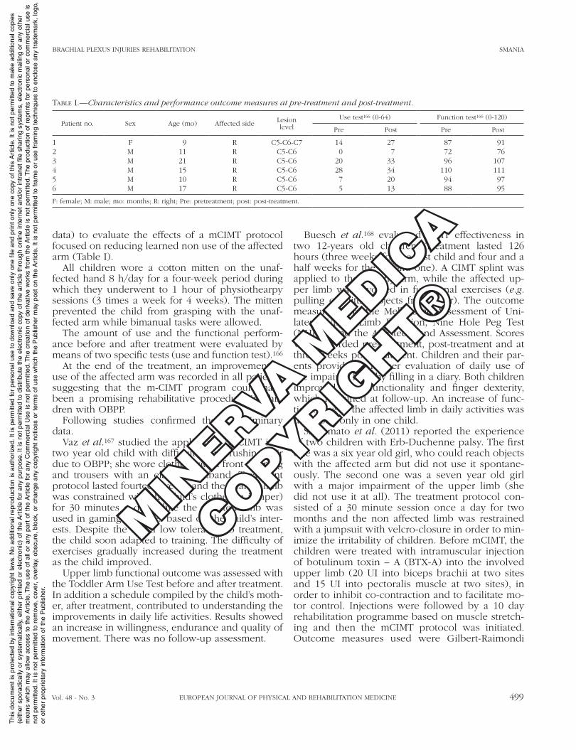
As regards rehabilitation, it is a long-lasting treat-ment because improvements are very slow. Limited muscle activity may be observed even months after the damage and improvements after surgery may only be seen after 3 or 4 years of treatment. Time of recovery depends on many variables, such as the complexity of injury and timing of diagnosis and treatment. When dealing with children, the time for reinnervation is not shorter than in adults, and certainly not age related.5 Therefore, it requires a lot of resources in terms of economic investments and time, by both the multidisciplinary team and the patients’ caregiver.
Moreover, patient cooperation in the rehabilita-tion project is crucial to ensure the best outcome. In addition, adult patients usually have psychologi-cal issues, such as anxiety, depression, as well as difficulty in returning to work, thus having a de-creased quality of life.6, 7
In the present paper the literature concerning BPI deficits and rehabilitation in both adults and children was reviewed and critically discussed.
Epidemiology of brachial plexus injuries
As regards to the epidemiology of BPI, it is im-portant to make a distinction between adults and children.
Concerning epidemiology of adult brachial plex-us damages, there are scarce and old data regard-ing the rate of occurrence in North America. At the end of 1990s Midha provided a prevalence of BPIs in the multitrauma population as approximately 1.2%.8
Although recent and exact data are lacking, BPI usually affects young healthy adults, and in par-ticular male patients (89%). Age of patients ranges between 14 to 63 years, with a mean of 29 years and a median of 25 years of age. Fifty percent of patients are between 19 and 34 years old.8, 9
Between 44% and 70% of BPI are caused by traumatic injuries, mostly occurring in motorcycle accidents, during sporting activities, or at the work place.8, 10-12
In particular, motorcycle accidents account for 22% of injuries, since it is estimated that 4.2% of pa-tients involved in motorcycle accidents have plex-us damage.8, 11, 12 In adults, another notable cause of BPI is iatrogenic lesion. In this group, traumas are often possible causes, for example during sur-gical and anesthetic procedures.1 In these cases, the mechanism of trauma is due to stretching and compression secondary to inappropriate position-ing of the upper limb during surgery, or to a direct lesion by interventions on the shoulder, the pos-terior triangle of the neck, and axilla.10,13, 14 Even though traumatic mechanism accounts for a signifi-cant number of brachial plexus iatrogenic lesions, there are some non traumatic less frequent causes of damage, such as adjuvant radiation.10, 15 The in-cidence of iatrogenic BPIs as a whole is difficult to estimate. Nonetheless they can be a significant cause of major disability.16-20
In regards to epidemiology of brachial plexus damage in children, OBPP is by far the most fre-quent etiological condition. Incidence of OBPP in the available literature ranges between 0.38 and 1.56 per 1000 live births. This wide variability may depend on the difference in the type of obstetric care and in the average birth weight of infants in different geographic regions.21
For instance, a recent study reported an incidence of OBPP in USA of about 1.51 cases per 1000 live births.22 Similar results are reported by a Canadian study, where the incidence is estimated to be be-tween 0.5 and 3 injuries per 1000 live Birth.23 As to European countries, a Dutch study reported an in-cidence of 4.6 per 1000 births. A further interesting finding of this study is that a complete neurological recovery occurred in 72.6% of cases.24
OBPP represents a frequent cause of litigation against physicians or other professionals involved in health care. At present, 4.2% of all medical mal-practice claims against obstetricians are related to trauma during birth.25
Some perinatal risk factors for OBPP are birth weight >4 kg, maternal diabetes mellitus, maternal obesity or excessive weight gain, prolonged preg-nancy, prolonged second stage of labor, persistent fetal malposition, and breech delivery.26 Other risk factors for OBPP include multiparous pregnancies, previous deliveries resulting in brachial plexus birth palsy, and difficult operative delivery.21
There is no consensus regarding caesarean sec-
MIN
ERVA M
EDICA
COPYRIGHT®
Thi
s do
cum
ent
is p
rote
cted
by
inte
rnat
iona
l cop
yrig
ht la
ws.
No
addi
tiona
l rep
rodu
ctio
n is
aut
horiz
ed.I
t is
per
mitt
ed fo
r pe
rson
al u
se t
o do
wnl
oad
and
save
onl
y on
e fil
e an
d pr
int
only
one
cop
y of
thi
s A
rtic
le.I
t is
not
per
mitt
ed t
o m
ake
addi
tiona
l cop
ies
(eith
er s
pora
dica
lly o
r sy
stem
atic
ally
, ei
ther
prin
ted
or e
lect
roni
c) o
f th
e A
rtic
le fo
r an
y pu
rpos
e.It
is n
ot p
erm
itted
to
dist
ribut
e th
e el
ectr
onic
cop
y of
the
art
icle
thr
ough
onl
ine
inte
rnet
and
/or
intr
anet
file
sha
ring
syst
ems,
ele
ctro
nic
mai
ling
or a
ny o
ther
mea
ns w
hich
may
allo
w a
cces
s to
the
Art
icle
.The
use
of
all o
r an
y pa
rt o
f th
e A
rtic
le fo
r an
y C
omm
erci
al U
se is
not
per
mitt
ed.T
he c
reat
ion
of d
eriv
ativ
e w
orks
fro
m t
he A
rtic
le is
not
per
mitt
ed.T
he p
rodu
ctio
n of
rep
rints
for
pers
onal
or
com
mer
cial
use
isno
t pe
rmitt
ed.I
t is
not
per
mitt
ed t
o re
mov
e, c
over
, ov
erla
y, o
bscu
re,
bloc
k, o
r ch
ange
any
cop
yrig
ht n
otic
es o
r te
rms
of u
se w
hich
the
Pub
lishe
r m
ay p
ost
on t
he A
rtic
le.I
t is
not
per
mitt
ed t
o fr
ame
or u
se f
ram
ing
tech
niqu
es t
o en
clos
e an
y tr
adem
ark,
logo
,or
oth
er p
ropr
ieta
ry in
form
atio
n of
the
Pub
lishe
r.
BRACHIAL PLExUS INJURIES REHABILITATION SMANIA
Vol. 48 - No. 3 EUROPEAN JOURNAL OF PHYSICAL AND REHABILITATION MEDICINE 485
As far as outcome is concerned, in supraclavicu-lar lesions upper limb recovery is worse than in infraclavicular lesions. Moreover, supraclavicular injuries more frequently require surgical explora-tion (52% of cases vs. 17%).8
As regards children, historically, the first classifi-cation was based on the observations carried out by Erb and Klumpke. Erb described both C5 and C6 root avulsions and upper trunk lesions, whereas Klumpke observed the C8-T1 injury pattern, which is not common as an isolated form.27
Subsequently, Narakas et al. suggested a clas-sification which partially overlapped with the pre-vious findings. They identified four types of le-sions.28 In type I (upper Erb’s), C5 and C6 roots are involved with spontaneous recovery in over 80% of cases. In these children weakness of shoul-der abductors, external rotators and elbow flexors is generally observed. Type II lesion (extended Erb’s) involves C5-C7 level and clinical presenta-tion is similar to type I with the addition of drop wrist. These children show a good recovery in 60% of cases.
Type III injury is characterized by involvement of C5-T1 roots with a complete flaccid paralysis. Spontaneous recovery occurs in 30-50% of cases with a functional hand in many patients.
In type IV clinical findings are the same as in type III, but with the addition of Horner syndrome. The roots involved are C5-T1. These children show the worst spontaneous recovery.
In addition, Brunelli and Brunelli described le-sions of the C7 root as “intermediate palsy”, usually due to anterior to posterior traction.29 An associat-ed variable involvement of the upper or the lower plexus was found, depending on the mechanism of trauma. Indeed, trauma in abduction causes prima-rily lower plexus injury, whereas downward trac-tion provokes predominantly upper plexus dam-age.
Al-Qattan et al. recently introduced a modifica-tion in Narakas’classification, subdividing type II into two categories: IIa and IIb.30 Type IIa includes children with extended Erb’s palsy and recovery of early (within two months) wrist extension, where-as in type IIb children do not show early wrist extension. Patients who belong to type IIa show a significantly higher percentage of good spontane-ous upper limb recovery than those belonging to type IIb.30
tions and the risk of OBPP. Some studies report it to be a protective factor, while in other studies it accounts for 1% to 4% of cases of lesion.22, 26
Similar risk factors have been noticed in a Swed-ish study for shoulder dystocia, which is frequently associated to OBPP, probably due to the supple-mentary effort needed during delivery in these conditions. However, a high percentage of babies with OBPP (54%) without known risk factors be-fore birth or during delivery was also reported.22
Classification
Classification of brachial plexus damages has been historically developed in relation to traumatic etiologies causing upper limb traction. Sequelae of traction injuries can be subdivided into rootlet avulsion or rupture.3
Two possible types of avulsion are peripheral and central avulsion. In peripheral avulsion, trac-tion forces overcome the resistance of the supports that keep rootlets attached. On the other hand, in central avulsion, the movement of the spinal cord leads to spinal bending and therefore rootlet avul-sion.4
Ruptures can be located both at a pre- or post-ganglionic level.
Since causes and mechanism of traumas are very different in adults and children, different types of classifications in these two groups have been put forward. Usually available literature classifies adult lesions on the basis of lesion site, distinguishing supraclavicular, retroclavicular or infraclavicular plexus lesions.5 In children, on the contrary, clas-sification is based on which roots are involved.
As to adults, supraclavicular injuries involve roots and/or trunks, and account for approximately 75% of brachial plexus lesions; true avulsions with subclavian artery rupture can be associated. Often these lesions are caused by a trauma that produces violent head and neck movement away from the ipsilateral shoulder. Retroclavicular lesions involve brachial plexus divisions and are the least com-mon; vascular damage is often present.10
In infraclavicular injury distal branches are in-volved. Vascular lesions are associated in about 30% of cases. The mechanism of trauma is often a violent injury to the shoulder girdle, particularly ab-duction trauma and anterior shoulder dislocation.
MIN
ERVA M
EDICA
COPYRIGHT®
Thi
s do
cum
ent
is p
rote
cted
by
inte
rnat
iona
l cop
yrig
ht la
ws.
No
addi
tiona
l rep
rodu
ctio
n is
aut
horiz
ed.I
t is
per
mitt
ed fo
r pe
rson
al u
se t
o do
wnl
oad
and
save
onl
y on
e fil
e an
d pr
int
only
one
cop
y of
thi
s A
rtic
le.I
t is
not
per
mitt
ed t
o m
ake
addi
tiona
l cop
ies
(eith
er s
pora
dica
lly o
r sy
stem
atic
ally
, ei
ther
prin
ted
or e
lect
roni
c) o
f th
e A
rtic
le fo
r an
y pu
rpos
e.It
is n
ot p
erm
itted
to
dist
ribut
e th
e el
ectr
onic
cop
y of
the
art
icle
thr
ough
onl
ine
inte
rnet
and
/or
intr
anet
file
sha
ring
syst
ems,
ele
ctro
nic
mai
ling
or a
ny o
ther
mea
ns w
hich
may
allo
w a
cces
s to
the
Art
icle
.The
use
of
all o
r an
y pa
rt o
f th
e A
rtic
le fo
r an
y C
omm
erci
al U
se is
not
per
mitt
ed.T
he c
reat
ion
of d
eriv
ativ
e w
orks
fro
m t
he A
rtic
le is
not
per
mitt
ed.T
he p
rodu
ctio
n of
rep
rints
for
pers
onal
or
com
mer
cial
use
isno
t pe
rmitt
ed.I
t is
not
per
mitt
ed t
o re
mov
e, c
over
, ov
erla
y, o
bscu
re,
bloc
k, o
r ch
ange
any
cop
yrig
ht n
otic
es o
r te
rms
of u
se w
hich
the
Pub
lishe
r m
ay p
ost
on t
he A
rtic
le.I
t is
not
per
mitt
ed t
o fr
ame
or u
se f
ram
ing
tech
niqu
es t
o en
clos
e an
y tr
adem
ark,
logo
,or
oth
er p
ropr
ieta
ry in
form
atio
n of
the
Pub
lishe
r.
SMANIA BRACHIAL PLExUS INJURIES REHABILITATION
486 EUROPEAN JOURNAL OF PHYSICAL AND REHABILITATION MEDICINE September 2012
by head and neck position, as well as shoulder and upper limb posture. For example, Erb’s or Klumpke’s palsy sometimes can be suspected by observing the placement and movement of the affected arm.23
As to shoulder girdle, it is necessary to rule out shoulder dislocation and winging of the scapula (cor-related to long thoracic nerve injury). Furthermore, segmental range of motion and movements have to be observed. For the evaluation of single movements, with and without gravity, the Active Movement Scale 33 is frequently used in children with OBPP.23
Instrumental examination
Instrumental examination supports clinical evalu-ation in BPI detection, both in adults and children.
As to adults, in the past years, myelography with water – soluble agents was found to be more ac-curate than MRI in the detection of nerve rootlets inside the spinal canal. Currently, computed tom-ography - myelography (CT – myelography) is con-sidered the gold standard to identify the site of le-sion and the amount of damage.34 Radiograms are used to detect fractures and/or dislocations of ribs (especially the first rib), elevation of ipsilateral he-midiafragm (in the latter case it could be a possible damage of C5), shoulder girdle depression, and dis-location of the scapula.
Neurophysiological studies are helpful, before and during surgery. Intraoperative sensory evoked potentials (SEPs), combined with paraspinal mus-cle electromyography (EMG), can be helpful in the identification of possible selective avulsion of ven-tral or dorsal rootlets and to distinguish between pre- and post-ganglionic lesions. Indeed, the ampli-tude of sensory nerve action potentials (SNAPs) is normal in pre-ganglionic injuries whereas is absent or reduced in postganglionic injuries.35, 36 As regards EMG, it can provide information about muscle den-ervation from axonal damage and prognostic infor-mation regarding reinnervation. The ideal timing to perform EMG ranges between 3 and six weeks after injury. As to reinnervation, EMG is characterized by polyphasic motor units, with low amplitude configu-ration; new terminal sprouts can be identified within 1-2 months after damage.37
As to OBPP, magnetic resonance imaging (MRI) and ultrasound scan (US) are often used, basing on the low compliance of newborns and the need to use less invasive techniques.
Assessment
Clinical evaluation
Early diagnosis of BPI is of most importance, both in newborns and in adults, since secondary damage could lead to disability and functional impairment. Indeed, muscular atrophy and visco – elastic chang-es in muscles, ligaments, and joints can hamper nor-mal use of limbs, even when reinnervation occurs.
Despite diagnosis is typically quite simple and clinical evaluation immediately leads to a correct identification of the injury, sometimes other medi-cal conditions, such as sepsis with localization at the glenohumeral joint or infant cerebral palsy, can show a similar clinical pattern.10
In the acute phase, clinical history is useful in identification of the traumatic mechanism of injury in adult patients. In addition, diagnosis could be supported by acute pain onset on the upper limb, with a distribution that often reflects the line of ap-plication of force during trauma. Skin bruising and focal ecchymosis can also be observed.10 A detailed neurological clinical examination can suggest a gen-eral identification of extent of lesion; the area of motor paralysis and sensory loss generally correlate with site and extension of BPI (see also somatosen-sory deficits for details).
Sympathetic damage is often associated with a C8-T1 lesion.10
It is also important to evaluate a possible appear-ance of a tingling sensation (with a precise neuro-anatomical distribution) exacerbated by percussion of a nerve trunk in the posterior triangle of the neck (positive Tinel’s sign). Generally it correlates with a post-ganglionic damage.31
In regards to children with OBPP, a detailed and thorough neurological examination, as in adults, is of most importance to support clinical diagnosis.
It is necessary to look for associated signs, such as torticollis, cutaneous colour and trophic changes, contralateral arm involvement and bone fractures (especially ipsilateral clavicle fractures).23 Usually newborns with OBPP and associated torticollis tend to look away from the affected arm.
Somatosensory deficits, as well as facial nerve pal-sy, phrenic nerve damage (seen as thoracic and ab-dominal asymmetry), sympathetic involvement and Horner syndrome must be carefully detected.32
The site and extent of the lesion can be suggested
MIN
ERVA M
EDICA
COPYRIGHT®
Thi
s do
cum
ent
is p
rote
cted
by
inte
rnat
iona
l cop
yrig
ht la
ws.
No
addi
tiona
l rep
rodu
ctio
n is
aut
horiz
ed.I
t is
per
mitt
ed fo
r pe
rson
al u
se t
o do
wnl
oad
and
save
onl
y on
e fil
e an
d pr
int
only
one
cop
y of
thi
s A
rtic
le.I
t is
not
per
mitt
ed t
o m
ake
addi
tiona
l cop
ies
(eith
er s
pora
dica
lly o
r sy
stem
atic
ally
, ei
ther
prin
ted
or e
lect
roni
c) o
f th
e A
rtic
le fo
r an
y pu
rpos
e.It
is n
ot p
erm
itted
to
dist
ribut
e th
e el
ectr
onic
cop
y of
the
art
icle
thr
ough
onl
ine
inte
rnet
and
/or
intr
anet
file
sha
ring
syst
ems,
ele
ctro
nic
mai
ling
or a
ny o
ther
mea
ns w
hich
may
allo
w a
cces
s to
the
Art
icle
.The
use
of
all o
r an
y pa
rt o
f th
e A
rtic
le fo
r an
y C
omm
erci
al U
se is
not
per
mitt
ed.T
he c
reat
ion
of d
eriv
ativ
e w
orks
fro
m t
he A
rtic
le is
not
per
mitt
ed.T
he p
rodu
ctio
n of
rep
rints
for
pers
onal
or
com
mer
cial
use
isno
t pe
rmitt
ed.I
t is
not
per
mitt
ed t
o re
mov
e, c
over
, ov
erla
y, o
bscu
re,
bloc
k, o
r ch
ange
any
cop
yrig
ht n
otic
es o
r te
rms
of u
se w
hich
the
Pub
lishe
r m
ay p
ost
on t
he A
rtic
le.I
t is
not
per
mitt
ed t
o fr
ame
or u
se f
ram
ing
tech
niqu
es t
o en
clos
e an
y tr
adem
ark,
logo
,or
oth
er p
ropr
ieta
ry in
form
atio
n of
the
Pub
lishe
r.
BRACHIAL PLExUS INJURIES REHABILITATION SMANIA
Vol. 48 - No. 3 EUROPEAN JOURNAL OF PHYSICAL AND REHABILITATION MEDICINE 487
of ultra-slow Na+ inactivation results in the capacity of voltage-dependent ion channels to operate in a condition of chronic depolarization.52 Another adap-tive change in a denervated muscle is the develop-ment of supersensitivity to acetylcholine (ACh). After 5-10 days acetylcholine receptors increase their ex-trajunctional density and distribution.53 In about two weeks the increased sensibility to ACh in extrajunc-tional membrane reaches a peak, and then begins to decrease at about three to four weeks.54
It is interesting to note that the different types of muscle fibers are not homogeneously affected by atrophy. For instance, a faster rate of atrophy was found in the soleus (typically slow-twitch) with re-spect to the extensor digitorum longus (typically fast-twitch) muscle in rats,55 while in dogs the gas-trocnemius underwent a slower atrophy than the an-terior tibial muscle.56 Other studies reported a pref-erential atrophy of histochemical type 2 (fast) with respect to histochemical type 1 (slow) fibers in fast-twitch,57-60 but not in slow-twitch muscles.58, 61 In humans a difference was found between early and later effects of denervation.60 Two to four days after denervation the most commonly reported change is an alteration of contractile properties, mainly due to an altered muscle activation process.48, 62-64 In particular, a few days after denervation there is a reduction of developed tension in the stimulated muscle. This observation can be explained by altera-tions of the electrical properties of the muscle cell membrane, which preclude the generation of high frequency action potentials, and thus a complete ac-tivation of the muscle.47, 63 Furthermore, fibrillation activity starts in the first few days, with spontaneous and uncoordinated contractile activity of the dener-vated muscle, occurring in coincidence with the fall in resting membrane potential.65 Fibrillation activity reaches a peak in a few days, and then decreases, especially in slow type muscles.66, 67 Afterwards, changes in the pattern of myofibrillar protein ex-pression and muscle fiber composition are observed some weeks later.60
A temporal progression in the development of hu-man muscle atrophy has been described.60 Between 2 and 3 months a decrease of 50% in fibers diam-eter appears, while streaks remain intact. At 2 and 4 months, morphological activation of satellite cells is found, simultaneously with a decrease in fibril number, myofiber and capillary death, and deposi-tion of massive amounts of interstitial collagen.
Fast spin-echo sequence MRI is considered more suitable than CT myelography, because of its rapid-ity of execution and the reduced doses of sedation required. Fast spin-echo MRI can also identify as-sociated pseudomeningoceles, indicating possible rootlets avulsions.38
In US technique, anatomical landmarks such as the anterior tubercles of transverse vertebral proc-esses can lead to a proper identification of roots.39 A root injury can be inferred by the demonstration of an increased nerve cross sectional area.40
In addition, differential diagnosis between pre- and post-ganglionic injury can be accomplished through US.41
Sequelae of brachial plexus injury
Muscle atrophy
Development of muscle atrophy is a remarkable result of denervation. Denervated muscles undergo several structural and neurophysiological alterations, which cause contractility deficits and changes in ex-citability, resulting in muscle atrophy. This topic has been widely discussed in literature based on studies on animal models.
In rats, after denervation, a progressive loss of weight and reduction of muscle fibre diameter oc-curs within two weeks.42, 43 At a microscopic level, muscular fiber structure is subverted. In particular, sarcomeric disorganization, myofibrillar disruption, changes in the number of ribosomes and in the number and size of mitochondria, and changes in sar-coplasmic reticulum morphology are reported44. As a result, altered structural properties lead to a slowed contraction speed. Besides morphologic alterations, changes in excitability contribute to pathological contractile activity. After only 7 days from denerva-tion, a reduction in the resting membrane potential by 10-15 mV was observed in rats, with smaller and slower action potentials than in controls.45-49 Further-more, a reduced velocity in the impulse conduction was recorded.46, 48 Action potential changes were explained by a decreased Na+ current as a conse-quence of an increased Na+ channel inactivation during the impulse.50, 51 Effectiveness of ultra-slow processes of Na+ conductance inactivation tended to decrease after 6 days of denervation, as a mechanism of membrane adaptation. That is, the partial removal
MIN
ERVA M
EDICA
COPYRIGHT®
Thi
s do
cum
ent
is p
rote
cted
by
inte
rnat
iona
l cop
yrig
ht la
ws.
No
addi
tiona
l rep
rodu
ctio
n is
aut
horiz
ed.I
t is
per
mitt
ed fo
r pe
rson
al u
se t
o do
wnl
oad
and
save
onl
y on
e fil
e an
d pr
int
only
one
cop
y of
thi
s A
rtic
le.I
t is
not
per
mitt
ed t
o m
ake
addi
tiona
l cop
ies
(eith
er s
pora
dica
lly o
r sy
stem
atic
ally
, ei
ther
prin
ted
or e
lect
roni
c) o
f th
e A
rtic
le fo
r an
y pu
rpos
e.It
is n
ot p
erm
itted
to
dist
ribut
e th
e el
ectr
onic
cop
y of
the
art
icle
thr
ough
onl
ine
inte
rnet
and
/or
intr
anet
file
sha
ring
syst
ems,
ele
ctro
nic
mai
ling
or a
ny o
ther
mea
ns w
hich
may
allo
w a
cces
s to
the
Art
icle
.The
use
of
all o
r an
y pa
rt o
f th
e A
rtic
le fo
r an
y C
omm
erci
al U
se is
not
per
mitt
ed.T
he c
reat
ion
of d
eriv
ativ
e w
orks
fro
m t
he A
rtic
le is
not
per
mitt
ed.T
he p
rodu
ctio
n of
rep
rints
for
pers
onal
or
com
mer
cial
use
isno
t pe
rmitt
ed.I
t is
not
per
mitt
ed t
o re
mov
e, c
over
, ov
erla
y, o
bscu
re,
bloc
k, o
r ch
ange
any
cop
yrig
ht n
otic
es o
r te
rms
of u
se w
hich
the
Pub
lishe
r m
ay p
ost
on t
he A
rtic
le.I
t is
not
per
mitt
ed t
o fr
ame
or u
se f
ram
ing
tech
niqu
es t
o en
clos
e an
y tr
adem
ark,
logo
,or
oth
er p
ropr
ieta
ry in
form
atio
n of
the
Pub
lishe
r.
SMANIA BRACHIAL PLExUS INJURIES REHABILITATION
488 EUROPEAN JOURNAL OF PHYSICAL AND REHABILITATION MEDICINE September 2012
recreation can relieve it.10 The intensity of pain after brachial plexus avulsion injury and prior to surgical treatment depends on the extent of nerve injury.78 Particularly, severity of pain is proved to be related to the number of spinal avulsed roots.78
The classical theory for the physiopathology of this neuropathic pain refers to central pain due to root avulsion and spinal cord deafferentation.73, 77, 79,
80 Sudden nerve rootlets avulsion involves interrup-tion of afferent pathways and degeneration of the central projections of cells in the dorsal root gan-glion, resulting in a loss of sensory afferent input into the spinal cord. Consequent cellular and neu-rochemical changes cause disinhibition of neurons within the substantia gelatinosa and the generation of abnormal activity of neurones in the laminae I, II, and V of the dorsal horn, which fire spontane-ously.73, 76, 81, 82 Constant pain seems to be related to spontaneous aberrant activity of the neurons in the denervated posterior horn. Moreover, sudden outbursts of ectopic electrical activity in the injured part of the dorsal horn lead to convulsive pain. This neuronal hyperexcitability was shown to be gradu-ally transmitted rostrally, with spontaneous impulses found in neurons in the Lateral Ventral Posterior nu-cleus of the thalamus, and in the medial thalamus.10 Thus, a central sensitization develops as a form of pathological plasticity of the Central Nervous Sys-tem.83, 84
Furthermore, inflammatory processes play a role in the pathophysiology of neuropathic pain. After peripheral and central nerve lesions, activation of macrophages into the nerve and dorsal root gan-glion and of microglia within the CNS is responsible for the release of immune modulators. Proinflam-matory cytokines as tumour necrosis factor-a and neurotrophic factors are shown to be involved in the onset and maintenance of the long-lasting neu-ropathic pain behavior in animal models of brachial plexus avulsion.83, 85, 86
Recently, an alternative hypothesis has been pro-posed. Based on previous data, Bertelli et al. esti-mated that in almost 80% of patients with total palsy of the brachial plexus there is at least one root not avulsed which is available for grafting.87, 88 Further-more, they considered that pain quickly subsided after early surgical repair by root grafting, and con-tinuous significant amelioration of pain was noticed over the following months and years after surgery. In addition, pain relief was not related to the number
At the same time, condensation and fragmenta-tion of nuclear chromatin appear in some nuclei, while later there is a loss in normal staining density of mitochondrial matrix. With increasing denerva-tion time, mitochondria become smaller and more globular in shape, with sparsely arrayed cristae. All these changes contribute to reduce the restorative capacity of the denervated muscle.60 Gross muscle and muscle fiber atrophy continues in the period between 2 and 7 months of denervation, with dense mats of collagen fibers surrounding remaining mus-cle fibers. Initial streak rarefaction begins around 1 year after denervation, while by 18 months a sub-stantial reduction in cross sectional area and char-acteristic changes in the sarcoplasmic reticulum are observed. Sarcoplasmic reticulums become more ir-regular and lose their close relationship to the A-I junctions.60 At two years muscular fiber fragmenta-tion and disintegration is evident. Substitution with adipose and fibrous connective tissue occurs be-tween 1 and 3 years postdenervation, leading to a loss and replacement of muscle fibers.68
Pain
Pain is a major complaint in adult patients with a brachial plexus avulsion or traction injury. Aside from nociceptive pain resulting from ligament and joint injuries at the time of initial trauma, the main concern is neuropathic pain derived from nerve in-jury. This kind of pain usually appears after pre-ganglionic injuries of the brachial plexus and is more frequent in patients with total palsy.69-72 It is reported that nearly 80% of patients with complete brachial plexus palsy after avulsion injuries suffer from pain.69, 71, 73, 74 Brachial plexus avulsion pain is highly characteristic. It usually arises immediately or within the first days after the trauma and it tends to persist for months and years after the triggering le-sion.75-77 Patients typically complain of two types of unbearable pain.10, 76 First, a continuous, crushing, burning, and compressing pain localized in the in-sensitive hand and/or in the forearm. Second, there are superimposed paroxysmal bursts of extremely severe pain shooting into the territory of the injured nerve distribution. These intermittent pains last a few seconds and appear with a high frequency dur-ing the day.10, 76 Elapsing factors such as cold weath-er, illness, emotional disturbances, and depression worsen pain, while a proper distraction at work or
MIN
ERVA M
EDICA
COPYRIGHT®
Thi
s do
cum
ent
is p
rote
cted
by
inte
rnat
iona
l cop
yrig
ht la
ws.
No
addi
tiona
l rep
rodu
ctio
n is
aut
horiz
ed.I
t is
per
mitt
ed fo
r pe
rson
al u
se t
o do
wnl
oad
and
save
onl
y on
e fil
e an
d pr
int
only
one
cop
y of
thi
s A
rtic
le.I
t is
not
per
mitt
ed t
o m
ake
addi
tiona
l cop
ies
(eith
er s
pora
dica
lly o
r sy
stem
atic
ally
, ei
ther
prin
ted
or e
lect
roni
c) o
f th
e A
rtic
le fo
r an
y pu
rpos
e.It
is n
ot p
erm
itted
to
dist
ribut
e th
e el
ectr
onic
cop
y of
the
art
icle
thr
ough
onl
ine
inte
rnet
and
/or
intr
anet
file
sha
ring
syst
ems,
ele
ctro
nic
mai
ling
or a
ny o
ther
mea
ns w
hich
may
allo
w a
cces
s to
the
Art
icle
.The
use
of
all o
r an
y pa
rt o
f th
e A
rtic
le fo
r an
y C
omm
erci
al U
se is
not
per
mitt
ed.T
he c
reat
ion
of d
eriv
ativ
e w
orks
fro
m t
he A
rtic
le is
not
per
mitt
ed.T
he p
rodu
ctio
n of
rep
rints
for
pers
onal
or
com
mer
cial
use
isno
t pe
rmitt
ed.I
t is
not
per
mitt
ed t
o re
mov
e, c
over
, ov
erla
y, o
bscu
re,
bloc
k, o
r ch
ange
any
cop
yrig
ht n
otic
es o
r te
rms
of u
se w
hich
the
Pub
lishe
r m
ay p
ost
on t
he A
rtic
le.I
t is
not
per
mitt
ed t
o fr
ame
or u
se f
ram
ing
tech
niqu
es t
o en
clos
e an
y tr
adem
ark,
logo
,or
oth
er p
ropr
ieta
ry in
form
atio
n of
the
Pub
lishe
r.
BRACHIAL PLExUS INJURIES REHABILITATION SMANIA
Vol. 48 - No. 3 EUROPEAN JOURNAL OF PHYSICAL AND REHABILITATION MEDICINE 489
a better recovery and an increased ability to regain function, with lower chance of developing pain.91 Several mechanisms are proposed to explain differ-ences between children and adults. Enhanced nerve regeneration, a distinct inflammatory response and scar tissue maturation, delayed maturation of in-jured fibers and peripheral nerves, neuronal plas-ticity, highly rich synaptogenesis, and long-lasting cortical reorganization may account for chronic neuropathic pain absence in children with OBPP.10,
88, 90, 91 In children, lack of long-term pain behavior goes together with excellent restoration of sensory function in avulsed spinal root dermatomes and normal pain sensation reported to external stimuli in unaffected regions. All these elements support the evidence of exquisite CNS plasticity during de-velopment.90, 92
Somatosensory deficit
After BPI, somatosensory afference plays an im-portant role on functional recovery of the upper limb, and in particular of the hand.93
In the following section we focus on clinical fea-tures of sensory deficits, on sensory testing, neuro-physiological changes, sensory recovery and treat-ment after nerve injury.
Changes in dermatomeriC representation
After BPI, the extent of sensory impairment on the upper limb generally depends on which roots have been involved, with an area of sensory deficit which follows dermatomeric distribution, both in adults,94 and in children.95 However, some studies in adults and infants showed how segmental sensory innerva-tion presented peculiar characteristics which did not correspond to classic dermatomeric representation.96 Bertelli et al. evaluated the distribution of sensory dis-turbance in 150 adult patients with brachial plexus root injury matching clinical evaluations of upper limb sensation, CT myelo-scans and surgical observa-tions.88 They found out that topography of sensory disturbance did not correspond perfectly to common dermatomeric distribution, possibly due to an over-lapping of cutaneous innervation of the radial side of the hand. For instance, after C5-C6 root injury, distur-bance in touch perception was localized to the lateral side of the forearm and to deltoid area and when C7 and C8 were also affected hand sensation was some-
of avulsed roots. In patients with chronic palsies and persistent pain despite surgery, they found that pain was temporarily alleviated by anesthetics injection close to the non-avulsed roots. Transient pain relief via anesthetic neural blocks is highly suggestive of pain arising within the peripheral nervous system. Based on these observations, Bertelli et al. raised the hypothesis that brachial plexus pain is maybe due to the presence of a non-avulsed ruptured root rather than avulsion and spinal cord deafferentation.88, 89 That is, in patients with subacute and chronic com-plete brachial plexus palsy pain arises from a pre-served root or radicular remnants which remain in contact with the spinal cord and are immersed into an adverse microenvironment. After trauma, local inflammatory processes and release of neurotrophic factors contribute to pain development and force the axons to regenerate into a scar tissue. Eventu-ally there is a neuroma formation. Therefore, root section/neuroma resection would account for early pain relief after surgery before nerve regeneration and end-organ contact. Furthermore, root grafting would create a healthy tissue where root stumps are relocated, thus preventing the recurrence of neuro-ma. Successful regrowth of axons and gradual re-myelination of the regenerating fibers would then contribute to continual improvement of pain over time. Conversely, a large release of neurotrophic fac-tors seems to be the cause of pain persistence in that minority of patients suffering from chronic pain despite grafting.71, 86 Neurotrophic factors should promote nerve regeneration of axons reaching end organs in the periphery. However, because of the wide distance and time required, neurotrophic fac-tors’ secondary painful effect may prevail. Other-wise aberrant reinnervation and misdirected sensory axons regenerating in muscles are further possible explanations suggested for chronic pain after bra-chial plexus grafting.
Whatever the actual origin of pain, of interest is that chronic neuropathic pain rarely appears in children after brachial plexus birth injuries.90 Neu-ropathic pain generally does not affect the pediat-ric population.91 This considerable difference from adults likely derives from higher plasticity of both the central and peripheral nervous system through-out early development in the neonatal period and at the youngest ages. In particular, after spinal root avulsion injury and nerve trauma, rapid and effi-cient changes in the young nervous system allow
MIN
ERVA M
EDICA
COPYRIGHT®
Thi
s do
cum
ent
is p
rote
cted
by
inte
rnat
iona
l cop
yrig
ht la
ws.
No
addi
tiona
l rep
rodu
ctio
n is
aut
horiz
ed.I
t is
per
mitt
ed fo
r pe
rson
al u
se t
o do
wnl
oad
and
save
onl
y on
e fil
e an
d pr
int
only
one
cop
y of
thi
s A
rtic
le.I
t is
not
per
mitt
ed t
o m
ake
addi
tiona
l cop
ies
(eith
er s
pora
dica
lly o
r sy
stem
atic
ally
, ei
ther
prin
ted
or e
lect
roni
c) o
f th
e A
rtic
le fo
r an
y pu
rpos
e.It
is n
ot p
erm
itted
to
dist
ribut
e th
e el
ectr
onic
cop
y of
the
art
icle
thr
ough
onl
ine
inte
rnet
and
/or
intr
anet
file
sha
ring
syst
ems,
ele
ctro
nic
mai
ling
or a
ny o
ther
mea
ns w
hich
may
allo
w a
cces
s to
the
Art
icle
.The
use
of
all o
r an
y pa
rt o
f th
e A
rtic
le fo
r an
y C
omm
erci
al U
se is
not
per
mitt
ed.T
he c
reat
ion
of d
eriv
ativ
e w
orks
fro
m t
he A
rtic
le is
not
per
mitt
ed.T
he p
rodu
ctio
n of
rep
rints
for
pers
onal
or
com
mer
cial
use
isno
t pe
rmitt
ed.I
t is
not
per
mitt
ed t
o re
mov
e, c
over
, ov
erla
y, o
bscu
re,
bloc
k, o
r ch
ange
any
cop
yrig
ht n
otic
es o
r te
rms
of u
se w
hich
the
Pub
lishe
r m
ay p
ost
on t
he A
rtic
le.I
t is
not
per
mitt
ed t
o fr
ame
or u
se f
ram
ing
tech
niqu
es t
o en
clos
e an
y tr
adem
ark,
logo
,or
oth
er p
ropr
ieta
ry in
form
atio
n of
the
Pub
lishe
r.
SMANIA BRACHIAL PLExUS INJURIES REHABILITATION
490 EUROPEAN JOURNAL OF PHYSICAL AND REHABILITATION MEDICINE September 2012
Only in a small group (20% of patients) referred sen-sation became stronger or did not change at all.
Furthermore, a different temporal “right-way” and “wrong-way” referred sensation onset was recorded. While the “right way” occurred at least six months af-ter injury, the “wrong-way” was found at early stages from injury (in the first six months).
A lack of central plasticity and a successful periph-eral nerve regeneration could explain the “right way” type. On the other hand, the “wrong way” referred sensation may be explained by the CNS reorganiza-tion after deafferentation. Since face, trunk and hand are represented in adjacent cortical areas, a dorsal root ganglia or column injury could result in an ex-pansion of input from face and trunk areas to the hand cortical territory.76
On the contrary, in children a perfect localization of restored sensation routed via nerves transposed from other anatomical sites (no referred sensation ex-perience), demonstrating evidence of exquisite CNS plasticity.90
sensory examination
Although sensory deficit in infants with OBPP is difficult to ascertain,98 response to touch and painful stimuli in newborns with OBPP and pinprick sensa-tion in very young children could be useful to assess sensory conduction integrity.95, 98
In previous studies, standardized tests are used to evaluate sensory function and thresholds in children and adults:
Evaluation of pain and touch sensation, with pin-prick and with cotton wool, respectively. Presence or absence of each stimulus perception is recorded (the site of these tests follow specific dermatomes as established by the Medical Research Council UK memorandum in 1976).76, 90, 101
Joint position sense: proprioceptive sensation is evaluated at different levels (hand, wrist, elbow and shoulder).76, 90
Two point discrimination test (2-PD): defined as the distance necessary to feel two contacts.100-103 A distance of 3 mm between the points on the fingers is considered appropriate for normal levels of sen-sibility.92 Pressure administrated is not standardized and children sometimes demonstrated difficulties in understanding the principle of 2-PD.102
Cutaneous pressure threshold: Semmen-Weinstein monofilament testing is used for the determination
what partially preserved.88 In classical dermatomeric distribution, sensory innervation of hand is provid-ed by C6, C7 and C8 roots. C6 distribution interests thumb and radial side of hand; C7 distribution in-volves second, third finger and central area of hand; finally C8 distribution reaches fifth finger and ulnar area of hand. Consequently, after C6 to C8 lesion, a complete hand anesthesia would be expected.97
Colon et al. examined sensory function using so-matosensory evoked potentials (SEPs) three to seven months after birth in children with upper (C5-C7) OBPP.96 They stimulated the thumb and the third dig-it and demonstrated how sensory innervation of the hand was more extensive than they expected, with alternative nervous pathways implied in the conduc-tion of sensory sensations (i.e., sensory inputs from the third digit, normally vehiculated by C7 root, were transmitted by C6 or C8 roots).96
CliniCal features
Patients with BPI at times present “indirected clini-cal signs” of sensory impairment or failure of restora-tion of sensation. Self mutilation, finger biting and poor use of the hand, even when the motor function was preserved, were found in children with soma-tosensory deficit due to OBPP.98, 99 In adult patients with sensory deficit, skin lesions and infections were noticed more commonly in the ulnar side of the hand and in the tip of the little finger (C7-T1 roots). These cutaneous areas resulted more susceptible to injury because often scraped against damaging surfaces during hand manipulation.100
referred sensations
Besides sensory loss, adults with BPI sometimes develop referred sensation experience.76, 99
Htut et al. divided referred sensation into main two categories, the “right way”, when sensation was “re-ferred from the affected arm to the original source of the donor nerve afferent fibers” and the “wrong way,” when sensation was referred “from a region that was not to a source of re-directed donor nerve fibers” (e.g., referral sensation in the affected arm when the patient is shaving or drinking something cold).76 In this study 56% of patients with BPI developed re-ferred sensation at some time after injury.
This phenomenon generally decreased over time becoming stationary or completely disappearing.
MIN
ERVA M
EDICA
COPYRIGHT®
Thi
s do
cum
ent
is p
rote
cted
by
inte
rnat
iona
l cop
yrig
ht la
ws.
No
addi
tiona
l rep
rodu
ctio
n is
aut
horiz
ed.I
t is
per
mitt
ed fo
r pe
rson
al u
se t
o do
wnl
oad
and
save
onl
y on
e fil
e an
d pr
int
only
one
cop
y of
thi
s A
rtic
le.I
t is
not
per
mitt
ed t
o m
ake
addi
tiona
l cop
ies
(eith
er s
pora
dica
lly o
r sy
stem
atic
ally
, ei
ther
prin
ted
or e
lect
roni
c) o
f th
e A
rtic
le fo
r an
y pu
rpos
e.It
is n
ot p
erm
itted
to
dist
ribut
e th
e el
ectr
onic
cop
y of
the
art
icle
thr
ough
onl
ine
inte
rnet
and
/or
intr
anet
file
sha
ring
syst
ems,
ele
ctro
nic
mai
ling
or a
ny o
ther
mea
ns w
hich
may
allo
w a
cces
s to
the
Art
icle
.The
use
of
all o
r an
y pa
rt o
f th
e A
rtic
le fo
r an
y C
omm
erci
al U
se is
not
per
mitt
ed.T
he c
reat
ion
of d
eriv
ativ
e w
orks
fro
m t
he A
rtic
le is
not
per
mitt
ed.T
he p
rodu
ctio
n of
rep
rints
for
pers
onal
or
com
mer
cial
use
isno
t pe
rmitt
ed.I
t is
not
per
mitt
ed t
o re
mov
e, c
over
, ov
erla
y, o
bscu
re,
bloc
k, o
r ch
ange
any
cop
yrig
ht n
otic
es o
r te
rms
of u
se w
hich
the
Pub
lishe
r m
ay p
ost
on t
he A
rtic
le.I
t is
not
per
mitt
ed t
o fr
ame
or u
se f
ram
ing
tech
niqu
es t
o en
clos
e an
y tr
adem
ark,
logo
,or
oth
er p
ropr
ieta
ry in
form
atio
n of
the
Pub
lishe
r.
BRACHIAL PLExUS INJURIES REHABILITATION SMANIA
Vol. 48 - No. 3 EUROPEAN JOURNAL OF PHYSICAL AND REHABILITATION MEDICINE 491
of the somatosensory cortical map after a peripheral nerve injury. Nerve injury at distal sites of the up-per limb was considered by the CNS as sudden deaf-ferentation with influence to cortical hand and arm representation.
After a median nerve injury, the development of a “black hole” in the somatosensory brain cortex corre-sponding to the median nerve projectional areas was demonstrated. As a result, the adjacent cortical areas could overlap the “black hole” in a few minutes; this phenomenon was probably due to unmasking syn-aptic connection normally inhibited.
If Schwann cell tubes were preserved after a trau-matic nerve injury, axons re-innerved the original peripheral target and the cortical map was exactly restored. On the contrary, a reorganization of the cortical hand map was found after a complete nerve transection followed by surgical repair. Even if micro-surgical techniques have been refined with time, af-ter nerve repair an axonal misdirection appeared.93 This misdirection caused a mismatched reinnervation of cutaneous areas by axons growing; this phenom-enon was followed by a synaptic reorganization of the somatosensory representation areas of hand and upper limb with overlapped cortical zones, like a “mosaic”.107
Synaptic changes occurred also in subcortical sites as such as the dorsal horn of spinal tract, cuneate and gracile nuclei in brainstem, ventroposterolateral and ventroposteralmedial nuclei of thalamus, according to somatosensory afferent ways.106
Altered sensory perception could be also ex-plained by modifications in physiology and biochem-istry both after distal and proximal pheripheral nerve injury 107-109 with a shift in metabolism of neuronal cell body from a stance status to a growth one. In as-sociation with Wallerian degeneration, Schawnn cells started to product growth factors (INGF, CTNF, BDNF, NT3, NT4/5 and NT5-6).
Apoptosis of a large number of sensory neurons at the dorsal root ganglia was described after pe-ripheral nerve injury and the amount of cellular loss seemed to correlate with a poor functional re-covery.
A correlation was also found between sensory cell loss and the level of injury along the nerve with more proximal injuries causing a wider loss of neu-rons.
Cellular loss started immediately after a nerve ax-otomy with a great amount of death sensory cells in
of cutaneus pressure threshold.104 Monofilaments are numbered from 1.65 to 6.65 and are placed on the skin with a pressure just adequate to bend the filament.103 Abnormal values are considered as >no.3 monofilaments (0.0479 g) for adults and children.76,
90, 100, 103 Filament 2.83 has been found to be a good predictor of normal pressure sensation on most areas of the human body.102
Vibration perception threshold: tested with a tun-ing fork or a biothensiometer placed on bony promi-nences (proximal interphalageal joint, ulnar or radial styloid process and elbow) of the investigated arm. When a tuning fork is used, the perception of vibra-tion is assessed for 30-, 128-, and 256-cycles/second stimuli.103 For the biothensiometer evaluation abnor-mal values are considered >8 V for children and >10 V for adults.76, 90, 101
Thermal threshold: cold and warm sensations are evaluated from a baseline temperature (e.g., 30°-32 °C) and the threshold of painful cold and warm are recorded. Normal threshold values in adults are 2.5 °C for cold sensation and 3 °C for warm sensation from the baseline.92 Abnormal values are considered in some studies as >3.9 °C for warm sensation and >2.6 °C and for cold sensation in adults aged 20-30 years (children: >3.8 °C and > 2.3 °C for warm and cold sensation, respectively).76, 90 Hattori et al. tested the perception of warmth using a steel bar warmed in 50°C hot water and asking the patients if the stimulus applied is perceived.103
Some studies 96, 98, 105 used Somatosensory Evoked Potentials (SEPs) for the assessment of integrity of sensory conduction from the periphery to CNS in children with OBPP.
neurophysiologiCal Changes
Clinical evidences have demonstrated the pres-ence of sensory disturbances and an anomalous dis-tribution of touch perception in the upper limbs of patients with BPI. This is thought to be related to somatosensory system modifications after deafferen-tation.76
To our knowledge no neurophysiological data con-cerning neuroplasticity or cortical reorganization after BPI are available. However, some studies described cortical reorganization of hand sensory representa-tion after distal nerve injuries (median or ulnar nerve at the wrist).106, 107
Lundborg 106, 107 described a rapid reorganization
MIN
ERVA M
EDICA
COPYRIGHT®
Thi
s do
cum
ent
is p
rote
cted
by
inte
rnat
iona
l cop
yrig
ht la
ws.
No
addi
tiona
l rep
rodu
ctio
n is
aut
horiz
ed.I
t is
per
mitt
ed fo
r pe
rson
al u
se t
o do
wnl
oad
and
save
onl
y on
e fil
e an
d pr
int
only
one
cop
y of
thi
s A
rtic
le.I
t is
not
per
mitt
ed t
o m
ake
addi
tiona
l cop
ies
(eith
er s
pora
dica
lly o
r sy
stem
atic
ally
, ei
ther
prin
ted
or e
lect
roni
c) o
f th
e A
rtic
le fo
r an
y pu
rpos
e.It
is n
ot p
erm
itted
to
dist
ribut
e th
e el
ectr
onic
cop
y of
the
art
icle
thr
ough
onl
ine
inte
rnet
and
/or
intr
anet
file
sha
ring
syst
ems,
ele
ctro
nic
mai
ling
or a
ny o
ther
mea
ns w
hich
may
allo
w a
cces
s to
the
Art
icle
.The
use
of
all o
r an
y pa
rt o
f th
e A
rtic
le fo
r an
y C
omm
erci
al U
se is
not
per
mitt
ed.T
he c
reat
ion
of d
eriv
ativ
e w
orks
fro
m t
he A
rtic
le is
not
per
mitt
ed.T
he p
rodu
ctio
n of
rep
rints
for
pers
onal
or
com
mer
cial
use
isno
t pe
rmitt
ed.I
t is
not
per
mitt
ed t
o re
mov
e, c
over
, ov
erla
y, o
bscu
re,
bloc
k, o
r ch
ange
any
cop
yrig
ht n
otic
es o
r te
rms
of u
se w
hich
the
Pub
lishe
r m
ay p
ost
on t
he A
rtic
le.I
t is
not
per
mitt
ed t
o fr
ame
or u
se f
ram
ing
tech
niqu
es t
o en
clos
e an
y tr
adem
ark,
logo
,or
oth
er p
ropr
ieta
ry in
form
atio
n of
the
Pub
lishe
r.
SMANIA BRACHIAL PLExUS INJURIES REHABILITATION
492 EUROPEAN JOURNAL OF PHYSICAL AND REHABILITATION MEDICINE September 2012
Anand et al. demonstrated exquisite sensory re-covery in patients with OBPP surgically operated, with good localization of restored sensation.90 However, children who were non-operated did not show significant sensory recovery. On the contrary, Strombeck et al. found no differences in recovery of sensation of the hand in operated and non-op-erated children with OBPP when testing two point discrimination (2-PD).105 Similar results were found by Palmgren et al., suggesting that surgery does not improve sensory recovery of the hand after OBPP.102
Furthermore, Strombeck et al., showed in 70 pa-tients with OBPP that sensory loss is less severe than motor impairment.92 Among the various sen-sory modalities, discriminative sensibility appeared the most impaired, while the thermal sensibility showed the best recovery. Strombeck 105 and Smith 110 showed how children with total palsy presented normal hand protective sensation but impairments with 2-PD.
Even though children with OBPP apparently showed a better sensory recovery than adults, this improvement after nerve graft appeared slower, con-sidering the distance through which the axons must grow. Sensory recovery, in particular nociception, in some cases was not seen until the age of 5 years in children. The slowness of sensory recovery in infants could be attributed to delayed maturation of periph-eral nerve injuries, such as at the nodes of Ranvier.90
Learned non-use and developmental disregard
Before sensory recovery a phenomenon termed learned non-use (LNU) in adults and developmental disregard in children could occur.
Learned non use is a concept developed by Taub et al. in 1976.111 They studied monkeys with loss of sensory perception after deafferentation. As a consequence of sensory deprivation, they found impairments in motor control, even when monkeys preserved muscle strength and visual-related control of movement in the affected limb. It was observed that, after deafferentation, monkeys commonly did not use their insensate limb in spontaneous activi-ties and restriction of the healthy forearm, associ-ated with a training period for the insensate limb, enabled monkeys to restart using their affected limb. Subsequently, the LNU theory was generalized in humans after a CNS injury, in particular in stroke patients.111
dorsal root ganglia at one week and a value of 35-40% of total cell bodies reached at two months after injury.109 Neural survival is essential for regeneration and an early surgical repair is recommended. When surgical treatment is not possible, pharmacological approaches with neuroprotective molecules could be useful.109
sensory reCovery after injury and surgiCal repair
After evaluation, sensory recovery could be classi-fied into 8 grades using the Highet Scale.101, 103
Sensory recovery after BPI appears generally quite poor. Htut et al. evaluated sensory recovery in 76 patients with traumatic BPI before and after surgical treatment. They found a very poor sensory recovery in most patients (with or without surgical repair), with altered stimuli localization. Only the C5 dermatome had a better recovery for thermal sensa-tion, probably due to nerve sprouting from overlap-ping adjacent dermatomeric areas.76
Similar results were found by Hattori et al.103 They investigated sensory recovery of the hand after in-tercostal nerve transfer to median or ulnar nerve in 17 patients following complete avulsion of the bra-chial plexus. According to the Highet scale, they re-corded recovery of superficial touch and pain sensa-tion with some over-response (S2+) in two patients, nine patients recovery of superficial touch and pain in nine patients (S2) and recovery of deep cutane-ous pain sensibility and autonomous activity of the nerve (S1) in six patients.
Hattori et al. (2008) found that patients with a shorter interval from injury to surgery obtained better sensory recovery, however no statistical correlation resulted between time elapsed before surgical intervention and the score on the Highet Scale.103
Most studies on children with OBPP examined motor recovery, however data about somatosensory function are scant.90
Palmgren et al. evaluated sensory changes in 95 patients with OBPP (14 surgically operated) and found a good recovery of sensation in the hand of the affected limb. An altered fine perception (Sem-mer-Weisten monofilaments test) on the affected hand was recorded in only 11% of children with upper OBPP while rates of children with sensory impairments increased (26% to 33%) when the mid-dle and lower plexus were involved.102
MIN
ERVA M
EDICA
COPYRIGHT®
Thi
s do
cum
ent
is p
rote
cted
by
inte
rnat
iona
l cop
yrig
ht la
ws.
No
addi
tiona
l rep
rodu
ctio
n is
aut
horiz
ed.I
t is
per
mitt
ed fo
r pe
rson
al u
se t
o do
wnl
oad
and
save
onl
y on
e fil
e an
d pr
int
only
one
cop
y of
thi
s A
rtic
le.I
t is
not
per
mitt
ed t
o m
ake
addi
tiona
l cop
ies
(eith
er s
pora
dica
lly o
r sy
stem
atic
ally
, ei
ther
prin
ted
or e
lect
roni
c) o
f th
e A
rtic
le fo
r an
y pu
rpos
e.It
is n
ot p
erm
itted
to
dist
ribut
e th
e el
ectr
onic
cop
y of
the
art
icle
thr
ough
onl
ine
inte
rnet
and
/or
intr
anet
file
sha
ring
syst
ems,
ele
ctro
nic
mai
ling
or a
ny o
ther
mea
ns w
hich
may
allo
w a
cces
s to
the
Art
icle
.The
use
of
all o
r an
y pa
rt o
f th
e A
rtic
le fo
r an
y C
omm
erci
al U
se is
not
per
mitt
ed.T
he c
reat
ion
of d
eriv
ativ
e w
orks
fro
m t
he A
rtic
le is
not
per
mitt
ed.T
he p
rodu
ctio
n of
rep
rints
for
pers
onal
or
com
mer
cial
use
isno
t pe
rmitt
ed.I
t is
not
per
mitt
ed t
o re
mov
e, c
over
, ov
erla
y, o
bscu
re,
bloc
k, o
r ch
ange
any
cop
yrig
ht n
otic
es o
r te
rms
of u
se w
hich
the
Pub
lishe
r m
ay p
ost
on t
he A
rtic
le.I
t is
not
per
mitt
ed t
o fr
ame
or u
se f
ram
ing
tech
niqu
es t
o en
clos
e an
y tr
adem
ark,
logo
,or
oth
er p
ropr
ieta
ry in
form
atio
n of
the
Pub
lishe
r.
BRACHIAL PLExUS INJURIES REHABILITATION SMANIA
Vol. 48 - No. 3 EUROPEAN JOURNAL OF PHYSICAL AND REHABILITATION MEDICINE 493
oped.112 In this critical period, children learned strat-egies for the use of the non-affected limb during the execution of daily activities, discovering that tasks were easier using only the non-impaired arm.113
Sometimes children are unable to move the affect-ed upper limb, even after reinnervation occurs. One possible explanation of this clinical finding is that at the onset of brachial plexus palsy there is a tem-porary interruption in peripheral nerves which im-pairs normal motor patterning and organization of body schema. So there could be a critical time after which lesions cannot cause long lasting damage and complete recovery can occur. A study by Zalis et al. examined 29 rabbits from birth to about one year of age. Temporary sciatic nerve interruption, by means of section followed by primary anastomosis or al-cohol injections or nerve crushing, was performed at three different times: at birth, at two months and in adulthood. Recovery was very poor when nerve interruption occurred at birth, whereas it was bet-ter when nerve interruption was performed at later stages. In all three groups there was evidence of an-atomic reconstitution (normal muscular endplates, normal anterior horn cells, good axis cylinders) and functional recovery (disappearance of fibrillation potentials and normal conduction velocity). This can suggest that a critical period of time is crucial to
The LNU basis consisted in the fact that motor deficit, resulting from injury of the CNS, is “the result not of the damage per se but of a learning phenom-enon stemming from the damage, but whose core is the learned suppression of movement”.111
The LNU theory was described as a consequence of reciprocally connected behavioural phenomena.
In the early period after a CNS injury, a reduced response of motor neurons resulted in a poor abil-ity in performing functional movements with the af-fected limb. The repetitive failures during the use of the affected limb induced the subject to attempt different motor strategies with a preference of the non-affected limb during the execution of daily ac-tivities. Achievements in motor skills encouraged the individual to use more the unaffected limb, while excluding the affected one (Figure 1).112
A peculiar type of LNU phenomenon was found even in children with CNS injury (occurred in pre-natal, perinatal o early-postnatal period), named de-velopmental disregard by Taub et al. in 2004 and defined as a “special case of learned non-use”. This phenomenon presented the peculiarity that motor activities (that in adults are lost after a CNS injury) were never experienced by children so they did not lose a function but disregarded the affected limb during their growth, while the motor function devel-
Figure 1.—Learned non-use theory (Taub 2006).
MIN
ERVA M
EDICA
COPYRIGHT®
Thi
s do
cum
ent
is p
rote
cted
by
inte
rnat
iona
l cop
yrig
ht la
ws.
No
addi
tiona
l rep
rodu
ctio
n is
aut
horiz
ed.I
t is
per
mitt
ed fo
r pe
rson
al u
se t
o do
wnl
oad
and
save
onl
y on
e fil
e an
d pr
int
only
one
cop
y of
thi
s A
rtic
le.I
t is
not
per
mitt
ed t
o m
ake
addi
tiona
l cop
ies
(eith
er s
pora
dica
lly o
r sy
stem
atic
ally
, ei
ther
prin
ted
or e
lect
roni
c) o
f th
e A
rtic
le fo
r an
y pu
rpos
e.It
is n
ot p
erm
itted
to
dist
ribut
e th
e el
ectr
onic
cop
y of
the
art
icle
thr
ough
onl
ine
inte
rnet
and
/or
intr
anet
file
sha
ring
syst
ems,
ele
ctro
nic
mai
ling
or a
ny o
ther
mea
ns w
hich
may
allo
w a
cces
s to
the
Art
icle
.The
use
of
all o
r an
y pa
rt o
f th
e A
rtic
le fo
r an
y C
omm
erci
al U
se is
not
per
mitt
ed.T
he c
reat
ion
of d
eriv
ativ
e w
orks
fro
m t
he A
rtic
le is
not
per
mitt
ed.T
he p
rodu
ctio
n of
rep
rints
for
pers
onal
or
com
mer
cial
use
isno
t pe
rmitt
ed.I
t is
not
per
mitt
ed t
o re
mov
e, c
over
, ov
erla
y, o
bscu
re,
bloc
k, o
r ch
ange
any
cop
yrig
ht n
otic
es o
r te
rms
of u
se w
hich
the
Pub
lishe
r m
ay p
ost
on t
he A
rtic
le.I
t is
not
per
mitt
ed t
o fr
ame
or u
se f
ram
ing
tech
niqu
es t
o en
clos
e an
y tr
adem
ark,
logo
,or
oth
er p
ropr
ieta
ry in
form
atio
n of
the
Pub
lishe
r.
SMANIA BRACHIAL PLExUS INJURIES REHABILITATION
494 EUROPEAN JOURNAL OF PHYSICAL AND REHABILITATION MEDICINE September 2012
out internal rotation contracture.117 Furthermore, these children have a higher risk of developing a dysfunctional limb.118
With regard to deformities, children with OBPP can develop an increased retroversion of the humer-al head and adaptive changes of the glenoid (gleno-humeral dysplasia) 119 with permanent disturbance of joint growth and instability.69 Another frequent bony abnormality is the scapular hypoplasia, eleva-tion, and rotation deformity (SHEAR) which is de-fined as the extension and elevation of the scapula above the clavicle. It usually occurs in severe impair-ment of the C5 nerve root, which supplies all muscles that stabilize the scapula. In this case, the unusual inclination of the acromioclavicular joint displaces the humeral head from the glenoid fossa.120, 121 Ra-diological examination can objectively detect this condition by using three-dimensional computerized tomography.121 Other common deformities can in-volve both the elbow and the hand. Elbow deformi-ties consist of angulation (secondary to an abducted and medially rotated shoulder), loss of supination (secondary to C6 root injury and weakness of the biceps muscle), shortened arm and anterior luxation of the radial head. Hand deformities are represented by abnormalities of the metacarpophalangeal joints of the 2nd through 5th fingers called the “claw hand deformity”.
Rehabilitation of brachial plexus injury
Adult patients with brachial plexus palsy require early and prolonged rehabilitation treatment. It is important to try to avoid range of motion limitations, muscular contractures, stiffness of the joints and the development of secondary deformities. Physiothera-pists and physicians should provide appropriate in-formation to patients since their compliance to treat-ment can be crucial to achieve planned goals.
It is important not to overlook a patient’s psycho-logical status, as psychological treatment should be carried out in case of depression or associated head injury. As to patients’ return to work, occupational therapists can try to improve a patient’s functional ability by simulating the workplace setting and by means of adaptations or orthoses.122
The main goals in the rehabilitation of BPIs are prevention of muscle atrophy, prevention and re-straint of secondary deformities, pain suppression,
develop motor programming which can be severely impaired in case of a lack of peripheral sensory – motor inputs.114 Brown et al. observed consistent findings in a study on 16 children with OBPP.115 In patients with persistent disability, they found the presence of significant weakness and clumsiness in the affected arm despite an adequate muscle rein-nervation. They hypothesized that the reduction of motor skills in the affected arm was not due only to poor nerve regeneration but was a result of in-ability to recruit motor units. They interpreted this phenomenon like a form of developmental apraxia. During the first months after birth, healthy children generally changed their ability in the execution of a reaching movement, from an inaccurate and afinal-istic form to a more precise and visual-guided one. Arm paralysis in this critical period, in association with a lack of sensory inputs from upper limb mus-cles, joint and skin, led to a failure of motor areas of CNS in construction of a motor program.
Secondary deformities
During growth children affected by OBPP are at risk for the development of muscle contractures 24 and imbalance between agonist and antagonist.116 As a consequence, a dysfunctional limb and deform-ities of bones and joints are frequently reported.
As regards muscular imbalance and contractures, they are probably caused by the combination of rapid limb growth and asymmetry in the degrees of nerve involvement. Healthy muscle groups are not counterbalanced and this leads to a further limita-tion in range of motion and to an abnormal pattern of movement of the upper limb. In particular, as a consequence of co-contractions and contractures, one of the most frequent pattern is characterized by an increased pronation of the forearm along with restricted abduction and elbow flexion, and to typi-cal posture of the arms with the shoulder abducted, flexed elbow and forearm pronated.116 Muscular im-balance can also arise between paralyzed external rotators (injury to the suprascapular nerve) and the strong group of internal rotators thus leading to up-per limb limitation of shoulder range of motion.
As to contractures, a common pattern consists of severe medial upper limb rotation contracture; the acromion is pressed down on the humeral head causing limitations of shoulder movements and more inadequate hand function than patients with-
MIN
ERVA M
EDICA
COPYRIGHT®
Thi
s do
cum
ent
is p
rote
cted
by
inte
rnat
iona
l cop
yrig
ht la
ws.
No
addi
tiona
l rep
rodu
ctio
n is
aut
horiz
ed.I
t is
per
mitt
ed fo
r pe
rson
al u
se t
o do
wnl
oad
and
save
onl
y on
e fil
e an
d pr
int
only
one
cop
y of
thi
s A
rtic
le.I
t is
not
per
mitt
ed t
o m
ake
addi
tiona
l cop
ies
(eith
er s
pora
dica
lly o
r sy
stem
atic
ally
, ei
ther
prin
ted
or e
lect
roni
c) o
f th
e A
rtic
le fo
r an
y pu
rpos
e.It
is n
ot p
erm
itted
to
dist
ribut
e th
e el
ectr
onic
cop
y of
the
art
icle
thr
ough
onl
ine
inte
rnet
and
/or
intr
anet
file
sha
ring
syst
ems,
ele
ctro
nic
mai
ling
or a
ny o
ther
mea
ns w
hich
may
allo
w a
cces
s to
the
Art
icle
.The
use
of
all o
r an
y pa
rt o
f th
e A
rtic
le fo
r an
y C
omm
erci
al U
se is
not
per
mitt
ed.T
he c
reat
ion
of d
eriv
ativ
e w
orks
fro
m t
he A
rtic
le is
not
per
mitt
ed.T
he p
rodu
ctio
n of
rep
rints
for
pers
onal
or
com
mer
cial
use
isno
t pe
rmitt
ed.I
t is
not
per
mitt
ed t
o re
mov
e, c
over
, ov
erla
y, o
bscu
re,
bloc
k, o
r ch
ange
any
cop
yrig
ht n
otic
es o
r te
rms
of u
se w
hich
the
Pub
lishe
r m
ay p
ost
on t
he A
rtic
le.I
t is
not
per
mitt
ed t
o fr
ame
or u
se f
ram
ing
tech
niqu
es t
o en
clos
e an
y tr
adem
ark,
logo
,or
oth
er p
ropr
ieta
ry in
form
atio
n of
the
Pub
lishe
r.
BRACHIAL PLExUS INJURIES REHABILITATION SMANIA
Vol. 48 - No. 3 EUROPEAN JOURNAL OF PHYSICAL AND REHABILITATION MEDICINE 495
Electrical stimulation through subcutaneous im-pulse generator with intramuscular electrodes dis-plays was proposed with promising results. Im-plantable systems would allow continuous muscle stimulation for extended periods thus improving functional results.128
After continuous intensive ES, a satisfactory re-generation of the severed nerve was also noted at the clinical examination and during electromyo-graphic studies.128, 129 Conversely, others believe that ES may be not only ineffective but may even inhibit the repair process if not properly used in terms of stimulation protocol and time of application.125 A decreased muscle excitability and accentuated mus-cle fiber atrophy was observed in crushed nerve ani-mal models, maybe due to an inhibition in release of growth factors next to nervous terminations and a suppression of terminal sprouting and reinnerva-tion.125 Thus, the effectiveness of very early muscle ES on injured nerve regeneration and reinnervation remains open to question.
Finally, animal and human studies tested a tech-nique involving short-term low-frequency electrical stimulation (ESTIM) of proximal peripheral nerve stumps.130 In rats, ESTIM at the experimental condi-tions (1 h, 20 Hz, 0.3 mA) was sufficient to acceler-ate long-distance regeneration and myelination of motor and sensory axons, especially in combination with nerve autotransplantation.130
In conclusion, ES could be worth starting as soon as possible and performed many times a day. The gold-standard for treatment would be the availability of home-based or portable equipments.
Management of pain
Neuropathic pain after BPI is a considerable con-cern in adult patients, because pain can be very debilitating and resistant to treatment. Patients with intractable pain usually have poor outcomes and a compromised quality of life. Moreover, pain is a ma-jor obstacle in rehabilitation. Another critical point is that there are no sufficient and conclusive data regarding the effects of various pain treatments in patients with a brachial plexus lesion. An adequate treatment for neuropathic pain is still needed, as pain management is a primary goal in the therapeu-tic approach to adult patients with BPI.
A multidisciplinary team management is advocat-ed in these patients.36
recovery of somato-sensory deficits, developmental disregard restraint, and postoperative care.
Muscle atrophy
Development of muscular atrophy in patients with BPI leads to muscle imbalance and secondary de-formities of the upper limb. Moreover, the temporal progression of structural and neurophysiological al-terations in muscles after denervation is a consider-able matter while nervous regeneration is occurring after a lesion. That is, considering the wide distance and time required for reinnervation, a fundamental goal in rehabilitation of brachial plexus palsy is to maintain adequate muscle trophism until the nerve reaches periphery.
A first approach to limit atrophy development is passive muscle stretching. Histochemical analy-sis on rats’muscles showed that the cross-sectional area of denervated soleus muscle fibers after re-petitive stretching was significantly larger than the controls.123 Stretch is an important mechani-cal signal acting on the production of more actin and myosin filaments and on the addition of new sarcomeres. Stretch-related mechanical loading of the muscle acts through upregulation of the signal pathway ending in the transcription of the appro-priate genes.124
Another therapeutic approach is electrical stimu-lation (ES). Electrical stimulation can be applied ei-ther to the injured nerve or to the denervated mus-cle, both having effects on muscular plasticity and on nerve regeneration and reinnervation.125 If an intensive daily regimen of ES is initiated as soon as possible after injury, a substantial recovery of den-ervated muscle size and function can be achieved, with significant improvement in wet weight, cross-sectional area, tetanic tension, shortening veloc-ity, and power.68, 126 The most effective stimulation method and timing are still open to question in re-habilitation, and there is no standardized program of stimulation. Comparisons between low (< 20 Hz) and high (40<Hz<100) frequency showed that the level of stimulation needed to restore muscle mass and counteract muscular atrophy, fibrosis and fat deposition is relatively low68,127. Biphasic rectangular long-lasting (120 ms) current pulses were necessary to recruit fibers and increase excitability throughout denervated muscles, which become less responsive even to direct stimulation.126
MIN
ERVA M
EDICA
COPYRIGHT®
Thi
s do
cum
ent
is p
rote
cted
by
inte
rnat
iona
l cop
yrig
ht la
ws.
No
addi
tiona
l rep
rodu
ctio
n is
aut
horiz
ed.I
t is
per
mitt
ed fo
r pe
rson
al u
se t
o do
wnl
oad
and
save
onl
y on
e fil
e an
d pr
int
only
one
cop
y of
thi
s A
rtic
le.I
t is
not
per
mitt
ed t
o m
ake
addi
tiona
l cop
ies
(eith
er s
pora
dica
lly o
r sy
stem
atic
ally
, ei
ther
prin
ted
or e
lect
roni
c) o
f th
e A
rtic
le fo
r an
y pu
rpos
e.It
is n
ot p
erm
itted
to
dist
ribut
e th
e el
ectr
onic
cop
y of
the
art
icle
thr
ough
onl
ine
inte
rnet
and
/or
intr
anet
file
sha
ring
syst
ems,
ele
ctro
nic
mai
ling
or a
ny o
ther
mea
ns w
hich
may
allo
w a
cces
s to
the
Art
icle
.The
use
of
all o
r an
y pa
rt o
f th
e A
rtic
le fo
r an
y C
omm
erci
al U
se is
not
per
mitt
ed.T
he c
reat
ion
of d
eriv
ativ
e w
orks
fro
m t
he A
rtic
le is
not
per
mitt
ed.T
he p
rodu
ctio
n of
rep
rints
for
pers
onal
or
com
mer
cial
use
isno
t pe
rmitt
ed.I
t is
not
per
mitt
ed t
o re
mov
e, c
over
, ov
erla
y, o
bscu
re,
bloc
k, o
r ch
ange
any
cop
yrig
ht n
otic
es o
r te
rms
of u
se w
hich
the
Pub
lishe
r m
ay p
ost
on t
he A
rtic
le.I
t is
not
per
mitt
ed t
o fr
ame
or u
se f
ram
ing
tech
niqu
es t
o en
clos
e an
y tr
adem
ark,
logo
,or
oth
er p
ropr
ieta
ry in
form
atio
n of
the
Pub
lishe
r.
SMANIA BRACHIAL PLExUS INJURIES REHABILITATION
496 EUROPEAN JOURNAL OF PHYSICAL AND REHABILITATION MEDICINE September 2012
conducted for other neuropathic pain disorders.135 Most recent international guidelines recommend that in clinical practice the choice for chronic neuro-pathic pain should be made between tricyclic anti-depressant and gabapentin or pregabalin as first-line treatment, while opioid analgesics and tramadol are second- or third-line options.136, 137 Another pos-sibility could be the combination of two or more medications, in an attempt to achieve either an ad-ditive beneficial effect or a reduction in the adverse effects. Yet, a major concern regards resistance to treatment for conditions involving central pain com-pared to peripheral neuropathic pain. This could be the case of brachial plexus avulsion pain. The anticonvulsant medicine gabapentin was reported to be ineffective in traumatic neuropathic pain.138 Moreover, in anecdotal observations high doses of gabapentin for long periods before brachial plexus reconstruction were found to be related to poorer outcomes, because of a possible suppressant effect on nerve regeneration.134 Cannabis-based medicines were tested for treatment of chronic pain associated with brachial plexus root avulsion, showing an im-provement in pain severity, pain related quality of life, and measures of sleep.139 When shooting pains are prevalent, they could be alleviated by anticon-vulsivants such as carbamazepine, phenytoin, and sodium valproate.10
Extensive physical therapy, delivered as part of an individual rehabilitation program, is indicated among the first-line treatments to prevent pain or to alleviate it by creating an appropriate distraction.10,
131, 135 It has been shown that a rehabilitation ap-proach aimed at returning the patient back to work and recreation activities as soon as possible is ef-fective in distracting patients from pain.10, 131 Physi-cal therapy in addition to use of orthoses is also necessary in preventing development of secondary contractures.
Transcutaneous electrical nerve stimulation (TENS) has been applied in several painful condi-tions, as it is a simple, cheap, and non-invasive an-algesic intervention.140, 141 However, real effective-ness is still inconclusive for chronic and neuropathic pain.142, 143 Furthermore, in patients with BPI, this technique is conceptually adequate only for post-ganglionic lesions or in cases in which there is preservation of some fibers.10 That is, at the basis of TENS effect there is the “gate control” theory of pain. Hence, nerve stimulation could not work if the
First of all, a crucial role is played by early ex-ploration and reconstruction of the brachial plexus, which is imperative not only for functional improve-ment of the arm, but also for the treatment of neuro-pathic pain.71, 78, 131 Pain relief following successful surgical nerve repair has been widely reported,131-133 with a better and prompter resolution compared to patients who did not undergo to nerve repair.74, 76, 78,
134 Pain amelioration has been related to motor and/or sensory recovery, with pain improvement preced-ing recovery of cutaneous sensation and palsy.74, 78,
90, 132, 131 The mechanisms at the basis of pain relief after surgical nerve repair are undefined. Successful regeneration of nerve fibres in muscle, with periph-eral reinnervation by both efferent and afferent fib-ers are likely involved in pain reduction. End organ contacts with restoration of peripheral inputs from muscle, and/or central connections seem to contrib-ute to modulation of pain in the spinal cord or at higher levels.76, 78, 88 Furthermore, a good recovery would reflect the successful treatment of neuroma in a ruptured root.88 Another significant correlation was found between the delay before nerve repair and the improvement of neuropathic pain. A shorter interval between injury and surgical repair led to a better functional outcome and higher improvement of pain.69, 131 Kato N. et al. added several possible explanations for these results. First, nerve transfer may inhibit abnormal electrical activity within the substantia gelatinosa. Second, surgical intervention could have a non-specific effect depending on anaes-thetic and analgesics used, or on suggestion alone. Third, sectioning of functioning axons of the pos-terior root system, which impulses may have been reaching the CNS, could contribute to pain relief.131 Moreover, patients with chronic lesions undergoing late surgical repair, despite initial pain relief, have a strong chance of recurrence and have a poorer outcome, maybe due to the diminished capacity of nervous regeneration among chronically damaged neurons.88 Of importance is also the fact that pain relief after surgery allows for early upper limb reha-bilitation and therefore for a better prognosis.131
Pharmacotherapy is another cornerstone of neu-ropathic pain treatment in many pathological condi-tions. However, a considerable matter is the almost absolute lack of data on drug therapy in patients with BPI. Thus, a pharmacological approach is worth trying, employing different drug regimens based on evidences extrapolated from clinical trials
MIN
ERVA M
EDICA
COPYRIGHT®
Thi
s do
cum
ent
is p
rote
cted
by
inte
rnat
iona
l cop
yrig
ht la
ws.
No
addi
tiona
l rep
rodu
ctio
n is
aut
horiz
ed.I
t is
per
mitt
ed fo
r pe
rson
al u
se t
o do
wnl
oad
and
save
onl
y on
e fil
e an
d pr
int
only
one
cop
y of
thi
s A
rtic
le.I
t is
not
per
mitt
ed t
o m
ake
addi
tiona
l cop
ies
(eith
er s
pora
dica
lly o
r sy
stem
atic
ally
, ei
ther
prin
ted
or e
lect
roni
c) o
f th
e A
rtic
le fo
r an
y pu
rpos
e.It
is n
ot p
erm
itted
to
dist
ribut
e th
e el
ectr
onic
cop
y of
the
art
icle
thr
ough
onl
ine
inte
rnet
and
/or
intr
anet
file
sha
ring
syst
ems,
ele
ctro
nic
mai
ling
or a
ny o
ther
mea
ns w
hich
may
allo
w a
cces
s to
the
Art
icle
.The
use
of
all o
r an
y pa
rt o
f th
e A
rtic
le fo
r an
y C
omm
erci
al U
se is
not
per
mitt
ed.T
he c
reat
ion
of d
eriv
ativ
e w
orks
fro
m t
he A
rtic
le is
not
per
mitt
ed.T
he p
rodu
ctio
n of
rep
rints
for
pers
onal
or
com
mer
cial
use
isno
t pe
rmitt
ed.I
t is
not
per
mitt
ed t
o re
mov
e, c
over
, ov
erla
y, o
bscu
re,
bloc
k, o
r ch
ange
any
cop
yrig
ht n
otic
es o
r te
rms
of u
se w
hich
the
Pub
lishe
r m
ay p
ost
on t
he A
rtic
le.I
t is
not
per
mitt
ed t
o fr
ame
or u
se f
ram
ing
tech
niqu
es t
o en
clos
e an
y tr
adem
ark,
logo
,or
oth
er p
ropr
ieta
ry in
form
atio
n of
the
Pub
lishe
r.
BRACHIAL PLExUS INJURIES REHABILITATION SMANIA
Vol. 48 - No. 3 EUROPEAN JOURNAL OF PHYSICAL AND REHABILITATION MEDICINE 497
uable treatment options which can be considered. The DREZ procedure involves surgical interruption of the afferent sensory input into the spinal cord, by lesioning the medial portion of the dorsal root, the Lissauer’s tract, and the posterior horn of the spinal cord,152 at a level depending on the location of the radicular avulsion. The DREZ region is an important centre of integration of painful stimuli, where par-oxysmal neuronal hyperactivity might be the physi-opathological basis of deafferentation pain.153 The DREZ procedure has demonstrated good outcomes with significant pain improvement in the treatment of intractable deafferentation pain after brachial plexus avulsion.154-157 In particular, DREZ appeared to be more effective on the paroxysmal component than on the continuous component of pain.158
Sensory re-education
In normal subjects, sensory impulses affering form the hand reach the cortex and thus are as-sociated with previous memories and experiences, turning into conscious perception. After a periph-eral nerve injury and nerve repair, altered profiles of neural impulses are elicited and not matched with previous experiences in the association cortex: the experience is not recognized and can result as new or pass unnoticed.93
Some studies have examined the effect of sensory re-education (SR) after peripheral nerve injury, con-centrating particularly on distal nerve lesions. Sen-sory re-education is based on cortical plasticity, with cortex re-mapping by experience.
Jerosch-Herold et al. (2011) proposed a defini-tion of SR: “A gradual and progressive process of reprogramming the brain through the use of cogni-tive learning techniques such as visualization and verbalization, the use of alternate senses such as vi-sion or hearing and the use of graded tactile stimuli designed to maintain and/or restore sensory areas affected by nerve injury or compression to improve tactile gnosis”.159
Lundborg et al. (2007) divided SR into two tem-poral phases after injury. The earlier one focused on cortical hand representation maintenance using visuo-tactile (e.g., mirror therapy) and audio-tactile interactions, and the later phase dedicated to en-hancing sensory re-education results.107
While classical SR methods were designed to im-prove sensory function after reinnervation (later
fibers of the dorsal columns are degenerated or rup-tured, as in cases of avulsion injuries. The selection of parameters combinations (intensity, frequency, and stimulations site) and type of experimental pain model are fundamental in achieving hypoalgesic ef-fects, with moderate evidence of efficacy for intense TENS.144 Considering the favourable benefit to risk ratio, TENS is still a therapeutic option in patients with neuropathic pain.145
Psychosocial intervention is another important feature of a therapeutic approach for patients with brachial plexus lesions. Patients who returned to work showed greater pain relief. Even patients who did not experience pain amelioration after surgery found their pain alleviated shortly after their return to work. These observations suggest that psycho-logical factors are important.131
In cases of patients refractory to more conservative treatment, interventional management is considered as a second-line option. Several neurostimulation in-terventions have been applied showing variable lev-els of evidence.83, 135 Spinal cord stimulation (SCS) produced positive results in several neuropathic pain conditions, in particular for those characterized by complete de-afferentation. Methods of cortical stimulation including epidural motor cortex stimula-tion (MCS), repetitive transcranial magnetic stimula-tion (rTMS), transcranial direct current stimulation (tDCS), and deep brain stimulation (DBS) were pro-posed as emerging alternatives for chronic medi-cally-refractory pain disorders.146 These techniques are based on different types of delivery of electric current to specific cortical areas of the brain, thus inducing excitability modification of neuronal activ-ity within the neural circuits responsible for pain processing and perception.146 In particular, DBS cur-rently appears to be effective for pain resulting from brachial avulsion with long-term success.147-149 MCS is a quite expensive invasive technique, with the major benefits of a prolonged duration of stimula-tion (hours a day for years) and positive effects on de-afferentation pain.146, 150 rTMS is a non-invasive similarly excellent technique, while tDCS offers a less focal method of brain stimulation, being easier to apply and having low risks.146 A recent Cochrane review states that there is insufficient and conflicting evidence regarding the efficacy of rTMS and tDCS for the treatment of chronic pain.151
The neurosurgical procedures for coagulation le-sioning in the dorsal root entry zone (DREZ) are val-
MIN
ERVA M
EDICA
COPYRIGHT®
Thi
s do
cum
ent
is p
rote
cted
by
inte
rnat
iona
l cop
yrig
ht la
ws.
No
addi
tiona
l rep
rodu
ctio
n is
aut
horiz
ed.I
t is
per
mitt
ed fo
r pe
rson
al u
se t
o do
wnl
oad
and
save
onl
y on
e fil
e an
d pr
int
only
one
cop
y of
thi
s A
rtic
le.I
t is
not
per
mitt
ed t
o m
ake
addi
tiona
l cop
ies
(eith
er s
pora
dica
lly o
r sy
stem
atic
ally
, ei
ther
prin
ted
or e
lect
roni
c) o
f th
e A
rtic
le fo
r an
y pu
rpos
e.It
is n
ot p
erm
itted
to
dist
ribut
e th
e el
ectr
onic
cop
y of
the
art
icle
thr
ough
onl
ine
inte
rnet
and
/or
intr
anet
file
sha
ring
syst
ems,
ele
ctro
nic
mai
ling
or a
ny o
ther
mea
ns w
hich
may
allo
w a
cces
s to
the
Art
icle
.The
use
of
all o
r an
y pa
rt o
f th
e A
rtic
le fo
r an
y C
omm
erci
al U
se is
not
per
mitt
ed.T
he c
reat
ion
of d
eriv
ativ
e w
orks
fro
m t
he A
rtic
le is
not
per
mitt
ed.T
he p
rodu
ctio
n of
rep
rints
for
pers
onal
or
com
mer
cial
use
isno
t pe
rmitt
ed.I
t is
not
per
mitt
ed t
o re
mov
e, c
over
, ov
erla
y, o
bscu
re,
bloc
k, o
r ch
ange
any
cop
yrig
ht n
otic
es o
r te
rms
of u
se w
hich
the
Pub
lishe
r m
ay p
ost
on t
he A
rtic
le.I
t is
not
per
mitt
ed t
o fr
ame
or u
se f
ram
ing
tech
niqu
es t
o en
clos
e an
y tr
adem
ark,
logo
,or
oth
er p
ropr
ieta
ry in
form
atio
n of
the
Pub
lishe
r.
SMANIA BRACHIAL PLExUS INJURIES REHABILITATION
498 EUROPEAN JOURNAL OF PHYSICAL AND REHABILITATION MEDICINE September 2012
strained Induced Movement Therapy (CIMT), was developed.161
The goal of CIMT is to reduce the behavioural suppression of the motor activity in the impaired limb. The use of the non-affected upper limb is lim-ited by using a glove or a sling and intensive activi-ties of the affected one could be associated for the 90% of waking hours. In addition, a repetitive train-ing of the affected limb for 6 hours a day for 2-3 weeks was suggested for adult patients.162
Modified approaches of CIMT (modified-CIMT, m-CIMT) have been proposed in order to increase the feasibility and tolerability, especially in children, by changing the type of restraint used and the frequen-cy and intensity of intervention.162, 163
Studies on CIMT were initially performed on mon-keys subsequently applied to humans, focusing on patients with upper limb impairment after stroke.164,
165
The two mechanisms proposed to explain CIMT effectiveness are changes of the behavioural ap-proaches and the consequent cortical reorganization (Figure 2).162
To date, studies regarding the application of CIMT and m-CIMT in children concentrated on children with hemiplegia due to cerebral palsy,162, 166 while only a few case reports on OBPP are available.
Smania et al. enrolled six children with OBPP (aged: 6-22 months) in a pilot study (unpublished
phase), new sensory re-educational programmes were created to maintain a correct cortical reorgani-zation (earlier phase).159
Exercises to regain tactile gnosis in adult patients include perceiving of different textures and shapes with eyes opened or closed, and training the capacity to localize a stimulus. Other cortical functions, such as attention and memory, are also involved during training.93 Recently, new techniques were developed using enhanced treatments, like the “sensor glove system” 160 or selective temporary anesthesia.108
Oud et al. (2007) reviewed studies focused on the rehabilitation of sensory impairments of the hand after peripheral nerve injury and found limited evi-dence of the effectiveness of sensory re-education stressing the need of further clinical trials.93
More recently, Jerosch-Herold et al. (2011) re-viewed re-education techniques for recovery of hand sensibility after peripheral nerve injury. They found that classical methods (retraining of tactile gnosis using different shapes, textures) were gen-erally used more frequently than innovative tech-niques (mirrors and sensory substitution) developed in the last 10 years.159
Treatment of developmental disregard
In order to contrast LNU or developmental dis-regard phenomena, a targeted technique, the Con-
Figure 2.—CI therapy applied to LNU theory (Taub 2006).
MIN
ERVA M
EDICA
COPYRIGHT®
Thi
s do
cum
ent
is p
rote
cted
by
inte
rnat
iona
l cop
yrig
ht la
ws.
No
addi
tiona
l rep
rodu
ctio
n is
aut
horiz
ed.I
t is
per
mitt
ed fo
r pe
rson
al u
se t
o do
wnl
oad
and
save
onl
y on
e fil
e an
d pr
int
only
one
cop
y of
thi
s A
rtic
le.I
t is
not
per
mitt
ed t
o m
ake
addi
tiona
l cop
ies
(eith
er s
pora
dica
lly o
r sy
stem
atic
ally
, ei
ther
prin
ted
or e
lect
roni
c) o
f th
e A
rtic
le fo
r an
y pu
rpos
e.It
is n
ot p
erm
itted
to
dist
ribut
e th
e el
ectr
onic
cop
y of
the
art
icle
thr
ough
onl
ine
inte
rnet
and
/or
intr
anet
file
sha
ring
syst
ems,
ele
ctro
nic
mai
ling
or a
ny o
ther
mea
ns w
hich
may
allo
w a
cces
s to
the
Art
icle
.The
use
of
all o
r an
y pa
rt o
f th
e A
rtic
le fo
r an
y C
omm
erci
al U
se is
not
per
mitt
ed.T
he c
reat
ion
of d
eriv
ativ
e w
orks
fro
m t
he A
rtic
le is
not
per
mitt
ed.T
he p
rodu
ctio
n of
rep
rints
for
pers
onal
or
com
mer
cial
use
isno
t pe
rmitt
ed.I
t is
not
per
mitt
ed t
o re
mov
e, c
over
, ov
erla
y, o
bscu
re,
bloc
k, o
r ch
ange
any
cop
yrig
ht n
otic
es o
r te
rms
of u
se w
hich
the
Pub
lishe
r m
ay p
ost
on t
he A
rtic
le.I
t is
not
per
mitt
ed t
o fr
ame
or u
se f
ram
ing
tech
niqu
es t
o en
clos
e an
y tr
adem
ark,
logo
,or
oth
er p
ropr
ieta
ry in
form
atio
n of
the
Pub
lishe
r.
BRACHIAL PLExUS INJURIES REHABILITATION SMANIA
Vol. 48 - No. 3 EUROPEAN JOURNAL OF PHYSICAL AND REHABILITATION MEDICINE 499
Buesch et al.168 evaluated CIMT effectiveness in two 12-years old children. Treatment lasted 126 hours (three weeks for the first child and four and a half weeks for the second one). A CIMT splint was applied to the healthy arm, while the affected up-per limb was involved in functional exercises (e.g. pulling out little objects from a jar). The outcome measures were the Melbourne Assessment of Uni-lateral Upper Limb Function, Nine Hole Peg Test (NHPT), and the Assisted Hand Assessment. Scores were recorded pre-treatment, post-treatment and at three weeks post treatment. Children and their par-ents provided a further evaluation of daily use of the impaired limb by filling in a diary. Both children improved hand functionality and finger dexterity, which remained at follow-up. An increase of func-tional use of the affected limb in daily activities was described only in one child.
Santamato et al. (2011) reported the experience of two children with Erb-Duchenne palsy. The first one was a six year old girl, who could reach objects with the affected arm but did not use it spontane-ously. The second one was a seven year old girl with a major impairment of the upper limb (she did not use it at all). The treatment protocol con-sisted of a 30 minute session once a day for two months and the non affected limb was restrained with a jumpsuit with velcro-closure in order to min-imize the irritability of children. Before mCIMT, the children were treated with intramuscular injection of botulinum toxin – A (BTx-A) into the involved upper limb (20 UI into biceps brachii at two sites and 15 UI into pectoralis muscle at two sites), in order to inhibit co-contraction and to facilitate mo-tor control. Injections were followed by a 10 day rehabilitation programme based on muscle stretch-ing and then the mCIMT protocol was initiated. Outcome measures used were Gilbert-Raimondi
data) to evaluate the effects of a mCIMT protocol focused on reducing learned non use of the affected arm (Table I).
All children wore a cotton mitten on the unaf-fected hand 8 h/day for a four-week period during which they underwent to 1 hour of physiothearpy sessions (3 times a week for 4 weeks). The mitten prevented the child from grasping with the unaf-fected arm while bimanual tasks were allowed.
The amount of use and the functional perform-ance before and after treatment were evaluated by means of two specific tests (use and function test).166
At the end of the treatment, an improvement in use of the affected arm was recorded in all patients, suggesting that the m-CIMT program could have been a promising rehabilitative procedure in chil-dren with OBPP.
Following studies confirmed these preliminary data.
Vaz et al.167 studied the application of CIMT in a two year old child with difficulties in brushing hair due to OBPP; she wore clothes with a front opening and trousers with an elastic waistband. Treatment protocol lasted fourteen weeks and the healthy limb was constrained with the child’s clothes (a jumper) for 30 minutes a day while the affected limb was used in gaming activities based on the child’s inter-ests. Despite the initial low tolerance to treatment, the child soon adapted to training. The difficulty of exercises gradually increased during the treatment as the child improved.
Upper limb functional outcome was assessed with the Toddler Arm Use Test before and after treatment. In addition a schedule compiled by the child’s moth-er, after treatment, contributed to understanding the improvements in daily life activities. Results showed an increase in willingness, endurance and quality of movement. There was no follow-up assessment.
table i.—�Characteristics and performance outcome measures at pre-treatment and post-treatment.
Patient no. Sex Age (mo) Affected side Lesionlevel
Use test166 (0-64) Function test166 (0-120)
Pre Post Pre Post
1 F 9 R C5-C6-C7 14 27 87 912 M 11 R C5-C6 0 7 72 763 M 21 R C5-C6 20 33 96 1074 M 15 R C5-C6 28 34 110 1115 M 10 R C5-C6 7 20 94 976 M 17 R C5-C6 5 13 88 95
500 EUROPEAN JOURNAL OF PHYSICAL AND REHABILITATION MEDICINE September 2012
massage, can improve muscular contractures and prevent nerve compression.
Agonist and antagonist muscles cocontraction can cause abnormal and fixed joint positions and impair functional recovery. Biofeedback can be a useful ap-proach 134 as well as BTx-A inoculation in the target muscles. BTx-A injection has been demonstrated to be useful in the treatment of imbalanced muscle con-traction in children with OBPP. In particular BTx-A has been used in combination with other conserva-tive treatment, such as physiotherapy, occupational therapy, CIMT and serial casting, and functional surgery for the reduction of altered posture of the affected limb.169, 171 Furthermore, the use of BTx-A could play a role in motor control and neuroplastic-ity facilitation by the inhibition of muscle co-contrac-tion.169 Despite the available literature studies, fur-ther randomized controlled trials are recommended in order to support indications and utility about the use of BTx-A in children with OBPP.171, 172
As regards OBPP rehabilitation, the aim of reha-bilitative treatment is the prevention of a range of motion impairment and avoidance of muscular con-tractures or joint dysmorphism. To achieve this it is necessary to move the affected limb cautiously early after birth. As a consequence, timing to start the treatment is still subject of debate, but a primary home program can start after 7 to 10 days after birth, and if no results are achieved after one month, an intensive treatment is needed. In particular gleno-humeral joint movements and the stabilization of the scapula should be performed. To this purpose abduction and external rotation splints can be use-ful to maintain range of motion, but a high level of compliance by the patient is required and joint de-velopment impairment could occur.
To avoid shoulder internal rotation deformity and glenohumeral dysplasia, it is important to monitor the function of passive external rotation of the limb with adduction limitations, which is correlated with the degree of joint abnormalities, and may direct to diagnostic deepening like MRI scan, or surgical ap-proaches. External rotator muscles strength is very important since its alteration is associated with pa-tients’ severe functional impairment.21
Patients who undergo surgery should be treated immediately in order to achieve long term goals. Custom made braces are built immediately after sur-gical treatment to keep the upper limb in abduction. Movement of the head is also limited so that nerve
score for elbow function, Gilbert shoulder score for shoulder movements, Raimondi Hand score and NHPT for dexterity, and Medical Research Council (MRC) for muscle strength. Patients were evaluated before treatment, after treatment and at two-month follow-up.
Results showed an improvement of extension, ab-duction and adduction of the affected limb, while no modification in MRC scores were found. Func-tional improvement persisted 6 months after the end of treatment without further injection of BTx-A and parents reported important improvements in the ex-ecution of daily activities and in bimanual tasks.
Despite these initial positive results, further studies are necessary to evaluate the effectiveness of CIMT in clinical practice, both in children with OBPP and in adults with brachial plexus injuries.
Treatment of secondary deformities and post-opera-tive care
As to physiotherapy, at first it consists of exercises that maintain upper limb passive range of motion, then gravity – assisted exercises are carried out. As muscle strength starts to develop, resistence and the use of isokinetic machines can be introduced.
Patients generally tend to neglect injured limbs, therefore therapists and clinicians must instruct them to take care of the injured limb and avoid trau-mas and disuse.
Thermic hypo-anesthesia as a result of lesions can lead to the application of an abnormal amount of heat to the patient’s injured limb and to potential harmful consequences for the patient. Therefore, specific care for the affected upper limb should be recommended to patients.
Upper limb passive movements and physiother-apy can be combined with orthoses, such as the whole arm flail splint, and elbow and hand splints which can contribute avoiding stiffness and range of motion impairment. The flail arm splint, for instance, is a sort of artificial upper limb that comprises a shoulder support, an elbow ratchet, a forearm sup-port and a gauntlet part which can be attached to a number of devices applied to improve patient’s function like split hook and tool holder.170 Physiot-erapists and physicians should also pay attention to the development of muscle contractures which can be painful and alter posture and function. Therapies like ultrasound and TENS, associated with a scar
MIN
ERVA M
EDICA
COPYRIGHT®
Thi
s do
cum
ent
is p
rote
cted
by
inte
rnat
iona
l cop
yrig
ht la
ws.
No
addi
tiona
l rep
rodu
ctio
n is
aut
horiz
ed.I
t is
per
mitt
ed fo
r pe
rson
al u
se t
o do
wnl
oad
and
save
onl
y on
e fil
e an
d pr
int
only
one
cop
y of
thi
s A
rtic
le.I
t is
not
per
mitt
ed t
o m
ake
addi
tiona
l cop
ies
(eith
er s
pora
dica
lly o
r sy
stem
atic
ally
, ei
ther
prin
ted
or e
lect
roni
c) o
f th
e A
rtic
le fo
r an
y pu
rpos
e.It
is n
ot p
erm
itted
to
dist
ribut
e th
e el
ectr
onic
cop
y of
the
art
icle
thr
ough
onl
ine
inte
rnet
and
/or
intr
anet
file
sha
ring
syst
ems,
ele
ctro
nic
mai
ling
or a
ny o
ther
mea
ns w
hich
may
allo
w a
cces
s to
the
Art
icle
.The
use
of
all o
r an
y pa
rt o
f th
e A
rtic
le fo
r an
y C
omm
erci
al U
se is
not
per
mitt
ed.T
he c
reat
ion
of d
eriv
ativ
e w
orks
fro
m t
he A
rtic
le is
not
per
mitt
ed.T
he p
rodu
ctio
n of
rep
rints
for
pers
onal
or
com
mer
cial
use
isno
t pe
rmitt
ed.I
t is
not
per
mitt
ed t
o re
mov
e, c
over
, ov
erla
y, o
bscu
re,
bloc
k, o
r ch
ange
any
cop
yrig
ht n
otic
es o
r te
rms
of u
se w
hich
the
Pub
lishe
r m
ay p
ost
on t
he A
rtic
le.I
t is
not
per
mitt
ed t
o fr
ame
or u
se f
ram
ing
tech
niqu
es t
o en
clos
e an
y tr
adem
ark,
logo
,or
oth
er p
ropr
ieta
ry in
form
atio
n of
the
Pub
lishe
r.
BRACHIAL PLExUS INJURIES REHABILITATION SMANIA
Vol. 48 - No. 3 EUROPEAN JOURNAL OF PHYSICAL AND REHABILITATION MEDICINE 501
cerns in adults with BPI. Since it is primarily related to nerve injury, the first line therapy is represented by surgical nerve repair (when possible) and by pharmacological treatment. However, it is important to consider that pain is a multidimensional phenom-enon involving both an objective (tissue damage) and subjective (perceptual, affective, cognitive and behavioral components) dimension. In addition, no-ciceptive/inflammatory processes may contribute to increase pain and disability. Thus, pain relief in BPI requires a multi-professional approach and a com-prehensive treatment plan consisting of both physical and medical procedures. In particular, future studies are needed to evaluate the effectiveness of partially unexplored non-pharmacological approaches (i.e.: TENS, US, Laser therapy, acupuncture) combined with surgery and pharmacological treatments.
Finally, the effect of deafferentation of the af-fected limb and the learned non use behavior (in adults) or developmental disregard (in children) are frequent results in BPI. Several studies involving pa-tients with CNS damage and preliminary studies in OBPP patients showed that a specific rehabilitation protocol, such as the CIMT protocol, might reduce these negative consequences. In particular, in chil-dren, future studies are recommended in order to evaluate the effectiveness of specific rehabilitation programs focused on promoting both recovery of function and use of the affected limb.
To conclude, research on the effectiveness of combined treatment protocols in the management of BPI is an important field of neurological rehabili-tation, which is recommended for the future.
References
1. Kim DH, Murovic JA, Tiel RL, Kline DG. Infraclavicular bra-chial plexus stretch injury. Neurosurg Focus 2004;16):E4.
2. Boome RS, Kaye JC. Obstetric traction injuries of the brachial plexus. Natural history, indications for surgical repair and re-sults. The Journal of Bone and Joint Surgery 1988;70:571-6.
3. Singhal S, Rao VV, Ravindranath R. Variations in brachial plexus and the relationship of median nerve with the axil-lary artery: a case report. J Brachial Plex Peripher Nerve Inj 2007;2:21.
5. Gilbert A, Pivato G, Kheiralla T. Long-term results of primary repair of brachial plexus lesions in children. Microsurgery 2006;26:334-42.
6. Kretschmer T, Ihle S, Antoniadis G, Seidel JA, Heinen C, Borm W et al. Patient satisfaction and disability after brachial plexus surgery. Neurosurgery 2009;65(4 Suppl):A189-96.
coaptation can be prevented. Patients keep wearing the brace for 6-8 weeks and then use a sling. Passive stretching in external rotation should be performed to prevent secondary deformities and in particular to correct imbalance between strong arm internal rota-tor muscles and weak external rotators. In case of dislocation of the radial head and consequent supi-nation of the forearm with intrinsic minus posture of the hand, correct repositioning and ligamentous reinforcement can help to reduce consequences and patient’s functional impairment.179
When neurotization is performed patients can also practice so-called induction exercises which can help the donor nerve to fire in case the reinner-vated muscle is active.134
Six months after the intervention the first follow-up evaluation in carried out. Tinel’s sign is important to assess reinnervation progress. Indeed its advance-ment rate along the nerve provides important prog-nostic information and reflects progressive nerve regeneration. Needle electromyographic studies are also good prognostic predictors.134
Conclusions
The present paper is the first review of the litera-ture on the epidemiology, classification and rehabili-tation issues of BPI in both adults and children. It is worth noting that this is an exclusive paper in its field, giving clinicians important feedbacks on the existing procedures potentially useful in the man-agement of patients with BPI.
According to the literature some major concerns must be acknowledged.
First, muscle atrophy is one of the major issues in these patients, and therefore it is one of the main goals of rehabilitation. In order to prevent and/or reduce the amount of muscle atrophy, studies on both animals and humans have shown that ES is an inexpensive, simple and effective tool in both adults and children. In particular, ES may reduce atrophy and maintain muscles until neural recovery occurs.
Future randomized control trials with large sample sizes and follow-up evaluations on both adults and children are required. In addition, it would be inter-esting to perform studies that use imaging (i.e.: MRI, US) to study how the visco-elastic muscle properties may be preserved over time due to ES.
Second, pain is one of the main challenging con-
MIN
ERVA M
EDICA
COPYRIGHT®
Thi
s do
cum
ent
is p
rote
cted
by
inte
rnat
iona
l cop
yrig
ht la
ws.
No
addi
tiona
l rep
rodu
ctio
n is
aut
horiz
ed.I
t is
per
mitt
ed fo
r pe
rson
al u
se t
o do
wnl
oad
and
save
onl
y on
e fil
e an
d pr
int
only
one
cop
y of
thi
s A
rtic
le.I
t is
not
per
mitt
ed t
o m
ake
addi
tiona
l cop
ies
(eith
er s
pora
dica
lly o
r sy
stem
atic
ally
, ei
ther
prin
ted
or e
lect
roni
c) o
f th
e A
rtic
le fo
r an
y pu
rpos
e.It
is n
ot p
erm
itted
to
dist
ribut
e th
e el
ectr
onic
cop
y of
the
art
icle
thr
ough
onl
ine
inte
rnet
and
/or
intr
anet
file
sha
ring
syst
ems,
ele
ctro
nic
mai
ling
or a
ny o
ther
mea
ns w
hich
may
allo
w a
cces
s to
the
Art
icle
.The
use
of
all o
r an
y pa
rt o
f th
e A
rtic
le fo
r an
y C
omm
erci
al U
se is
not
per
mitt
ed.T
he c
reat
ion
of d
eriv
ativ
e w
orks
fro
m t
he A
rtic
le is
not
per
mitt
ed.T
he p
rodu
ctio
n of
rep
rints
for
pers
onal
or
com
mer
cial
use
isno
t pe
rmitt
ed.I
t is
not
per
mitt
ed t
o re
mov
e, c
over
, ov
erla
y, o
bscu
re,
bloc
k, o
r ch
ange
any
cop
yrig
ht n
otic
es o
r te
rms
of u
se w
hich
the
Pub
lishe
r m
ay p
ost
on t
he A
rtic
le.I
t is
not
per
mitt
ed t
o fr
ame
or u
se f
ram
ing
tech
niqu
es t
o en
clos
e an
y tr
adem
ark,
logo
,or
oth
er p
ropr
ieta
ry in
form
atio
n of
the
Pub
lishe
r.
SMANIA BRACHIAL PLExUS INJURIES REHABILITATION
502 EUROPEAN JOURNAL OF PHYSICAL AND REHABILITATION MEDICINE September 2012
30. Al-Qattan MM, El-Sayed AA, Al-Zahrani AY, Al-Mutairi SA, Al-Harbi MS, Al-Mutairi AM et al. Narakas classification of ob-stetric brachial plexus palsy revisited. J Hand Surg Eur Vol 2009;34:788-91.
31. Landi A, Copeland SA, Parry CB, Jones SJ. The role of somato-sensory evoked potentials and nerve conduction studies in the surgical management of brachial plexus injuries. The Journal of bone and joint surgery. British Volume 1980;62-B(4):492-6.
33. Curtis C, Stephens D, Clarke HM, Andrews D. The active movement scale: an evaluative tool for infants with obstetrical brachial plexus palsy. J Hand Surg Am 2002;27:470-8.
34. Carvalho GA, Nikkhah G, Matthies C, Penkert G, Samii M. Diagnosis of root avulsions in traumatic brachial plexus in-juries: value of computerized tomography myelography and magnetic resonance imaging. J Neurosurg 1997;86:69-76.
35. Harper CM. Preoperative and intraoperative electrophysiologic assessment of brachial plexus injuries. Hand Clin 2005;21:39-46, vi.
36. Giuffre JL, Kakar S, Bishop AT, Spinner RJ, Shin AY. Current concepts of the treatment of adult brachial plexus injuries. J Hand Surg Am 2010;35:678-88; quiz 88.
37. O’Shea K, Feinberg JH, Wolfe SW. Imaging and electrodiag-nostic work-up of acute adult brachial plexus injuries. J Hand Surg Eur 2011;36:747-59.
38. Francel PC, Koby M, Park TS, Lee BC, Noetzel MJ, Mackin-non SE et al. Fast spin-echo magnetic resonance imaging for radiological assessment of neonatal brachial plexus injury. J Neurosurg 1995;83:461-6.
39. Martinoli C, Bianchi S, Santacroce E, Pugliese F, Graif M, Der-chi LE. Brachial plexus sonography: a technique for assessing the root level. AJR Am J Roentgenol 2002;179:699-702.
40. Haun DW, Cho JC, Kettner NW. Normative cross-sectional area of the C5-C8 nerve roots using ultrasonography. Ultrasound Med Biol 2010;36:1422-30.
41. Chen DZ, Cong R, Zheng MJ, Zhu T, Coles G, Feng H et al. Differential diagnosis between pre- and postganglionic adult traumatic brachial plexus lesions by ultrasonography. Ultra-sound Med Biol 2011;37:1196-203.
42. Ohira Y. Effects of denervation and deafferentation on mass and enzyme activity in rat skeletal muscles. Jpn J Physiol 1989;39:21-31.
43. Gutmann E. Effect of delay of innervation on recovery of mus-cle after nerve lesions. J Neurophysiol 1948;11:279-94.
44. Midrio M. The denervated muscle: facts and hypotheses. A historical review. Eur J Appl Physiol 2006;98:1-21.
45. Albuquerque Ex, Thesleff S. A comparative study of mem-brane properties of innervated and chronically denervated fast and slow skeletal muscles of the rat. Acta Physiol Scand 1968;73:471-80.
46. Germinario E, Esposito A, Megighian A, Midrio M, Biral D, Betto R et al. Early changes of type 2B fibers after denervation of rat EDL skeletal muscle. J Appl Physiol 2002;92:2045-52.
47. Kotsias BA, Muchnik S. Mechanical and electrical properties of denervated rat skeletal muscles. Exp Neurol 1987;97:516-28.
48. Lewis DM. The effect of denervation on the mechanical and electrical responses of fast and slow mammalian twitch mus-cle. J Physiol 1972;222:51-75.
49. Midrio M, Danieli-Betto D, Megighian A, Betto R. Early effects of denervation on sarcoplasmic reticulum properties of slow-twitch rat muscle fibres. Pflugers Arch 1997;434:398-405.
50. Kotsias BA, Venosa RA. Sodium influx during action poten-tial in innervated and denervated rat skeletal muscles. Muscle Nerve 2001;24:1026-33.
51. Pappone PA. Voltage-clamp experiments in normal and denervated mammalian skeletal muscle fibres. J Physiol
7. Ciaramitaro P, Mondelli M, Logullo F, Grimaldi S, Battiston B, Sard A et al. Traumatic peripheral nerve injuries: epidemio-logical findings, neuropathic pain and quality of life in 158 patients. J Peripher Nerv Syst 2010;15:120-7.
8. Midha R. Epidemiology of brachial plexus injuries in a multi-trauma population. Neurosurgery 1997;40:1182-8; discussion 88-9.
9. Stewart RL, Black GB. Snowmobile trauma: 10 years’ ex-perience at Manitoba’s tertiary trauma centre. Can J Surg 2004;47:90-4.
10. Birch R. Management of brachial plexus injuries.. In: Green-wood R, Barnes M, McMillan T, Ward C, editors. Handbook of neurological rehabilitation. Second ed. New York, NY: Psy-chology Press; 2003. p. 663-94.
11. Wee AS, Truitt NR, Smith LD. Type and frequency of periph-eral nerve injuries encountered in a clinical neurophysiology laboratory. J Miss State Med Assoc 2006;47:67-71.
12. Kim DH, Murovic JA, Tiel RL, Kline DG. Mechanisms of in-jury in operative brachial plexus lesions. Neurosurg Focus 2004;16:E2.
13. Coppieters MW, Van de Velde M, Stappaerts KH. Positioning in anesthesiology: toward a better understanding of stretch-in-duced perioperative neuropathies. Anesthesiology 2002;97:75-81.
14. Ngamprasertwong P, Phupong V, Uerpairojkit K. Brachial plexus injury related to improper positioning during general anesthesia. J Anesth 2004;18:132-4.
15. Chen AM, Hall WH, Li J, Beckett L, Farwell DG, Lau DH et al. Brachial Plexus-Associated Neuropathy After High-Dose Ra-diation Therapy for Head-and-Neck Cancer. Int J Radiat Oncol Biol Phys 2012.
16. Zhang J, Moore AE, Stringer MD. Iatrogenic upper limb nerve injuries: a systematic review. ANZ J Surg 2011;81:227-36.
26. Doumouchtsis SK, Arulkumaran S. Are all brachial plexus injuries caused by shoulder dystocia? Obstet Gynecol Surv 2009;64:615-23.
27. Alfonso I, Alfonso DT, Papazian O. Focal upper extremity neu-ropathy in neonates. Semin Pediatr Neurol 2000;7:4-14.
28. Narakas A. Obstetrical brachial plexus injuries. In: Lamb D, editor. The paralysed hand. Edinburgh: Churchill Livingstone; 1987. p. 116-35.
29. Brunelli GA, Brunelli GR. A fourth type of brachial plexus le-sion: the intermediate (C7) palsy. J Hand Surg Br 1991;16:492-4.
MIN
ERVA M
EDICA
COPYRIGHT®
Thi
s do
cum
ent
is p
rote
cted
by
inte
rnat
iona
l cop
yrig
ht la
ws.
No
addi
tiona
l rep
rodu
ctio
n is
aut
horiz
ed.I
t is
per
mitt
ed fo
r pe
rson
al u
se t
o do
wnl
oad
and
save
onl
y on
e fil
e an
d pr
int
only
one
cop
y of
thi
s A
rtic
le.I
t is
not
per
mitt
ed t
o m
ake
addi
tiona
l cop
ies
(eith
er s
pora
dica
lly o
r sy
stem
atic
ally
, ei
ther
prin
ted
or e
lect
roni
c) o
f th
e A
rtic
le fo
r an
y pu
rpos
e.It
is n
ot p
erm
itted
to
dist
ribut
e th
e el
ectr
onic
cop
y of
the
art
icle
thr
ough
onl
ine
inte
rnet
and
/or
intr
anet
file
sha
ring
syst
ems,
ele
ctro
nic
mai
ling
or a
ny o
ther
mea
ns w
hich
may
allo
w a
cces
s to
the
Art
icle
.The
use
of
all o
r an
y pa
rt o
f th
e A
rtic
le fo
r an
y C
omm
erci
al U
se is
not
per
mitt
ed.T
he c
reat
ion
of d
eriv
ativ
e w
orks
fro
m t
he A
rtic
le is
not
per
mitt
ed.T
he p
rodu
ctio
n of
rep
rints
for
pers
onal
or
com
mer
cial
use
isno
t pe
rmitt
ed.I
t is
not
per
mitt
ed t
o re
mov
e, c
over
, ov
erla
y, o
bscu
re,
bloc
k, o
r ch
ange
any
cop
yrig
ht n
otic
es o
r te
rms
of u
se w
hich
the
Pub
lishe
r m
ay p
ost
on t
he A
rtic
le.I
t is
not
per
mitt
ed t
o fr
ame
or u
se f
ram
ing
tech
niqu
es t
o en
clos
e an
y tr
adem
ark,
logo
,or
oth
er p
ropr
ieta
ry in
form
atio
n of
the
Pub
lishe
r.
BRACHIAL PLExUS INJURIES REHABILITATION SMANIA
Vol. 48 - No. 3 EUROPEAN JOURNAL OF PHYSICAL AND REHABILITATION MEDICINE 503
72. Bertelli JA, Ghizoni MF. Brachialis muscle transfer to recon-struct finger flexion or wrist extension in brachial plexus pal-sy. J Hand Surg Am 2006;31:190-6.
73. Parry CB. Thoughts on the rehabilitation of patients with bra-chial plexus lesions. Hand Clin 1995;11:657-75.
74. Waikakul S, Waikakul W, Pausawasdi S. Management of pain after brachial plexus injury: A parallel study. Pain Clin 2003;15:125-32.
75. Rodrigues-Filho R, Santos AR, Bertelli JA, Calixto JB. Avulsion injury of the rat brachial plexus triggers hyperalgesia and al-lodynia in the hindpaws: a new model for the study of neuro-pathic pain. Brain Res 2003;982:186-94.
76. Htut M, Misra P, Anand P, Birch R, Carlstedt T. Pain phenom-ena and sensory recovery following brachial plexus avulsion injury and surgical repairs. J Hand Surg Br 2006;31:596-605.
77. Narakas AO. Pain syndromes in brachial plexus injuries. In: Brunelli G, editor. Textbook of Microsurgery. Milan: Masson, 1988:809-16.
78. Berman JS, Birch R, Anand P. Pain following human brachial plexus injury with spinal cord root avulsion and the effect of surgery. Pain 1998;75:199-207.
79. Djouhri L, Koutsikou S, Fang x, McMullan S, Lawson SN. Spon-taneous pain, both neuropathic and inflammatory, is related to frequency of spontaneous firing in intact C-fiber nociceptors. J Neurosci 2006;26:1281-92.
80. Sindou MP, Blondet E, Emery E, Mertens P. Microsurgical le-sioning in the dorsal root entry zone for pain due to brachial plexus avulsion: a prospective series of 55 patients. J Neuro-surg 2005;102:1018-28.
81. Parry CB. Pain in avulsion lesions of the brachial plexus. Pain 1980;9:41-53.
82. Loeser JD, Ward AA, Jr., White LE, Jr. Chronic deafferentation of human spinal cord neurons. J Neurosurg 1968;29:48-50.
83. Baron R, Binder A, Wasner G. Neuropathic pain: diagnosis, pathophysiological mechanisms, and treatment. Lancet Neurol 2010;9:807-19.
84. Carvalho GA, Nikkhah G, Samii M. [Pain management after post-traumatic brachial plexus lesions. Conservative and surgi-cal therapy possibilities]. Orthopade 1997;26:621-5.
85. Quintao NL, Balz D, Santos AR, Campos MM, Calixto JB. Long-lasting neuropathic pain induced by brachial plexus injury in mice: Role triggered by the pro-inflammatory cytokine, tu-mour necrosis factor alpha. Neuropharmacology 2006;50:614-20.
86. Quintao NL, Santos AR, Campos MM, Calixto JB. The role of neurotrophic factors in genesis and maintenance of mechani-cal hypernociception after brachial plexus avulsion in mice. Pain 2008;136:125-33.
87. Bertelli JA, Ghizoni MF. Use of clinical signs and computed tomography myelography findings in detecting and excluding nerve root avulsion in complete brachial plexus palsy. J Neu-rosurg 2006;105:835-42.
88. Bertelli JA, Ghizoni MF, Loure Iro Chaves DP. Sensory distur-bances and pain complaints after brachial plexus root injury: a prospective study involving 150 adult patients. Microsurgery 2011;31:93-7.
89. Bertelli JA, Ghizoni MF. Pain after avulsion injuries and com-plete palsy of the brachial plexus: the possible role of nona-vulsed roots in pain generation. Neurosurgery 2008;62:1104-13; discussion 13-4.
90. Anand P, Birch R. Restoration of sensory function and lack of long-term chronic pain syndromes after brachial plexus injury in human neonates. Brain 2002;125(Pt 1):113-22.
91. Walco GA, Dworkin RH, Krane EJ, LeBel AA, Treede RD. Neu-ropathic pain in children: Special considerations. Mayo Clin Proc 2010;85(3 Suppl):S33-41.
92. Strombeck C, Remahl S, Krumlinde-Sundholm L, Sejersen T.
of sarcolemmal action potential mechanisms to chronic de-polarization in denervated skeletal muscle. Exp Neurol 1988;100:403-15.
53. Guzzini M, Raffa S, Geuna S, Nicolino S, Torrisi MR, Tos P et al. Denervation-related changes in acetylcholine receptor density and distribution in the rat flexor digitorum sublimis muscle. Ital J Anat Embryol 2008;113:209-16.
54. Ringel SP, Bender AN, Engel WK. Extrajunctional acetylcholine receptors. Alterations in human and experimental neuromus-cular diseases. Arch Neurol 1976;33:751-8.
55. al-Amood WS, Lewis DM, Schmalbruch H. Effects of chronic electrical stimulation on contractile properties of long-term denervated rat skeletal muscle. J Physiol 1991;441:243-56.
56. Cotlar AM, Thrasher JP, Harris AS. Effect of muscle length on denervation atrophy. J Bone Joint Surg Am 1963;45:1234-40.
57. Davis DD, Smith GR, Guillory WA. Infrared laser photoly-sis: a new tool for the study of prebiotic chemistry. Orig Life 1980;10:237-45.
58. Jaweed MM, Herbison GJ, Ditunno JF. Denervation and rein-nervation of fast and slow muscles. A histochemical study in rats. J Histochem Cytochem 1975;23:808-27.
59. Karpati G, Engel WK. Correlative histochemical study of skele-tal muscle after suprasegmental denervation, peripheral nerve section, and skeletal fixation. Neurology 1968;18:681-92.
60. Lu Dx, Huang SK, Carlson BM. Electron microscopic study of long-term denervated rat skeletal muscle. Anat Rec 1997;248:355-65.
61. Dedkov EI, Borisov AB, Carlson BM. Dynamics of postdener-vation atrophy of young and old skeletal muscles: differential responses of fiber types and muscle types. J Gerontol A Biol Sci Med Sci 2003;58:984-91.
62. Dulhunty AF. Excitation-contraction coupling and contractile properties in denervated rat EDL and soleus muscles. J Muscle Res Cell Motil 1985;6:207-25.
63. Finol HJ, Lewis DM, Owens R. The effects of denervation on contractile properties or rat skeletal muscle. J Physiol 1981;319:81-92.
64. Midrio M, Danieli Betto D, Betto R, Noventa D, Antico F. Cor-dotomy-denervation interactions on contractile and myofibril-lar properties of fast and slow muscles in the rat. Exp Neurol 1988;100:216-36.
65. Hník P, Skorpil V. Fibrillation activity in denervated muscle. In: Gutmann E, editor. The denervated muscle. Prague: Pub-lishing House of the Czechoslovak Academy of Sciences, 1962:136-50.
66. Midrio M, Caldesi-Valeri V, Princi T, Ruzzier F, Velussi C. Dif-ferential effects of disuse preceding denervation on the onset and development of fibrillation in fast and slow muscles. Ex-perientia 1977;33:209-11.
67. Robinson A, Tufft N, Lewis DM. A comparison of fibrillation in denervated skeletal muscle of the anaesthetized rat and guinea-pig. J Muscle Res Cell Motil 1991;12:271-80.
68. Ashley Z, Sutherland H, Russold MF, Lanmuller H, Mayr W, Jarvis JC et al. Therapeutic stimulation of denervated muscles: the influence of pattern. Muscle Nerve 2008;38:875-86.
69. Birch R, Bonney G, Wynn Parry CB. Surgical Disorders of the Peripheral Nerves. London: Churchill Livingstone; 1998.
70. Bertelli JA, Ghizoni MF. Reconstruction of C5 and C6 brachial plexus avulsion injury by multiple nerve transfers: spinal ac-cessory to suprascapular, ulnar fascicles to biceps branch, and triceps long or lateral head branch to axillary nerve. J Hand Surg Am 2004;29:131-9.
71. Bertelli JA, Ghizoni MF. The possible role of regenerating ax-ons in pain persistence after brachial plexus grafting. Micro-surgery 2010;30:532-6.
MIN
ERVA M
EDICA
COPYRIGHT®
Thi
s do
cum
ent
is p
rote
cted
by
inte
rnat
iona
l cop
yrig
ht la
ws.
No
addi
tiona
l rep
rodu
ctio
n is
aut
horiz
ed.I
t is
per
mitt
ed fo
r pe
rson
al u
se t
o do
wnl
oad
and
save
onl
y on
e fil
e an
d pr
int
only
one
cop
y of
thi
s A
rtic
le.I
t is
not
per
mitt
ed t
o m
ake
addi
tiona
l cop
ies
(eith
er s
pora
dica
lly o
r sy
stem
atic
ally
, ei
ther
prin
ted
or e
lect
roni
c) o
f th
e A
rtic
le fo
r an
y pu
rpos
e.It
is n
ot p
erm
itted
to
dist
ribut
e th
e el
ectr
onic
cop
y of
the
art
icle
thr
ough
onl
ine
inte
rnet
and
/or
intr
anet
file
sha
ring
syst
ems,
ele
ctro
nic
mai
ling
or a
ny o
ther
mea
ns w
hich
may
allo
w a
cces
s to
the
Art
icle
.The
use
of
all o
r an
y pa
rt o
f th
e A
rtic
le fo
r an
y C
omm
erci
al U
se is
not
per
mitt
ed.T
he c
reat
ion
of d
eriv
ativ
e w
orks
fro
m t
he A
rtic
le is
not
per
mitt
ed.T
he p
rodu
ctio
n of
rep
rints
for
pers
onal
or
com
mer
cial
use
isno
t pe
rmitt
ed.I
t is
not
per
mitt
ed t
o re
mov
e, c
over
, ov
erla
y, o
bscu
re,
bloc
k, o
r ch
ange
any
cop
yrig
ht n
otic
es o
r te
rms
of u
se w
hich
the
Pub
lishe
r m
ay p
ost
on t
he A
rtic
le.I
t is
not
per
mitt
ed t
o fr
ame
or u
se f
ram
ing
tech
niqu
es t
o en
clos
e an
y tr
adem
ark,
logo
,or
oth
er p
ropr
ieta
ry in
form
atio
n of
the
Pub
lishe
r.
SMANIA BRACHIAL PLExUS INJURIES REHABILITATION
504 EUROPEAN JOURNAL OF PHYSICAL AND REHABILITATION MEDICINE September 2012
sy: a randomised controlled trial methods paper. BMC Neurol 2010;10:58.
114. Zalis OS, Zalis AW, Barron KD, Oester YT. Motor patterning following transitory sensory-motor deprivations. Arch Neurol 1965;13:487-94.
115. Brown T, Cupido C, Scarfone H, Pape K, Galea V, McComas A. Developmental apraxia arising from neonatal brachial plexus palsy. Neurology 2000;55:24-30.
116. Nath RK, Karicherla P, Mahmooduddin F. Shoulder function and anatomy in complete obstetric brachial plexus palsy: long-term improvement after triangle tilt surgery. Childs Nerv Syst 2010;26:1009-19.
117. Maillet M, Romana C. Complete obstetric brachial plexus pal-sy: surgical improvement to recover a functional hand. J Child Orthop 2009;3:101-8.
119. Bahm J, Wein B, Alhares G, Dogan C, Radermacher K, Schuind F. Assessment and treatment of glenohumeral joint deformi-ties in children suffering from upper obstetric brachial plexus palsy. Journal of pediatric orthopaedics. Part B 2007;16:243-51.
120. Nath RK, Lyons AB, Melcher SE, Paizi M. Surgical correction of the medial rotation contracture in obstetric brachial plexus palsy. The Journal of bone and joint surgery. British volume 2007;89:1638-44.
121. Nath RK, Paizi M. Scapular deformity in obstetric brachial plexus palsy: a new finding. Surgical and radiologic anatomy. SRA 2007;29:133-40.
122. Bonney G. Iatropathic. In: Birch R, Bonney G, Wynn Parry C, editors. Surgical disorders of the peripheral nerves. London: Churchill Livingstone; 1998. p. 293-333.
123. Agata N, Sasai N, Inoue-Miyazu M, Kawakami K, Hayakawa K, Kobayashi K et al. Repetitive stretch suppresses denerva-tion-induced atrophy of soleus muscle in rats. Muscle Nerve 2009;39:456-62.
124. Goldspink G. Changes in muscle mass and phenotype and the expression of autocrine and systemic growth factors by muscle in response to stretch and overload. J Anat 1999;194(Pt 3):323-34.
125. Gigo-Benato D, Russo TL, Geuna S, Domingues NR, Salvini TF, Parizotto NA. Electrical stimulation impairs early functional recovery and accentuates skeletal muscle atrophy after sciatic nerve crush injury in rats. Muscle Nerve 2010;41:685-93.
127. Gargiulo P, Vatnsdal B, Ingvarsson P, Knutsdottir S, Gud-mundsdottir V, Yngvason S et al. Restoration of muscle volume and shape induced by electrical stimulation of denervated de-generated muscles: qualitative and quantitative measurement of changes in rectus femoris using computer tomography and image segmentation. Artif Organs 2008;32:609-13.
128. Nicolaidis SC, Williams HB. Muscle preservation using an im-plantable electrical system after nerve injury and repair. Micro-surgery 2001;21:241-7.
129. Lee TH, Pan H, Kim IS, Hwang SJ, Kim SJ. Functional re-generation of severed peripheral nerve using an implant-able electrical stimulator. Conf Proc IEEE Eng Med Biol Soc 2010;2010:1511-4.
130. Haastert-Talini K, Schmitte R, Korte N, Klode D, Ratzka A, Grothe C. Electrical stimulation accelerates axonal and func-tional peripheral nerve regeneration across long gaps. J Neu-rotrauma 2011;28:661-74.
131. Kato N, Htut M, Taggart M, Carlstedt T, Birch R. The effects of operative delay on the relief of neuropathic pain after injury to the brachial plexus: a review of 148 cases. The Journal of bone and joint surgery. British volume 2006;88:756-9.
Long-term follow-up of children with obstetric brachial plexus palsy II: neurophysiological aspects. Dev Med Child Neurol 2007;49:204-9.
93. Oud T, Beelen A, Eijffinger E, Nollet F. Sensory re-education after nerve injury of the upper limb: a systematic review. Clin Rehabil 2007;21:483-94.
94. Russell S. Examination of peripheral nerve injuries: an ana-tomical approach. New York: Thieme; 2006.
95. Benjamin K. Part 2. Distinguishing physical characteristics and management of brachial plexus injuries. Adv Neonatal Care 2005;5:240-51.
96. Colon AJ, Vredeveld JW, Blaauw G, Slooff AC, Richards R. Extensive somatosensory innervation in infants with obstetric brachial palsy. Clin Anat 2003;16:25-9.
97. Ropper AH, Brown RH. Adams and Victor’s Principles of Neu-rology New York, NY: Mc Graw Hill Publ; 2005.
99. Anand P, Terenghi G, Birch R, Wellmer A, Cedarbaum JM, Lindsay RM et al. Endogenous NGF and CNTF levels in human peripheral nerve injury. Neuroreport 1997;8:1935-8.
100. Bertelli JA. Distal sensory nerve transfers in lower-type injuries of the brachial plexus. J Hand Surg Am 2012;37:1194-9.
101. Ihara K, Doi K, Sakai K, Kuwata N, Kawai S. Restoration of sensibility in the hand after complete brachial plexus injury. J Hand Surg Am 1996;21:381-6.
102. Palmgren T, Peltonen J, Linder T, Rautakorpi S, Nietosvaara Y. Sensory evaluation of the hands in children with brachial plexus birth injury. Dev Med Child Neurol 2007;49:582-6.
103. Hattori Y, Doi K, Sakamoto S, Yukata K. Sensory recovery of the hand with intercostal nerve transfer following com-plete avulsion of the brachial plexus. Plast Reconstr Surg 2009;123:276-83.
104. Jerosch-Herold C. Assessment of sensibility after nerve injury and repair: a systematic review of evidence for validity, relia-bility and responsiveness of tests. J Hand Surg Br 2005;30:252-64.
105. Strombeck C, Krumlinde-Sundholm L, Forssberg H. Functional outcome at 5 years in children with obstetrical brachial plexus palsy with and without microsurgical reconstruction. Dev Med Child Neurol 2000;42:148-57.
106. Lundborg G. Richard P. Bunge memorial lecture. Nerve injury and repair a challenge to the plastic brain. J Peripher Nerv Syst 2003;8:209-26.
107. Lundborg G, Rosen B. Hand function after nerve repair. Acta Physiol (Oxf) 2007;189:207-17.
108. Rosen B, Bjorkman A, Lundborg G. Improved sensory relearn-ing after nerve repair induced by selective temporary anaes-thesia - a new concept in hand rehabilitation. J Hand Surg Br 2006;31:126-32.
109. Terenghi G, Hart A, Wiberg M. The nerve injury and the dy-ing neurons: diagnosis and prevention. J Hand Surg Eur Vol 2011;36:730-4.
110. Smith NC, Rowan P, Benson LJ, Ezaki M, Carter PR. Neonatal brachial plexus palsy. Outcome of absent biceps function at three months of age. J Bone Joint Surg Am 2004;86-A:2163-70.
111. Taub E, Uswatte G, Mark VW, Morris DM. The learned nonuse phenomenon: implications for rehabilitation. Eura Medico-phys 2006;42:241-56.
112. Taub E, Ramey SL, DeLuca S, Echols K. Efficacy of constraint-induced movement therapy for children with cerebral palsy with asymmetric motor impairment. Pediatrics 2004;113:305-12.
113. Hoare BJ, Imms C, Rawicki HB, Carey L. Modified constraint-induced movement therapy or bimanual occupational therapy following injection of Botulinum toxin-A to improve bimanual performance in young children with hemiplegic cerebral pal-
MIN
ERVA M
EDICA
COPYRIGHT®
Thi
s do
cum
ent
is p
rote
cted
by
inte
rnat
iona
l cop
yrig
ht la
ws.
No
addi
tiona
l rep
rodu
ctio
n is
aut
horiz
ed.I
t is
per
mitt
ed fo
r pe
rson
al u
se t
o do
wnl
oad
and
save
onl
y on
e fil
e an
d pr
int
only
one
cop
y of
thi
s A
rtic
le.I
t is
not
per
mitt
ed t
o m
ake
addi
tiona
l cop
ies
(eith
er s
pora
dica
lly o
r sy
stem
atic
ally
, ei
ther
prin
ted
or e
lect
roni
c) o
f th
e A
rtic
le fo
r an
y pu
rpos
e.It
is n
ot p
erm
itted
to
dist
ribut
e th
e el
ectr
onic
cop
y of
the
art
icle
thr
ough
onl
ine
inte
rnet
and
/or
intr
anet
file
sha
ring
syst
ems,
ele
ctro
nic
mai
ling
or a
ny o
ther
mea
ns w
hich
may
allo
w a
cces
s to
the
Art
icle
.The
use
of
all o
r an
y pa
rt o
f th
e A
rtic
le fo
r an
y C
omm
erci
al U
se is
not
per
mitt
ed.T
he c
reat
ion
of d
eriv
ativ
e w
orks
fro
m t
he A
rtic
le is
not
per
mitt
ed.T
he p
rodu
ctio
n of
rep
rints
for
pers
onal
or
com
mer
cial
use
isno
t pe
rmitt
ed.I
t is
not
per
mitt
ed t
o re
mov
e, c
over
, ov
erla
y, o
bscu
re,
bloc
k, o
r ch
ange
any
cop
yrig
ht n
otic
es o
r te
rms
of u
se w
hich
the
Pub
lishe
r m
ay p
ost
on t
he A
rtic
le.I
t is
not
per
mitt
ed t
o fr
ame
or u
se f
ram
ing
tech
niqu
es t
o en
clos
e an
y tr
adem
ark,
logo
,or
oth
er p
ropr
ieta
ry in
form
atio
n of
the
Pub
lishe
r.
BRACHIAL PLExUS INJURIES REHABILITATION SMANIA
Vol. 48 - No. 3 EUROPEAN JOURNAL OF PHYSICAL AND REHABILITATION MEDICINE 505
Cochrane Database Syst Rev 2010;(9):CD008208.152. Nashold BS, Jr., Ostdahl RH. Dorsal root entry zone lesions for
pain relief. J Neurosurg 1979;51:59-69.153. Sindou M, Quoex C, Baleydier C. Fiber organization at the
posterior spinal cord-rootlet junction in man. J Comp Neurol 1974;153:15-26.
154. Prestor B. Microsurgical junctional DREZ coagulation for treatment of deafferentation pain syndromes. Surg Neurol 2001;56:259-65.
155. Prestor B. Microcoagulation of junctional dorsal root entry zone is effective treatment of brachial plexus avulsion pain: long-term follow-up study. Croat Med J 2006;47:271-8.
156. Zhang xH, Li YJ, Hu YS, Tao W, Zheng Z. Dorsal root entry zone coagulation for treatment of deafferentation pain syn-dromes. Chin Med J (Engl) 2008;121:1089-92.
157. Kanpolat Y, Tuna H, Bozkurt M, Elhan AH. Spinal and nucleus caudalis dorsal root entry zone operations for chronic pain. Neurosurgery 2008;62(3 Suppl 1):235-42; discussion 42-4.
158. Aichaoui F, Mertens P, Sindou M. Dorsal root entry zone le-sioning for pain after brachial plexus avulsion: results with special emphasis on differential effects on the paroxysmal versus the continuous components. A prospective study in a 29-patient consecutive series. Pain 2011;152:1923-30.
159. Jerosch-Herold C. Sensory relearning in peripheral nerve dis-orders of the hand: a web-based survey and delphi consensus method. J Hand Ther 2011;24:292-8; quiz 99.
160. Lundborg G, Bjorkman A, Hansson T, Nylander L, Nyman T, Rosen B. Artificial sensibility of the hand based on cortical audiotactile interaction: a study using functional magnetic resonance imaging. Scand J Plast Reconstr Surg Hand Surg 2005;39:370-2.
161. Fritz SL, Butts RJ, Wolf SL. Constraint-induced movement therapy: from history to plasticity. Expert Rev Neurother 2012;12:191-8.
162. Hoare B, Imms C, Carey L, Wasiak J. Constraint-induced move-ment therapy in the treatment of the upper limb in children with hemiplegic cerebral palsy: a Cochrane systematic review. Clin Rehabil 2007;21:675-85.
163. Gordon AM, Charles J, Wolf SL. Methods of constraint-induced movement therapy for children with hemiplegic cerebral pal-sy: development of a child-friendly intervention for improving upper-extremity function. Arch Phys Med Rehabil 2005;86:837-44.
164. Smania N, Gandolfi M, Paolucci S, Iosa M, Ianes P, Recchia S et al. Reduced-Intensity Modified Constraint-Induced Movement Therapy Versus Conventional Therapy for Upper Extremity Rehabilitation After Stroke: A Multicenter Trial. Neurorehabil Neural Repair 2012 [Epub ahead of print].
165. Caimmi M, Carda S, Giovanzana C, Maini ES, Sabatini AM, Smania N et al. Using kinematic analysis to evaluate con-straint-induced movement therapy in chronic stroke patients. Neurorehabil Neural Repair 2008;22:31-9.
166. Smania N, Aglioti SM, Cosentino A, Camin M, Gandolfi M, Tinazzi M et al. A modified constraint-induced movement therapy (CIT) program improves paretic arm use and func-tion in children with cerebral palsy. Eur J Phys Rehabil Med 2009;45:493-500.
167. Vaz DV, Mancini MC, Do Amaral MF, de Brito Brandao M, de Fran Drummond A, da Fonseca ST. Clinical changes during an intervantion based on constraint-induced movement therapy principles on use of the affected arm of a child with obstretic brachial plexus injury: a case report. Occup Ther Int. 2010; 17:159-67.
168. Buesch FE, Schlaepfer B, de Bruin ED, Wohlrab G, Ammann-Reiffer C, Meyer-Heim A. Constraint-induced movement thera-py for childre with obstetric brachial plexus palsy: two single-case series. Int J Reahbil Res. 2010;33:187-92.
132. Berman J, Anand P, Chen L, Taggart M, Birch R. Pain relief from preganglionic injury to the brachial plexus by late inter-costal nerve transfer. The Journal of bone and joint surgery. British volume 1996;78:759-60.
133. Carlstedt T, Anand P, Hallin R, Misra PV, Noren G, Seferlis T. Spinal nerve root repair and reimplantation of avulsed ventral roots into the spinal cord after brachial plexus injury. J Neuro-surg 2000;93(2 Suppl):237-47.
134. Terzis JK, Kostopoulos VK. The surgical treatment of brachial plexus injuries in adults. Plast Reconstr Surg 2007;119:73e-92e.
135. van Dongen R, Cohen SP, van Kleef M, Mekhail N, Huygen F. 22. Traumatic plexus lesion. Pain Pract 2011;11:414-20.
136. O’Connor AB, Dworkin RH. Treatment of neuropathic pain: an overview of recent guidelines. Am J Med 2009;122(10 Suppl):S22-32.
137. Dworkin RH, O’Connor AB, Audette J, Baron R, Gourlay GK, Haanpaa ML et al. Recommendations for the pharmacological management of neuropathic pain: an overview and literature update. Mayo Clin Proc 2010;85(3 Suppl):S3-14.
138. Attal N, Cruccu G, Baron R, Haanpaa M, Hansson P, Jensen TS et al. EFNS guidelines on the pharmacological treatment of neuropathic pain: 2010 revision. Eur J Neurol 2010;17:1113-e88.
139. Berman JS, Symonds C, Birch R. Efficacy of two cannabis based medicinal extracts for relief of central neuropathic pain from brachial plexus avulsion: results of a randomised con-trolled trial. Pain 2004;112:299-306.
140. Sluka KA, Walsh D. Transcutaneous electrical nerve stimula-tion: basic science mechanisms and clinical effectiveness. J Pain 2003;4:109-21.
141. Oosterhof J, Samwel HJ, de Boo TM, Wilder-Smith OH, Oos-tendorp RA, Crul BJ. Predicting outcome of TENS in chronic pain: a prospective, randomized, placebo controlled trial. Pain 2008;136:11-20.
142. Dubinsky RM, Miyasaki J. Assessment: efficacy of transcutane-ous electric nerve stimulation in the treatment of pain in neu-rologic disorders (an evidence-based review): report of the Therapeutics and Technology Assessment Subcommittee of the American Academy of Neurology. Neurology 2010;74:173-6.
143. Carroll D, Moore RA, McQuay HJ, Fairman F, Tramer M, Lei-jon G. Transcutaneous electrical nerve stimulation (TENS) for chronic pain. Cochrane Database Syst Rev 2001;(3):CD003222.
144. Claydon LS, Chesterton LS, Barlas P, Sim J. Dose-specific effects of transcutaneous electrical nerve stimulation (TENS) on ex-perimental pain: a systematic review. Clin J Pain 2011;27:635-47.
145. Binder A, Baron R. Utility of transcutaneous electrical nerve stimulation in neurologic pain disorders. Neurology 2010;74:104-5.
146. Zaghi S, Heine N, Fregni F. Brain stimulation for the treatment of pain: A review of costs, clinical effects, and mechanisms of treatment for three different central neuromodulatory ap-proaches. J Pain Manag 2009;2:339-52.
147. Bittar RG, Kar-Purkayastha I, Owen SL, Bear RE, Green A, Wang S et al. Deep brain stimulation for pain relief: a meta-analysis. J Clin Neurosci 2005;12:515-9.
148. Hamani C, Schwalb JM, Rezai AR, Dostrovsky JO, Davis KD, Lozano AM. Deep brain stimulation for chronic neuropathic pain: long-term outcome and the incidence of insertional ef-fect. Pain 2006;125:188-96.
149. Levy RM. Deep brain stimulation for the treatment of intracta-ble pain. Neurosurg Clin N Am 2003;14:389-99, vi.
150. Brill S, Aryeh IG. Neuromodulation in the management of pain from brachial plexus injury. Pain Physician 2008;11:81-5.
506 EUROPEAN JOURNAL OF PHYSICAL AND REHABILITATION MEDICINE September 2012
MH, Becher JG. Indications and effects of botulinum toxin A for obstetric brachial plexus injury: a systematic literature review. Dev Med Child Neurol 2010;52:517-28.
173. Bahm J, Ocampo-Pavez C, Disselhorst-Klug C, Sellhaus B, Weis J. Obstetric brachial plexus palsy: treatment strategy, long-term results, and prognosis. Dtsch Arztebl Int 2009;106:83-90.
The topic of this review was presented as invited communication by professor N. Smania at the first European NeuroRehabilitation Congress (ENRC), October 20-22, 2011 at Kurhaus Meran, Merano, Italy.
169. Santamato A, Panza F, Ranieri M, Fiore P. Effect of botulinum toxin type A and modified constraint induced movement ther apy on motor function of upper limb in children with ob stet-rical brachial plexuspalsy. Childs Nerv Syst 2011;27:2187-92.
170. Wynn Parry C, Frampton V, Monteith A. Rehabilitation of patients following traction lesion of the brachial plexus. In: Terzis J, editor. Microreconstruction of nerve injuries. Philadel phia, PA: WB Saunders; 1987.p.483-95.
171. Intiso D. Therapeutic use of botulinum toxin in neurorehabili tation. J Toxicol 2012;2012:802893.
172. Gobets D, Beckerman H, de Groot V, Van Doorn-Loogman