Parmar et al. IEJDTR, 2016; 5(2): 364-366 364 ISSN: 2454-311X REHABILITATION OF HEMISECTED MOLAR: A RAY OF HOPE PARMAR VISHAL 1 , KADAM MANOJ 2 , SHAH ANKIT 3 1 Dept. of Prosthodontics and Crown & Bridge), 2 Dept. of Pedodontics and Preventive Dentistry, 3 Dept. of Public Health Dentistry. K.M. Shah Dental College & Hospital, Sumandeep Vidyapeeth University, Piparia, Waghodia, Vadodara (Gujarat) India., Corresponding Author :- Dr. Parmar Vishal, Prosthodontics and Crown & Bridge, K.M. Shah Dental College & Hospital, Sumandeep Vidyapeeth University, Piparia, Waghodia, Vadodara (Gujarat) India. E-Mail:- [email protected](M):- +91-9904858682. ABSTRACT The most commonly teeth that are extracted due to dental caries and periodontal disease are the mandibular first molars. Mandibular molars occupy an important position in the dentition with a wide pericemental area. Thus, any defect in either the mesial or distal root may lead to total extraction. Thus under certain conditions, hemisection can be a suitable treatment option when the decay is restricted to one root and the other root is healthy. This article describes and illustrates a case in which the decayed mandibular molarwas part of a fixed prosthesis. Hemisection and prosthetic rehabilitation yielded a satisfactory result. Keywords: Rehabilitation, hemisected Molar. INRODUCTION A terminal abutment molar with extensive decay may be unsuitable for restoration. In such cases, the treatment options are limited and may include a removable partial denture or a dental implant to replace the missing tooth. 1 Alternatively, if the decay is limited to one root, a hemisection procedure may be possible. Thus this procedure is helpful in retaining the healthy root by the conservative approach. 2 This treatment can yield predictable results using proper diagnostic, endodontic, Surgical and prosthetic techniques. 3 Periodontal, Prosthodontics and endodontic assessment for appropriate selection of cases is important. From a periodontal perspective, this procedure is indicated if there is severe bone loss limited to one root or involvement of a Class III furcation that could produce a stable root after hemisection. This procedure is also appropriate if the patient is unable to perform appropriate oral hygiene in the area. Extensive exposure of the roots because of dehiscence is another indication for excision of one root. 3 From a restorative standpoint, treatment by hemisection is indicated for failure of an abutment within a fixed prosthesis, provided a portion of the tooth can be retained to act as the abutment for the prosthesis. Other indications include vertical root fracture confined to a single root of a multirooted tooth or any severe destructive process that is confined to a single root, including caries, external root resorption and trauma. Contraindications include the presence of a strong abutment tooth adjacent to the proposed hemisection, which could act as an abutment to prosthesis. The remaining root may be inoperable for the necessary root canal treatment. Also, fusion or proximity of the roots may prevent their separation. 3 Hemisection is a conservative way of preserving tooth. The term “hemi section” or “root amputation” are synonyms for “root sectioning” or “bisection” and is a treatment modality, whichallows the preservation of tooth structure, alveolar bone and cost savings over other treatment options. 4 CASE REPORT:- A 48-year-old male patient, reported to the Department of Prosthodontics and Crown & Bridge with the chief complaint of replacement of his root canal treated tooth. The patient gave history of food lodgement distally irt 36. IOPA radiograph revealed periodontal bone loss which obliterated the coronal third of the distal root (Fig.1). Periodontal support of mesial root of 36 was good. Periodontal prognosis with 36 was good and the vitality test was positive. TREATMENT OBJECTIVES 1) Extraction of 36 and Implant irt 36.
Transcript
Parmar et al. IEJDTR, 2016; 5(2): 364-366 364
ISSN: 2454-311X
REHABILITATION OF HEMISECTED MOLAR: A RAY OF HOPE
PARMAR VISHAL1, KADAM MANOJ2, SHAH ANKIT3
1Dept. of Prosthodontics and Crown & Bridge), 2Dept. of Pedodontics and Preventive Dentistry,3Dept. of Public Health Dentistry. K.M. Shah Dental College & Hospital, Sumandeep Vidyapeeth University, Piparia, Waghodia, Vadodara (Gujarat) India., Corresponding Author :- Dr. Parmar Vishal, Prosthodontics and Crown & Bridge, K.M. Shah Dental College & Hospital, Sumandeep Vidyapeeth University, Piparia, Waghodia, Vadodara (Gujarat) India. E-Mail:- [email protected] (M):- +91-9904858682. ABSTRACT The most commonly teeth that are extracted due to dental caries and periodontal disease are the mandibular first molars. Mandibular molars occupy an important position in the dentition with a wide pericemental area. Thus, any defect in either the mesial or distal root may lead to total extraction. Thus under certain conditions, hemisection can be a suitable treatment option when the decay is restricted to one root and the other root is healthy. This article describes and illustrates a case in which the decayed mandibular molarwas part of a fixed prosthesis. Hemisection and prosthetic rehabilitation yielded a satisfactory result. Keywords: Rehabilitation, hemisected Molar. INRODUCTION A terminal abutment molar with extensive decay may be unsuitable for restoration. In such cases, the treatment options are limited and may include a removable partial denture or a dental implant to replace the missing tooth.1Alternatively, if the decay is limited to one root, a hemisection procedure may be possible. Thus this procedure is helpful in retaining the healthy root by the conservative approach.2This treatment can yield predictable results using proper diagnostic, endodontic, Surgical and prosthetic techniques.3 Periodontal, Prosthodontics and endodontic assessment for appropriate selection of cases is important. From a periodontal perspective, this procedure is indicated if there is severe bone loss limited to one root or
involvement of a Class III furcation that could produce a stable root after hemisection. This procedure is also appropriate if the patient is unable to perform appropriate oral hygiene in the area. Extensive exposure of the roots because of dehiscence is another indication for excision of one root.3From a restorative standpoint, treatment by
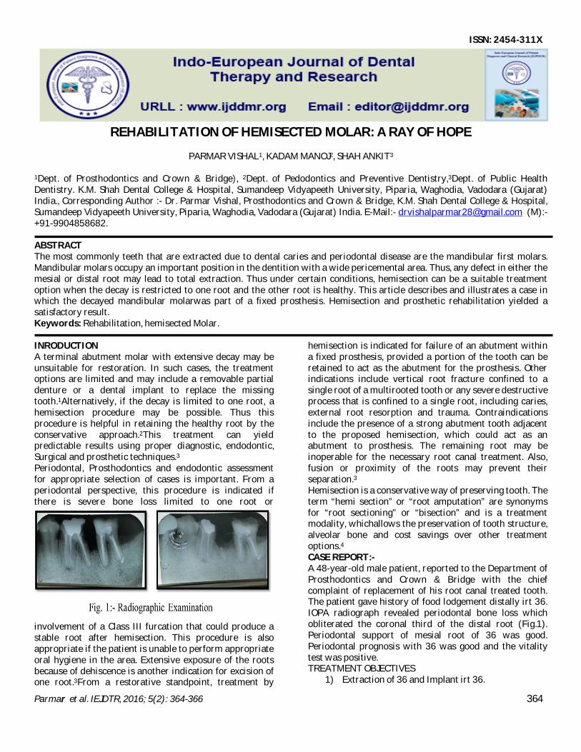
hemisection is indicated for failure of an abutment within a fixed prosthesis, provided a portion of the tooth can be retained to act as the abutment for the prosthesis. Other indications include vertical root fracture confined to a single root of a multirooted tooth or any severe destructive process that is confined to a single root, including caries, external root resorption and trauma. Contraindications include the presence of a strong abutment tooth adjacent to the proposed hemisection, which could act as an abutment to prosthesis. The remaining root may be inoperable for the necessary root canal treatment. Also, fusion or proximity of the roots may prevent their separation.3 Hemisection is a conservative way of preserving tooth. The term “hemi section” or “root amputation” are synonyms for “root sectioning” or “bisection” and is a treatment modality, whichallows the preservation of tooth structure, alveolar bone and cost savings over other treatment options.4 CASE REPORT:- A 48-year-old male patient, reported to the Department of Prosthodontics and Crown & Bridge with the chief complaint of replacement of his root canal treated tooth. The patient gave history of food lodgement distally irt 36. IOPA radiograph revealed periodontal bone loss which obliterated the coronal third of the distal root (Fig.1). Periodontal support of mesial root of 36 was good. Periodontal prognosis with 36 was good and the vitality test was positive. TREATMENT OBJECTIVES
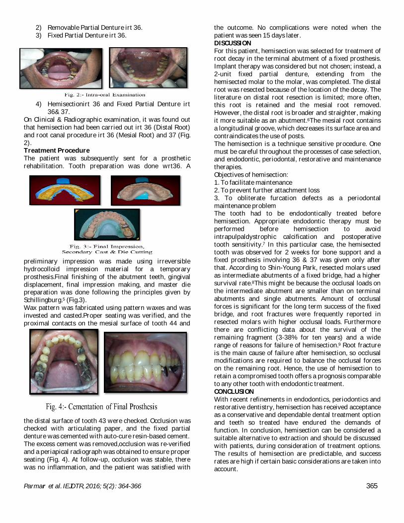
4) Hemisectionirt 36 and Fixed Partial Denture irt 36& 37.
On Clinical & Radiographic examination, it was found out that hemisection had been carried out irt 36 (Distal Root) and root canal procedure irt 36 (Mesial Root) and 37 (Fig. 2). Treatment Procedure The patient was subsequently sent for a prosthetic rehabilitation. Tooth preparation was done wrt36. A
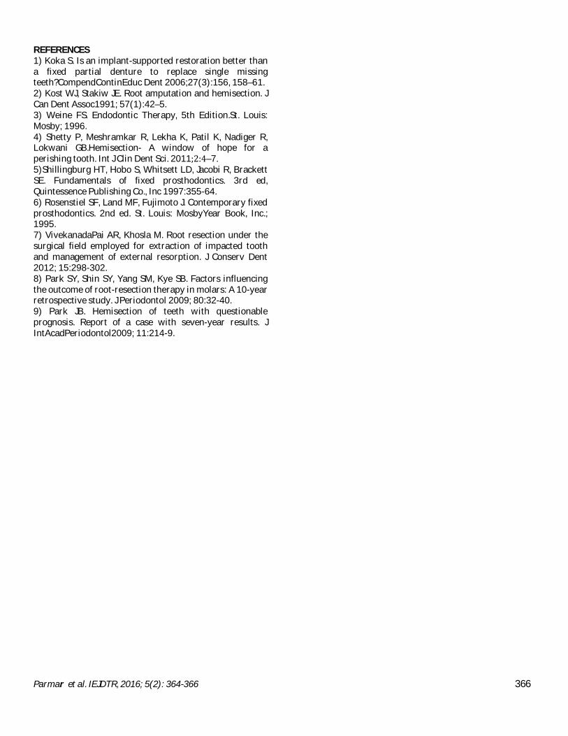
preliminary impression was made using irreversible hydrocolloid impression material for a temporary prosthesis.Final finishing of the abutment teeth, gingival displacement, final impression making, and master die preparation was done following the principles given by Schillingburg.5 (Fig.3). Wax pattern was fabricated using pattern waxes and was invested and casted.Proper seating was verified, and the proximal contacts on the mesial surface of tooth 44 and
the distal surface of tooth 43 were checked. Occlusion was checked with articulating paper, and the fixed partial denture was cemented with auto-cure resin-based cement. The excess cement was removed,occlusion was re-verified and a periapical radiograph was obtained to ensure proper seating (Fig. 4). At follow-up, occlusion was stable, there was no inflammation, and the patient was satisfied with
the outcome. No complications were noted when the patient was seen 15 days later. DISCUSSION For this patient, hemisection was selected for treatment of root decay in the terminal abutment of a fixed prosthesis. Implant therapy was considered but not chosen; instead, a 2-unit fixed partial denture, extending from the hemisected molar to the molar, was completed. The distal root was resected because of the location of the decay. The literature on distal root resection is limited; more often, this root is retained and the mesial root removed. However, the distal root is broader and straighter, making it more suitable as an abutment.6The mesial root contains a longitudinal groove, which decreases its surface area and contraindicates the use of posts. The hemisection is a technique sensitive procedure. One must be careful throughout the processes of case selection, and endodontic, periodontal, restorative and maintenance therapies. Objectives of hemisection: 1. To facilitate maintenance 2. To prevent further attachment loss 3. To obliterate furcation defects as a periodontal maintenance problem The tooth had to be endodontically treated before hemisection. Appropriate endodontic therapy must be performed before hemisection to avoid intrapulpaldystrophic calcification and postoperative tooth sensitivity.7 In this particular case, the hemisected tooth was observed for 2 weeks for bone support and a fixed prosthesis involving 36 & 37 was given only after that. According to Shin-Young Park, resected molars used as intermediate abutments of a fixed bridge, had a higher survival rate.8This might be because the occlusal loads on the intermediate abutment are smaller than on terminal abutments and single abutments. Amount of occlusal forces is significant for the long term success of the fixed bridge, and root fractures were frequently reported in resected molars with higher occlusal loads. Furthermore there are conflicting data about the survival of the remaining fragment (3-38% for ten years) and a wide range of reasons for failure of hemisection.9 Root fracture is the main cause of failure after hemisection, so occlusal modifications are required to balance the occlusal forces on the remaining root. Hence, the use of hemisection to retain a compromised tooth offers a prognosis comparable to any other tooth with endodontic treatment. CONCLUSION With recent refinements in endodontics, periodontics and restorative dentistry, hemisection has received acceptance as a conservative and dependable dental treatment option and teeth so treated have endured the demands of function. In conclusion, hemisection can be considered a suitable alternative to extraction and should be discussed with patients, during consideration of treatment options. The results of hemisection are predictable, and success rates are high if certain basic considerations are taken into account.
Parmar et al. IEJDTR, 2016; 5(2): 364-366 366
REFERENCES 1) Koka S. Is an implant-supported restoration better than a fixed partial denture to replace single missing teeth?CompendContinEduc Dent 2006;27(3):156, 158–61. 2) Kost WJ, Stakiw JE. Root amputation and hemisection. J Can Dent Assoc1991; 57(1):42–5. 3) Weine FS. Endodontic Therapy, 5th Edition.St. Louis: Mosby; 1996. 4) Shetty P, Meshramkar R, Lekha K, Patil K, Nadiger R, Lokwani GB.Hemisection- A window of hope for a perishing tooth. Int J Clin Dent Sci. 2011;2:4–7. 5)Shillingburg HT, Hobo S, Whitsett LD, Jacobi R, Brackett SE. Fundamentals of fixed prosthodontics. 3rd ed, Quintessence Publishing Co., Inc 1997:355-64. 6) Rosenstiel SF, Land MF, Fujimoto J. Contemporary fixed prosthodontics. 2nd ed. St. Louis: MosbyYear Book, Inc.; 1995. 7) VivekanadaPai AR, Khosla M. Root resection under the surgical field employed for extraction of impacted tooth and management of external resorption. J Conserv Dent 2012; 15:298-302. 8) Park SY, Shin SY, Yang SM, Kye SB. Factors influencing the outcome of root-resection therapy in molars: A 10-year retrospective study. J Periodontol 2009; 80:32-40. 9) Park JB. Hemisection of teeth with questionable prognosis. Report of a case with seven-year results. J IntAcadPeriodontol2009; 11:214-9.