Research ArticleRelationship between Serum Vitamin D and Calcium Levels andVitamin D Receptor Gene Polymorphisms in Colorectal Cancer
Ayat B. Al-Ghafari ,1,2,3,4 Khadijah S. Balamash ,1 and Huda A. Al Doghaither 1
1Biochemistry Department, Faculty of Science, King Abdulaziz University, Jeddah, Saudi Arabia2Cancer Metabolism and Epigenetics Unit, Faculty of Science, King Abdulaziz University, Jeddah, Saudi Arabia3Experimental Biochemistry Unit, King Fahd Medical Research Centre, King Abdulaziz University, Jeddah, Saudi Arabia4Cancer and Mutagenesis Unit, King Fahd Medical Research Centre, King Abdulaziz University, Jeddah, Saudi Arabia
Correspondence should be addressed to Ayat B. Al-Ghafari; [email protected]
Received 28 May 2019; Accepted 25 July 2019; Published 26 August 2019
Background. Many epidemiological studies have shown that vitamin D deficiency is associatedwith various types of human cancers.The biological action of vitamin D and its metabolites is mediated by the transcription factor vitamin D receptor (VDR). TheVDR gene is highly expressed in the colon and is involved in many biological functions. The aim of the current study was toassess the relationship between serum vitamin Dmetabolite and calcium levels with VDR polymorphisms in normal and colorectalcancer (CRC) patients.Methods. Fifty Saudi CRC patients and fifty controls were enrolled in the study. The levels of total vitaminD, 25(OH)D
3, and calcium were measured in serum. Results. The homozygous genotype (aa) of the ApaI VDR polymorphism
(rs7975232) was found to correlate with total serum vitamin D levels of CRC patients, while the heterozygous (Tt) TaqI VDRpolymorphism (rs731236)was associatedwith serumcalcium levels. In contrast, theBsmI and FokI VDR polymorphisms (rs1544410and rs2228570, resp.) did not affect the serum levels of total vitaminD, 25-hydroxyvitaminD
3, and calcium.Conclusion. Appropriate
vitamin D levels were shown to be important in preventing the onset of CRC.
1. Introduction
Vitamin D is an effective regulator of several physiologicprocesses, such as calcium (Ca) homeostasis and innate andadaptive immunity [1, 2]. On a molecular level, the effects ofvitamin D are mediated by the vitamin D receptor (VDR)[3, 4]. The VDR is a transcription factor and a member ofthe nuclear receptor superfamily [5]. Epidemiological andexperimental studies reveal that the VDR and its ligands arepromising targets for the prevention and possible treatmentof cancer, autoimmune diseases, and infections, as well as ofbone and mineral disorders [6, 7].
Serum levels of 25-hydroxyvitamin D [25(OH)D]induced by exposure to sunlight never exceed 60 ng/mL(149.76 nmol/L). According to the Mayo medical laboratoriesreference ranges, the total serum level of 25(OH)D rangesfrom 25 to 80 ng/ml, with a 25(OH)D level of less than30 ng/ml defined as vitamin D insufficiency, a 25(OH)D
level of less than 20 ng/ml defined as vitamin D deficiency,and a 25(OH)D level of 80 ng/ml or greater defined aspossible toxicity [8]. Interindividual differences in thehuman genome, often referred to as gene polymorphisms,are called variants when they appear in at least 1% ofthe population. A number of biological processes anddiseases, such as Alzheimer’s disease, autism, type 2 diabetesmellitus, obesity, and rheumatoid arthritis, associate withVDR polymorphisms [5, 9–12]. However, the molecularmechanism by which the variants of the VDR gene exertthese effects is unclear [1, 13]. This also applies to the role ofVDR polymorphisms in colorectal cancer (CRC).
According to the 14th cancer incidence report issuedby the Saudi Cancer Registry in 2014, in the Kingdom ofSaudi Arabia, CRC represents 10.4% of all cancers and isthe second most common cancer type after breast canceramong Saudi cancer patients. Among Saudi males, CRCis the most common cancer (11.8%) [14, 15]. Compared to
HindawiBioMed Research InternationalVolume 2019, Article ID 8571541, 7 pageshttps://doi.org/10.1155/2019/8571541
other types of cancers, CRC occurs sporadically [16] andpresents at a younger age in Saudis, especially in women[14]. The underlying etiology of CRC is not well determined,but it has been proposed that the onset of CRC results fromthe interaction between exogenous chemical and biologicalfactors such as age, diet, smoking, physical activity, andindividual genetic predisposition [17, 18].
Available treatment options for CRC depend on thestage of the disease. Treatment of CRC has systematicallyadvanced over the past few years with the introduction ofeffective chemotherapeutic agents. These drugs are oftenused in combination with biological therapy in patientswith advanced disease, such as enhancing the response ofimmune system and the use of minerals and vitamins [19].A number of studies have investigated the contribution ofvitamin D status andVDR gene variants (polymorphisms andmutations) in several types of cancer, including CRC.Most ofthese studies showed that insufficient vitamin D status maycontribute toCRCdevelopment [20–24].However, the resultsof studies on VDR gene polymorphisms and their relation toCRC development and prognosis are contradictory [25–28].In fact, these correlations mostly result from the interactionbetween VDR polymorphisms and other factors, such as Caand vitamin D intake, plasma levels of 1,25-dihydroxyvitaminD
3[1,25(OH)
2D
3], UV radiation exposure, obesity, and
energy intake [29–32].The aim of this study was to analyzethe relationship between serum total vitamin D, 25(OH)D
3,
and Ca levels and VDR polymorphisms in the manifestationof CRC.
2. Materials and Methods
2.1. Subjects. Fifty CRC patients (12 females and 38 males)aged 30–80 years were selected from the day care unit ofKing Abdulaziz University Hospital (Jeddah, Saudi Arabia).The same number of age- and sex-matched healthy controlswere randomly selected from the blood bank unit. All studyparticipants were of Saudi ethnicity and signed an ethicalconsent form, following the Declaration of Helsinki’s humanethical principles. CRC patients were accepted at any stageof the disease but had no primary tumors other than CRC.Members of the control group had no previous history of anysystemic illness or cancer. The study protocol as well as thequestionnaire and the informed consent were approved bythe ethics committee of King Abdulaziz University, Facultyof Medicine (reference no. 379-17). From each study partic-ipant, two blood samples were drawn for DNA and serumextraction.
2.2. Genotyping of Vitamin D Receptor (VDR) Variants.Extracted genomic DNA (100 ng/ml) was amplified in a 25𝜇l polymerase chain reaction (PCR) with 12.5 𝜇l HotStart-IT� FideliTaq� PCR Master Mix (2X) (Affymetrix/USB�,71156, USA), 9.5 𝜇l RNase free water (Affymetrix/ USB�,7732-18-5, USA), and 1 𝜇l of each primer (0.2 𝜇mol). Theprimers and the PCR thermocycler reactions were previouslypublished in Hajj et al. [29] for ApaI and BsmI, whereasfor TaqI and FokI in Rizk et al., [21]. The amplified PCR
products were genotyped with restriction fragment lengthpolymorphism (RFLP) using appropriate restriction endonu-cleases fromThermo Fisher Scientific (Waltham, MA, USA)[FastDigest ApaI (FD1414), FastDigest TaqI, (FD0674), Fast-Digest Mva1269I (FD0964), and FastDigest FokI (FD2144)]following themanufacturer’s instructions.Thedifferent geno-types for each variant were confirmed with 2% agarosegel electrophoresis and 10% of the samples were selectedrandomly for further DNA sequencing confirmation.
2.3. Measurement of Serum Total Vitamin D, 25(OH)𝐷3,and Ca Levels. The level of total vitamin D [25(OH)D
2and
25(OH)D3] in serum was measured using a commercial
LIAISON�25OHVitaminDTotal AssayKit (DiaSorin,USA).Serum Ca and 25(OH)D
3levels were measured by the
ADVIACentaur chemiluminescent reaction in the Biochem-istry laboratory at King Abdulaziz University Hospital.
2.4. Statistical Analysis. All statistical analyses were per-formed on GraphPad Prism version 7.00 (San Diego, Cali-fornia, USA). The Mann-Whitney test was used to comparedifferences between two independent groups with either onephysical or biochemical variable.TheKruskal-Wallis test withDunn’s multiple comparison was applied to correlate thegenotype distribution of each VDR variant with total vitaminD, 25(OH)D
3, and Ca levels measured in the serum. P values
< 0.05 were considered statistically significant. Values intables were represented as mean ± standard error of mean(SEM).
3. Results
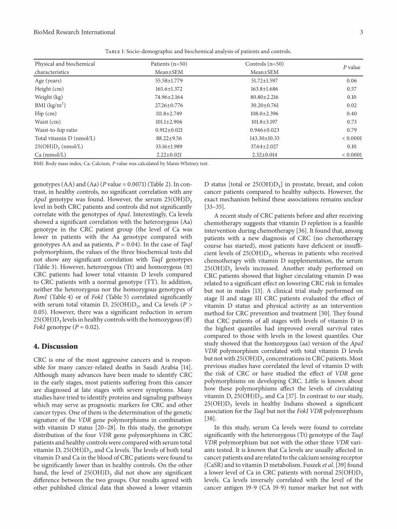
A total of 100 study participants were enrolled: fifty CRCpatients and fifty healthy controls. All CRC patients weretreatment-experienced and had completed the full courseof chemotherapy. The common chemotherapy regimens ofthe CRC cases were capecitabine (Xeloda) as neoadjuvanttherapy with radiation; oxaliplatin and capecitabine (Xelox)intravenously for 6 to 8 weeks; and, finally, irinotecanintravenously and capecitabine as tablets twice a day (Xeliri)for 2 to 3 weeks. The baseline of sociodemographic andlaboratory measurements for study participants is presentedin Table 1. There was a highly significant difference in bodymass index (BMI) between CRC patients and controls (P< 0.0001). This is due to poor food intake in the patientsas a consequence of chemotherapy, resulting in a loss ofappetite. Table 1 shows highly significant differences in serumtotal vitamin D and Ca levels between patients and controls(P < 0.0001). In contrast, serum 25(OH)D
3levels were not
significantly different between the two groups (P > 0.05).In order to find a possible correlation between the differ-
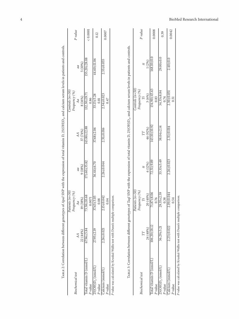
ent genotypes of the VDR gene polymorphisms (ApaI, TaqI,BsmI, and FokI) and the serum total vitamin D, 25(OH)D
3,
and Ca levels, a Kruskal-Wallis test was performed (Tables2–5). In the case of the ApaI polymorphism, only CRCpatients with homozygous ApaI (aa) genotypes showed ahigher level of total vitaminD in the serumcompared to other
BioMed Research International 3
Table 1: Socio-demographic and biochemical analysis of patients and controls.
Ca (mmol/L) 2.22±0.021 2.32±0.014 < 0.0001BMI: Body mass index, Ca: Calcium, P value was calculated by Mann-Whitney test.
genotypes (AA) and (Aa) (P value = 0.0071) (Table 2). In con-trast, in healthy controls, no significant correlation with anyApaI genotype was found. However, the serum 25(OH)D
3
level in both CRC patients and controls did not significantlycorrelate with the genotypes of ApaI. Interestingly, Ca levelsshowed a significant correlation with the heterozygous (Aa)genotype in the CRC patient group (the level of Ca waslower in patients with the Aa genotype compared withgenotypes AA and aa patients, P = 0.04). In the case of TaqIpolymorphism, the values of the three biochemical tests didnot show any significant correlation with TaqI genotypes(Table 3). However, heterozygous (Tt) and homozygous (tt)CRC patients had lower total vitamin D levels comparedto CRC patients with a normal genotype (TT). In addition,neither the heterozygous nor the homozygous genotypes ofBsmI (Table 4) or of FokI (Table 5) correlated significantlywith serum total vitamin D, 25(OH)D
3, and Ca levels (P >
0.05). However, there was a significant reduction in serum25(OH)D
3levels in healthy controls with the homozygous (ff)
FokI genotype (P = 0.02).
4. Discussion
CRC is one of the most aggressive cancers and is respon-sible for many cancer-related deaths in Saudi Arabia [14].Although many advances have been made to identify CRCin the early stages, most patients suffering from this cancerare diagnosed at late stages with severe symptoms. Manystudies have tried to identify proteins and signaling pathwayswhich may serve as prognostic markers for CRC and othercancer types. One of them is the determination of the geneticsignature of the VDR gene polymorphisms in combinationwith vitamin D status [20–28]. In this study, the genotypedistribution of the four VDR gene polymorphisms in CRCpatients and healthy controls were comparedwith serum totalvitamin D, 25(OH)D
3, and Ca levels. The levels of both total
vitamin D and Ca in the blood of CRC patients were found tobe significantly lower than in healthy controls. On the otherhand, the level of 25(OH)D
3did not show any significant
difference between the two groups. Our results agreed withother published clinical data that showed a lower vitamin
D status [total or 25(OH)D3] in prostate, breast, and colon
cancer patients compared to healthy subjects. However, theexact mechanism behind these associations remains unclear[33–35].
A recent study of CRC patients before and after receivingchemotherapy suggests that vitamin D repletion is a feasibleintervention during chemotherapy [36]. It found that, amongpatients with a new diagnosis of CRC (no chemotherapycourse has started), most patients have deficient or insuffi-cient levels of 25(OH)D
3, whereas in patients who received
chemotherapy with vitamin D supplementation, the serum25(OH)D
3levels increased. Another study performed on
CRC patients showed that higher circulating vitamin D wasrelated to a significant effect on lowering CRC risk in femalesbut not in males [13]. A clinical trial study performed onstage II and stage III CRC patients evaluated the effect ofvitamin D status and physical activity as an interventionmethod for CRC prevention and treatment [30]. They foundthat CRC patients of all stages with levels of vitamin D inthe highest quantiles had improved overall survival ratescompared to those with levels in the lowest quantiles. Ourstudy showed that the homozygous (aa) version of the ApaIVDR polymorphism correlated with total vitamin D levelsbut notwith 25(OH)D
3concentrations inCRCpatients.Most
previous studies have correlated the level of vitamin D withthe risk of CRC or have studied the effect of VDR genepolymorphisms on developing CRC. Little is known abouthow these polymorphisms affect the levels of circulatingvitamin D, 25(OH)D
3, and Ca [37]. In contrast to our study,
25(OH)D3levels in healthy Indians showed a significant
association for the TaqI but not the FokI VDR polymorphism[38].
In this study, serum Ca levels were found to correlatesignificantly with the heterozygous (Tt) genotype of the TaqIVDR polymorphism but not with the other three VDR vari-ants tested. It is known that Ca levels are usually affected incancer patients and are related to the calcium sensing receptor(CaSR) and to vitamin Dmetabolism. Fuszek et al. [39] founda lower level of Ca in CRC patients with normal 25(OH)D
3
levels. Ca levels inversely correlated with the level of thecancer antigen 19-9 (CA 19-9) tumor marker but not with
4 BioMed Research International
Table2:Correlatio
nbetweendifferent
geno
typeso
fApaIS
NPwith
thee
xpressionof
totalvitamin
D,25(OH)D
3,and
calcium
serum
levelsin
patie
ntsa
ndcontrols.
Biochemica
ltest
Patients(n=
50)
Controls(n=5
0)
Pvalue
Frequency(
%)
Frequency(
%)
AAAa
aaAA
Aaaa
22(44%
)19
(38%
)9(18%
)37
(74%
)8(16%
)5(10%
)To
talvitamin
D(nmol/L)
67.96±
5.84
73.58±
10.68
171.6
0±35.82
143.80±12.88
132.30±19.71
155.20±28.08
<0.00
01Pvalue
0.00
710.86
25(O
H)D
3(nmol/L)
27.95±
2.19
37.63±
3.05
36.44±
6.70
37.68±
2.06
33.13±5.28
44.60±
11.06
0.12
Pvalue
0.08
0.81
Calcium
(mmol/L)
2.26±0.021
2.15±0.04
22.26±0.04
42.31±0.016
2.34±0.023
2.33±0.055
0.00
07Pvalue
0.04
0.47
Pvaluew
ascalculated
byKruskal-W
allis
testwith
Dun
n’smultip
lecomparis
on.
Table3:Correlatio
nbetweendifferent
geno
typeso
fTaqIS
NPwith
thee
xpressionof
totalvitamin
D,25(OH)D
3,and
calcium
serum
levelsin
patie
ntsa
ndcontrols.
Biochemica
ltest
Patients(n=
50)
Controls(n=5
0)
Pvalue
Frequency(
%)
Frequency(
%)
TTTt
ttTT
Tttt
24(48%
)20
(40%
)6(12%
)46
(92%
)3(6%)
1(2%
)To
talvitamin
D(nmol/L)
106.20±18.12
71.87±
8.06
72.51±9.8
9143.10±10.92
134.90±45.63
168.10±0.0
0.00
08Pvalue
0.76
0.83
25(O
H)D
3(nmol/L)
36.29±
3.21
29.35±
2.59
33.33±5.49
38.04±
2.14
34.33±8.84
29.00±
0.0
0.39
Pvalue
0.38
0.74
Calcium
(mmol/L)
2.23±0.022
2.19±0.04
42.26±0.023
2.31±0.014
2.30±0.051
2.40±0.0
0.00
42Pvalue
0.54
0.51
Pvaluew
ascalculated
byKruskal-W
allis
testwith
Dun
n’smultip
lecomparis
on.
BioMed Research International 5
Table4:Correlatio
nbetweendifferent
geno
typeso
fBsm
ISNPwith
thee
xpressionof
totalvitamin
D,25(OH)D
3,and
calcium
serum
levelsin
patie
ntsa
ndcontrols.
Biochemica
ltest
Patients(n=
50)
Controls(n=5
0)
Pvalue
Frequency(
%)
Frequency(
%)
BBBb
bbBB
Bbbb
20(40%
)22
(44%
)8(16%
)6(12%
)28
(56%
)16
(32%
)To
talvitamin
D(nmol/L)
107.4
0±16.58
79.32±14.77
69.30±
9.71
124.0±
23.87
130.4±
14.40
172.4±
16.54
<0.00
01Pvalue
0.25
0.07
25(O
H)D
3(nmol/L)
36.50±
3.85
30.77±
2.58
31.38±
3.27
36.17±4.60
38.86±
2.46
36.06±
4.45
0.48
Pvalue
0.58
0.79
Calcium
(mmol/L)
2.23±0.021
2.18±0.04
12.27±0.035
2.34±0.013
2.31±0.021
2.31±0.023
0.0031
Pvalue
0.35
0.57
Pvaluew
ascalculated
byKruskal-W
allis
testwith
Dun
n’smultip
lecomparis
on.
Table5:Correlatio
nbetweendifferent
geno
typeso
fFokIS
NPwith
thee
xpressionof
totalvitamin
D,25(OH)D
3,and
calcium
serum
levelsin
patie
ntsa
ndcontrols.
Biochemica
ltest
Patients(n=
50)
Controls(n=5
0)
Pvalue
Frequency(
%)
Frequency(
%)
FFFf
ffFF
Ffff
33(66%
)13
(26%
)4(8%)
24(48%
)23
(46%
)3(
6%)
Totalvitamin
D(nmol/L)
91.62±
13.49
79.15±11.75
91.89±
26.26
154.0±
16.46
136.20±14.21
109.0±11.36
0.00
13Pvalue
0.87
0.57
25(O
H)D
3(nmol/L)
33.00±
2.69
32.69±
3.00
36.00±
6.88
42.83±
2.62
33.48±
2.98
28.00±
9.02
0.06
Pvalue
0.82
0.02
Calcium
(mmol/L)
2.22±0.029
2.21±0.023
2.19±0.058
2.33±0.018
2.31±0.019
2.22±0.095
0.00
12Pvalue
0.39
0.30
Pvaluew
ascalculated
byKruskal-W
allis
testwith
Dun
n’smultip
lecomparis
on.
6 BioMed Research International
the carcinoembryonic antigen (CEA) or alpha fetoprotein(AFP) tumor markers or the CaSR genotypes. A study with922 Korean CRC patients showed that Ca consumption wasinversely related to CRC risk [40]. Another recent studysuggested that lower serum Ca levels are correlated withNigerian CRC patients that show high CEA levels comparedto patients with low levels of the tumor marker [41].
5. Conclusion
This study found that the homozygous genotype (aa) of theVDR SNP ApaI correlates with total vitamin D level in theserum of CRC patients and that the heterozygous genotype(Tt) of theVDR SNP TaqI significantly associates with serumCa levels. Thus, our findings found that vitamin D status mayplay an important role in CRC tumorigenesis. However,morestudies on larger number of CRC patients in Saudi Arabiaor on cancerous tissues are needed to elucidate further thecurrent findings and study the molecular mechanisms.
Data Availability
The data used to support the findings of this study areavailable from the corresponding author upon request.
Ethical Approval
The study was approved by the Research Committee ofthe Biomedical Ethics Unit at Faculty of Medicine, KAU(reference no. 379-17).
Consent
Written informed consent was obtained from all participants.
Conflicts of Interest
The authors declare that there are no conflicts of interestregarding the publication of this paper.
Authors’ Contributions
Ayat B. Al-Ghafari and Khadijah S. Balamash designedthe study. Huda A. Al Doghaither and Ayat B. Al-Ghafariconducted the experiments, analyzed the data, and draftedthe manuscript. All authors revised the manuscript and readand approved the final version of the manuscript.
Acknowledgments
This project was funded by the Deanship of ScientificResearch (DSR), King Abdulaziz University, Jeddah, underGrant no. (RG-6-130-38). The authors, therefore, acknowl-edge with thanks DSR technical and financial support. Theauthors would like also to express their thanks for Prof.Carsten Carlberg, Professor of Biochemistry at University ofEastern Finland, for his critical contribution in reviewing themanuscript.
References
[1] J. Sun, “The role of vitamin D and vitamin D receptors in coloncancer,” Clinical and Translational Gastroenterology, vol. 8, p.e103, 2017.
[2] B. Prietl, G. Treiber, T. R. Pieber, and K. Amrein, “Vitamin Dand immune function,” Nutrients, vol. 5, no. 7, pp. 2502–2521,2013.
[3] S. Christakos, P. Dhawan, A. Verstuyf, L. Verlinden, and G.Carmeliet, “Vitamin D: metabolism, molecular mechanism ofaction, and pleiotropic effects,” Physiological Reviews, vol. 96,no. 1, pp. 365–408, 2016.
[4] V. Dimitrov, R. Salehi-Tabar, B.-S. An, and J. H. White, “Non-classical mechanisms of transcriptional regulation by the vita-min D receptor: Insights into calcium homeostasis, immunesystem regulation and cancer chemoprevention,”�e Journal ofSteroid Biochemistry and Molecular Biology, vol. 144, pp. 74–80,2014.
[5] W. Wang, A. Wu, Y. Zhou, Y. Wang, and K. Cao, “Associationbetween Vitamin D receptor polymorphisms and rheumatoidarthritis risk: A meta-analysis,” International Journal of Clinicaland Experimental Medicine, vol. 10, no. 2, pp. 4221–4233, 2017.
[6] L. A. Plum andH. F. Deluca, “Vitamin D, disease and therapeu-tic opportunities,”Nature Reviews Drug Discovery, vol. 9, no. 12,pp. 941–955, 2010.
[7] M.Choi andM.Makishima, “Therapeutic applications for novelnon-hypercalcemic vitaminD receptor ligands,”Expert Opinionon �erapeutic Patents, vol. 19, no. 5, pp. 593–606, 2009.
[8] K. A. Kennel, M. T. Drake, and D. L. Hurley, “Vitamin Ddeficiency in adults: when to test and how to treat,”Mayo ClinicProceedings, vol. 85, no. 8, pp. 752–758, 2010.
[9] B. Angel, L. Lera, C.Marquez, andC.Albala, “The association ofVDR polymorphisms and type 2 diabetes in older people livingin community in Santiago de Chile,” Nutrition & Diabetes, vol.8, no. 1, article 31, 2018.
[10] A. Cieslinska, E. Kostyra, B. Chwała et al., “Vitamin D receptorgene polymorphisms associated with childhood autism,” BrainSciences, vol. 7, no. 9, p. 15, 2017.
[11] M. Zaki, S. Kamal, W. Basha et al., “Association of vitaminD receptor gene polymorphism (VDR) with vitamin D defi-ciency, metabolic and inflammatorymarkers in Egyptian obesewomen,” Genes & Diseases, vol. 4, no. 3, pp. 176–182, 2017.
[12] H. R. Khorram Khorshid, E. Gozalpour, K. Saliminejad, M.Karimloo, M. Ohadi, and K. Kamali, “Vitamin D receptor(VDR) polymorphisms and late-onset Alzheimer’s disease: Anassociation study,” Iranian Journal of Public Health, vol. 42, no.11, pp. 1253–1258, 2013.
[13] M. L. McCullough, E. S. Zoltick, S. J. Weinstein et al., “Circu-lating vitamin D and colorectal cancer risk: an internationalpooling project of 17 cohorts,” Journal of the National CancerInstitute, vol. 111, no. 2, pp. 158–169, 2018.
[14] N. Alsanea, M. A. Almadi, A. S. Abduljabbar et al., “Nationalguidelines for colorectal cancer screening in Saudi Arabia withstrength of recommendations and quality of evidence,” Annalsof Saudi Medicine, vol. 35, no. 3, pp. 189–195, 2015.
[15] A. P. Shaik, A. S. Shaik, and Y. A. Al-Sheikh, “Colorectal cancer:a reviewof the genome-wide association studies in the kingdomof Saudi Arabia,” Saudi Journal of Gastroenterology, vol. 21, no.3, pp. 123–128, 2015.
[16] K. W. Jasperson, T. M. Tuohy, D. W. Neklason, and R. W. Burt,“Hereditary and familial colon cancer,” Gastroenterology, vol.138, no. 6, pp. 2044–2058, 2010.
BioMed Research International 7
[17] M. Raman, A. N. Milestone, J. R. F. Walters, A. L. Har, and S.Ghosh, “Vitamin D and gastrointestinal diseases: inflammatorybowel disease and colorectal cancer,” �erapeutic Advances inGastroenterology, vol. 4, no. 1, pp. 49–62, 2011.
[18] D. Cunningham,W. Atkin, H.-J. Lenz et al., “Colorectal cancer,”�e Lancet, vol. 375, no. 9719, pp. 1030–1047, 2010.
[19] M. Panczyk, “Pharmacogenetics research on chemotherapyresistance in colorectal cancer over the last 20 years,” WorldJournal of Gastroenterology, vol. 20, no. 29, p. 9775, 2014.
[20] J. P. Vayrynen, S. J. Mutt, K. Herzig et al., “Decreased pre-operative serum 25-Hydroxyvitamin D levels in colorectalcancer are associated with systemic inflammation and serratedmorphology,” Scientific Reports, vol. 6, no. 1, Article ID 36519,2016.
[21] M. M. Rizk, N. H. Zakaria, and W. G. Elshazely, “Studyof vitamin D receptor (VDR) gene polymorphisms amongegyptian cohort patients with different stages of colorectalcancer,” Journal of Cancer �erapy, vol. 05, no. 03, pp. 253–263,2014.
[22] M. Jenab, H. B. Bueno-de-Mesquita, P. Ferrari et al., “Associa-tion between pre-diagnostic circulating vitamin D concentra-tion and risk of colorectal cancer in European populations:anested case-control study,” BMJ, vol. 340, Article ID b5500,2010.
[23] C. G. Woolcott, L. R. Wilkens, A. M. Y. Nomura et al., “Plasma25-hydroxyvitamin D levels and the risk of colorectal cancer:Themultiethnic cohort study,”CancerEpidemiology, Biomarkers& Prevention, vol. 19, no. 1, pp. 130–134, 2010.
[24] L. Yin, N. Grandi, E. Raum, U. Haug, V. Arndt, and H.Brenner, “Meta-analysis: longitudinal studies of serum vitaminD and colorectal cancer risk,” Alimentary Pharmacology &�erapeutics, vol. 30, no. 2, pp. 113–125, 2009.
[25] K. A. Alkhayal, Z. H. Awadalia, M.-A. Vaali-Mohammed et al.,“Association of Vitamin D receptor gene polymorphisms withcolorectal cancer in a Saudi Arabian Population,” PLoS ONE,vol. 11, no. 6, Article ID e0155236, 2016.
[26] S. Budhathoki, T. Yamaji, M. Iwasaki et al., “Vitamin D receptorgene polymorphism and the risk of colorectal cancer: a nestedcase-control study,” PLoS ONE, vol. 11, no. 10, Article IDe0164648, 2016.
[27] V. Vidigal, T. da Silva, C. Pimenta, J. Oliveira, A. Felipe, andN. Forones, “P-220 * Genetic Polymorphism of Vitamin DReceptor BsmI, ApaI and CYP27B1, CYP24A1 genes and therisk of colorectal cancer,” Annals of Oncology, vol. 26, no. 4, pp.iv64–iv64, 2015.
[28] M. Sarkissyan, Y. Wu, Z. Chen et al., “Vitamin D receptor FokIgene polymorphisms may be associated with colorectal canceramong African American and Hispanic participants,” Cancer,vol. 120, no. 9, pp. 1387–1393, 2014.
[29] A.Hajj, R.Chedid, E.Chouery,A.Megarbane, andM.Gannage-Yared, “Relationship between vitamin D receptor gene poly-morphisms, cardiovascular risk factors and adiponectin in ahealthy young population,” Pharmacogenomics, vol. 17, no. 15,pp. 1675–1686, 2016.
[30] V. Morales-Oyarvide, J. A. Meyerhardt, and K. Ng, “VitaminD and physical activity in patients with colorectal cancer:epidemiological evidence and therapeutic implications,”CancerJournal, vol. 22, no. 3, pp. 223–231, 2016.
[31] E. M. John, G. G. Schwartz, J. Koo, D. Van Den Berg, and S. A.Ingles, “Sun exposure, vitaminD receptor gene polymorphisms,and risk of advanced prostate cancer,” Cancer Research, vol. 65,no. 12, pp. 5470–5479, 2005.
[32] E. M. John, G. G. Schwartz, J. Koo, W. Wang, and S. A.Ingles, “Sun exposure, vitaminD receptor gene polymorphisms,and breast cancer risk in a multiethnic population,” AmericanJournal of Epidemiology, vol. 166, no. 12, pp. 1409–1419, 2007.
[33] N. Shaukat, F. Jaleel, F. A. Moosa, and N. A. Qureshi, “Associa-tion between vitamin D deficiency and breast cancer,” PakistanJournal of Medical Sciences, vol. 33, no. 3, pp. 645–649, 2017.
[34] G. P. Levin, C. Robinson-Cohen, I. H. de Boer et al., “Geneticvariants and associations of 25-hydroxyvitamin D concentra-tions with major clinical outcomes,” Journal of the AmericanMedical Association, vol. 308, no. 18, pp. 1898–1905, 2012.
[35] J. Ahn, D. Albanes, S. I. Berndt et al., “Vitamin D-relatedgenes, serum vitamin D concentrations and prostate cancerrisk,” Carcinogenesis, vol. 30, no. 5, pp. 769–776, 2009.
[36] M. B. Savoie, A. Paciorek, L. Zhang et al., “Vitamin D levelsin patients with colorectal cancer before and after treatmentinitiation,” Journal of Gastrointestinal Cancer, 2018.
[37] V. Rai, J. Abdo, S. Agrawal, and D. K. Agrawal, “Vitamin Dreceptor polymorphism and cancer: An update,” AnticancerReseach, vol. 37, no. 8, pp. 3991–4003, 2017.
[38] A. A. Bhanushali, N. Lajpal, S. S. Kulkarni, S. S. Chavan,S. S. Bagadi, and B. R. Das, “Frequency of fokI and taqIpolymorphism of vitamin D receptor gene in Indian populationand its association with 25-hydroxyvitamin D levels,” IndianJournal of Human Genetics, vol. 15, no. 3, pp. 108–113, 2009.
[39] P. Fuszek, P. Lakatos, A. Tabak et al., “Relationship betweenserum calcium and CA 19-9 levels in colorectal cancer,” WorldJournal of Gastroenterology, vol. 10, no. 3, pp. 1890–1892, 2004.
[40] C.Han,A. Shin, J. Lee et al., “Dietary calcium intake and the riskof colorectal cancer: a case control study,” BMC Cancer, vol. 15,no. 1, p. 966, 2015.
[41] P. N. Wichendu and C. Amadi, “Relationship between plasmacalcium and carcinoembryonic antigen among colorectal can-cer patients,” International Surgery Journal, vol. 5, no. 6, pp.1995–1999, 2018.