Reliability and validity of manual palpationfor the assessment of patients with lowback pain: a systematic and critical reviewPaul S. Nolet1,2,3* , Hainan Yu4,5,9, Pierre Côté4,5, Anne-Laure Meyer6, Vicki L. Kristman7,8, Deborah Sutton4,5,Kent Murnaghan9 and Nadège Lemeunier5,10
Abstract: Background: Static or motion manual palpation of the low back is commonly used to assess painlocation and reproduction in low back pain (LBP) patients. The purpose of this study is to review the reliability andvalidity of manual palpation used for the assessment of LBP in adults.
Method: We systematically searched five databases from 2000 to 2019. We critically appraised internal validity ofstudies using QAREL and QUADAS-2 instruments. We stratified results using best-evidence synthesis. Validity studieswere classified according to Sackett and Haynes.
Results: We identified 2023 eligible articles, of which 14 were low risk of bias. Evidence suggests that reliability ofsoft tissue structures palpation is inconsistent, and reliability of bony structures and joint mobility palpation is poor.We found preliminary evidence that gluteal muscle palpation for tenderness may be valid in differentiating LBPpatients with and without radiculopathy.
Conclusion: Reliability of manual palpation tests in the assessment of LBP patients varies greatly. This isproblematic because these tests are commonly used by manual therapists and clinicians. Little is known about thevalidity of these tests; therefore, their clinical utility is uncertain. High quality validity studies are needed to informthe clinical use of manual palpation tests.
IntroductionLow back pain (LBP) is the most prevalent musculoskel-etal condition in the general population [1, 2]. The pointprevalence of LBP ranges between 1 to 58.1% and one-year prevalence ranges between 0.8 to 82.5% [3] depend-ing of the LBP definition and population. LBP is theleading cause of years lived with disability and is thesixth leading cause of disability adjusted life years glo-bally [4, 5] and it is associated with poor health-relatedquality of life and has a substantial economic burden to
society [6, 7]. Non-specific LBP is more common thanspecific LBP (e.g., cancer, fractures, infectious disorders,or ankylosing spondylitis) and it cannot be attributed toa specific underlying pathology [8].The clinical assessment of low back pain involves
completing a physical examination [9]. Manual palpationis a common tool used to assess patients with LBP [10].It includes static and dynamic palpation of soft tissue orjoints and aims to identify painful structures and bio-mechanical dysfunction of the spine [11]. However, theclinical utility of these tests is controversial.Previous systematic reviews have investigated the reli-
ability and validity of manual palpation for the assess-ment of patients with LBP [9, 11–13]. According tothese reviews, the inter-rater reliability of static joint and
* Correspondence: [email protected] of Graduate Education and Research, Canadian MemorialChiropractic College, Toronto, Ontario, Canada2School of Kinesiology, Lakehead University, Thunder Bay, Ontario, CanadaFull list of author information is available at the end of the article
Nolet et al. Chiropractic & Manual Therapies (2021) 29:33 https://doi.org/10.1186/s12998-021-00384-3
soft-tissue palpation to locate pain is poor (kappa (k) ≤0.40), and the inter-rater reliability of static palpation forsoft tissue changes (e.g., tension) is inconsistent [9, 11,13]. Furthermore, one review reported that motion pal-pation may be valid in detecting decreased motion, orlack of end-play in the lumbar spine [12]. However, mo-tion palpation may not be valid to detect aberrant mo-tion of the sacroiliac joints [12]. These reviews areoutdated and there is a need for an up-to-date system-atic review. The purpose of our systematic review was todetermine the reliability and validity of manual palpationused to assess adult patients with LBP.
MethodsEligibility criteriaPopulationWe included studies of adults (≥18 years) with LBP. LBPrefers to pain or discomfort below the costal margin andabove the inferior gluteal folds and can be with or with-out referred leg pain [14]. Our systematic review in-cludes patients with non-radicular low back pain,radicular low back pain, spinal stenosis, degenerative oristhmic spondylolisthesis, and failed back surgerysyndrome.
DefinitionsOur review focuses on studies assessing the reliability orvalidity of manual palpation for the assessment of pa-tients with LBP. Reliability describes the consistency ofmeasurements across people or instruments [15]. Valid-ity is the degree to which a test measures what it isintended to measure [15].Manual palpation is a diagnostic procedure where the
examiner feels with their hands to assess the mobilityand state of the soft and boney tissues [16]. Palpationtechniques include both static and dynamic (motion)methods, which are often used to identify areas of tissuepain and dysfunction, target manual and manipulativetherapies and determine effectiveness of the intervention[9]. Static palpation is used to identify bony asymmetryof bony landmarks, tender points, and trigger points toevaluate tissue texture, temperature and tone [17]. Mo-tion palpation is used to assess the quantity and qualityof movement through the lumbar spine and pelvis [17].Motion palpation assessment can be continuous withinthe normal range of motion with joint play, or dynamicsoft tissue palpation or end range assessment for end-feel or joint springing [17]. Palpation involving devicessuch as pressure algometry were excluded.
OutcomesWe aimed to evaluate clinical outcomes assessed by pal-pation. Outcomes include pain, segmental mobility andstiffness for static joint palpation; joint movement and
position assessed for motion joint palpation; and pain,tenderness, trigger points, muscle contraction assessedfor static soft tissue palpation.
Study characteristicsEligible studies met the following inclusion: 1) Englishor French language; 2) published in peer reviewed jour-nals between January 1, 2000 to July 11, 2019; 3) asses-sing the reliability or validity of manual palpation.Previously published systematic reviews on this topicwere included in our review. Comparing our systematicreview with previous systematic reviews examined find-ings of studies published before 2000. We excluded: 1)letters, guidelines, editorials, commentaries, unpublishedmanuscripts, dissertations, reports, book chapters, con-ference proceedings and abstracts, lectures, addresses,and consensus statements; 2) cadaveric and animal stud-ies; 3) literature reviews and case studies; 4) studies tar-geting individuals with serious pathology (e.g., fractures,dislocations, systemic disease, myelopathy, neoplasm andinfection; and 5) studies with sample size < 20 per group.
Search strategy and data sourcesThe search strategy was developed in consultation witha health sciences librarian and a second librarian wasconsulted to ensure accuracy and completeness usingthe Peer Review of Electronic Search Strategies PRESSchecklist [18]. We systematically searched the followingelectronic databases: MEDLINE, CINAHL, PubMed,Cochrane Central Register of Controlled Trials, andSPORTDiscus. Search terms consisted of subject head-ings specific to each database (e.g. MeSH in MEDLINE)and free text words relevant to LBP, diagnosis, reliability,validity, and palpation (Additional file 1).
Study selectionIdentified citations were exported into EndNote for ref-erence management and tracking of the screeningprocess. We screened articles in two stages. In stage one,titles and abstracts were screened for their relevance bypairs of independent reviewers (NL, PN, ALM). Stagetwo involved screening the full text article of all possiblyrelevant citations from stage one. Disagreements onscreening stages were discussed between reviewers toreach consensus. When consensus could not be reached,a third reviewer independently screened the citation anddiscussed with the two reviewers to reach consensus.
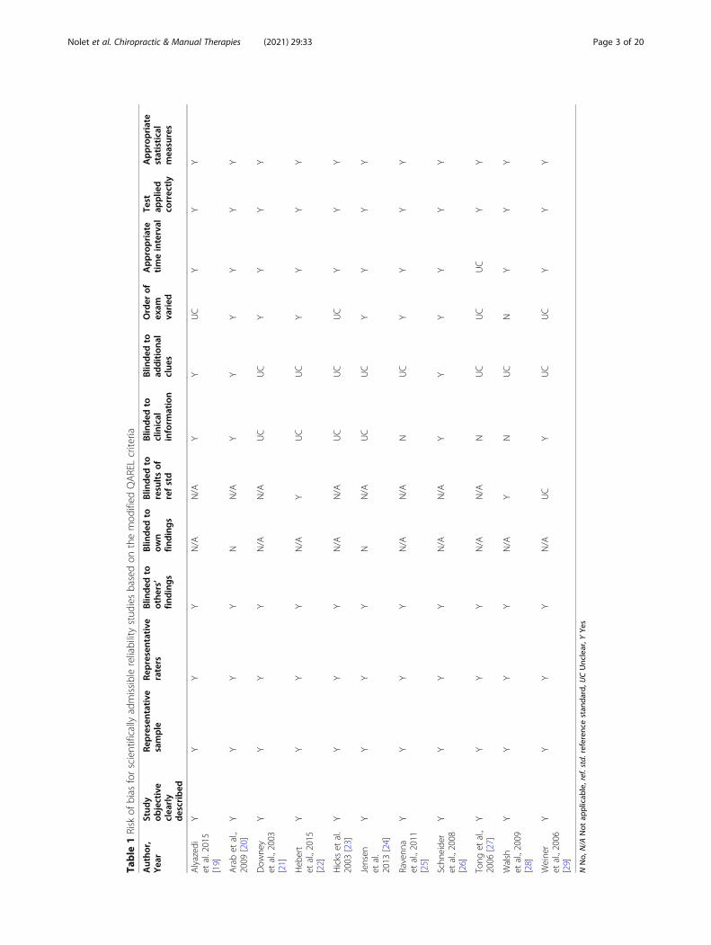
Assessment of risk of BiasThree reviewers (NL, PN, ALM) critically appraised allrelevant studies (Tables 1 and 2) using the modifiedQuality Appraisal Tool for Studies of Diagnostic Reli-ability (QAREL) [33] criteria to assess the internal valid-ity of the diagnostic reliability studies and the modified
Nolet et al. Chiropractic & Manual Therapies (2021) 29:33 Page 2 of 20
Table
1Risk
ofbias
forscientifically
admissiblereliabilitystud
iesbasedon
themod
ified
QARELcriteria
Autho
r,Yea
rStud
yob
jective
clea
rly
described
Representative
sample
Representative
raters
Blin
dedto
othe
rs’
findings
Blin
ded
toow
nfin
dings
Blin
ded
toresultsof
refstd
Blin
ded
toclinical
inform
ation
Blin
ded
toad
ditiona
lclue
s
Order
ofexam
varied
Appropriate
timeinterval
Test
applie
dco
rrectly
Appropriate
statistical
mea
sures
Alyazed
iet
al.2015
[19]
YY
YY
N/A
N/A
YY
UC
YY
Y
Arabet
al.,
2009
[20]
YY
YY
NN/A
YY
YY
YY
Dow
ney
etal.,2003
[21]
YY
YY
N/A
N/A
UC
UC
YY
YY
Heb
ert
etal.,2015
[22]
YY
YY
N/A
YUC
UC
YY
YY
Hicks
etal.
2003
[23]
YY
YY
N/A
N/A
UC
UC
UC
YY
Y
Jensen
etal.
2013
[24]
YY
YY
NN/A
UC
UC
YY
YY
Ravenn
aet
al.,2011
[25]
YY
YY
N/A
N/A
NUC
YY
YY
Schn
eide
ret
al.,2008
[26]
YY
YY
N/A
N/A
YY
YY
YY
Tong
etal.,
2006
[27]
YY
YY
N/A
N/A
NUC
UC
UC
YY
Walsh
etal.,2009
[28]
YY
YY
N/A
YN
UC
NY
YY
Weine
ret
al.,2006
[29]
YY
YY
N/A
UC
YUC
UC
YY
Y
NNo,
N/A
Not
applicab
le,ref.std.referen
cestan
dard,U
CUnclear,Y
Yes
Nolet et al. Chiropractic & Manual Therapies (2021) 29:33 Page 3 of 20
Table
2Risk
ofbias
forscientifically
admissiblevalidity
stud
iesbasedon
themod
ified
QUADAS-2criteria
Autho
r,Yea
rCon
secu
tive
sample
Case-co
ntrol
design
avoided
Avo
ided
inap
propriate
exclusions
Blin
ded
torefstd
results
Pre-
specified
threshold
Described
refstd
Appropriate
refstd
Blin
ded
toindex
test
results
Appropriate
timeinterval
Allpatients
received
ref
std
Allpatients
received
same
refstd
Allpatients
includ
edin
analysis
Ade
lmanesh
etal.,2016
[30]
YY
YY
N/A
YY
YY
YY
Y
Heb
ertet
al.,
2015
[22]
NY
YY
N/A
YY
YY
YY
UC
Kopp
enhaver
etal.,2013
[31]
NY
YY
N/A
YY
YUC
YY
Y
Soleim
anifar
etal.,2017
[32]
NY
YN
YY
YN
YY
YY
Walsh
etal.,
2009
[28]
YY
YY
N/A
YUC
YUC
YY
Y
Weine
ret
al.,
2006
[29]
NN
YN/A
N/A
N/A
N/A
N/A
N/A
N/A
N/A
UC
NNo,
N/A
Not
applicab
le,ref.std.referen
cestan
dard,U
CUnclear,Y
Yes
Nolet et al. Chiropractic & Manual Therapies (2021) 29:33 Page 4 of 20
Quality Assessment of Diagnostic Accuracy Studies-2(QUADAS-2) [34] criteria to assess diagnostic accuracy/validity studies (Additional files 2 and 3). The originalQAREL and QUADAS-2 instruments were modified toinclude: 1) not applicable options; 2) a question regard-ing the clarity of the study objective; and 3) the Sackettand Haynes classification (phases of validity studies inQUADAS-2 instrument). If a study was judged as “low”on all domains relating to bias or applicability then itwas appropriate to have an overall judgment of “low riskof bias” or “low concern regarding applicability” for thatstudy. If a study was judged “high” or “unclear” on oneor more domains then it may be judged “at risk of bias”or as having “concerns regarding applicability” [33, 34].We included low risk of bias studies in our best evidencesynthesis.Validity studies with low risk of bias were classified
into one for four phases of investigation following therecommendation of Sackett and Haynes [35]. The pur-pose of phase I studies is to determine if test results aredifferent for LBP patients and healthy controls. The pur-pose of Phase I studies is to determine whether test re-sults differ between LBP patients and healthy controls.This information is useful to justify Phase II studies.Phase II studies aim to determine whether patients witha positive palpation result are more likely to have de-creased functions, severe disability or structure changes(e.g., spinal stenosis) than patients with a negative result.Phase I and II studies provide preliminary evidence thata test should to be tested in phase III studies. On theirown, results from phase I and II studies cannot be usedto confirm the validity of tests. However, according toSackett and Haynes classification, phase I – II justify thata test should be further investigated. Phase III studiesaim to determine whether a test result can distinguishbetween LBP patients with suspected conditions (e.g.,radiculopathy). Finally, Phase IV studies aim to deter-mine whether patients who undergo a manual palpationtest have a better prognosis than similar patients whowere not tested [35]. Phase IV studies are a unique typeof studies that differ from phase I-III studies in examin-ing diagnostic accuracy. Low risk of bias of phase IVstudy would be assessed using the Scottish Intercollegi-ate Guidelines Network (SIGN) criteria [36].
Data extraction and synthesis of resultsOne reviewer (PN) extracted data from low risk of biasstudies and built evidence tables (Tables 3 and 4); andtwo reviewers (NL or HY) verified the accuracy andcompleteness of the data extraction. The reliability andvalidity studies were stratified according to targeted bodystructures (joint or soft tissue), technique (static or mo-tion palpation), and clinical outcome (pain provocation,mobility, or stiffness). We used qualitative synthesis to
synthesize the best evidence [37]. Eligible statistics in-clude 1) means, median and/or percent in phase I stud-ies; 2) correlations, sensitivity, specificity, positivepredictive value, negative predictive value and/or likeli-hood ratio in phase II or III studies; and 3) prevalence inphase III studies.No arbitrary classification was used to report the
strength of reliability or validity findings. Such classifica-tion used arbitrary cut-points that do not take intoaccount the level of misclassification that can be accept-able in specific context. Rather, values of kappa coeffi-cients, sensitivity, specificity etc. were reported. Theauthors interpreted the kappa and measurement errorsaccording to clinical settings and purposes of palpationtests in their context. Kappa scores of < 0.6 are consid-ered to have no, minimal or weak agreement and kappascores of > 0.6 are considered to have moderate, strongor almost perfect agreement [38]. This should be used asa rough guide when interpreting the kappa and measure-ment errors according to clinical settings and purposesof palpation tests in individual context.
Statistical analysesWe computed kappa coefficients (k) and 95% confidenceintervals (CI) to determine the inter-rater reliability ofour screening methodology of articles. We computedthe percentage agreement between reviewers for theclassification of articles into high or low risk of bias.
ReportingThis review complies with the Reporting Items forSystematic Reviews and Meta-Analyses (PRISMA) state-ment (Additional file 4) [39]. The Statement for Report-ing Studies of Diagnostic Accuracy (STARD) was usedto inform in the critical appraisal with the QAREL andQUADAS-2 [40].
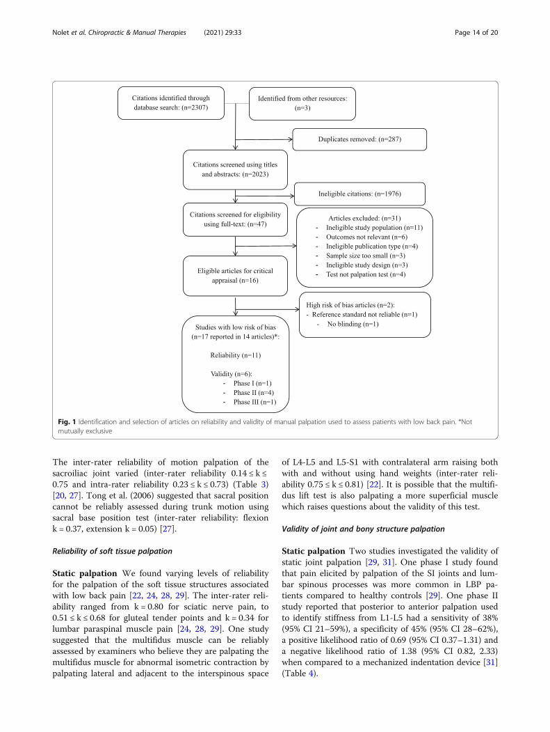
ResultsStudy selectionWe identified 2307 citations (plus 3 citations from otherresources) removed 287 duplicates, and reviewed 2023articles for eligibility (Fig. 1). In stage 1 screening, 1976citations were ineligible. Forty-seven papers werereviewed in stage 2, and 31 were excluded: ineligiblestudy population (n = 11) [41–51], inappropriate out-come measure (n = 6) [52–57], ineligible publicationtype (n = 4) [58–61], ineligible sample size (n = 3) [62–64], study design (n = 3) [65–67] and did not investigatemanual palpation (n = 4) [68–71]. Two authors werecontacted for publication type and age range, bothresponded [27, 59].We critically appraised 16 articles and 14 articles had
low risk of bias and were included in our evidence syn-thesis [19–32] (Fig. 1). Over the 16 articles appraised, 14
Nolet et al. Chiropractic & Manual Therapies (2021) 29:33 Page 5 of 20
Table 3 Evidence table for low risk of bias studies assessing the reliability of manual palpation tests in patients with low back pain
Authors,YearCountry
DesignSample Size(n)
Case Definition Index Test Reliability
Static Joint Palpation (n = 262)
Alyazediet al.2015[19]
USA
Inter-raterreliability(n = 40)
Recurrent LBP or chronic LBP (≥ 3months), 21–71 yrs. old.
Prone instability test was done in two parts: 1)relaxation phase: the subject was lying prone onthe examination table with feet on the floor. Theexaminer performed PA mobility testing to identifypainful lumbar segments with the subject’s musclesrelaxed. 2) co-contraction phase: the subjects thenraises their feet off the floor. If pain identified in therelaxation phase subsides at the co-contractionphase the test is considered positive.PA glide test: Subjects were lying prone andexaminers performs PA glide on the lumbar spinousprocesses. Lack of segmental hypomobility, isconsidered a positive test.Examiners: two physical therapists who werecertified as Orthopaedic Clinical SpecialistsTime between inter-rater assessments was at least15 min
Inter-rater reliabilityProne instability test forpain (relaxation phase);k (95% CI)k = 0.41 (0.18, 0.63)Prone instability test forpain (co-contractionphase);k (95% CI)k = 0.71 (0.45, 0.98)PA glide test forhypomobility; k (95% CI)k = − 0.02 (− 0.22, 0.18)
Palpation for the spinal level contributing most tothe patients’ LBP symptoms (abnormal end-feel, ab-normal quality of resistance to motion, andreproduction of pain, local or referred); patientprone, posterior to anterior pressure applied tospinal process and verbal communication betweenexaminer and patient about reproduction of pain.Examiners: three pairs of manipulativephysiotherapists with 7–15 yrs. experience and≥ 3yrs. experience after postgraduate qualifications inmanipulative physiotherapy.Time between inter-rater assessments unknown.
Inter-rater reliabilityPalpation to locate thespinal level; k (95% CI):Overall: k = 0.37 (0.20,0.54)Pair 1: k = 0.54 (0.26, 0.82)Pair 2: k = 0.45 (0.18, 0.72)Pair 3: k = 0.23 (0.00, 0.46)Palpation to name thespinal level; k (95% CIlower band):Overall: k = 0.09 (0.00,0.18)Pair 1: k = 0.41 (0.12, 0.70)Pair 2: k = 0.10 (0.00, 0.20)Pair 3: k = − 0.13 (0.00,0.26)
Hickset al.,2003[23]
USA
Inter-raterreliability(n = 63)[pair 1 n = 20,pair 2 n = 28,pair 3 n = 15]
Low back pain without radiation ofpain past the knee, symptomduration unknown, 20 to 66 yrs. old.
Prone instability test: The subject lies prone on theexamination table with their feet on the floor. Theexaminer performs passive intervertebral motiontesting for pain. The subject then lifts their feet offthe floor. A positive test is when pain provokedduring the first part of the test disappears when thelegs are lifted up.Passive intervertebral motion testing: with thesubject lying prone the examiner applied PApressure with their hypothenar eminence on eachlumbar spinous process. Segmental mobility isjudged as normal mobility, hypomobility (moremotion than normally expected) and hypermobility(less motion than normally expected). Painprovocation is judged as manual pressureproducing pain or not producing pain.Examiners were 4 physical therapists with a least 2yrs. experience. Examiners were placed in 3 separatepairs.Time between inter-rater assessments was at least15 min
Inter-rater reliabilityProne instability test; k(95% CI):k = 0.87 (0.80, 0.94)Pair 1 (n = 20): k = 1.0(1.0–1.0)Pair 2 (n = 28): k = 0.81(0.80–0.94)Passive intervertebralmotion tests; k (95% CI):Segmental mobility(dichotomous):Hypermobility k = 0.30(0.13, 0.47)Hypomobility k = 0.18(0.05–0.32)Segmental mobility(hypo/normal/hyper):L1 k = 0.26 (− 0.01, 0.53)L2 k = 0.17 (− 0.13, 0.47)L3 k = − 0.02 (− 0.25, 0.28)L4 k = 0.11 (− 0.26, 0.35)L5 k = 0.18 (− 0.03, 0.49)Pain provocation(positive/negative):L1 k = 0.36 (0.12, 0.59)L2 k = 0.45 (0.26, 0.63)L3 k = 0.30 (0.12, 0.47)L4 k = 0.25 (0.11, 0.40)L5 k = 0.55 (0.43, 0.67)
Nolet et al. Chiropractic & Manual Therapies (2021) 29:33 Page 6 of 20
Table 3 Evidence table for low risk of bias studies assessing the reliability of manual palpation tests in patients with low back pain(Continued)
Authors,YearCountry
DesignSample Size(n)
Case Definition Index Test Reliability
Ravennaet al., 2011[25]USA
Inter-raterreliability(n = 30)
Chronic and recurrent LBP, 18 to 60yrs. Old.
Prone Instability Test: patient prone with legs overthe edge and feet resting comfortably on the floor.The examiner palpates for pain. The patient thenraises their legs off the floor and examiner palpatesagain for pain. A positive test is when painprovoked during the first part of the test disappearsor decrease when the legs are lifted up.Examiners were a second-year Doctor of PhysicalTherapy student and a licensed physical therapistwith two years clinical experience.Time between inter-rater assessments was 20min.
Low back pain, symptom durationunknown, 18–65 yrs. old.
Palpation for lumbar segmental mobility, painprovocation and prone instability: patient pronewith 1) Prone mobility testing: posterior to anteriorjoint springing palpation by examiners of SIJs, alllumbar spinous processes and all lumbar facet jointsbilaterally; normal or restricted mobility was noted;2) prone pain provocation testing: patient notifiespain or discomfort provoked while repeating pronemobility test; 3) prone instability test: patient pronewith legs over the edge and feet restingcomfortably on the floor. The examiner palpates forpain. The patient then raises their legs off the floorand examiner palpates again for pain. A positivetest is when pain provoked during the first part ofthe test disappears when the legs are lifted up.Examiners: two doctors of chiropractic with 25 and10 years of clinical experience.All the examinations performed in one day.
Prone Mobility Testing;k (95%CI); PABAKLeft L4–5, and L5-S1 facetmobility: k = − 0.17 (−0.41,.06); PABAK = 0.08Right L4–5, and L5-S1facet mobility: k = − 0.12(− 0.41,0.18); PABAK = −0.09Spinous L4–5 and S1mobility: k = − 0.05 (−0.36,0.27); PABAK = 0.11Left L1–4 facet mobility:k = 0.17 (− 0.14,0.48);PABAK = 0.44Right L1–4 facet mobility:k = − 0.01 (− 0.33,0.30);PABAK = 0.44Spinous L1–4 mobility:k = 0.02 (− 0.27,0.32)PABAK = 0.07Sacroiliac mobility L: k =− 0.11 (− 0.21,-0.01);PABAK = 0.54Sacroiliac mobility R: k =− 0.10 (− 0.18,-0.02);PABAK = 0.64Prone Pain ProvocationTesting; k (95%CI):PABAKLeft L4–5, and L5-S1 pain:k = 0.73 (0.51,0.95);PABAK = 0.74Right L4–5, and L5-S1pain: k = 0.52 (0.25,0.79);PABAK = 0.54Spinous L4–5 and L5-S1pain: k = 0.57 (0.32,0.83);PABAK = 0.58Left L1–4 pain: k = 0.46(0.17,0.75); PABAK = 0.48Right L1–4 pain: k = 0.38(0.06,0.69); PABAK = 0.54Spinous L1–4 pain: k =0.21 (− 0.10,0.53);PABAK = 0.34Right Sacroiliac pain: k =0.14 (− 0.19,0.47);PABAK = 0.38Left Sacroiliac pain: k =0.33 (0.0,0.66); PABAK =0.54Prone Instability Test; k
Nolet et al. Chiropractic & Manual Therapies (2021) 29:33 Page 7 of 20
Table 3 Evidence table for low risk of bias studies assessing the reliability of manual palpation tests in patients with low back pain(Continued)
Authors,YearCountry
DesignSample Size(n)
Case Definition Index Test Reliability
(95%CI). PABAKTest 1: k = 0.54 (0.27,0.81);PABAK = 0.58Test 2: k = 0.46 (0.15,0.77);PABAK = 0.58
Weineret al., 2006[29]USA
Inter-raterreliability (n =30)
Chronic LBP, ≥3 months duration,≥60 yrs. old.
Palpation of the SI joints and lumbar spinousprocesses to identify pain: 1) SI joints: patientstanding on floor with shoes removed, examinerstanding behind patient exerts firm pressure oversacroiliac joint, palpation of right joint with rightthumb while standing to left side of patient; 2)lumbar spinous processes: examiner behind patient,firmly palpate spinous processes L1–L5 usingdominant thumbThe examiners underwent training in the protocolwith an expert physical therapists to refine andstandardize the physical examination proceduresTime between intra-rater assessments < 5min
SI palpation; k (95% CInot reported)k = 0.59Lumbar spinouspalpationk = 0.47
Motion Joint Palpation (n = 49)
Arabet al.,2009[20]
Iran
Intra-rater andinter-rater reli-ability (n = 25)
LBP around posterior superior iliacspine and buttock, symptomduration unknown, 20–65 yrs. old.
Standing flexion test: The subject is standing andthe examiner palpates the movement of PSIS as thesubject bends forward.Sitting flexion test: The subject is sitting and theexaminer palpates the movement of PSIS as thesubject bends forward.Gillett test: The subject is standing with theexaminer palpating the movement of PSIS as thesubject raises that knee toward their chest.Examiner: two physiotherapists with 1 yearexperience.Time between intra-rater assessments: 15 min.
Inter-rater reliability; k(95% CI). PABAKStanding flexion testR- k = 0.51 (0.08–0.95);PABAK = 0.68L- k = 0.55 (0.2–0.9);PABAK = 0.60Sitting flexion testR- k = 0.75 (0.42–1.08);PABAK = 0.84L- k = 0.64 (0.32–0.96);PABAK = 0.68Gillet testR- k = 0.41 (0.03–0.87);PABAK = 0.60L- k = 0.34 (− 0.06–0.7);PABAK = 0.44Intra-rater reliability; k(95% CI). PABAKStanding flexion testRater 1: R- k = 0.68 (0.35–1.01); PABAK = 0.76Rater 1: L- k = 0.61 (0.27–0.96); PABAK = 0.68Rater 2: R- k = 0.60 (0.18–1.02); PABAK = 0.76Rater 2: L- k = 0.51 (0.08–0.95); PABAK = 0.68Sitting flexion testRater 1: k = 0.73 (0.45–1.01); PABAK = 0.76Rater 1: k = 0.65 (0.34–0.96); PABAK = 0.68Rater 2: k = 0.65 (0.29–1.02); PABAK = 0.76Rater 2: k = 0.56 (0.21–0.90); PABAK = 0.60Gillett testRater 1: k = 0.42 (− 0.01–0.87); PABAK = 0.60Rater 1: k = 0.49 (0.09–0.89); PABAK = 0.60Rater 2: k = 0.25 (− 0.20–0.77); PABAK = 0.52
Nolet et al. Chiropractic & Manual Therapies (2021) 29:33 Page 8 of 20
Table 3 Evidence table for low risk of bias studies assessing the reliability of manual palpation tests in patients with low back pain(Continued)
Authors,YearCountry
DesignSample Size(n)
Case Definition Index Test Reliability
Rater 2: k = 0.23 (− 0.02–0.67); PABAK = 0.36
Tong et al.,2006 [27]USA
Inter-raterreliability (n =24)
LBP, symptom duration unknown, 32to 81 yrs. old.
Seated flexion test: the evaluator palpates thecephalad movement at PSISs. As the subject bendsforward, the evaluator’s thumbs follow the motionof the PSIS cephaladStanding stork test: the evaluator’s thumb palpatesthe unilateral movement of left PSIS, and the otherthumb palpates the midline of the sacrum. Thesubject then flexes the left hip and knee to aminimum of 90 degrees. The same is repeated onthe right PSIS with the subject flexing the right hip.Standing flexion test: the evaluator palpates themovement of unilateral PSIS. As the subject bendsforward to touch the floor, the evaluator’s thumbsfollow the PSIS cephalad. The test is repeated oneach side.Sacral base position: the subject is sitting, theevaluator palpates the sacral base with the subject’strunk forward flexed and backward flexed. Apositive test is when one side of the sacrum ismore anterior or posterior when compared to theother side of the sacrum on the spine motions.Examiners: four physicians.Time between inter-rater assessments unknown.
k (95% CI not reported);p valueSeated flexion test: k =0.06; p = 0.68Standing stork test: k =0.27; p = 0.07Standing flexion test:k = 0.14; p = 0.37Sacral base position:flexion k = 0.37; p = 0.002extension k = 0.05; p =0.74
Static Soft Tissue Palpation (n = 150)
Hebertet al.,2015[22]
USA
Inter-raterreliability(n = 32)
Low back pain with ≥20/100 onmodified ODI, median duration ofsymptoms = 205 days, 18 to 60 yrs.old.
Multifidus lift test: to identify lumbar multifiduscontraction; participants prone and contralateralarm lifted with/without a hand weight whilemultifidus muscle palpated immediately lateral andadjacent to the interspinous space of L4–L5 andL5–S1.Examiners: > 10 yrs. clinical experience andapproximately 5 yrs. research experience.Time between intra-rater assessments unknown.
Inter-rater reliability; k(95% CI)L4–L5 no weight: k = 0.75(0.52,0.97)L4–L5 weight: k = .0.79(0.57, 1.00)L5-S1 no weight: k = 0.81(0.62, 1.00)L5-S1 weight: k = 0.80(0.59, 1.00)
Jensenet al.,2013[24]
Denmark
Intrarater andinter-rater reli-ability (n = 43)
LBP with or without radiculopathy,variable duration of symptoms, 16–60 yrs. old.
Palpation of gluteal tender points: patient seated,tender points tested from right to left with 4 kgdigital pressure on upper outer quadrants ofbuttocks.Examiners: two consultants in rheumatology andrehabilitation.20 min between inter-rater assessments and 7 daysbetween intra-rater assessments.
Intra-rater reliability(95% CI not reported)Rater A: R k = 0.78; L k =0.69Rater B: R k = 0.79; L k =059.Inter-rater reliabilityDay 1: R k = 0.68; L k =0.53Day 2: R k = 0.51; L k =0.50
Walshet al.,2009[28]
Ireland
Inter-raterreliability (n =45)
Unilateral low-back related leg pain,mean duration of symptoms = 5.6months, 18–70 yrs. old.
Palpation of sciatic nerve: With the patient lyingprone they are asked if there is any pain ordiscomfort when the examiner applies gentlepressure at the sciatic nerve bilaterally at themidway point of a line from ischial tuberosity to thegreater trochanter of the femur.Examiner: two physiotherapists (eleven yrs.experience with a Masters in Manipulative Therapyand three months clinical experience, respectively).
Palpation of the lumbar paraspinal muscles, andpiriformis muscles to identify pain:; 1) paralumbarmuscles: patient standing on floor with shoesremoved, examiner stands behind to left side ofpatient and braces patient in front with left arm;palpate full extent of right paravertebral
Lumbar paraspinalpalpation k (95% CI notreported)k = 0.34Piriformis palpationk = 0.66
Nolet et al. Chiropractic & Manual Therapies (2021) 29:33 Page 9 of 20
articles including 17 studies were reported (three articlesincluded both reliability and validity in their study). Theinter-rater agreement for screening of articles wasKappa = 0.86 (95% CI 0.73–0.98). The percentage agree-ment for the admissibility of studies was 100% (17 agree-ments/17 studies over the 16 articles appraised).
Study characteristicsFourteen articles had a low risk of bias [19–32]. Ofthose, 11 reported on the reliability of palpation tests[19–29] and six reported on validity [22, 28–32].Three articles examined both reliability and validity[22, 28, 29].The eleven reliability studies with low risk of bias ex-
amined inter-rater reliability of manual palpation to as-sess joints mobility or motion [19–21, 23, 26, 27], pain[19, 21, 23–26, 28, 29] and muscle contraction [22]. Twoof the eleven studies also examined intra-rater reliabilityof manual palpation assessing joint motion [20] andmuscle tenderness [24]. The six validity studies includedone phase I study on palpation of joints and muscles toassess pain [29], four phase II on palpation of nerves toelicit pain [28], spinal stiffness [31] muscle contraction[22] and sacroiliac joint motion [32] and one phase IIIstudy on palpation of gluteal muscle for tenderness andpain [30].The 14 low risk of bias articles investigated: 1) static
joint palpation (n = 7) [19, 21, 23, 25, 26, 29, 31], 2) mo-tion joint palpation (n = 3) [20, 27, 32], and 3) static softtissue palpation (n = 5) [22, 24, 28–30] (Tables 3 and 4).They assessed various techniques: 1) joint pain provoca-tion [19, 21, 23, 26, 29], 2) pain or tenderness of muscles[24, 29, 30], 3) pain and tenderness of nerves [28], 4)joint stiffness/mobility [19, 21, 23, 25, 26, 31], 5) jointmotion [20, 27, 32], and 6) isometric muscle contraction[22]. Table 5 showed a glossary of definitions for all ofthe palpation tests included in the articles.
The duration of LBP varied across studies: < 7 weeks(1/14 articles) [21], > 4 weeks (1/14 articles) [24], ≥ lmonths (1/14 articles) [29], new episode to > 3 months(1/14 articles) [19] and unspecified duration (10/14 arti-cles) [20, 22, 23, 25–28, 30–32]. The studies were con-ducted in Australia [21], Canada [30], Denmark [24],Iran [20, 32], Ireland [28], and the United States [19, 22,23, 25–27, 29, 31] between 2003 and 2017.We did not perform a meta-analysis because of the
heterogeneity of studies in symptom duration, palpationtechnique, and outcome specification.
Assessment of risk of BiasTables 1 and 2 showed the risk of bias for scientificallyadmissible reliability and validity studies based on themodified QAREL and QUADAS-2 criteria respectively.The low risk of bias studies met the following criteria:
1) clearly described objective; 2) representative sample;3) representative raters; 4) blinding of the test results be-tween raters; 5) appropriate and valid standard test; and6) appropriate statistical analysis (Tables 1 and 2). How-ever, these studies had the following limitations: 1) un-clear time interval between tests (n = 1) [27]; 2) noblinding for intra-examiner reliability (n = 2) [20, 24]; 3)30 min rest period between the repeat testing betweenthe same examiner and no blinding to clinical informa-tion (n = 2) [27, 28]; 4) unclear blinding to clinical infor-mation or additional clues [23]; 5) no blinding to clinicalinformation and unclear blinding to additional clues (n=8) [21, 22, 23, 24, 25, 27, 28, 29] and 6) non-random orunclear administration of tests (n = 5) [19, 23, 27–29].Most validity studies had appropriate exclusion criteriaand blinding. However, validity studies had limitations:1) four studies did not use a consecutive or randomsample [22, 29, 31, 32]; and 2) two studies were unclearas to whether an appropriate time interval between testswere used [28, 31]; 3) one study was unclear as to
Table 3 Evidence table for low risk of bias studies assessing the reliability of manual palpation tests in patients with low back pain(Continued)
Authors,YearCountry
DesignSample Size(n)
Case Definition Index Test Reliability
musculature with right thumb. Exert approximately4 kgf. Repeated on the other side2) piriformis: patient supine flexes right hip andknee, keeping sole of foot on table. Cross bent legover opposite leg and again place sole on table andexert mild medially directed pressure on lateralaspect of knee to put piriformis in stretch. Exert firmpressure (4 kg) over middle extent of piriformis.Repeated on the other side.The examiners underwent training in the protocolwith an expert physical therapists to refine andstandardize the physical examination proceduresTime between intra-rater assessments < 5min
CI Confidence interval, k Cohen’s kappa, LBP Low back pain, ODI Oswestry Disability Index, PA Posterior to anterior, PABAK Prevalence-adjusted and bias-adjustedkappa, PSIS Posterior superior iliac spine, SE Standard error, SIJ Sacroiliac joint, yrs. years
Nolet et al. Chiropractic & Manual Therapies (2021) 29:33 Page 10 of 20
Table 4 Evidence table for low risk of bias studies assessing the validity of manual palpation tests in patient with low back pain
AuthorsYearCountry
Designa
Samplesize (n)
Population Index test Gold/Reference Standard Validity
Static Joint Palpation (n = 182)
Koppenhaveret al., 2014[31]
USA
Phase II(n = 51)
LBP with modified ODI≥20/100, medianduration of symptoms =184 days, 18–60 yrs. old.
Palpation of spinal stiffness: thespinous processes ofL1-L5 palpated with the subjectlying prone. The participant wasasked to relax as a posterior toanterior (PA) force was applied.Examiner: 1 clinician with 8 yrs.experience
Spinal stiffness was quantifiedusing a mechanized indentationdevice.
Palpation of the SI joints and,lumbar spinous processes toidentify pain:1) SI joints: patientstanding on floor with shoesremoved, examiner standingbehind patient exerts firmpressure over sacroiliac joint,palpation of right joint with rightthumb while standing to left sideof patient, repeated on the otherside; 2) lumbar spinous processes:examiner behind patient, firmlypalpate spinous processes L1–L5using dominant thumb;The examiners underwent trainingin the protocol with an expertphysical therapist to refine andstandardize the physicalexamination procedures.
N/A Differencebetween groups:Positive palpationn(%), (p value)SI palpation70 (59), p < 0.001Lumbar spinouspalpation59 (53.2), p < 0.001
Gillet test: the subjects standswhile the examiner sits behind thepatient and palpates each of thepatient’s PSIS, one at a time, withone thumb on the inferior aspectof the PSIS while palpating thesacrum with the other thumb. Thesubject stands on one leg whilepulling the opposite leg uptoward the chest. A positive test isif the PSIS on the side of the kneeflexion does not move or movesposterior-inferiorly only minimallyor even paradoxically movessuperiorlyStanding flexion test: the subjectstands while the examiner sitsbehind the patient and palpatesboth of the patient’s |PSIS on theirinferior margins. The subjectbends forward. A positive result ina standing flexion test indicateslimited movement of the ilium onthe sacrum.Sitting flexion test: the subject sitswhile the examiner sits behind thepatient and palpates both of thepatient’s |PSIS on their inferiormargins. The subject bendsforward. A positive result indicateslimited movement of the sacrumon the ilium.Examiner: one physical Therapist
Thigh thrust test: with the subjectlying supine the examiner flexesthe hip joint to 90 degree offlexion and slight adduction withthe knee flexed. The examinerwith one hand cups thesacrum and wraps the other armand hand around the flexed kneeapplying axial pressure. A test ispositive when pain is provokedover the posterior aspect of thesymptomatic SI joint.Faber test: The patient lies supineon the table. The examiner bringsthe ipsilateral hip into flexion,abduction and external rotation.The foot is rested on unaffectedknee. A positive test is whenbuttock or groin pain below L5 isreproducedResisted abduction test: Thesubject supine with the leg fullyextended as well as beingabducted to 30°. The examinerholds the ankle and pushesmedially while the subject pusheslaterally. The test is positive whenfamiliar pain is produced over theSIJ below L5.
Nolet et al. Chiropractic & Manual Therapies (2021) 29:33 Page 11 of 20
Table 4 Evidence table for low risk of bias studies assessing the validity of manual palpation tests in patient with low back pain(Continued)
AuthorsYearCountry
Designa
Samplesize (n)
Population Index test Gold/Reference Standard Validity
Adelmaneshet al., 2016[30]
Canada
Phase III(n =337)
LBP with or withoutradiculopathy of anyduration, > 18 yrs. old.
Palpation of the superior-lateralquadrant of the gluteal muscle toidentify GTrP representing thecombination of tenderness, tautband and pain: With the patientprone the gluteal muscle wascompressed with a flat thumb orindex finger against the under-lying tissue or bone.Examiner: 1 physician with 7 yrs.experience.
Multidisciplinary panel of expertsbased on examination of clinicalevaluations, MRI, and if needed,electrodiagnostic testing.Examiner: 1) clinical evaluation by1 clinician with 12 yrs. experience;2) MRI by 1 experiencedneuroradiologist and 1 physiatrist;3) electrodiagnostic testing by aphysiatrist with 15 yrs. experience.
Low back pain with≥20/100 on modifiedODI, median duration ofsymptoms = 205 days, 18to 60 yrs. old.
Multifidis lift test: to identifylumbar multifidus contraction;participants prone andcontralateral arm lifted with/without a hand weight whilemultifidus muscle palpatedimmediately lateral and adjacentto the interspinous space of L4–L5and L5–S1.Examiner: 2 examiners with > 10yrs. clinical experience andapproximately 5 yrs. researchexperience.
Lumbar multifidus musclethickness measures at the L4–L5and L5–S1 spinal levels, at rest andsubmaximal contraction duringcontralateral arm lift usingbrightness-mode real-time ultra-sound imaging.Examiner: 1 clinician with 5 yearsultrasound experience.
Changes in lumbarmultifidus thicknessat L4-L5 (r biserialcorrelation coeffi-cient); p value.Examiner 1L4–L5 no weight r0.59; p = 0.010L4–L5 weight r 0.71;p = 0.003L5–S1 no weight r0.73; p = 0.002L5–S1 weight r 0.62;p = 0.008Examiner 2L4–L5 no weight r0.71; p = 0.002L4–L5 weight r 0.69;p = 0.005L5–S1 no weight r0.69; p = 0.003L5–S1 weight r 0.63;p = 0.009Changes in lumbarmultifidus thicknessat L5-S1 (r biserialcorrelation coeffi-cient); p value.Examiner 1L4–L5 no weight r0.29; p = 0.201L4–L5 weight r 0.44;p = 0.063L5–S1 no weight r0.47; p = 0.040L5–S1 weight r 0.39;p = 0.097Examiner 2L4–L5 no weight r0.45; p = 0.053L4–L5 weight r 0.24;p = 0.0341L5–S1 no weight r0.44; p = 0.056L5–S1 weight r 0.17;p = 0.472
Walsh et al.,2009 [28]
Ireland
Phase II(n = 45)
Unilateral low-back re-lated leg pain, meanduration of symptoms =5.6 months, 18–70 yrs.old.
Palpation of sciatic nerve: patientprone lying is asked for any painor discomfort when examinerapplies gentle pressure at thesciatic nerve bilaterally at themidway point of a line from ischial
Straight leg raise (SLR) and slumptests conducted by onephysiotherapist with one yearexperience.
Nolet et al. Chiropractic & Manual Therapies (2021) 29:33 Page 12 of 20
whether an appropriate reference standard (slump testand straight leg raise) was used [28]; 4) in one study theexaminer was not blinded to the results of the index orreference test [32] and 5) in one study it was unclear asto whether all patients were included in the analysis[22].Two validity studies were excluded after critical ap-
praisal. Abbott et al. used flexion/extension radiographsas a reference standard without establishing the test-retest reliability of patient positioning when taking ofthe radiographs [72]. Telli et al. didn’t use blinding intheir reliability study [73].
Summary of evidenceReliability of joint and bony structure palpation
Static palpation Four studies investigated static palpa-tion to elicit pain. Overall, these studies suggest that im-portant measurement error is associated with elicitingpain from: 1) lumbar facet joints (inter-rater reliability0.38 ≤ k ≤ 0.73); 2) lumbar spinous processes (inter-rater
reliability 0.21 ≤ k ≤ 0.57); 3) sacro-iliac (SI) joints (inter-rater reliability 0.14 ≤ k ≤ 0.59) [19, 23, 26, 29] (Table 3).Similarly, the evidence suggests that static palpation usedto identify joint segmental mobility has low inter-rater re-liability (i.e., lumbar facet joints: − 0.17 ≤ k ≤ 0.17; andlumbar spinous processes; − 0.02 ≤ k ≤ 0.26 SI joints: −0.11 ≤ k ≤ − 0.10) [19, 23, 26]. The inter-rater reliability ofthe prone instability test for pain ranged from a kappa of0.30 [23], 0.41 [19] and 0.54 [26] in the relaxation phase ofthe test and a kappa of 0.46 [26], 0.71 [19] and 0.87 [23] inthe contraction phase of the test. In a study that combinedthe two phases of the test into a positive or negative find-ing reported a kappa of 0.10 [25] (Table 3). Furthermore,a third study by Downey et al. (2003) reported low inter-rater reliability of joint static palpation to locate the spinallevel (0.23 ≤ k ≤ 0.54) and name the spinal level (− 0.13 ≤k ≤ 0.41) in patients with LBP symptoms [21] (Table 3).
Motion palpation We found inconsistent evidence insupport of the reliability of motion palpation of the lum-bar spine and SI joints to assess joint motion [20, 27].
Table 4 Evidence table for low risk of bias studies assessing the validity of manual palpation tests in patient with low back pain(Continued)
AuthorsYearCountry
Designa
Samplesize (n)
Population Index test Gold/Reference Standard Validity
tuberosity to the greatertrochanter of the femur.Examiner: two physiotherapists(eleven yrs. experience with aMasters in Manipulative Therapyand three months clinicalexperience, respectively).
Palpation of the lumbar paraspinalmuscles, and piriformis muscles toidentify pain: 1) paralumbarmuscles: patient standing on floorwith shoes removed, examinerstands behind to left side ofpatient and braces patient in frontwith left arm; palpate full extent ofright paravertebral musculaturewith right thumb. Exertapproximately 4 kgf: 2) piriformis:patient supine flexes right hip andknee, keeping sole of foot ontable. Cross bent leg overopposite leg and again place soleon table and exert mild mediallydirected pressure on lateral aspectof knee to put piriformis in stretch.Exert firm pressure (4 kg) overmiddle extent of piriformis,The examiners underwent trainingin the protocol with an expertphysical therapist to refine andstandardize the physicalexamination procedures.
N/A Differencebetween groups:Positive palpationn(%), p valueLumbar paraspinalpalpation60(50), p < 0.001Piriformispalpation57(51.4), p < 0.001
GTrP Gluteal trigger point, LBP Low back pain, +LR Positive likelihood ration, −LR Negative likelihood ration, N/A Not Applicable, NPV Negative predictive value,ODI Oswestry Disability Index, PPT Pressure pain threshold, PPV Positive predictive value, Sens Sensitivity, SI Sacroiliac, Spec Specificity, yrs. yearsa: Design refers to Phase I-IV questions as described by Sackett & Haynes (2002). 2002;324(7336):539–41
Nolet et al. Chiropractic & Manual Therapies (2021) 29:33 Page 13 of 20
The inter-rater reliability of motion palpation of thesacroiliac joint varied (inter-rater reliability 0.14 ≤ k ≤0.75 and intra-rater reliability 0.23 ≤ k ≤ 0.73) (Table 3)[20, 27]. Tong et al. (2006) suggested that sacral positioncannot be reliably assessed during trunk motion usingsacral base position test (inter-rater reliability: flexionk = 0.37, extension k = 0.05) [27].
Reliability of soft tissue palpation
Static palpation We found varying levels of reliabilityfor the palpation of the soft tissue structures associatedwith low back pain [22, 24, 28, 29]. The inter-rater reli-ability ranged from k = 0.80 for sciatic nerve pain, to0.51 ≤ k ≤ 0.68 for gluteal tender points and k = 0.34 forlumbar paraspinal muscle pain [24, 28, 29]. One studysuggested that the multifidus muscle can be reliablyassessed by examiners who believe they are palpating themultifidus muscle for abnormal isometric contraction bypalpating lateral and adjacent to the interspinous space
of L4-L5 and L5-S1 with contralateral arm raising bothwith and without using hand weights (inter-rater reli-ability 0.75 ≤ k ≤ 0.81) [22]. It is possible that the multifi-dus lift test is also palpating a more superficial musclewhich raises questions about the validity of this test.
Validity of joint and bony structure palpation
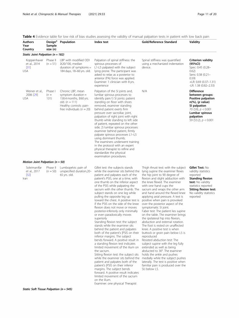
Static palpation Two studies investigated the validity ofstatic joint palpation [29, 31]. One phase I study foundthat pain elicited by palpation of the SI joints and lum-bar spinous processes was more common in LBP pa-tients compared to healthy controls [29]. One phase IIstudy reported that posterior to anterior palpation usedto identify stiffness from L1-L5 had a sensitivity of 38%(95% CI 21–59%), a specificity of 45% (95% CI 28–62%),a positive likelihood ratio of 0.69 (95% CI 0.37–1.31) anda negative likelihood ratio of 1.38 (95% CI 0.82, 2.33)when compared to a mechanized indentation device [31](Table 4).
Fig. 1 Identification and selection of articles on reliability and validity of manual palpation used to assess patients with low back pain. *Notmutually exclusive
Nolet et al. Chiropractic & Manual Therapies (2021) 29:33 Page 14 of 20
Table 5 Glossary of Manual Palpation Tests in Accepted Articles
Test Purpose of Test Description of Test
Static Joint Palpation
Lumbar spinous palpationWeiner et al., 2006 [29]
Palpation to identify pain The examiner is behind patient and firmly palpates the spinousprocesses of L1–L5 using their dominant thumb. A positive test ispain on palpation.
Passive intervertebral motiontests
Hicks et al. 2003 [23]
Lumbar palpation for segmental mobility andpain
With the subject lying prone the examiner applies AP pressure withtheir hypothenar eminence on each lumbar spinous process.Segmental mobility is judged as normal, hypomobile andhypermobile. Pain provocation is judged as pressure producing painor not producing pain.
Posterior/Anterior Glide TestAlyazedi et al. 2015 [19]
Palpation for identify lumbar spinal mobility. Subjects are lying prone and examiners performs PA glide on thelumbar spinous processes. Lack of segmental hypomobility, isconsidered a positive test.
Prone Instability TestAlyazedi et al. 2015 [19]Hicks et al. 2003 [23]Ravenna et al., 2011 [25]Schneider et al., 2008 [26]
Lumbar springing palpation for pain Patient prone with legs over the edge of table and feet restingcomfortably on the floor. The examiner palpates for pain. Thepatient then raises their legs off the floor and examiner againpalpates for pain. A positive test is indicated by painful segments inthe first position becoming nonpainful with contraction of the backextensors.
Prone Lumbar Palpation(comparable level and levelidentification)
Downey et al., 2003 [21]
Identification of spinal level mostcontributing to LBP and identification of thatlevel
Palpation for the spinal level contributing most to the patients’ LBPsymptoms (abnormal end-feel, abnormal quality of resistance to mo-tion, and reproduction of pain, local or referred); patient prone, pos-terior to anterior pressure applied to spinal process and verbalcommunication between examiner and patient about reproductionof pain.
Prone Mobility TestSchneider et al., 2008 [26]
Lumbar segmental mobility Posterior to anterior joint springing palpation by examiners of SIJs,all lumbar spinous processes and all lumbar facet joints bilaterally. Apositive test is the palpation of restricted motion.
Prone Pain Provocation TestSchneider et al., 2008 [26]
Lumbar spine pain Patient notifies the examiner of pain or discomfort provoked whilerepeating prone mobility test
Sacroiliac Joint PalpationWeiner et al., 2006 [29]
Palpation of the sacroiliac joints for pain The patient stands on floor with shoes removed and the examinerstands behind patient. The examiner exerts firm pressure oversacroiliac joint, palpation of right joint with right thumb whilestanding to left side of patient and palpation of the left joint withthe left thumb while standing to the right of the patient. A positivetest is the patient reporting pain in the back.
Spinous Palpation forStiffness
Koppenhaver et al., 2014 [31]
Joint springing of the lumbar spinousprocess
The spinous processes of L1-L5 are palpated with the subject lyingprone. The participant was asked to relax as a posterior to anterior(PA) force was applied. Each vertebral segment was judged to behypermobile, hypomobile or normal mobility.
Motion Joint Palpation
Gillet TestArab et al., 2009 [20]Soleimanifar et al., 2017 [32]
Palpation for movement at the PSIS whilepatient raises knee
The subject is standing with the examiner palpating the PSIS as thesubject raises that knee toward their chest.A positive test is when the PSIS on the side of the knee flexion doesnot move or moves posterior-inferiorly only minimally or even para-doxically moves superiorly.
Sacral Base Position TestTong et al., 2006 [27]
Palpation of the sacral base for position whilethe patient flexes then extends their spine
The subject is sitting, the evaluator palpates the sacral base with thesubject’s trunk forward flexed and backward flexed. A positive test iswhen one side of the sacrum is more anterior or posterior whencompared to the other side of the sacrum on the spine motions.
Seated Flexion TestTong et al., 2006 [27]
Bilateral palpation for cephalad movement atthe PSIS while patient forward bends spine
The evaluator palpates both PSISs. As the subject bends forward, theevaluator’s thumbs follow the motion of the PSIS cephalad. If oneside moves more cephalad than the other side by more than 1 cm,the side that moves more is considered abnormal.
Sitting Flexion TestArab et al., 2009 [20]Soleimanifar et al., 2017 [32]
Palpation for movement at the PSIS whilepatient forward bends spine
The subject is sitting and the examiner palpates the PSIS as thesubject bends forward. A positive result in this test indicates limitedmovement of the sacrum on the ilium.
Standing Flexion TestArab et al., 2009 [20]Soleimanifar et al., 2017 [32]Tong et al., 2006 [27]
Palpation for movement at the PSIS whilepatient forward bends spine
The subject is standing and the examiner palpates the PSIS as thesubject bends forward. A positive result in a standing flexion testindicates limited movement of the ilium on the sacrum.
Nolet et al. Chiropractic & Manual Therapies (2021) 29:33 Page 15 of 20
Motion palpation One phase II study investigated thevalidity of joint motion palpation tests for the sacroiliacjoints [32]. They examined the relationship betweensacroiliac tests for joint motion (Gillet test, sittingflexion test and standing flexion test) and sacroiliac painprovocation tests (Faber test, thigh thrust test andresisted abduction test) but did not use statistics for val-idity (Table 4).
Validity of soft tissue palpation
Static palpation Four studies investigated the validity ofstatic soft tissue palpation [22, 28–30]. One phase Istudy found that pain elicited by palpation of the lumbarparaspinal and piriformis muscles was more common inLBP patients compared to without LBP [29]. A phase IIstudy tested the validity of the multifidus lift test with
and without hand weights to identify abnormal isometricmultifidus muscle contraction when compared to meas-urement with real-time ultrasound imaging of lumbarmultifidus muscle thickness [22] (Table 4). The authorsreported that the multifidus lift test correlates withultrasound finding at the L4–5 level (r biserial correl-ation coefficient: 0.59 without hand weight and 0.73without hand weight) and weakly associated at the L5-S1level (r biserial correlation coefficient: 0.17 and 0.47)(Table 4) [31]. Another phase II study investigated thevalidity of sciatic nerve palpation between the ischial tu-berosity and the greater trochanter for pain using thestraight leg raise and slump test as reference standard toevaluate mechanosensitivity of the sciatic nerve [28].The authors found that sciatic nerve palpation had asensitivity of 85% (95% CI, 75–95%) and a specificity of60% (95% CI, 46–74%) [26]. Finally, one phase III study
Table 5 Glossary of Manual Palpation Tests in Accepted Articles (Continued)
Test Purpose of Test Description of Test
Standing Stork TestTong et al., 2006 [27]
Palpation for movement at the PSIS whilepatient raises knee
The evaluator’s thumb palpates the unilateral PSIS, and the otherthumb palpates the midline of the sacrum. The subject then flexesthe left hip and knee to a minimum of 90 degrees. The same isrepeated on the right PSIS with the subject flexing the right hip.The sacroiliac joint motion is considered normal if the thumb onthe PSISs moves caudal and abnormal if the thumb on the PSISsdoes not move or if it rises.
Static Soft Tissue Palpation
Palpation of Gluteal MuscleJensen et al., 2013 [24]
Gluteal muscle palpation for tenderness Patient seated, tender points tested from right to left with 4 kgdigital pressure on upper outer quadrants of buttocks. A positivetest was pain on palpation.
Multifidus Lift TestHebert et al., 2015 [22]
Palpation of the multifidus muscle forcontraction while patient raises while liftingcontralateral arm
Participants prone and contralateral arm lifted with/without a handweight while multifidus muscle palpated immediately lateral andadjacent to the interspinous space of L4–L5 and L5–S1. A test wasjudged as normal or abnormal lumbar multifidus contraction.
Palpation of Gluteal MuscleAdelmanesh et al., 2016 [30]
Palpation of the superior-lateral quadrant ofthe gluteal muscle to identify GTrP.
Palpation of the superior-lateral quadrant of the gluteal muscle toidentify GTrP representing the combination of tenderness, taut bandand pain: With the patient prone the gluteal muscle was com-pressed with a flat thumb or index finger against the underlying tis-sue or bone. The points were considered GTrP when thecombination of taut band, tenderness, and pain recognition werepresent.
Palpation of LumbarParaspinal Muscles
Weiner et al., 2006 [29]
Lumbar paraspinal muscle palpation for pain With the patient standing on the floor with shoes removed, theexaminer stands behind to left side of patient and braces patient infront with left arm; palpate full extent of right paravertebral withright thumb. Exert approximately 4 kgf. The test is repeated on theother side. A positive test is when the patient reports pain when themuscle is palpated.
Palpation of PiriformisMuscle
Weiner et al., 2006 [29]
Piriformis muscle palpation for pain With the patient supine they flex their right hip and knee, keepingsole of foot on table. The bent leg is crossed over opposite leg andagain the sole of the foot is placed on table. Mild pressure isexerted medially directed on the lateral aspect of knee to putpiriformis in stretch. Exert firm pressure (4 kg) over middle extent ofpiriformis. The test is repeated on the other side. A positive test iswhen the patient reports pain when the muscle is palpated.
Palpation of Sciatic NerveWalsh et al., 2009 [28]
Sciatic nerve palpation for pain With the patient lying prone they are asked if there is any pain ordiscomfort when the examiner applies gentle pressure at the sciaticnerve bilaterally at the midway point of a line from ischial tuberosityto the greater trochanter of the femur. A positive test is pain ordiscomfort over the sciatic nerve.
Nolet et al. Chiropractic & Manual Therapies (2021) 29:33 Page 16 of 20
investigated the validity of static palpation of glutealmuscle for taut band, tenderness and pain recognitioncompared to an expert panel confirmation of radicularLBP (informed by MRI and electro-diagnostic testing).The authors reported that static palpation of the glutealmuscle had a sensitivity of 74.1% (95% CI, 67.7–80.3%)and a specificity of 91.4% (95% CI, 86.8–96.0%) in identi-fying radicular pain [30].
DiscussionSummary of resultsWe reviewed the reliability and validity of manual palpa-tion used to assess patients with LBP. We retrievedeleven studies on the reliability of static and motion pal-pation of joint and soft tissue. Overall, the evidence sug-gest that static joint palpation is not reliable inidentifying pain and segmental mobility of the lumbarfacet joints, lumbar spinous processes and SI joints, andlocation of spinal level contributing LBP symptoms.However, static soft tissue palpation may help reliablyidentify gluteal tender points, sciatic nerve pain, andmultifidus contraction but not lumbar paraspinal musclepain. We identified six validity studies for the assessmentof LBP using static joint, joint motion and soft tissuepalpation. Gluteal muscle palpation for pain was able tohelp identify differentiate LBP patients with or withoutradiculopathy (phase III study). We found preliminaryevidence for the validity of the piriformis and lumbarparaspinal muscle palpation for pain (phase I study),spinous and sacroiliac joint palpation for pain (phase Istudy), sciatic nerve palpation for pain to identifymechanosensitivity of the sciatic nerve as determined bythe straight leg raise and slump test (phase II study) andthe multifidus lift test to help identify abnormal isomet-ric contraction (phase II study); and against posterior toanterior palpation used to identify stiffness from L1-L5spine levels (phase II study). Sacroiliac joint motion testswere not associated with sacroiliac pain provocationtests (phase II study). Overall, very little knowledge isavailable to support the usefulness of palpation of thelumbar and sacroiliac test when examining patient withlow back pain.
Comparison with previous systematic reviewsThe results of our systematic review differ from previoussystematic reviews [9, 11, 13]. Our finding that staticjoint palpation of the spinous processes, facet and sacro-iliac joints is not reliable to identify pain disagrees withprevious systematic reviews [9, 11, 13]. Three reviews re-ported that the reliability of static joint palpation forpain was acceptable, but the kappa used to make thisconclusion is low (k ≥ 0.4) [9, 11, 13]. Our review dis-agrees with the previous finding by Stochkendahl et al.et al. that found that static soft tissue palpation may help
reliably identify soft tissue pain (k ≤ 0.4) [11]. Our re-view found inconsistent reliability to identify soft tis-sue pain with the inclusion of three recent studies[22, 24, 28]. The different conclusions may be due todifferent search strategies, new evidence, inclusion ofsmall sample studies, use of self-developed checklists,or use of predefined cut-off points to differentiate lowand high quality studies in the four systematic re-views. However, our results are consistent with a sys-tematic review published in 2020 focusing only onsegmental motion palpation [74]. Poor evidence re-garding reliability and validity of segmental motiontesting were reported and clinical use of stand-alonetests cannot be recommended [74].
Strengths and limitationsOur systematic review has several strengths. First, ourcomprehensive search strategy of multiple databases wasdeveloped by a health sciences librarian in consultationwith content experts and was then reviewed by an inde-pendent health sciences librarian using the PRESSChecklist [18]. Second, we used detailed, predefined in-clusion and exclusion criteria to capture a diffuse rangeof possibly relevant citations. Third, we used paired in-dependent reviewers to screen and critically appraise ci-tations to minimize bias and error. The critical appraisalwas completed by trained reviewers using standardizedquality assessment tools (QAREL/QUADAS-2). Fourth,bias in reported results was minimized by performing abest-evidence synthesis that included only high-qualitystudies. Finally, we only included studies that testedsubjects with LBP. This makes our results moregeneralizable to the patients seen by practitioners inclinical practice.Our review also had limitations. First, our search was
limited to studies published in English and French lan-guages. It is possible that relevant studies in other lan-guages may have been excluded. Second, our search maynot have retrieved all relevant studies, although oursearch strategy was comprehensive and the search wasconducted in multiple major medical databases. Third,our search was limited to studies published after 2000.Fourth, it is possible that individual differences in scien-tific judgment could have resulted in varied critical ap-praisal outcomes among reviewers. This bias wasminimized using training with the standardized assess-ment tools and a consensus process for determining in-ternal validity of studies. Finally, studies examiningmotion palpation tests had smaller sample sizes (validitystudies n = 50; reliability studies n = 49) than studies ofstatic joint or muscle palpation. This may have limitedthe precision of the results and led to uncertainty in ourassessment of motion palpation tests.
Nolet et al. Chiropractic & Manual Therapies (2021) 29:33 Page 17 of 20
Clinical implicationsOur review found very little evidence for the use of man-ual palpation to assess low back pain patients. Manualpalpation tests suffered from misclassification error inthat they were unable to differentiate those with LBP tosubjects without LBP. Soft tissue palpation of the sciaticnerve, gluteal muscles for pain and the multifidusmuscle for isometric contraction were reliable but havenot been tested sufficiently for their validity for use inclinical practice. Although we did find that glutealmuscle palpation of trigger points and taut bands is validto differentiate LBP patients with or without radiculopa-thy in a clinical setting. We found very limited evidenceto support the use of joint palpation and clinician shouldreconsider its diagnostic value when assessing patientswith low back pain.
ConclusionWe synthesize the evidence on the reliability and validityof manual palpation to assess adults with LBP. The evi-dence does not support reliability of joint palpation butstatic soft tissue palpation is reliable. There is little evi-dence on the motion joint palpation used in LBP pa-tients. Gluteal muscle palpation for pain was able todifferentiate LBP patients with or without radiculopathy(phase III study). We found preliminary evidence fromPhases I and II validity studies for some palpation tests.High quality phase III and IV validity studies are re-quired to understand the diagnostic value of manual pal-pation tests in the assessment of adults with LBP.Clinicians must reconsider the usefulness of these testswhen examining patients.
Supplementary InformationThe online version contains supplementary material available at https://doi.org/10.1186/s12998-021-00384-3.
Additional file 1.
Additional file 2.
Additional file 3.
Additional file 4.
AcknowledgementsThe authors acknowledge and thank Mrs. Anne Taylor-Vaisey, librarian for hersuggestions and review of the search strategy. This research was undertaken,in part, thanks to funding from the Canada Research Chairs program to Dr.Pierre Côté, Canada Research Chair in Disability Prevention and Rehabilitationat the University of Ontario Institute of Technology.
Authors’ contributionsPC, NL developed the research question. The search strategy was developedby KM, citations and full text articles were screened and assessed for risk ofbias by NL, PN, ALM, HY. Data extraction and evidence tables built by PN, NL,HY. Statistical analysis was done by DS. Manuscript was written and editedby PN, NL, HY, DS, PC, VK. All authors approved the final manuscript assubmitted and agree to be accountable for all aspects of the work.
FundingThis study was funded by the Association Française de Chiropraxie in France.This association was not involved in the collection of data, data analysis,interpretation of data, or drafting of the manuscript.
Availability of data and materialsNot applicable.
Declarations
Ethics approval and consent to participateNot applicable.
Consent for publicationNot applicable.
Competing interestsThe authors declare that they have no conflict of interest.
Author details1Department of Graduate Education and Research, Canadian MemorialChiropractic College, Toronto, Ontario, Canada. 2School of Kinesiology,Lakehead University, Thunder Bay, Ontario, Canada. 3CAPHRI School forPublic Health and Primary Care, Faculty of Health, Medicine, and LifeSciences, Maastricht University, 6211 LM Maastricht, The Netherlands.4Institute for Disability and Rehabilitation Research, Ontario Tech University,Oshawa, Ontario, Canada. 5Faculty of Health Sciences, University of OntarioInstitute of Technology (UOIT), Oshawa, Ontario, Canada. 6InstitutFranco-Européen de Chiropraxie, Toulouse, France. 7EPID@Work ResearchInstitute, Department of Health Sciences, and the Division of HumanSciences, Northern Ontario School of Medicine, Lakehead University, ThunderBay, Ontario, Canada. 8Institute for Work and Health, Toronto, Ontario,Canada. 9Canadian Memorial Chiropractic College, Toronto, Ontario, Canada.10UMR1295, Université de Toulouse, UPS, Inserm, Toulouse, France.
Received: 14 February 2021 Accepted: 22 June 2021
References1. Vos T, Allen C, Arora M, Barber RM, Bhutta ZA, Brown A, et al. Global,
regional, and national incidence, prevalence, and years lived with disabilityfor 310 diseases and injuries, 1990–2015: a systematic analysis for the globalburden of disease study 2015. Lancet. 2016;388(10053):1545–602. https://doi.org/10.1016/S0140-6736(16)31678-6.
2. Cassidy JD, Côté P, Carroll LJ, Kristman V. Incidence and course of low backpain episodes in the general population. Spine. 2005;30(24):2817–23. https://doi.org/10.1097/01.brs.0000190448.69091.53.
3. Hoy D, Brooks P, Blyth F, Buchbinder R. The epidemiology of low back pain.Best Pract Res Clin Rheumatol. 2010;24(6):769–81. https://doi.org/10.1016/j.berh.2010.10.002.
4. Vos T, Flaxman AD, Naghavi M, Lozano R, Michaud C, Ezzati M, et al. Yearslived with disability (YLDs) for 1160 sequelae of 289 diseases and injuries1990–2010: a systematic analysis for the global burden of disease study2010. Lancet. 2012;380(9859):2163–96. https://doi.org/10.1016/S0140-6736(12)61729-2.
5. Murray CJ, Vos T, Lozano R, Naghavi M, Flaxman AD, Michaud C, et al.Disability-adjusted life years (DALYs) for 291 diseases and injuries in 21regions, 1990–2010: a systematic analysis for the global burden of diseasestudy 2010. Lancet. 2012;380(9859):2197–223. https://doi.org/10.1016/S0140-6736(12)61689-4.
6. Nolet PS, Kristman VL, Côté P, Carroll LJ, Cassidy JD. Is low back painassociated with worse health-related quality of life 6 months later? EurSpine J. 2015;24(3):458–66. https://doi.org/10.1007/s00586-014-3649-4.
7. Hoy D, March L, Brooks P, Blyth F, Woolf A, Bain C, et al. The global burdenof low back pain: estimates from the global burden of disease 2010 study.Ann Rheum Dis. 2014;73(6):968–74. https://doi.org/10.1136/annrheumdis-2013-204428.
8. Savigny P, Watson P, Underwood M. Early management of persistent non-specific low back pain: summary of NICE guidance. Bmj. 2009;338(jun04 3):b1805. https://doi.org/10.1136/bmj.b1805.
Nolet et al. Chiropractic & Manual Therapies (2021) 29:33 Page 18 of 20
9. Seffinger MA, Najm WI, Mishra SI, Adams A, Dickerson VM, Murphy LS, et al.Reliability of spinal palpation for diagnosis of back and neck pain: asystematic review of the literature. Spine. 2004;29(19):E413–25. https://doi.org/10.1097/01.brs.0000141178.98157.8e.
10. Najm WI, Seffinger MA, Mishra SI, Dickerson VM, Adams A, Reinsch S, et al.Content validity of manual spinal palpatory exams-a systematic review. BMCComplement Altern Med. 2003;3(1):1–4. https://doi.org/10.1186/1472-6882-3-1.
11. Stochkendahl MJ, Christensen HW, Hartvigsen J, Vach W, Haas M, HestbaekL, et al. Manual examination of the spine: a systematic critical literaturereview of reproducibility. J Manip Physiol Ther. 2006;29(6):475–85. https://doi.org/10.1016/j.jmpt.2006.06.011.
12. Hestœk L, Leboeuf-Yde C. Are chiropractic tests for the lumbo-pelvic spinereliable and valid? A systematic critical literature review. J Manip PhysiolTher. 2000;23(4):258–75. https://doi.org/10.1067/mmt.2000.106097.
13. Haneline MT, Young M. A review of intraexaminer and interexaminerreliability of static spinal palpation: a literature synthesis. J Manip PhysiolTher. 2009;32(5):379–86. https://doi.org/10.1016/j.jmpt.2009.04.010.
14. Duthey B. Background paper 6.24 low back pain. World Health Organization(WHO)(ed.) priority medicines for Europe and the world ‘a public healthapproach to innovation’. Geneva: WHO; 2012.
15. Fletcher RH, Fletcher SW, Fletcher GS. Clinical Epidemiology: The essentials.5th ed: Philadelphia, Pennsylvania, Lippincott Williams & Williams; 2012.
16. Jonas WB. Mosby’s dictionary of complementary and alternative medicine;St. Louis (Mo), Mosby, Elsevier, 2005.
17. Bergmann TF, Peterson DH. Chiropractic Technique: Principles andProcedures. 3rd ed: St. Louis (Mo) Mosby; 2002.
18. McGowan J, Sampson M, Salzwedel DM, Cogo E, Foerster V, Lefebvre C.PRESS peer review of electronic search strategies: 2015 guideline statement.J Clin Epidemiol. 2016;75:40–6. https://doi.org/10.1016/j.jclinepi.2016.01.021.
19. Alyazedi FM, Lohman EB, Wesley Swen R, Bahjri K. The inter-rater reliabilityof clinical tests that best predict the subclassification of lumbar segmentalinstability: structural, functional and combined instability. J Man Manip Ther.2015;23(4):197–204. https://doi.org/10.1179/2042618615Y.0000000002.
20. Arab AM, Abdollahi I, Joghataei MT, Golafshani Z, Kazemnejad A. Inter-andintra-examiner reliability of single and composites of selected motionpalpation and pain provocation tests for sacroiliac joint. Man Ther. 2009;14(2):213–21. https://doi.org/10.1016/j.math.2008.02.004.
21. Downey B, Taylor N, Niere K. Can manipulative physiotherapists agree onwhich lumbar level to treat based on palpation? Physiotherapy. 2003;89(2):74–81. https://doi.org/10.1016/S0031-9406(05)60578-0.
22. Hebert JJ, Koppenhaver SL, Teyhen DS, Walker BF, Fritz JM. The evaluationof lumbar multifidus muscle function via palpation: reliability and validity ofa new clinical test. Spine J. 2015;15(6):1196–202. https://doi.org/10.1016/j.spinee.2013.08.056.
23. Hicks GE, Fritz JM, Delitto A, Mishock J. Interrater reliability of clinicalexamination measures for identification of lumbar segmental instability.Arch Phys Med Rehabil. 2003;84(12):1858–64. https://doi.org/10.1016/S0003-9993(03)00365-4.
24. Jensen OK, Callesen J, Nielsen MG, Ellingsen T. Reproducibility of tenderpoint examination in chronic low back pain patients as measured byintrarater and inter-rater reliability and agreement: a validation study. BMJOpen. 2013;3(2):e002532.
25. Ravenna MM, Hoffman SL, Van Dillen LR. Low interrater reliability ofexaminers performing the prone instability test: a clinical test for lumbarshear instability. Arch Phys Med Rehabil. 2011;92(6):913–9. https://doi.org/10.1016/j.apmr.2010.12.042.
26. Schneider M, Erhard R, Brach J, Tellin W, Imbarlina F, Delitto A. Spinalpalpation for lumbar segmental mobility and pain provocation: aninterexaminer reliability study. J Manip Physiol Ther. 2008;31(6):465–73.https://doi.org/10.1016/j.jmpt.2008.06.004.
27. Tong HC, Heyman OG, Lado DA, Isser MM. Interexaminer reliability of threemethods of combining test results to determine side of sacral restriction,sacral base position, and innominate bone position. J Am Osteopath Assoc.2006;106(8):464–8.
28. Walsh J, Hall T. Reliability, validity and diagnostic accuracy of palpation ofthe sciatic, tibial and common peroneal nerves in the examination of lowback related leg pain. Man Ther. 2009;14(6):623–9. https://doi.org/10.1016/j.math.2008.12.007.
29. Weiner DK, Sakamoto S, Perera S, Breuer P. Chronic low back pain in olderadults: prevalence, reliability, and validity of physical examination findings. J
Am Geriatr Soc. 2006;54(1):11–20. https://doi.org/10.1111/j.1532-5415.2005.00534.x.
30. Adelmanesh F, Jalali A, Shirvani A, Pakmanesh K, Pourafkari M, Raissi GR,et al. The diagnostic accuracy of gluteal trigger points to differentiateradicular from nonradicular low back pain. Clin J Pain. 2016;32(8):666–72.https://doi.org/10.1097/AJP.0000000000000311.
31. Koppenhaver SL, Hebert JJ, Kawchuk GN, Childs JD, Teyhen DS, Croy T, et al.Criterion validity of manual assessment of spinal stiffness. Man Ther. 2014;19(6):589–94. https://doi.org/10.1016/j.math.2014.06.001.
32. Soleimanifar M, Karimi N, Arab AM. Association between composites ofselected motion palpation and pain provocation tests for sacroiliac jointdisorders. J Bodyw Mov Ther. 2017;21(2):240–5. https://doi.org/10.1016/j.jbmt.2016.06.003.
33. Lucas NP, Macaskill P, Irwig L, Bogduk N. The development of a qualityappraisal tool for studies of diagnostic reliability (QAREL). J Clin Epidemiol.2010;63(8):854–61. https://doi.org/10.1016/j.jclinepi.2009.10.002.
34. Whiting PF, Rutjes AW, Westwood ME, Mallet S, Deeks JJ, Reitsma JB,et al. Research and reporting methods accuracy studies. Ann InternMed. 2011;155(4):529–36. https://doi.org/10.7326/0003-4819-155-8-201110180-00009.
35. Sackett DL, Haynes RB. The architecture of diagnostic research. BMJ. 2002;324(7336):539–41. https://doi.org/10.1136/bmj.324.7336.539.
36. Harbour R, Miller J. A new system for grading recommendations inevidence based guidelines. BMJ. 2001;323(7308):334–6. https://doi.org/10.1136/bmj.323.7308.334.
37. Slavin RE. Best evidence synthesis: an intelligent alternative to meta-analysis.J Clin Epidemiol. 1995;48(1):9–18. https://doi.org/10.1016/0895-4356(94)00097-A.
38. McHugh ML. Interrater reliability: the kappa statistic. Biochemia Medica.2012;22(3):276–82.
39. Moher D, Liberati A, Tetzlaff J, Altman DG, Prisma Group. Preferred reportingitems for systematic reviews and meta-analyses: the PRISMA statement.PLoS Med. 2009;6(7):e1000097.
40. Bossuyt PM, Reitsma JB, Bruns DE, Gatsonis CA, Glasziou PP, Irwig LM, et al.Toward complete and accurate reporting of studies of diagnostic accuracy:the STARD initiative. Am J Clin Pathol. 2003;119(1):18–22. https://doi.org/10.1309/8EXCCM6YR1THUBAF.
41. Hsieh CY, Hong CZ, Adams AH, Platt KJ, Danielson CD, Hoehler FK, et al.Interexaminer reliability of the palpation of trigger points in the trunk andlower limb muscles. Arch Phys Med Rehabil. 2000;81(3):258–64. https://doi.org/10.1016/S0003-9993(00)90068-6.
42. Leboeuf-Yde C, van Dijk J, Franz C, Hustad SA, Olsen D, Pihl T, et al. Motionpalpation findings and self-reported low back pain in a population-basedstudy sample. J Manip Physiol Ther. 2002;25(2):80–7. https://doi.org/10.1067/mmt.2002.122330.
44. Chakraverty R, Pynsent P, Isaacs K. Which spinal levels are identified bypalpation of the iliac crests and the posterior superior iliac spines? J Anat.2007;210(2):232–6. https://doi.org/10.1111/j.1469-7580.2006.00686.x.
45. Holmgren U, Waling K. Inter-examiner reliability of four static palpation testsused for assessing pelvic dysfunction. Man Ther. 2008;13(1):50–6. https://doi.org/10.1016/j.math.2006.09.009.
46. Liu Y, Palmer JL. Iliacus tender points in young adults: a pilot study. J AmOsteopathic Assoc. 2012;112(5):285–9.
47. Monnier A, Heuer J, Norman K, Äng BO. Inter-and intra-observer reliability ofclinical movement-control tests for marines. BMC Musculoskelet Disord.2012;13(1):1–1.
48. Kurosawa D, Murakami E, Ozawa H, Koga H, Isu T, Chiba Y, et al. Adiagnostic scoring system for sacroiliac joint pain originating from theposterior ligament. Pain Med. 2017;18(2):228–38. https://doi.org/10.1093/pm/pnw117.
49. Ferreira AP, Póvoa LC, Zanier JF, Machado DC, Ferreira AS. Sensitivity forpalpating lumbopelvic soft-tissues and bony landmarks and its associatedfactors: a single-blinded diagnostic accuracy study. J Back MusculoskeletalRehabil. 2017;30(4):735–44. https://doi.org/10.3233/BMR-150356.
50. Holt K, Russell D, Cooperstein R, Young M, Sherson M, Haavik H.Interexaminer reliability of a multidimensional battery of tests used to assessfor vertebral subluxations. Chiropractic J Aust. 2018;46(1):101-7.
Nolet et al. Chiropractic & Manual Therapies (2021) 29:33 Page 19 of 20
51. Holt K, Russell D, Cooperstein R, Young M, Sherson M, Haavik H.Interexaminer reliability of seated motion palpation for the stiffest spinalsite. J Manip Physiol Ther. 2018;41(7):571–9. https://doi.org/10.1016/j.jmpt.2017.08.009.
52. Pollard HP, Bablis P, Bonello R. Can the ileocecal valve point predict low backpain using manual muscle testing? Chiropractic J Aust. 2006;36(2):58–62.
53. Robinson HS, Brox JI, Robinson R, Bjelland E, Solem S, Telje T. The reliabilityof selected motion-and pain provocation tests for the sacroiliac joint. ManTher. 2007;12(1):72–9. https://doi.org/10.1016/j.math.2005.09.004.
54. Degenhardt BF, Johnson JC, Snider KT, Snider EJ. Maintenance andimprovement of interobserver reliability of osteopathic palpatory tests overa 4-month period. J Am Osteopathic Assoc. 2010;110(10):579–86.
55. Merz O, Wolf U, Robert M, Gesing V, Rominger M. Validity of palpationtechniques for the identification of the spinous process L5. Man Ther. 2013;18(4):333–8. https://doi.org/10.1016/j.math.2012.12.003.
56. Mainka T, Lemburg SP, Heyer CM, Altenscheidt J, Nicolas V, Maier C.Association between clinical signs assessed by manual segmentalexamination and findings of the lumbar facet joints on magnetic resonancescans in subjects with and without current low back pain: a prospective,single-blind study. PAIN. 2013;154(9):1886–95. https://doi.org/10.1016/j.pain.2013.06.018.
57. Adhia DB, Milosavljevic S, Tumilty S, Bussey MD. Innominate movementpatterns, rotation trends and range of motion in individuals with low backpain of sacroiliac joint origin. Man Ther. 2016;21:100–8. https://doi.org/10.1016/j.math.2015.06.004.
58. Fransoo P, Legand D. Inter-observer reliability of clinical sacroiliac tests. LesAnnales Kinesitherapie. 2004;32:33–8.
59. Sebastian D, Chovvath R. Reliability of palpation assessment in non-neutraldysfunctions of the lumbar spine. Orthop Phys Ther Pract. 2004;16:23–6.
60. Adelmanesh F, Jalali A, Shirvani A. Comparison between the sensitivity ofstraight leg raising test and gluteal trigger point to detect radicular lowback pain: A diagnostic accuracy study. J Rehabil Med. 2016;48:94.
61. Ridehalgh C, Moore A, Hough A. Relationship of straight leg raise andslump tests to nerve palpation in individuals with spinally referred leg pain.Man Ther. 2016;100(25):e49.
62. Abbott JH, Mercer SR. Lumbar segmental hypomobility: criterion-relatedvalidity of clinical examination items (a pilot study). N Z J Physiother. 2003;31(1):3–10.
63. Billis EV, Foster NE, Wright CC. Reproducibility and repeatability: errors ofthree groups of physiotherapists in locating spinal levels by palpation. ManTher. 2003;8(4):223–32. https://doi.org/10.1016/S1356-689X(03)00017-1.
64. Calvo-Lobo C, Diez-Vega I, Martínez-Pascual B, Fernández-Martínez S, de laCueva-Reguera M, Garrosa-Martín G, et al. Tensiomyography,sonoelastography, and mechanosensitivity differences between active,latent, and control low back myofascial trigger points: a cross-sectionalstudy. Medicine. 2017;96(10):e6287.
65. Skorupska E. Muscle atrophy measurement as assessment method for lowback pain patients. Muscle Atrophy. 2018:437–61. https://doi.org/10.1007/978-981-13-1435-3_20 .
66. Thawrani DP, Agabegi SS, Asghar F. Diagnosing sacroiliac joint pain. JAAOS-J Am Acad Orthop Surg. 2019;27(3):85–93.
67. Hunter C, Dubois M, Zou S, Oswald W, Coakley K, Shehebar M, et al. A newmuscle pain detection device to diagnose muscles as a source of back and/or neck pain. Pain Med. 2010;11(1):35–43. https://doi.org/10.1111/j.1526-4637.2009.00773.x.
68. Gerhardt A, Eich W, Janke S, Leisner S, Treede RD, Tesarz J. Chronicwidespread back pain is distinct from chronic local back pain: evidencefrom quantitative sensory testing, pain drawings, and psychometrics. Clin JPain. 2016;32(7):568–79. https://doi.org/10.1097/AJP.0000000000000300.
69. Adachi S, Nakano A, Kin A, Baba I, Kurokawa Y, Neo M. The tibial nervecompression test for the diagnosis of lumbar spinal canal stenosis—asimple and reliable physical examination for use by primary care physicians.Acta Orthop Traumatol Turc. 2018;52(1):12–6. https://doi.org/10.1016/j.aott.2017.04.007.
70. Alqarni AM, Manlapaz D, Baxter D, Tumilty S, Mani R. Test procedures toassess somatosensory abnormalities in individuals with back pain: asystematic review of psychometric properties. Phys Ther Rev. 2018;23(3):178–96. https://doi.org/10.1080/10833196.2018.1479212.
71. Esmailiejah AA, Abbasian M, Bidar R, Esmailiejah N, Safdari F, Amirjamshidi A.Diagnostic efficacy of clinical tests for lumbar spinal instability. Surg NeurolInt. 2018;9:17.
72. Abbott JH, McCane B, Herbison P, Moginie G, Chapple C, Hogarty T. Lumbarsegmental instability: a criterion-related validity study of manual therapyassessment. BMC Musculoskelet Disord. 2005;6(1):1–0.
73. Telli H, Telli S, Topal M. The validity and reliability of provocation tests in thediagnosis of sacroiliac joint dysfunction. Pain Physician. 2018;21(4):E367–76.
74. Stolz M, von Piekartz H, Hall T, Schindler A, Ballenberger N. Evidence andrecommendations for the use of segmental motion testing for patients withLBP–A systematic review. Musculoskeletal Sci Pract. 2020;45:102076. https://doi.org/10.1016/j.msksp.2019.102076.
Publisher’s NoteSpringer Nature remains neutral with regard to jurisdictional claims inpublished maps and institutional affiliations.
Nolet et al. Chiropractic & Manual Therapies (2021) 29:33 Page 20 of 20