Qatar Univ. Sci. J. (1992), 12: 128-137 RENAL STRUCTURAL AND PHYSIOLOGICAL ALTERATIONS SUBSEQUENT TO UNILATERAL URETERAL OBSTRUCTION By ZEINAB M. A. EL GOHARY •Zoology Department, Faculty of Science, Mansoura University, Mansoura, A.R. Egypt Present Address: Zoology Department, Faculty of Science, University of Qatar, Doha, Qatar ,; => •II ;,;WJI J ·'<II J s!t.;J ;;_,WJI _,t:;yl t ...... I_,..JI .:.lA .:.'=-" r" r .li_, . J>i.JI Lo l.o$ .:.'=-" I -- -:q •t.:JI .jj L1 L...o ti.JWI •I .. <lJ - · 1 1 ':?'"' ct"' lJ - - •. u i.Sjhll FIJ : l.o llA 4-=J! US:_, t \..i:;) Lo_:,J4 · 1..q J ... 1. •• t ...... l ..JI 11.... $. <'11 L -·'1 · .<- u.:r:-' - U"'r-'"" -> lJ - J • .J.J:" J.a.o (1_?.1 J.a.o ..bl .....tu ..W dl.j J! (1_?.1 (.L.l..UI _;.s: Renal angiotensim- I --Converting enzyme . • - . Key Words: Renal kidney structure, function, Ureteral obstruction. ABSTRACT Complete obstruction of the right ureter of male rabbits for eight weeks resulted in filling of the obstructed kidney with clear urine, no precipitation or stone formation. The calyces and pelvis were distinctly dilated with parenchymal atrophy, the medulla was almost completely destroyed and the cortex was reduced to a thin extensively sclerotic rim. When compared with the contralateral control kidney, the parenchymal weight and water content of the obstructed kidney were distinctly greater. From the histological point of view, some of the renal tubules were dilated, others atrophied and showed chronic interstitial inflammatory changes more sever than those in the glomeruli. Urinary sodium concentration of the obstructed kidney was significantly greater ( P< 0.01) while that of potassium and urea were lower (P < 0.01) than those of the controls. In the obstructed kidney, the tissue concentration gradients of sodium, potassium and urea between the cortex and papillary tip were significantly lower (P < 0.01) in comparison to the control kindney. Also, renal Na-K-ATPase activity in the different zones of the obstructed kidney was greatly lower (P < 0.01) than those in control for all renal zones. While renal angiotensin-1-converting enzyme activity was higher in the obstructed kidney than that in control. INTRODUCTION Obstruction of the urinary tract occurs often and is a relatively common cause of renal failure (Klahr, 1983). Because obstruc- tion generally is a remediable cause of kidney failure, early and accurate diagnosis and prompt implementation of appropriate therapeutic maneuvers assume considerable importance in the preservation and avoiding irreversible loss of renal function (Paltiel and Lebowitz, 1989), that occurs with prolonged and complete obstruction. Urinary tract obstruction has been widely utilized as a tool for studying certain aspects of renal function. Diminished renin production and/or storage capacity, hyper- kalemia and damage to the renal medulla and distal tubules are demonstrated in hydronephrotic rat kidneys (Susie et al., 1975). 128 Urinary tract obstruction leads to many alternations in renal hemodynamics arid tubular function including progressive azotemia due to decreased glomerular filtration rate (GFR) (Bander et al., 1985), a defect in urinary concentrating ability (Susie et al., 1975). Also, obstruction leads to changes in sodium, potassium excretion and urinary acidification (Berlyne, 1961; McDougal and Persky, 1975; Susie et al., 1975; Klahr, 1983). Furthermore, increase in the intraluminal pressure of the ureter and renal tubules is one of the earliest detectable effects of urinary tract obstruction (Dalcanton et al., 1980; and Wright, 1982). The present study was designed to reveal the structural and
Transcript
Qatar Univ. Sci. J. (1992), 12: 128-137
RENAL STRUCTURAL AND PHYSIOLOGICAL ALTERATIONS SUBSEQUENT TO UNILATERAL URETERAL OBSTRUCTION
By
ZEINAB M. A. EL GOHARY •Zoology Department, Faculty of Science, Mansoura University, Mansoura, A.R. Egypt
Present Address: Zoology Department, Faculty of Science, University of Qatar, Doha, Qatar
Complete obstruction of the right ureter of male rabbits for eight weeks resulted in filling of the obstructed kidney with clear urine, no precipitation or stone formation. The calyces and pelvis were distinctly dilated with parenchymal atrophy, the medulla was almost completely destroyed and the cortex was reduced to a thin extensively sclerotic rim. When compared with the contralateral control kidney, the parenchymal weight and water content of the obstructed kidney were distinctly greater. From the histological point of view, some of the renal tubules were dilated, others atrophied and showed chronic interstitial inflammatory changes more sever than those in the glomeruli. Urinary sodium concentration of the obstructed kidney was significantly greater ( P< 0.01) while that of potassium and urea were lower (P < 0.01) than those of the controls. In the obstructed kidney, the tissue concentration gradients of sodium, potassium and urea between the cortex and papillary tip were significantly lower (P < 0.01) in comparison to the control kindney. Also, renal Na-K-ATPase activity in the different zones of the obstructed kidney was greatly lower (P < 0.01) than those in control for all renal zones. While renal angiotensin-1-converting enzyme activity was higher in the obstructed kidney than that in control.
INTRODUCTION
Obstruction of the urinary tract occurs often and is a relatively common cause of renal failure (Klahr, 1983). Because obstruction generally is a remediable cause of kidney failure, early and accurate diagnosis and prompt implementation of appropriate therapeutic maneuvers assume considerable importance in the preservation and avoiding irreversible loss of renal function (Paltiel and Lebowitz, 1989), that occurs with prolonged and complete obstruction. Urinary tract obstruction has been widely utilized as a tool for studying certain aspects of renal function. Diminished renin production and/or storage capacity, hyperkalemia and damage to the renal medulla and distal tubules are demonstrated in hydronephrotic rat kidneys (Susie et al., 1975).
128
Urinary tract obstruction leads to many alternations in renal hemodynamics arid tubular function including progressive azotemia due to decreased glomerular filtration rate (GFR) (Bander et al., 1985), a defect in urinary concentrating ability (Susie et al., 1975). Also, obstruction leads to changes in sodium, potassium excretion and urinary acidification (Berlyne, 1961; McDougal and Persky, 1975; Susie et al., 1975; Klahr, 1983).
Furthermore, increase in the intraluminal pressure of the ureter and renal tubules is one of the earliest detectable effects of urinary tract obstruction (Dalcanton et al., 1980; and Wright, 1982).
The present study was designed to reveal the structural and
ZEINAB M. A. EL GOHARY
physiological renal response of adult male rabbits to complete unilateral proximal obstruction of the right ureter.
MATERIALS AND METHODS
1. Animals and experimental design
Fifteen adult male rabbits 6-9 month old, each weighting 1-1.5Kg. were used. Under light ether anesthesia, the right ureter of each animal was exposed through an abdominal incision and completely obstructed below the ureteropelvic junction without interference with the renal vessels. Then the abdomen was closed and the operated animals were allowed free access to water and food for eight weeks, after which they were sacrificed. Kidney size was determined by water replacement method. Urine samples were collected from the renal pelvis of both obstructed and control kidneys of each animal to measure sodium, potassium and urea concentrations. Both kidneys of each animal were removed rapidly and weighed separately. Kidneys of eight rabbits were divided into two halves, one half was used for histological studies, while the other half for measuring the renal concentration gradient of sodium, potassium and urea in cortical, medullary and papillary renal tissues. Kidneys of the other seven rabbits were used for measuring water content, renal angiotensin converting enzyme activity and Na-K-ATPase activity in cortical, medullary and papillary regions.
2. Analytical procedures
a. VVater content
Known weight of fresh renal tissue from obstructed and control kidneys were dried to constant weight at 90°C for 48h. The percentage of water content was calculated from the difference between the weight of fresh and dried renal tissue.
b. Sodium and potassium concentrations in renal tissue and urine
Equal weights (0.5g) of renal tissues from different zones; cortex, medulla and papilla were dried at 90°C to constant weight. The dried tissue was then digested completely by concentrated nitric acid. The digested renal homogenate was diluted to a definite volume by bidistilled water for measuring renal sodium and potassium concentrations using an atomic absorption spectrophotometer.
c. Renal urea concentrations
Urea concentration in urine as well as in the different zones of renal tissue was determined colorimetrically by the method of (Foster and Hochhlozer, 1971).
d. Renal Na-K-ATPase and angiotensin-1-converting enzyme activities
Crude kidney homogenate preparation, Na-K-ATPase in the cortical and medullary zones angiotensin-1-converting enzyme activity assays performed as previously described (El Gohary, 1986 and 1990).
3. Histological preparation
Histological sections 61Jm thick were prepared from formalsaline fixed,· paraffin-wax embedded material and stained with haematoxylin and eosin.
129
4. Statistical analysis
The means and standard errors were calculated for each criterian and the data were compared using paired Student "t" test. Values of P < 0.05 were considered to indicate significant difference between the means.
RESULTS
1. Macroscopic observations
No mortality occurred after surgery. The obstructed kidney size (9.01 em) was 223.5% greater than the control size (4.03 em). The obstructed kidney was rounded with flattened renal papilla. The renal capsule was markedly stretched and tightly adhering to the kidney. The renal parenchyma was markedly fragile, yellowish grey in colour. Fibrous tissue proliferation was obvious in the vicinity of the obstructed kidney. The great hydronephrosis was easily detected in the obstructed kidney. It was so dilated that its wall was transparent while the renal papilla was narrow and cystic spaces occurred in the renal parenchyma, typically at the cortico-medullary junction.
2. Kidney weight and water content
Obstructed kidney (9.5g+/-0.02) was significantly heavier and its water content (78%+/-1.3) was significantly higher than the corresponding values in the contralateral unobstructed kidney (4.8g+/-0.3, 62%+/-0.8) respectively, as shown in Table 1.
Table 1 Kidney weight (g) and water content (%) of obstructed and
control kidney.
Kidney weight (g) Water content (%)
Obstructed kidney
Control kidney
9.5+/-0.2
4.8+/-0.3
3. Histological observations
78+/-1.3
62+/-0.8
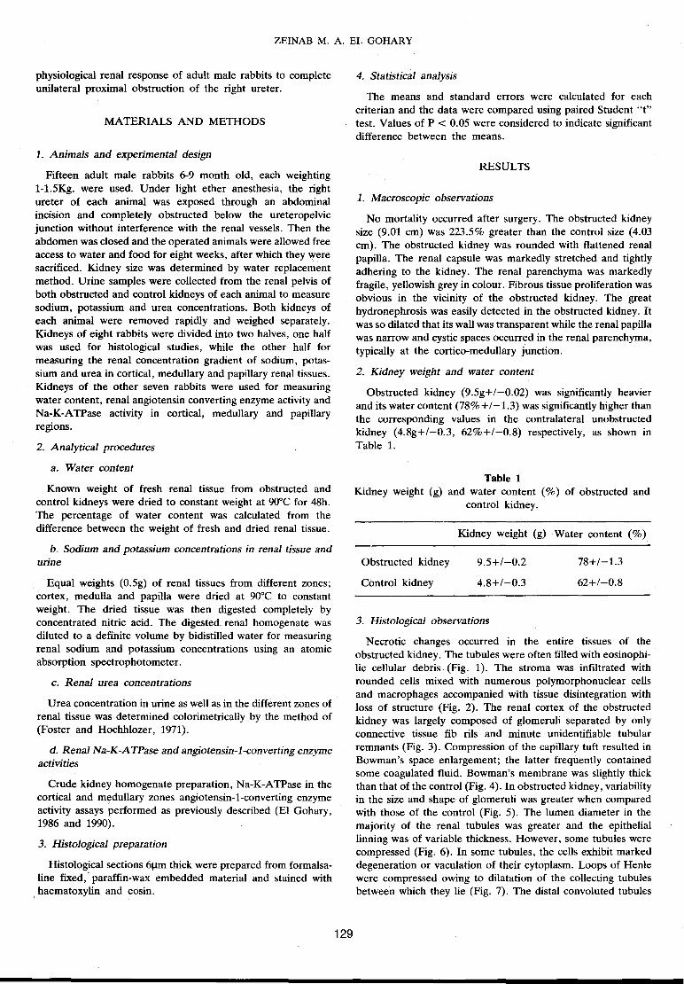
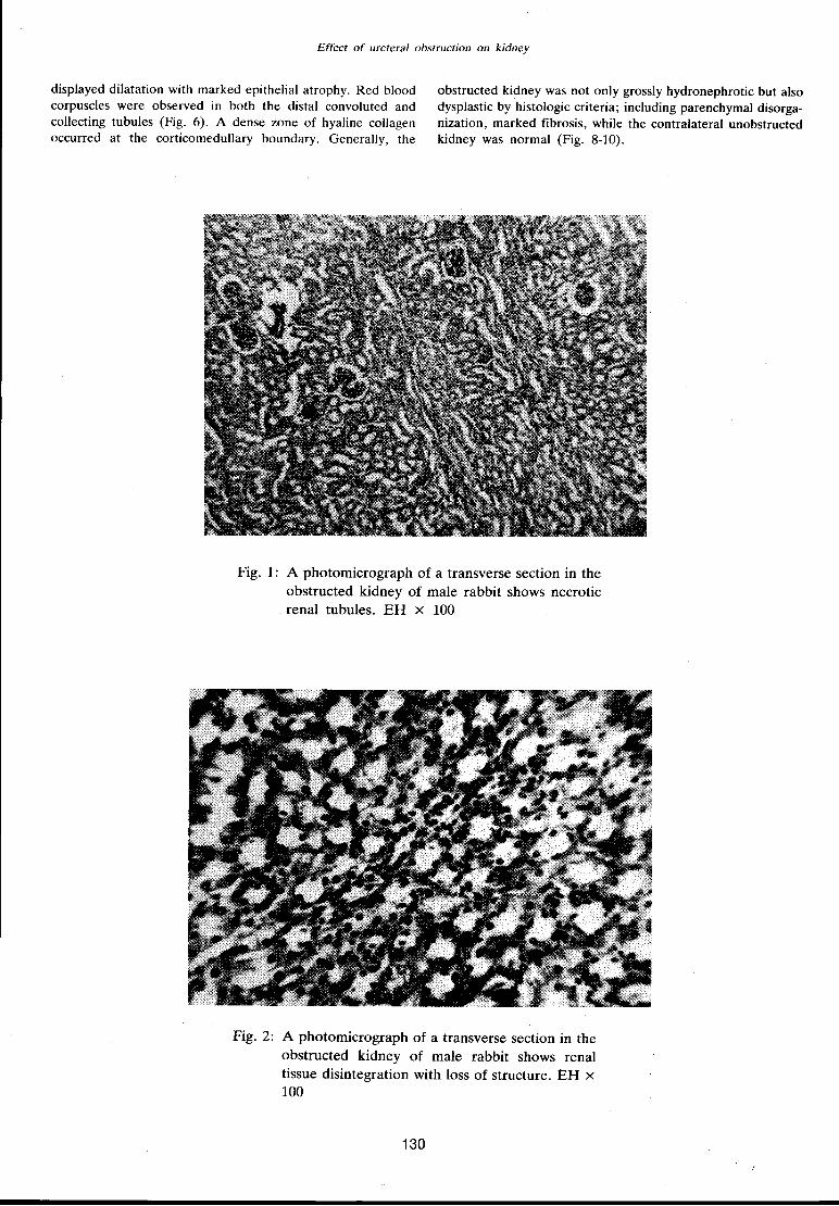
Necrotic changes occurred in the entire tissues of the obstructed kidney. The tubules were often filled with eosinophilic cellular debris (Fig. 1). The stroma was infiltrated with rounded cells mixed with numerous polymorphonuclear cells and macrophages accompanied with tissue disintegration with loss of structure (Fig. 2). The renal cortex of the obstructed kidney was largely composed of glomeruli separated by only connective tissue fib rils and minute unidentifiable tubular remnants (Fig. 3). Compression of the capillary tuft resulted in Bowman's space enlargement; the latter frequently contained some coagulated fluid. Bowman's membrane was slightly thick than that of the control (Fig. 4). In obstructed kidney, variability in the size and shape of glomeruli was greater when compared with those of the control (Fig. 5). The lumen diameter in the majority of the renal tubules was greater and the epithelial tinning was of variable thickness. However, some tubules were compressed (Fig. 6). In some tubules, the cells exhibit marked degeneration or vaculation of their cytoplasm. Loops of Henle were compressed owing to dilatation of the collecting tubules betwee·n which they lie (Fig. 7). The distal convoluted tubules
Effect of ureteral obstruction on kidney
displayed dilatation with marked epithelial atrophy. Red blood corpuscles were observed in both the distal convoluted and collecting tubules (Fig. 6). A dense zone of hyaline collagen occurred at the corticomedullary boundary. Generally, the
obstructed kidney was not only grossly hydronephrotic but also dysplastic by histologic criteria; including parenchymal disorganization, marked fibrosis, while the contralateral unobstructed kidney was normal (Fig. 8-10).
Fig. 1: A photomicrograph of a transverse section in the obstructed kidney of male rabbit shows necrotic renal tubules. EH x 100
Fig. 2: A photomicrograph of a transverse section in the obstructed kidney of male rabbit shows renal tissue disintegration with loss of structure. EH x 100
130
ZEINAB M. A. EL GOHARY
Fig. 3: A photomicrograph of a transverse section in the obstructed kidney of male rabbit shows the renal cortex of the obstructed kidney which was largely composed of glomeruli separated by only copnective tissue fibrils and minute unidentifiable tubular remnants. EH x 100
Fig. 4: A photomicrograph of a transverse section in the obstructed kidney of male rabbit shows enlargement of Bowman's space due to compression of its capillary tuft. EH x 100
131
Effect of ureteral obstruction on kidney
Fig. 5: A photomicrograph of a transverse section in the obstructed kidney of male rabbit shows great variability in the size and shape of glomeruli. EH X 100
Fig. 6: A photomicrograph of a transverse section in the obstructed kidney of male rabbit shows relative great diameter of the majority of renal tubules. EH X 100
132
ZEINAB M. A. EL GOHARY
Fig. 7: A photomicrograph of a transverse section in the obstructed kidney of male rabbit shows dilatation of the collecting ducts and compression of the loops of Henle with marked epithdial necrosis. EH X 100
Fig. 8: A photomicrograph of a transverse section in the control kidney of male rabbit shows normal structure of renal cortex. EH x 100
133
Effect of ureteral obstruction on kidney
Fig. 9: A photomicrograph of a transverse section in the control kidney of male rabbit shows normal appearance of glomeruli, proximal and distal convoluted tubules. EH x 400
Fig. 10: A photomicrograph of a transverse section in the control kidney of male rabbit shows normal structure of renal medulla. EH x 100
134
ZEINAB M. A. EL GOHARY
4. Electrolyte concentrations
a. Urine
Urine sodium concentration from the obstructed kidney (120+/-2.3 mmol/L) was significantly higher (P < 0.001) than that from control kidney (90+/-1.4 mmol/L) (Table 2). On the other hand, both urine potassium (92+/-2.4 mmol/L) and urea (84.5+/-4.8 mM) concentrations from the obstructed kidney were significantly (P < 0.001) lower than the corresponding values (160+/-3.1 mmol/L and 110+/-1.1 mM respectively) in urine from control kidney (Table 2).
Table 2 Sodium (Na), potassium (K) and urea concentrations in urine from obstructed kidney (Obst. K.) and control kidney (Con. K.) as well as in different renal zones (Renal Z.); cortex (Cort.), medulla (Med.) and papilla (Pap.) of obstructed and control
kidney. All data are expressed as means +1-SE.
Exp. parameter
Urine Obst. K.
Con. K.
Renal Z. Obst. K.
Cort.
Med.
Pap.
Con. K.
Cort.
Med.
Pap.
b. Renal tissue
Na. (mmol/L)
120+/-2.3
90+/-1.4
35+/-1.3
72+/-2.6
65.4+/-0.5
52.7+/-1.4
98.9+/-2.1
185.4+/-2.3
K. Urea (mmol/L) (mM)
92+/-2.4 84.5+/-4.8
160+/-3.1 110+/-1.1
60.4+/-2.6 18.3+/-1.1
75.9+/-2.8 120+/-2.1
88.7+/-1.6 140+/-1.5
95.7+/-2.4 29.4+/-1.3
87+/-3.1 103.9+/-2.1
76.9+/-2.5 333.6+/-1.6
In the cortical, medullary and papillary zones, concentration of sodium (35+/-1.3, 72+/-2.6 and 65.4+/-0.5 mmol/g respectively), potassium (60.4+/-2.6, 75.9+/-2.8, 88.7+/-1.6 mmol/g respectively) and urea (18.3+/-1.1, 120.4+/-2.1 and 140+/-1.5 mM respectively) were consistently lower (P < 0.01) for the obstructed kidney than the corresponding values for the control kidney (Sodium, 52.7+/-1.4, 98.9+/-2.1, 185.4+/-2.3 mmol/g respectively; potassium, 95.7+/-2.4, 87+/-3.1, 76.9+/ -2.5 mmol/g respectively; urea, 29.4+/-1.3, 103.9+/-2.1, 333.6+/-1.6 mM respectively) (Table 2).
5. Renal Na-K-ATPase and angiotensine-converting enzyme activities
Renal Na-K-ATPase activity in crude renal homogenate from the cortex and the entire medulla were 3.5+/-0.1 j.lmole Pi/mg protein/hand 6.3+/-1.2 j.lmole Pi/mg protein/h respectively for obstructed kidney. These values were significantly lower (P < 0.001) than the corresponding values (6~8+/-0.1 j.lmole Pi/mg ·protein/h and 22.5+/-0.3 j.lmole Pi/mg protein/h respectively) for control kidney. In contrast, renal angiotensin-1-converting
enzyme (AC) activity (12.3+/-0.3 !Jmole hippuric acid/mg protein/h) in the obstructed kidney homogenate was significantly higher (P < 0.01) than that in the control one (5.1 +/-0.2 !Jmole hippuric acid/mg protcin/h, (Table 3).
ln the present study, obstruction of one ureter provided adequate functional renal mass in the contralateral kidney for survival of the rabbit. This technique also minimized the compensatory hypertrophy occurring in response to the initial reduction in total renal mass. Also, the unobstructed contralateral kidney provided an excellent intra-individual control in the present model.
In the present study, obvious alterations subsequent to this obstruction included marked increase in the obstructed kidney weight probably owing to congestion, edema and accumulation of fluid in the dilated tubules as demonstrated in the high water content.
Also, histological investigations revealed that the interstitial tissue of the obstructed kidney was edematous and exhibited cellular infiltration and fibrosis within the parenchyma of both cortex and atrophied medulla. These histological changes are similar to those described by (Cylwik et al., 1985) in the dog and (Fortyne et al., 1987) in the rat and pig. Elevated pressure in the renal pelvis and calyces consequent to obstruction (Wright, 1982), may be directly responsible for the demonstrated medullary atrophied.
The higher sodium concentration urine from the obstructed rat kidney in the present study is in-agreement with the results of (Klahr, 1983), and such elevation may be attributed to impairment of deep nephron function and distal nephron reabsorption (Wilson, 1972, 1974 and Wilson and Honrath, 1975), as a consequence to destruction of the medullary region. Enhanced prostaglandin (PG) biosynthesis in hydronephrotic kidney (Nishikawa et al., 1977), may explain the reduction in sodium
·reabsorption and naturesis associated with ureter obstruction since prostaglandins are known to be naturetic (Johnson et al., 1967). Elevated sodium in the obstructed kidney urine is paralled with low renal Na-K-ATPase activity, since Na-KATPase is involved in sodium reabsorption (Katz and Epstein, 1968).
135
The lower urinary potassium concentration of the urine from the obstructed kidney may be related to reduction in sodium delivery and in tubular flow rate in the distal nephron (McDougal and Wright, 1972).
Effect of ureteral obstruction on kidney
The low urea concentration in urine from the obstructed kidney reflects its poor concentrating ability. Also increased urea reabsorption in the distal nephron may explain the fall in urea concentration in the urine from obstructed kidney.
In the present study, tissue concentration gradient of sodium, potassium and urea between the cortex and papillary tip was abolished in the obstructed kidney. The results are in agreement with the findings of (Bedyne and Macken, 1962; Eknoyan et al., 1970; and Suki et al., 1971) in dogs and Honda et al., (1971) in rabbits. Further support comes from the study of (Hanley and Davidson, 1982), who reported a marked decrease in chloride transport in the thick ascending limb of Henle's loops microdissected from obstructed rat kidneys and perfused in vitro.
Lowered renal Na, K and urea concentrations in the different renal zones of the obstructed kidney is related either to decrease in the amount of sodium and potassium transported into the interstitium as a result of the non-filtering nephrons, a consequence to the elevated ureteral pressure (Malvin et al., 1964; DalCanton et al., 1980 and Wright, 1982), or to washout of solute from interstitium due to elevated medullary blood flow (Wilson, 1980 and Klahr, 1983). Also, a defect in the transtubular transport of NaCI in the ascending limb of the loop of Henle in the obstructed kidney (McDougal and Persky, 1975) would lower the tonicity of the medullary interstitium and, thus, the osmotic driving force for water movement from the collecting duct into the interstitium (Schmidt-Nielsen and O'Dell, 1961).
The results of this study demonstrate a striking inhibition of renal Na-K-ATPase activity in both cortex and medulla of the obstructed kidney, which was more marked in the medullary zone. Such reduction is consistent with the demonstrated severe necrotic tubular epithelium, where the enzyme localized. The detected decrease in renal Na-K-ATPase activity of obstructed kidney are comparable with the results of Williams et al. (1976), on dogs and Wilson et al. ( 1974, 1978) on rats. In agreement with the present results, Eknoyan et al. (1970) and Buerkert et al. (1977) reported impairment in the maximal concentrating ability and capacity to reabsorb solute -free water in obstructed rat kidney whereas Na-K-ATPase plays an important role in the bulk reabsorption of sodium by the kidney (Katz and Epstein, 1968).
The demonstrated elevation in the angiotensin-1-converting enzyme activity in the obstructed rat kidney in the present study is comparable with the results of Kaloyanides et al., (1973), who reported increased renal renin release with ureteral obstruction in rats and with the findings of Wideman and Gregg, (1988), who reported reduction in glomerular filtration rate (GFR), consequence to ureteral obstruction in the chick. Also, the present results are consistent with the studies of Nishikawa et al., (1977), who demonstrated increased prostaglandin synthesis in the hydronephrotic isolated perfused kidney and with the findings of Yarger and Harris, (1978) and Yarger et al., (1980), who noted improvement of GFR and renal blood flow in the obstructed rat kidney after administration of an angiotensin-1-converting enzyme inhibitor, captopril.
REFERENCES
Bander, S.G., J.E. Buerkert; D. Marttin and S. Klahr, 1985. Long-term effect of 24 hours unilateral ureteral obstruction on renal function in the rat. Kidney Int. 28: 614-620.
136
Berlyne, G.M. and A. Macken, 1962. On the mechanism of renal inability to produce a concentrated urine in chronic hydronephrosis. Clin. Sci. 22: 315-321.
Buerkert, J., M. Head and S. Klahr, 1977. Effects of acute bilateral ureteral obstruction on deep nephron and terminal collecting duct function in the young rat. J. Clin. Invest. 59: 1055-1064.
Cylwik, B., J. Darewicz and B. Karasewicz, 1985. Morphometric investigations of dog kidney infracts after embolization of the renal artery with spongostan. Int. Urol. Nephrol. 17: 109-112.
Dal Canton, A., A. Corradi and R. Stanziale, 1980. Glomerular hemodynamics before and after release of 24 hours bilateral ureteral obstruction. Kidney Int. 17: 491-496.
Eknoyan, G., W.N. Suki, M. Martinez-Maldonado and M.A. Anhalt, 1970. Chronic hydronephrosis: observations on the mechanism of the defect in urine concentration. Proc. Soc. Exp. Bioi. Med. 134: 634-639.
El Gohary, Z.M.A., 1986. Water and electrolytes turnovers with special reference to high dietary sodium intake. Ph. D. Thesis Sheffield Univ. England.
El Gohary, Z.M.A., 1990. Anatomical adaptation of green toad (Bufo-viridis) to a ride environment. J. Agric. Sci. Mansoura Univ. 15: 170-177.
Fortyn, K., J. Hradecky, V. Hruban, P. Dvorak and J. Tichy, 1987. Morphology of regressive changes in the kidney following experimental ischaemia. Int. Urology Neph. 19: 9-19.
Foster, L.B. and J.M. Hochholzer, 1971. A single reagent annual method for directly determining urea nitrogen in serum. J. Clin. Chern., 17: 971.
Hanley, M.J. and K. Davidson, 1982. Isolated nephron segments from rabbit models of obstructive nephropathy. J. Clin. Invest. 69: 165-174.
Honda, N., C. Aizawa, A. Morikawa andY. Yoshitoshi, 1971. Effect of elevated ureteral pressure on renal medullary osmolal concentration in hydronephrotic rabbits. Am. J. Physiol. 221: 698-703.
Johnson, H.H., J.P. Herzog and D.P. Lanier, 1967. Effect of prostaglandin El on renal hemodynamics, sodium and water excretion. Am. J. Physiol. 213: 939-946.
Kaloyanides, G.J., R.D. Bastron and G. DiBona, 1973. Effect of ureteral clamping and increased renal arterial pressure on renin release. Am. J. Physiol. 225: 95-104.
Katz, A. I. and F .H. Epstein, 1968. Physiological role of sodium-potassium-activated adenosine triphosphatase in the transport of cations across biologic membranes. N. England. J. Med. 278: 253-262.
Malvin, R.L., H. Kutchai and F. Ostermann, 1964. Decreased nephron population resulting from increased ureteral pressure. Am. J. Physiol. 207: 835-839.
McDougal, W.S. and F.S. Wright, 1972. Defect in proximal and distal sodium transport in post-obstructive diuresis. Kidney. Int. 2: 304-317.
McDougal, W.S. and L. Persky, 1975. Renal functional abnormalities in post-unilateral ureteral obstruction in man: A comparison of these defects to post-obstructive diuresis. J. Urol. 113: 601-109.
Nishikawa, K., A. Morrison and P. Needleman, 1977. Exaggerated prostaglandin biosynthesis and its influence on renal resistance in the isolated hydronephrotic rabbit kidney. J. Clin. Invest. 59: 1143-1150.
ZEINAB M. A. EL GOHARY
Paltiel, H.J. and R.L. Lebowitz, 1989. Neonatal hydronephrosis due to primary vesicoureteral reflux: trends in diagnosis and treatment. Pediatric Radiology, 170: 787-789.
Schmidt-Nielsen, B and R. O'Dell, 1961. Structure and concentrating mechanism in the mammal kidney. Am. J. Physiol., 200: 1119-1124.
Suki, W.N., A.G. Guthrie and M. Martinez-Maldonado, 1971. Effect of ureteral pressure elevation on renal hemodynamics and urine concentration. Am. J. Physiol. 220: 28-32.
Susie, D., J.C. Sparks and D. Kentera, 1975. The renin angiotensin system in rats with hereditary hydronephrosis. Pflugers Arch. 358: 265-274.
Wideman, R.F. and C.M. Gregg, 1988. Model for evaluating avian renal hemodynamics and glomerular filtration rate autoregulation. Am. J. Physiol. 254: 925-932.
Williams, R.D., D.D. Fanestil and C.E. Blackard, 1976. Etiology of postobstructive diuresis: Oubain-sensitive adenosine triphosphatase deficit and elevated solute excretion in the postobstructed dog kidney. Invest. Urol. 14: 148-152.
Wilson, D.R., 1972. Micropuncture study of chronic obstructive nephropathy before and after release of obstruction. Kidney Int. 2: 119-122.
Wilson, D.R., 1974. The influence of volume expansion on renal function after relief of chronic unilateral ureteral obstruction. Kidney Int. 5: 402-410.
Wilson, D.R., W. Knox, E. Hall and A.K. Sen, 1974. Renal sodium -and potassium -activated adenosin triphosphatase deficiency during post-obstructive diuresis in the rat. Cand. J. Physiol. Pharmacol. 52: 105-113.
Wilson, D.R. and U. Honrath, 1975. Nephron functional heterogeneity in the postobstructive kidney. Kidney Int. 7: 19-23.
Wilson, D.R., W. Knox, E. Hall and A.K. Sen, 1978. Postobstructive nephropathy in the rat. Relationship between Na-K-ATPase activity and renal function. Nephron 22:55-62.
Wright, F.S., 1982. Effects of urinary tract obstruction on glomerular filtration rate and renal blood flow. In Obstructive Uropathy. Edit¢d by Klahr, S. New York Grune and Stratton Inc. 1982, pp. 5-16.
Yarger, W.E. and R.H. Harris, 1978. Angiotensin 11, a vasoconstrictor in the postobstructive kidney of the rat ( abst). Kidney Int. 14: 735.
Yarger, W.E., D.D. Shocken and R.H. Harris, 1980. Obstructive nephropathy in the rat: possible roles for the renin -angiotensin system, prostaglandins, and thromboxanes in postobstructive renal function. J. Clin. Invest. 65: 400-409.