Hindawi Publishing CorporationJournal of TransplantationVolume 2011, Article ID 581485, 5 pagesdoi:10.1155/2011/581485
Clinical Study
Renal Transplantation in Hepatitis C Positive Patients:A Single Centre Experience
P. R. Shah,1 A. V. Vanikar,2 M. R. Gumber,1 H. V. Patel,1
V. B. Kute,1 S. M. Godara,1 and H. L. Trivedi1
1 Department of Nephrology and Transplantation Medicine, IKDRC-ITS, Ahmedabad 380016, India2 Department of Pathology, Laboratory Medicine, Transfusion Services and Immunohematology, IKDRC-ITS,Ahmedabad 380016, India
Introduction. Hepatitis C virus (HCV) infection is an independent risk factor for renal transplantation (RTx). Immunosuppressionminimization can render better quality of life to these patients. Methods. We analyzed 132 HCV-positive RTx patients (group A)transplanted under tolerance induction protocol (TIP) and compared them with 79 controls (group B) transplanted using standardtriple drugs. TIP consisted of 1 donor-specific transfusion, peripheral blood stem cell infusion, portal infusion of bone marrow, andtarget-specific irradiation. Their immunosuppression was cyclosporin, 2 ± 1 mg/kgBW/day + prednisone, 10 mg/day. Results. TIPhad no side effects. Although unequal in size, the groups were well balanced. Group A patient survival at 1, 5, and 10 years was92.4%, 70.4%, and 63.7%, respectively, versus 75.6%, 71.7%, and 55.7% in later, and graft survival was 92.9%, 81.5%, and 79.1%versus 91.7%, 75.7%, and 67.7%, respectively. Mean serum creatinine (mg/dL) at these time periods in former was 1.38, 1.72, and1.87, versus 1.3, 1.75, and 2.1 in later. Altered liver functions were noted in 22% patients in former versus 31% in later. Group Ahad lesser rejection episodes. Conclusion. RTx using TIP in HCV-positive patients is a viable option with acceptable outcome.
1. Introduction
Hepatitis C virus (HCV) infection affects 20–50% of end-stage renal disease (ESRD) patients and contributes signifi-cantly to morbidity and mortality following renal transplan-tation (RTx) [1, 2]. Approximately 8–28% of RTx patientsdie due to chronic liver disease [3]. HCV infection has aprevalence of about 2.6–66% among RTx patients in differentcountries with great genotype diversity in different parts ofthe world [3]. Antiviral drugs used for the management ofHCV can have graft-threatening effects. In addition immu-nosuppressants themselves can be life threatening to suchpatients thereby putting the treating doctor in a seriousdilemma, and the patient in precarious position. In such sit-uation transplantation after adequate antiviral therapy follo-wed by minimal immunosuppression can be a good option.
This is a retrospective analysis of RTx carried out inour center between 1998 to 2006 in HCV-positive patientsusing specially designed tolerance induction protocol (TIP).
Standard RTx were compared to evaluate graft function andgraft/patient survival.
2. Materials and Methods
We analyzed medical records of HCV-positive patients(tested by third-generation enzyme linked immunoassay(ELISA)) who underwent RTx in our center from April1998–2006 after adequate treatment. Patients were dividedinto 2 groups; group 1 who were transplanted after TIP withlow-dose immunosuppression and group 2 who opted outof TIP and were transplanted under standard triple drug im-munosuppression.
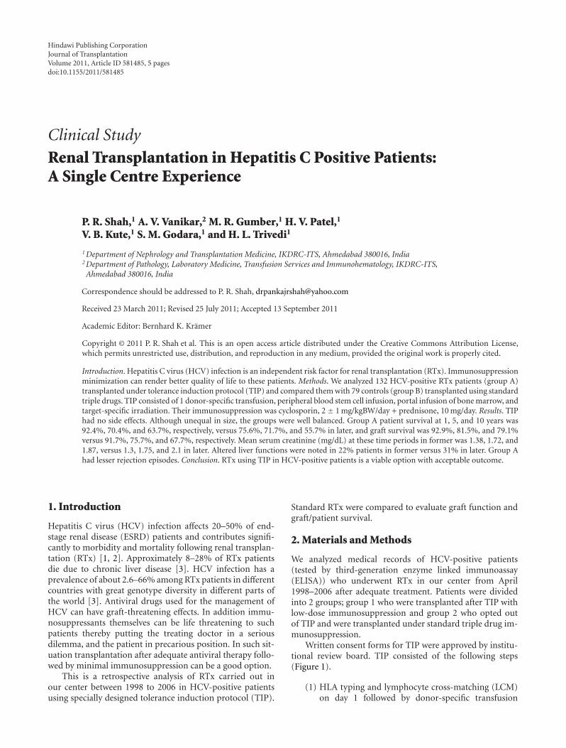
Written consent forms for TIP were approved by institu-tional review board. TIP consisted of the following steps(Figure 1).
(1) HLA typing and lymphocyte cross-matching (LCM)on day 1 followed by donor-specific transfusion
2 Journal of Transplantation
TSI800 CGy200× 4
Portal infusion-BM
PBSC
Renal Tx
5–8 101 153 12
LCMDays
Steroid, CsA 3
LCM,DST
periphery
mg/kg BW/day
Figure 1: Tolerance induction paradigm for HCV-positive patients.
(DST; buffy coat infusion) in to RTx patient afterbleeding 330 mL of blood from donor.
(2) Mobilization of donor stem cells by leucophoresis ofdonor on day 3 after stimulating with granulocytecolony stimulating factor (G-CSF), 7.5 microgram/kg BW/day subcutaneously for 2 days (days 1, 2) andinfusing the collected peripheral blood stem cells(PBSC) in to recipient’s periphery.
(3) Target-specific irradiation to subdiaphragmatic lym-ph nodes, spleen, part of pelvic bones, and lumbarvertebrae (200 CGY× 4 days) on days 5 to 8. Unmod-ified cytokine stimulated donor bone marrow (BM;200 mL) infusion intraportally on day 10. Portal infu-sion technique of BM was carried out by omental veincannulation under general anesthesia.
(4) LCM on day 12.
(5) RTx on day 15 with favorable LCM.
(6) Peritransplant immunosuppression induction of in-travenous methylprednisone, 500 mg on day beforetransplant, on day of transplant, 500 mg on 1st post-operative day (POD), and then switched over tooral prednisone, 30 mg/day, tapered to 10 mg/day bythe end of 3 months to be continued thereafter.Cyclosporin (CsA), 3 mg/kg BW/day from day beforetransplant to be continued thereafter by monitoringtrough levels.
No immunological preconditioning of the recipient wasdone. Graft-versus-host disease (GvHD) was ruled out bymonitoring absence of skin rashes, gastrointestinal symp-toms, abnormal liver function tests, and evidence of BM sup-pression.
2.1. HLA Typing and LCM. HLA typing and LCM were doneby conventional serological technique (one lambda predottrays were used for HLA A, B, and DR typing). LCM was doneby serological method using auto dithiothreitol and standardcytotoxicity methods with T and B lymphocytes each.
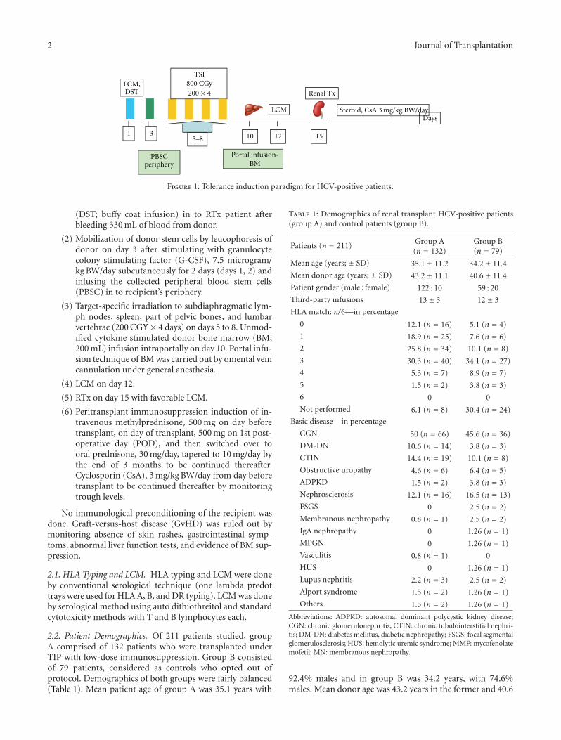
2.2. Patient Demographics. Of 211 patients studied, groupA comprised of 132 patients who were transplanted underTIP with low-dose immunosuppression. Group B consistedof 79 patients, considered as controls who opted out ofprotocol. Demographics of both groups were fairly balanced(Table 1). Mean patient age of group A was 35.1 years with
Table 1: Demographics of renal transplant HCV-positive patients(group A) and control patients (group B).
Patients (n = 211) Group A(n = 132)
Group B(n = 79)
Mean age (years; ± SD) 35.1± 11.2 34.2± 11.4
Mean donor age (years; ± SD) 43.2± 11.1 40.6± 11.4
92.4% males and in group B was 34.2 years, with 74.6%males. Mean donor age was 43.2 years in the former and 40.6
Journal of Transplantation 3
0 1 2 3 4 5 6 7 8 9 10 11 12 13
Time (years)
0
0.1
0.2
0.3
0.4
0.5
0.6
0.7
0.8
0.9
1
Cu
msu
rviv
al
Group BGroup A
Group B censoredGroup A censored
(a)
Group BGroup A
Group B censoredGroup A censored
0 1 2 3 4 5 6 7 8 9 10 11 12 13
Time (years)
0
0.1
0.2
0.3
0.4
0.5
0.6
0.7
0.8
0.9
1
Cu
msu
rviv
al
(b)
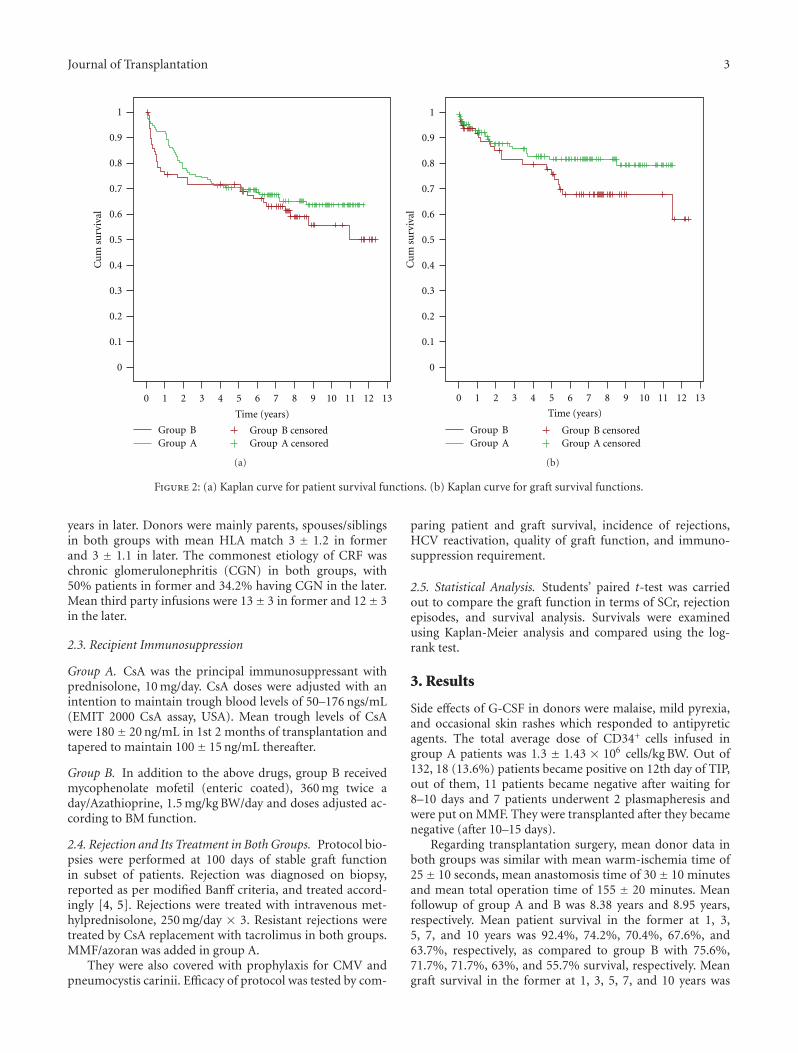
Figure 2: (a) Kaplan curve for patient survival functions. (b) Kaplan curve for graft survival functions.
years in later. Donors were mainly parents, spouses/siblingsin both groups with mean HLA match 3 ± 1.2 in formerand 3 ± 1.1 in later. The commonest etiology of CRF waschronic glomerulonephritis (CGN) in both groups, with50% patients in former and 34.2% having CGN in the later.Mean third party infusions were 13± 3 in former and 12± 3in the later.
2.3. Recipient Immunosuppression
Group A. CsA was the principal immunosuppressant withprednisolone, 10 mg/day. CsA doses were adjusted with anintention to maintain trough blood levels of 50–176 ngs/mL(EMIT 2000 CsA assay, USA). Mean trough levels of CsAwere 180± 20 ng/mL in 1st 2 months of transplantation andtapered to maintain 100± 15 ng/mL thereafter.
Group B. In addition to the above drugs, group B receivedmycophenolate mofetil (enteric coated), 360 mg twice aday/Azathioprine, 1.5 mg/kg BW/day and doses adjusted ac-cording to BM function.
2.4. Rejection and Its Treatment in Both Groups. Protocol bio-psies were performed at 100 days of stable graft functionin subset of patients. Rejection was diagnosed on biopsy,reported as per modified Banff criteria, and treated accord-ingly [4, 5]. Rejections were treated with intravenous met-hylprednisolone, 250 mg/day × 3. Resistant rejections weretreated by CsA replacement with tacrolimus in both groups.MMF/azoran was added in group A.
They were also covered with prophylaxis for CMV andpneumocystis carinii. Efficacy of protocol was tested by com-
paring patient and graft survival, incidence of rejections,HCV reactivation, quality of graft function, and immuno-suppression requirement.
2.5. Statistical Analysis. Students’ paired t-test was carriedout to compare the graft function in terms of SCr, rejectionepisodes, and survival analysis. Survivals were examinedusing Kaplan-Meier analysis and compared using the log-rank test.
3. Results
Side effects of G-CSF in donors were malaise, mild pyrexia,and occasional skin rashes which responded to antipyreticagents. The total average dose of CD34+ cells infused ingroup A patients was 1.3 ± 1.43 × 10⁶ cells/kg BW. Out of132, 18 (13.6%) patients became positive on 12th day of TIP,out of them, 11 patients became negative after waiting for8–10 days and 7 patients underwent 2 plasmapheresis andwere put on MMF. They were transplanted after they becamenegative (after 10–15 days).
Regarding transplantation surgery, mean donor data inboth groups was similar with mean warm-ischemia time of25± 10 seconds, mean anastomosis time of 30± 10 minutesand mean total operation time of 155 ± 20 minutes. Meanfollowup of group A and B was 8.38 years and 8.95 years,respectively. Mean patient survival in the former at 1, 3,5, 7, and 10 years was 92.4%, 74.2%, 70.4%, 67.6%, and63.7%, respectively, as compared to group B with 75.6%,71.7%, 71.7%, 63%, and 55.7% survival, respectively. Meangraft survival in the former at 1, 3, 5, 7, and 10 years was
4 Journal of Transplantation
Table 2: Results.
Patients (n = 211) Group A (n = 132) Group B (n = 79) P value
Study period Jan 99–Dec 06 Jan 98–Dec 06
Mean followup (years; range) 8.38± 2.2 (3.8–11.8) 8.95± 2.2 (4–12.6)
92.9%, 85.6%, 81.5%, 81.5%, 79.1%, respectively, as com-pared to group B with 91.7%, 81.2%, 75.7%, 67.7%, 67.7%,respectively. Kaplan Meier graphs of patient and graft sur-vival are shown in Figures 2(a) and 2(b).
Graft function in terms of SCr (in mg/dL) in both groupsat 1, 3, 5, 7, and 10 years was 1.38± 0.29, 1.55± 0.34, 1.72±0.47, 1.8±0.39, and 1.87±0.69, versus 1.3±0.37, 1.58±0.64,1.75±0.61, 1.97±0.73, and 2.1±0.81 in group B. Liver func-tion status was deranged and accompanied by presence ofHCV-RNA (tested by PCR and 1 to 5 million copies/mL withmean rate of 1.65 ± 0.75 copies/mL in TIP and 2.3 ±2.05 copies/mL in controls were noted) in 22% (n = 29) pa-tients of group A out of which 8.3% (n = 11) succumbedto chronic liver failure and in 31% (25 patients) of group B,16.5% (13 patients) died of chronic liver failure. Group Apatients had better graft and patient survival along with graftfunction status as compared to group B (statistically not sig-nificant).
In terms of rejection there was statistically significantdecrease in T-cell-mediated rejections and chronic changesin the former as compared to the later (Table 2). Majorityof the patients responded to antirejection therapy. However4/132 (about 3%) in TIP group and 4/79 (about 5%) incontrol group did not respond and eventually lost their grafts
to chronic dysfunction and eventually succumbed to secon-dary infections and septicemia. Incidence of reactivation ofHCV was also significantly less in the former as comparedto controls. In TIP group totally 45 (34%) patients and incontrols 32 (40.5%) were lost over a followup of 12 years.Out of these 11 in TIP and 13 in controls succumbed to liverfailure, others to chronic graft dysfunction-related morbidityor to septicemia. The other advantage in group A was signifi-cantly less requirement of maintenance immunosuppressionin the form of CsA, 2 ± 1 mg/kg BW and prednisone, 5–10 mg/day versus group B with standard triple drug im-munosuppression.
4. Discussion
The effect of pretransplant HCV infection on survival of pa-tients and grafts in RTx is controversial [6]. However, sur-vival is better in HCV RTx patients as compared to dialysis[7].The goals of pretransplantation HCV therapy are todecrease the risk for progression of HCV-associated liverdisease, stabilize renal function in patients with HCV-rela-ted glomerulopathy, and prevent development of HCV-as-sociated disease after transplantation [8]. The use of immu-nosuppression predisposes RTx patients to risks of deranged
Journal of Transplantation 5
liver functions and mortality [9, 10]. A meta-analysis ofnatural history of HCV in 6365 RTx patients showed thatanti-HCV antibody was an independent risk factor for deathand graft failure with relative risk of 1.79 [11]. In our centerwe offer TIP to all patients, and we start the protocol onlyafter informed consent form is signed by patient, donor,and witness. However, all donors are not willing to undergostimulation protocols, abdominal fat resection, BM aspira-tion, and above all they are not willing to wait till renaltransplantation. We explain to them that it may take a monthor little longer for transplantation, to finish the protocol andif patient becomes lymphocyte cross-match positive, waitingperiod can become longer. Many patients and donors cannotget leave from their work for such a long period even if theydo not have to stay in hospital, they need to visit us fre-quently which they are not willing. With minimization of im-munosuppression, patients are at lower risk of infections andhence return to better quality of life. Secondly lowering ofrejection incidence and severity automatically saves financialburden, especially in India where there is no financial sup-port from government medicare/medical insurance. WithTIP, use of less number and low-dose of drugs brings downthe cost, though we have not touched upon this aspect here.We have more than 10 years of experience of using TIP inabout 1500 patients and hence we modified it for HCV-positive patients and implemented it [12]. Our study showsthat with use of tolerance induction protocol for HCV-posi-tive patients, quality of life, graft function, and survival arereasonably good even for a long period of ten years. Our con-trol (group B) patients have reasonable quality of graft func-tion and survival as found in other studies [6–11]. Interest-ingly, tolerance induction protocol yielded significantly lesschances of reactivation of HCV as compared to controls. Thiscould be attributed to better immune competence in thesepatients since they require less immunosuppression.
5. Conclusion
RTx is an acceptable option for HCV-positive patients withESRD, and tolerance induction protocol is preferable overstandard triple drug immunosuppression in these group ofpatients.
Acknowledgment
The authors are indebted to Ms. Priyardarshini Shah and Ms.Shobhanarani Sengunthar for data acquisition and analysis.
References
[1] B. J. G. Pereira and A. S. Levey, “Hepatitis C virus infection indialysis and renal transplantation,” Kidney International, vol.51, no. 4, pp. 981–999, 1997.
[2] D. Roth, “Hepatitis C virus: the nephrologist’s view,” AmericanJournal of Kidney Diseases, vol. 25, no. 1, pp. 3–16, 1995.
[3] S. M. H. Moghaddam, S. M. Alavian, and N. A. Kermani,“Hepatitis C and renal transplantation: a review on historicalaspects and current issues,” Reviews in Medical Virology, vol.18, no. 6, pp. 375–386, 2008.
[4] L. C. Racusen, R. B. Colvin, K. Solez et al., “Antibody-me-diated rejection criteria—an addition to the Banff ’97 classifi-cation of renal allograft rejection,” American Journal of Trans-plantation, vol. 3, no. 6, pp. 708–714, 2003.
[5] K. Solez, R. B. Colvin, L. C. Racusen et al., “Banff ’05 meetingreport: differential diagnosis of chronic allograft injury andelimination of chronic allograft nephropathy (‘CAN’),” Ameri-can Journal of Transplantation, vol. 7, no. 3, pp. 518–526, 2007.
[6] B. Einollahi, B. Hajarizadeh, S. Bakhtiari et al., “Pretransplanthepatitis C virus infection and its effect on the post-transplantcourse of living renal allograft recipients,” Journal of Gastroen-terology and Hepatology, vol. 18, no. 7, pp. 836–840, 2003.
[7] G. A. Knoll, M. R. Tankersley, J. Y. Lee, B. A. Julian, and J.J. Curtis, “The impact of renal transplantation on survival inhepatitis C- positive end-stage renal disease patients,” Amer-ican Journal of Kidney Diseases, vol. 29, no. 4, pp. 608–614,1997.
[8] N. A. Terrault and D. B. Adey, “The kidney transplant recipientwith hepatitis C infection: pre- and posttransplantation treat-ment,” Clinical Journal of the American Society of Nephrology,vol. 2, no. 3, pp. 563–575, 2007.
[9] J. M. Morales and J. M. Campistol, “Transplantation in the pa-tient with hepatitis C,” Journal of the American Society of Neph-rology, vol. 11, no. 7, pp. 1343–1353, 2000.
[10] F. Fabrizi, P. Martin, and C. Ponticelli, “Hepatitis C virus infec-tion and renal transplantation,” American Journal of KidneyDiseases, vol. 38, no. 5, pp. 919–934, 2001.
[11] F. Fabrizi, P. Martin, V. Dixit, S. Bunnapradist, and G.Dulai, “Hepatitis C virus antibody status and survival afterrenal transplantation: meta-analysis of observational studies,”American Journal of Transplantation, vol. 5, no. 6, pp. 1452–1461, 2005.
[12] A. V. Vanikar, K. R. Goplani, A. Feroz et al., “Operational tol-erance in living-related renal transplantation: a single-centerexperience,” Transplantation Proceedings, vol. 43, no. 5, pp.1551–1558, 2011.