Repair of replication errors by the MisMatch Repair System: Marking newly synthesized DNA in E. coli * GATC normally methylated on the A CTAG * • Newly synthesized strands not methylated right away, delayed for ~10 minutes: gives hemi -methylated DNA * GATC CTAG Hemi-methylated DNA: 1. Not recognized by the oriC activation system 2. Recognized by the Mismatch Repair System
Transcript
Repair of replication errors by the MisMatch Repair System: Marking newly synthesized DNA in E. coli
*GATC normally methylated on the ACTAG *• Newly synthesized strands not methylated right away, delayed for ~10 minutes: gives hemi-methylated DNA
* GATC CTAG
Hemi-methylated DNA:1. Not recognized by the oriC activation system2. Recognized by the Mismatch Repair System
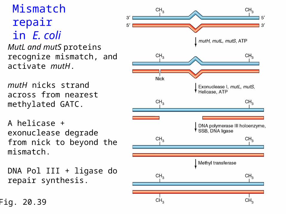
Fig. 20.39
Mismatch repairin E. coli
MutL and mutS proteins recognize mismatch, and activate mutH.
mutH nicks strand across from nearest methylated GATC.
A helicase + exonuclease degrade from nick to beyond the mismatch.
DNA Pol III + ligase do repair synthesis.
Mismatch Repair
• Repairs replication errors that create mismatches• In E. coli, new DNA not methylated right away
– mismatch recognized by mutS, then mutL binds and attracts mutH (endonuclease that cleaves nearest CTAG that is not methylated)
• Eucaryotes have mutS and mutL homologues, but no mutH
– also have the requisite exonuclease, but not clear how the strand specificity is determined
Mismatch Repair and Colon Cancer
• Hereditary nonpolyposis colon cancer (HNPCC) • 1/200 Americans is affected (15% of colon cancers)• Characterized by microsatellite instability:
1. Microsatellites are tandem repeats of 1-4 bp sequences that change during lifetime of HNPCC patients
2. Microsatellites are prone to replication slippage resulting in insertions or deletions, which are normally repaired by the Mismatch Repair (MMR) System
• Mutations in one of 5 mismatch repair (MMR) genes increase susceptibility to HNPCC
Mammalian Mitochondrial DNA (MtDNA)
1. Multi-copy, circular molecule of ~16,000 bp. Uniparental-maternal inheritance.
2. Encodes genes for respiration (13 proteins) and translation (22 tRNAs, 2 rRNAs).
3. 2 promoters (1 on each strand); the STOP codons for the protein genes, UAA, created post-transcriptionally by polyadenylation
4. Some genetic diseases caused by mutations in mtDNA. Also, MtDNA mutations accumulate during aging.
5. MtDNA used to define phylogenetic relationships between species, subspecies, etc., or define breeding populations.
Mammalian Mt DNA
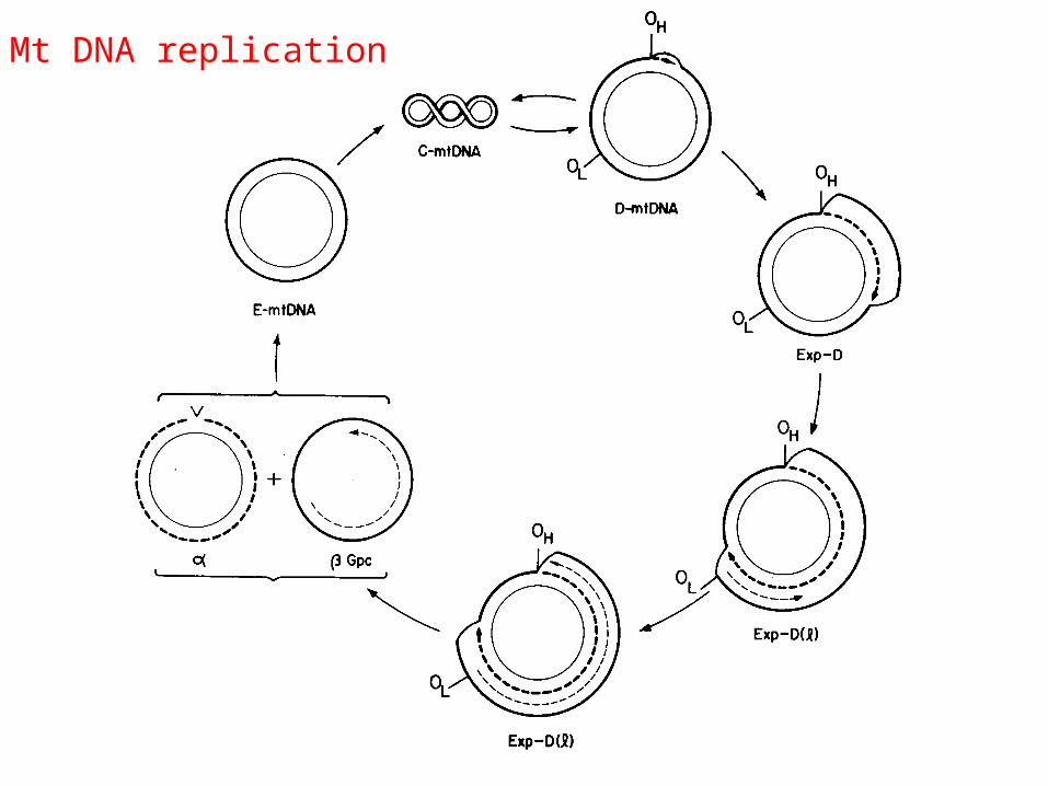
Mt DNA replication
1. Two origins of replication: H (for heavy strand) and L (for light strand) that are used sequentially for
unidirectional replication (from each origin).
2. Persistent D-loop at H ori, which is extended to start replication of the H strand.
3. Once ~2/3 of H strand is replicated, L ori is exposed and replication of L strand starts.
4. The lagging L strand replication gives 2 type of molecules: and is gapped on L strand.
5 L strand finishes replicating, and then both and are converted to supercoiled forms.
Mammalian (mouse) mtDNA Replication
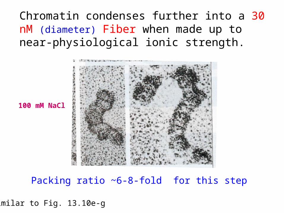
Condensing and Packaging of DNA into a small space is a universal feature
of cells and other genetic systems.
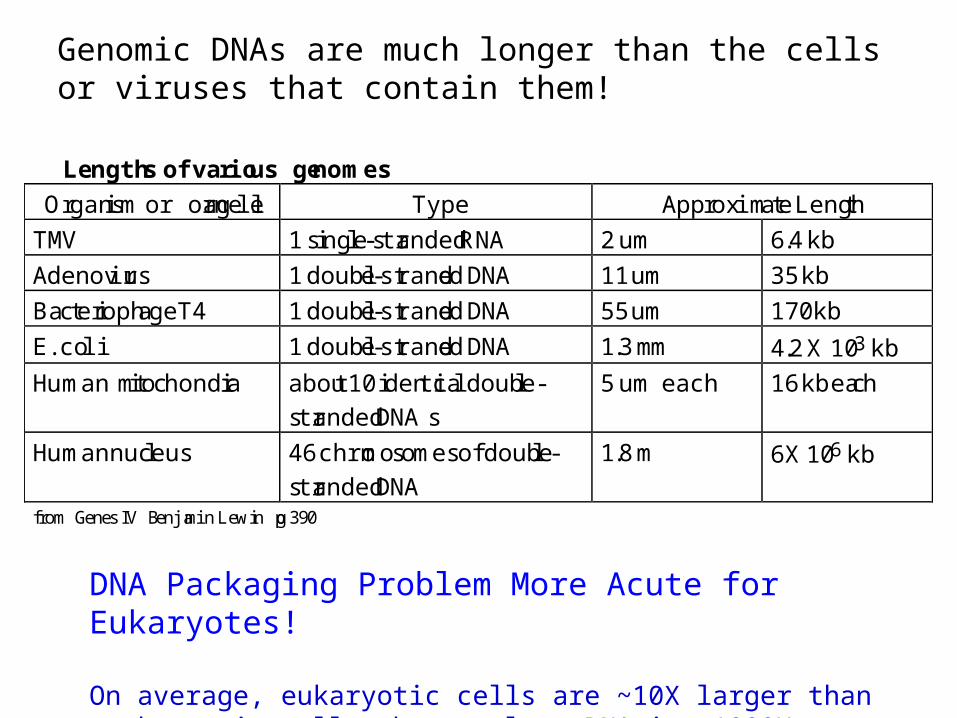
Lengths of various genomes
Organism or organelle Type Approximate Length
TMV 1 single-stranded RNA 2 um 6.4 kb
Adenovirus 1 double-stranded DNA 11 um 35 kb
Bacteriophage T4 1 double-stranded DNA 55 um 170 kb
E. coli 1 double-stranded DNA 1.3 mm 4.2 X 10³ kb
Human mitochondria about 10 identical double-
stranded DNAs
5 um each 16 kb each
Human nucleus 46 chromosomes of double-
stranded DNA
1.8 m 6X 10⁶ kb
from Genes IV Benjamin Lewin pg 390
Genomic DNAs are much longer than the cells or viruses that contain them!
DNA Packaging Problem More Acute for Eukaryotes!
On average, eukaryotic cells are ~10X larger than prokaryotic cells, but nuclear DNA is ~1000X larger than bacterial DNA.
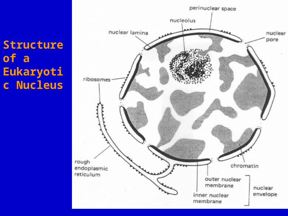
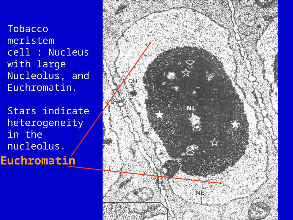
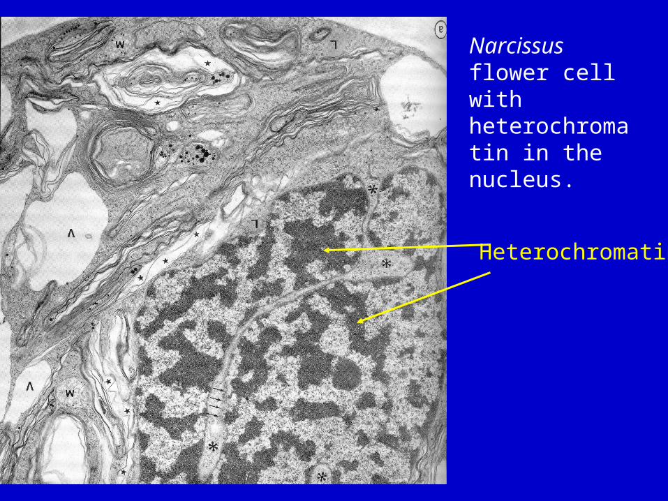
Structure of a Eukaryotic Nucleus
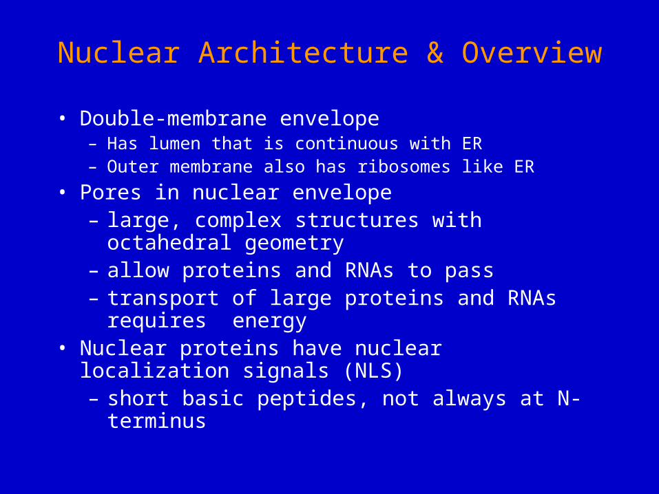
Nuclear Architecture & Overview
• Double-membrane envelope– Has lumen that is continuous with ER– Outer membrane also has ribosomes like ER
• Pores in nuclear envelope– large, complex structures with octahedral
geometry – allow proteins and RNAs to pass– transport of large proteins and RNAs requires
energy• Nuclear proteins have nuclear localization signals
(NLS)– short basic peptides, not always at N-terminus
Nuclear architecture (cont.)
• nuclear skeleton (or lamina)– intermediate filaments (lamins)– anchor DNA and proteins (i.e., chromatin) to
envelope• Nucleolus
– site of pre-rRNA synthesis and ribosome assembly
Electron microscopic views of pores in the nuclear envelope.Freeze-fracture EM Transmission EM (TEM)


![Reversible Manipulation of Supramolecular Chirality using ... · molecular system is based on discovery of serendipity among numerous newly synthesized molecules,[6b,7a] anditisstilla](https://static.documents.pub/doc/80x56/5f64e54208025e205533969c/reversible-manipulation-of-supramolecular-chirality-using-molecular-system-is.jpg)
