53
Reproductive Reproductive Anatomy and Physiology Anatomy and Physiology Presented by Ann Hearn
| Date post: | 31-Dec-2015 |
| Category: |
Documents |
| Upload: | edan-odonnell |
| View: | 16 times |
| Download: | 1 times |

ReproductiveReproductive
Anatomy and PhysiologyAnatomy and Physiology
Presented byAnn Hearn

Menstrual CycleMenstrual Cycle Varies every 28 - 35 days
The time between ovulation and menstruation is relatively constant (14 days). Follicular phase.
The variable is from menses to ovulation. It can not be predicted, the luteal phase.
Affected by various physical and emotional factors

Reproductive A&PReproductive A&P
• You are responsible for reviewing the female and male reproductive structures and functions that make childbearing possible.

Ovarian Cycle
Hormonal Cycle
Endometrial Cycle
MENSTRUAL CYCLESMENSTRUAL CYCLES

Ovarian CycleOvarian Cycle
Development of the Graafian Follicle
Ovulation
Formation of Corpus Luteum

Signs and SymptomsSigns and Symptomsof Ovulationof Ovulation
1. Body Temperature increase
2. Mittelschmerz
3. Cervical Mucus ChangesIncrease in amountBecomes thin, watery, and clearFerning Stretchable: SpinnbarkheitAlkaline

Hormonal CycleHormonal Cycle• FSH -- Follicle Stimulating Hormone
– Begins Growth and Maturation of graafian
follicle• LH -- Luteinizing Hormone
– assists in continued growth of graafian follicle
• ESTROGEN – responsible for proliferation of endometrium
• PROGESTERONE– Pro-gestation. Corpus luteum produces
progesterone so endometrium won’t slough

Endometrial CycleEndometrial Cycle• Proliferation Phase
•marked growth of glands and stroma• Secretory / Progestational Phase
• endometrium secretes nourishment for the ovum
• endometrium becomes thick and soft• getting ready for implantation
• Menstrual Phase– without fertilization, both
progesterone and estrogen drop--sloughing occurs. Corpus luteum degenerates.

MatchingMatching
• FSH
• LH
• Progesterone
• Estrogen
a. proliferation of the endometrium
b. begins the growth of the graafian follicle
c. prevents sloughing of the endometrium
d. stimulates continued growth of the graafian follicle

Critical ThinkingCritical Thinking
• The phase of the menstrual cycle when the endometrium is getting thick and soft and preparing for implantation is known as:a. secretory phaseb. menstrual phasec. proliferative phase

ConceptionConception
Maturation of Ovum and Sperm Cells– Pregnancy comes about
from the union of a female germ cell, ovum with a male germ cell, the spermatozoon.

– One ovum per month is discharged from the ovary. It is transported into the fallopian tube where it begins its journey through the tube in search for the sperm. Viable for 6 - 26 hours

FertilizationFertilization When intercourse occurs, millions of
sperm travel in search of an ova. Sperm release an enzyme as they swarm around the ova and one sperm is able to penetrate -- fertilization

FertilizationFertilizationZONA

Usually occurs in the distal portion of the fallopian tube
Once sperm penetrates ova, physiological barrier renders the ova impenetrable by other sperm, thus only one sperm enters a single ova
Each contributes 23Chromosomes making aTotal of 46 chromosomes
Sex of baby determinedat this time. X =female, Y = male

Fertilized Ovum begins Fertilized Ovum begins its travel to the uterusits travel to the uterus

Intrauterine Intrauterine DevelopmentDevelopment
• Two main phases are:– 1. Cellular multiplication
– 2. Implantation

Cellular MultiplicationCellular Multiplication The fertilized zygote begins its travel
through the fallopian tube toward the uterus.
Cell / mitotic division (cleavage) occurs
Morula eventually forms a fluid filled cavity within the cell mass. – Inner solid cell mass is called Blastocyst– Outer cell mass that surrounds the cavity
is the Trophoblast

TrophoblastOuter layer of cells
PlacentaChorion
BlastocystINNER CELL MASS
FetusAmnion
CHANGES

Implantation
Zygote travels for about 7 days
Small finger-like projections extend from the trophoblast And burrows into the endometriumImplantation enables the blastocyst to absorb nutrients

DeciduaDecidua
• Decidua Basalis part directly under the blastocyst
• Decidua capsularisportion that is pushed out by thegrowing blastocyst and covers the blastocyst
• Decidua Vera --portion which is not in immediate contact with the ovum
After implantation, the endometrium becomes more thickened,the cells enlarge, and is now called the Decidua.

Cellular DifferentiationCellular Differentiation
• At 10 – 14 days of age, the blastocyst or beginning zygote begins cellular differentiation into the primary germ layers.
• All tissues, organs, and systems develop from these layers.

Germ LayersGerm LayersEctoderm•nervous• skin, hair, nails• sensory organs
• Mesoderm• muscle• connective tissue• blood vessels• bone marrow
• Endoderm• Genitourinary• Respiratory--larynx, trachea, lungs• Digestive


Ask Yourself ??Ask Yourself ??
• The thickened endometrium in which the fertilized embryo implants is called the:a. endodermb. deciduac. amniond. chorion

Answer this ...Answer this ...
• The fetal nervous system is formed by the germ layer known as the:a. ectodermb. mesodermc. entodermd. endoderm
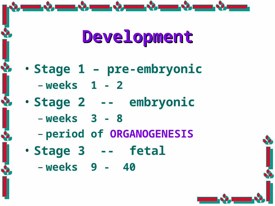
DevelopmentDevelopment
• Stage 1 – pre-embryonic– weeks 1 - 2
• Stage 2 -- embryonic– weeks 3 - 8– period of ORGANOGENESIS
• Stage 3 -- fetal– weeks 9 - 40

Pre-embryonic PeriodPre-embryonic PeriodWeeks 1-2Weeks 1-2
• Traveling in the fallopian tube where rapid cellular multiplication and
differentiation occurs.• The establishment of the embryonic
membranes and the germ layers.• Groove formed along middle of the back for the neural tube.

Embryonic PeriodEmbryonic PeriodWeeks 3-8Weeks 3-8
• Week 3 – anterior end of neural tube closes to form the brain and the posterior end closes to form the spinal cord– Heart begins to beat– Eyes appear– Limb Buds for arms and legs– CR = 4 mm

• Week 5– Head grows larger– Hand and feet plates develop– Facial features begin to develop– CR = 8 mm.

• Week 6– Fetal circulation is established– Chambers form in the heart– Upper lip and palate start fusing– Eyes move to front of face– Fingers are webbed– External ear develops

• Week 7– Eyelids start to form– Fingers develop; elbows visible– Diaphragm separates abdomen from chest– Bronchi develop– Arms and legs move

• Week 8– Fingers and toes distinct– Skeletal ossification begins– Testes and ovaries are distinguishable– Heart has four chambers– Circulation through umbilical cord occurs
– *** ALL essential external and internal structures are present and now will continue to grow

Fetal PeriodFetal PeriodWeeks 9-40Weeks 9-40
12 weeks18 weeks
16 weeks 24 weeks
32 weeks gestation

Weeks 9-12 Weeks 9-12
• Head size increases• Face is well formed• Nails appear • Eyelids appear and close and fuse shut• Kidneys excrete urine• Intestines are forming; peristalsis
begins• Heartbeat can be heard via ultrasound• Tooth buds appear for the baby teeth

Weeks 13-16Weeks 13-16
• Lips form, facial contour develops
• Ossification of bone begins• Meconium begins to form in
the intestines• Hair present on scalp• Sex can be determined visually

Weeks 17-20Weeks 17-20• Hair abundant on head• Lanugo covers the body• Vernix begins to form• Myelination of spinal cord begins• Suck and swallow begin• Quickening occurs ~ 18 weeks

Weeks 21-24 weeksWeeks 21-24 weeks• Respiratory movement with air sacs
formed • Surfactant production begins ~ 24
weeks• Brain appears mature• Eyebrows and eyelashes can be seen• Reacts to sudden noise with active movement

Weeks 25 - 28Weeks 25 - 28
• Eyelids open and close• Capillaries proliferate around
the lungs’ alveoli making gas exchange possible
• Skin has wrinkled red appearance
• Rapid brain development

Weeks 29- 32 Weeks 29- 32
• Subcutaneous fat forms• Testes start descending• Fingernails and toenails are
complete• Bones are fully developed, but
still soft and pliable

Weeks 33 - 40Weeks 33 - 40
• Limbs start to flex• Muscle tone is developed• Lanugo disappears• Body begins to store fat• Maternal antibodies transfer to
the fetus• Exhibits sleep and awake
patterns

Functions of the Functions of the PlacentaPlacentaFetal Respirations
Fetal Nutrition
Endocrine Functions
Elimination of Wastes
Barrier against certain substances

Embryonic MembranesEmbryonic Membranes
• At the time of Implantation, the embryonic membranes begin to form.
• The two main membranes are the:– 1. Chorion– 2. Amnion

ChorionChorion
• Thick membrane with finger-like projections called chorionic villi.
• Chorionic villi contain blood vessels that are main connection with mother.
• Chorionic villi produce human chorionic gonadotropin (HCG)
• Merges with the decidua basalis to form the PLACENTA.

AmnionAmnion
• Smooth, glistening membrane know as the AMNION is the lining of a fluid filled space that develops around the embryo.

Functions of Amniotic Functions of Amniotic FluidFluid
Keeps the fetus at an even temperature
Cushions the fetus against possible injury
Provides place for the fetus to move easily and grow symmetrically
Fetus drinks the fluid

Umbilical Vessels
• 1 Veins – O2 & nutrients• 2 Artery – CO2 & waste
products rich• Wharton’s jelly

TeratogensTeratogens
• Risk factors such as environmental substances– Smoking– Alcohol– Drugs– Viruses– Occupational hazards

ReviewReview
•Describe the components of the process of fertilization.

• Ovum released into fallopian tube—viable for 24 hr.
• Sperm deposited into vagina—viable for 48 to 72 hr (highly fertile for 24 hr).
• Sperm must undergo capacitation and acrosomal reaction.
• Sperm penetration causes a chemical reaction that blocks more sperm penetration.
• Fertilization occurs in the distal end of the fallopian tube.
• Sperm enters ovum. The nuclei of the ovum and sperm unite and become a diploid zygote.

ReviewReview
• How can knowledge of the normal fertilization process assist in helping couples conceive?

ReviewReview
• How can knowing the gestational age of the fetus help in assessment for the potential effects of a teratogen?

The End
return


