Original Article
Autophagy in Normal and Abnormal EarlyHuman Pregnancies
Laura Avagliano, MD, PhD1, Laura Terraneo, PhD1,Eleonora Virgili, BD1, Carla Martinelli PhD2, Patrizia Doi, MS1,Michele Samaja, PhD1, Gaetano Pietro Bulfamante, MD1,and Anna Maria Marconi, MD1
AbstractAutophagy is an inducible catabolic process by which cells degrade and recycle materials to survive stress, starvation, and hypoxia.The aim of this study was to evaluate autophagy at the fetal–maternal interface, to assess autophagy involvement during the earlyphase of human gestation, and to explore autophagic modification in case of early abnormal pregnancy outcome. Specimens werecollected from first-trimester normal gestations undergoing legal termination of pregnancy and first-trimester sporadic sponta-neous miscarriages. Autophagy was studied in villous and decidual samples by transmission electron microscopy, immunohisto-chemistry, immunofluorescence, and Western blotting. Autophagy markers were found in cytotrophoblast, syncytiotrophoblast,extravillous trophoblast, and decidual stromal cells. Autophagy is physiologically involved in early normal gestation. Comparedwith normal pregnancy, spontaneous miscarriage presents an increase in autophagy expression in villous specimens due to anincrement in concentration of autophagic vacuole in syncytiotrophoblast, suggesting a cytoprotective mechanism of the cellsto respond to microenvironmental challenge.
Keywordsautophagy, apoptosis, pregnancy, trophoblast, miscarriage
Introduction
Autophagy is an evolutionary conserved, intracellular inducible
catabolic process by which injured organelles, damaged proteins,
and pathogens are sequestered into autophagosomes, double
membrane-bound vesicles that fuse with lysosomes for proteoly-
tic degradation and material recycling.1 Microtubule-associated
protein light chain 3 (LC3), the mammalian homologue of yeast
autophagy-related gene 8, intervenes in the late stages of autop-
hagosome formation. Among LC3 isoforms, LC3-II, the phos-
phatidylethanolamine conjugated product of LC3-I obtained
after LC3 activation,1,2 is currently used as a specific marker
of autophagy for its role during autophagosome genesis.3
Previous studies have investigated the occurrence of autop-
hagy during the early stages of mammalian pregnancy in the
preimplantation period for oocyte-to-embryo transition4-6 but
no extensive in vivo assessment of autophagy at the fetomater-
nal interface after implantation has been performed in humans.
At the site of implantation, apoptosis permits placenta develop-
ment,7 and a relatively hypoxic environment warrants an ade-
quate embryonic and placental growth.8 Although many
cellular and molecular events are expected to regulate the var-
ious steps of fetomaternal interactions, the mechanisms that
regulate the early stage of pregnancy are not yet fully
understood. The aim of the present study was to evaluate the
occurrence of autophagy during the first trimester of pregnancy
to verify whether it is involved in the early phase of gestation
and whether autophagy expression differs between normal
pregnancy (NP) and spontaneous miscarriage (SM).
Materials and Methods
Sample Collection
Normal pregnancies. Tissues were obtained from 10 healthy,
nonsmokers women (age 28.5 + 8.1 years) undergoing legal
termination of pregnancy within 12 weeks of amenorrhea
(mean gestational age 9.4 + 1.1 weeks; range 8-11 weeks);
1 Department of Health Sciences, San Paolo Hospital Medical School,
Universita degli Studi di Milano, Milan, Italy2 Department of Biomedical Sciences for Health, Universita degli Studi di
Milano, Milan, Italy
Corresponding Author:
Laura Avagliano, Unit of Obstetrics and Gynecology, Department of Health
Sciences, San Paolo Hospital Medical School. University of Milano, Via A. di
Rudinı 8. 20142 Milano, Italy.
Email: [email protected]
Reproductive Sciences2015, Vol. 22(7) 838-844ª The Author(s) 2014Reprints and permission:sagepub.com/journalsPermissions.navDOI: 10.1177/1933719114565036rs.sagepub.com
by guest on June 24, 2015rsx.sagepub.comDownloaded from
in all cases gestational age and fetal viability were confirmed
by ultrasound before suction curettage. All women gave their
consent to the study. All procedures were performed in general
anesthesia. At the time of suction curettage, the gestational tis-
sues were removed in sterile conditions and macroscopically
assessed, through several washes in sterile phosphate-
buffered saline solution. For each woman, samples of decidua
and villi were collected and stored separately: immediately
after the washes, tissues collected for immunohistochemical
and immunofluorescence analysis were fixed in 10% formalin
solution; samples for electron microscopy were fixed in 2.5%glutaraldehyde; samples for Western blotting were frozen in
liquid nitrogen and stored at �80�C for protein extraction.
Spontaneous miscarriages. Tissues were obtained from archive
specimens from our previous work as described.9 All cases
were sporadic miscarriage. Only embryonic pregnancies were
included and all cases had normal embryonic karyotype. Smo-
kers were excluded, therefore 10 cases were eligible for this
study. Spontaneous miscarriage and NPs were comparable for
gestational age (gestational age 9.2 + 0.6 weeks; P ¼ .76) and
maternal age (age 30.9 + 4.8 years; P ¼ .43).
Immunohistochemistry
Immunohistochemical studies were carried out in all samples
(n ¼ 20: 10 NPs and 10 SMs) on 4-mm thick tissue sections
as described previously.10-12 Primary antibodies were as fol-
lows: rabbit polyclonal LC3 (NB100-2220, Novus Biologicals,
Littleton, Colorado; dilution 1:1000), mouse monoclonal anti-
CK-7 (OV-TL 12/30, Dako, Glostrup, Denmark; dilution
1:100), mouse monoclonal anti-HLA-G (SC-21799, Santa Cruz
Biotechnology, Dallas, Texas; dilution 1:2000), and rabbit mono-
clonal cleaved caspase-3 (#9664, Cell Signaling Technology,
Danvers, Massachusetts; dilution 1:800).
Immunofluorescence
Immunofluorescence studies were conducted in the same
samples used for immunohistochemistry (n ¼ 20: 10 NPs
and 10 SMs), as described previously11 using rabbit antiLC3
(NB100-2220, Novus Biologicals, Littleton, Colorado; dilution
1:500). Fluorophore-conjugated secondary antibodies were
used and nuclei were subsequently counterstained with 40,6-
diamidino-2-phenylindole (Invitrogen, Carlsbad, California).
Fluorescence images were viewed and captured using Imager.Z1
microscope (Zeiss, Feldbach, Switzerland).
Transmission Electron Microscopy
Tissues from 8 cases (4 NPs and 4 SMs) were fixed in 2.5%glutaraldehyde in 0.13 mol/L phosphate buffer at pH 7.2-7.4;
specimens were postfixed in 1% OsO4, dehydrated in ethanol
plus propylene oxide, and embedded in epoxy resin. Ultrathin
sections of 50 to 60 nm were counterstained with uranyl acetate
and lead citrate. A Jeol JEM 1010 (Tokyo, Japan) electron
microscope was used for examination. Cases used for
transmission electron microscopy evaluation were a subgroup
of the total population due to tissue availability restrictions.
Autophagic vacuoles were identified, in villi and decidua, as
double membrane compartments containing material in various
stages of degradation. For each case, the area of autophagic
vacuoles was calculated by 2 observers (LA and CM) on
60000� printed micrographs (previously converted in digital
bitmap images by Image Processing and Analysis in Java-Ima-
geJ) in 3 randomly selected areas of villi, both in cytotropho-
blast and in syncytiotrophoblast. Quantification of autophagy
was measured as a ratio of autophagic vacuole area to the total
cytoplasmic area. The mean ratio from the 3 areas was consid-
ered as the final value for each case.
Western Blotting
Villous (n¼ 20: 10 NPs and 10 SMs) and decidual (n¼ 20: 10s
NP and 10 SMs) samples were homogenized in a glass potter in
a 1:5 ratio (weight:volume) with 50 mmol/L Tris-HCl pH 7.4,
5 mmol/L EDTA, 1 mmol/L DTT, 2% SDS, and Protease Inhi-
bitor Cocktail (Sigma Aldrich, St. Louis, Missouri) and centri-
fuged at 14 000g for 15 minutes. Protein concentration within
the supernatant was measured by the Coomassie Plus Protein
Assay reagent Kit (Pierce, Rockford, Illinois). An equal
amount of protein from each sample (30 mg) was subjected to
electrophoresis in a 10% polyacrylamide gel for LC3 and Bcl2
or in 8% polyacrylamide gel for hypoxia-inducible factor 1a(HIF-1a), caspase-3, Bax, and transferred onto nitrocellulose
membrane. Membranes were incubated in a blocking solution
(Tris-buffered saline [TBS] containing 5% powdered nonfat
milk and 0.01% Tween-20) at room temperature for 1 hour.
Membranes were incubated overnight at 4�C with the pri-
mary antibody, followed by incubation with horseradish
peroxidase-conjugated secondary antibody. The used primary
antibodies and dilutions were as follows: rabbit polyclonal
anti-HIF-1a (H-206X: sc-10790, Santa Cruz Biotechnology,
Dallas, Texas; 1:300 in TBS-Tween 1�), rabbit polyclonal
LC3 (NB100-2220, Novus Biologicals, Littleton, Colorado;
1:1000 in 5% bovine serum albumin [BSA]), mouse monoclo-
nal Bcl2 (C-2: sc-7382, Santa Cruz Biotechnology, Dallas,
Texas; 1:500 in TBS-Tween 1X), mouse monoclonal b-actin
(A5316, Clone AC-74, Sigma Aldrich, St Louis, Missouri,
1:5000 in 5% BSA), mouse monoclonal Bax (B-9: sc-7480,
Santa Cruz Biotechnology, Dallas, Texas; 1:500 in TBS-
Tween 1�), and rabbit monoclonal caspase-3 (#9665, Cell Sig-
naling, Danvers, Massachusetts; 1:1000 in 5% BSA). After
washes in TBS-Tween 1�, membranes were incubated with the
secondary antibodies at room temperature for 1 hour.
Secondary antibodies used were as follows: peroxidase-
conjugated goat antirabbit immunoglobulin (IgG; AffiniPure,
Jackson Immunoresearch, West Grove, Pennsylvania), diluted
1:10 000 in 5% milk for LC3, Beclin-1, and HIF-1a, or 1:10
000 in TBS-Tween 1� for caspase-3, and goat anti-mouse IgG
(AffiniPure, Jackson Immunoresearch, West Grove, Pennsyl-
vania), diluted 1:10 000 in 5% milk for b-actin or 1:10000 in
TBS-Tween 1X for Bax and Bcl2.
Avagliano et al 839
by guest on June 24, 2015rsx.sagepub.comDownloaded from
Chemiluminescence was detected by incubating the mem-
brane with LiteAblot Chemiluminescent substrate (Lite Ablot,
EuroClone, Milano, Italy) followed by X-ray film exposure
(Kodak X-Omat Blue XB-1 Film, Eastman Kodak Company,
Rochester, New York).
Protein expression was quantified densitometrically (OD/
mm2) by Quantity One 4.2.1 image analysis software (Bio-
Rad, Segrate, Italy). To correlate the blot intensity with the
actual protein amount, calibration curves were run in blots
within the dynamic range of the instrument. After the back-
ground signal was subtracted, densitometry data were nor-
malized with respect to a single reference extract that was
inserted in every run, thereby representing the positive con-
trol to allow normalization and encompass intra- and interrun
variability. Protein levels are expressed as densitometry ratio
with b-actin. Western blotting analysis permits to distinguish
cytosolic and membrane-bound forms of LC3, LC3-I (18
kDa), and LC3-II (16 kDa), respectively.
Statistical Analysis
Analysis of immunoblots is reported as mean and standard
error of the mean. The Wilcoxon matched-pairs test and
Mann-Whitney U test were used, as appropriated, to compare
protein levels in villi and decidua and to compare autophago-
some content in cytotrophoblast and syncytiotrophoblast. The
P values �.05 were considered significant. Statistical tests
were performed using Instat 3, GraphPad software.
Results
Normal Pregnancies
Signs of autophagy were detected both in villous and decidual
specimens of first-trimester pregnancies (Figure 1C, E, and G):
LC3 staining was observed both in villous syncytiotrophoblast
and in cytotrophoblast by immunohistochemical method; LC3
immunostaining was also observed in trophoblast-anchoring
columns and extravillous trophoblast (EVT). Moreover, decid-
ual stromal cells displayed a positive immunoreactivity both in
basalis and in parietalis decidua.
We confirmed the localization of autophagy by elec-
tron microscopy in cytotrophoblast, syncytiotrophoblast, and
decidua by observing autophagosomes, a double-membrane
structures containing cytoplasmic material at various stage of
degradation (Figure 1I-L). To detect differences in autophagy
between villous and decidual samples, LC3-II protein expres-
sion was measured by Western blotting analysis. The expression
of LC3-II was comparable in decidual and villous specimens.
Spontaneous Miscarriages
Localization of LC3 staining in SM was comparable to NP, but
in syncytiotrophoblast an increased intensity of staining was
observed by immunofluorescence, finding an increase in punc-
tate dots (Figure 2A). We confirmed the increase of autophagy
in syncytiotrophoblast by semiquantitative electron micro-
scopy analysis, detecting a higher concentration of
autophagosome in syncytiotrophoblast of SMs compared to
NPs (Figure 2D and E). We detected an increased LC3-II
expression in villi from SMs than villi from NPs by Western
blotting analysis (Figure 2B and C).
Autophagy Expression and Expression of Markersof Hypoxia or Apoptosis
A proper oxygen level and cells turnover are necessary at the
implantation site to warrant an adequate evolution of preg-
nancy. Therefore, to explore the possible correlation of autop-
hagy with hypoxia, we measured the expression of HIF-1a, a
transcription factor stabilized in hypoxic conditions, by West-
ern blotting. The expression of HIF-1a was similarly low in vil-
lous and decidual samples of NP, whereas in cases of SM, the
expression of HIF-1a increased both in villi and in decidua
with the highest level in decidual samples (Figure 3A).
Moreover, to compare the expression of autophagy to apop-
tosis, we measured the expression of Bax (that promotes pro-
grammed cells death) and Bcl-2 (that inhibits programmed
cells death) in villous and decidual specimens by Western blot-
ting analysis, using for each case the same sample of tissue pre-
viously used to investigate autophagy. The ratio of Bax–Bcl-2
protein was used to indicate the susceptibility to apoptosis.
Comparable low level of apoptosis was observed both in villi
and in decidua of NP, whereas an increase in apoptosis was
observed in decidua of SM although without statistical signifi-
cance (Figure 3B). Cleaved caspase-3 expression was also
evaluated to detect apoptosis and it was observed in all samples
of decidua of miscarriage, according to the trend of Bax/Bcl-2
expression (Figure 3C) and in agreement with immunohisto-
chemical staining (Figure 3D).
Discussion
The present study assess autophagy expression during early
human pregnancy. Few previous studies have evaluated autop-
hagy in placental villi13,14 or decidua15 of the first trimester of
gestation. A novelty of our study is the analysis, at the same
time, of both the sides of blastocyst implantation, considering
the embryonic side by evaluating villi and the maternal side
by evaluating decidua.
Interestingly, the results of the present study show that
autophagy at the fetomaternal interface is constitutively
expressed in normal early human pregnancy. During normal
conditions, in villous specimens, we observed autophagy
expression in both the trophoblastic layers, with higher expres-
sion in cytotrophoblast than in syncytiotrophoblast. This obser-
vation is in agreement with a previous report,13 confirming the
physiological occurrence of autophagy in placental villi during
the first weeks of gestation.
The strength of our study is the novelty of the evaluation of
autophagy in SM. We observed an increased autophagy expres-
sion in villous samples due to an increment of autophagic
vacuoles concentration in syncytiotrophoblast. Syncytiotro-
phoblast is the outer trophoblastic layer, directly in contact with
840 Reproductive Sciences 22(7)
by guest on June 24, 2015rsx.sagepub.comDownloaded from
maternal blood. Since autophagy can intervene in placental
trophoblasts during stressor conditions as a cytoprotective
process,16 we suggest that the increase of autophagy in syncy-
tiotrophoblast could reflect a prosurvival tentative of the cells
to respond to the environmental changes.
Moreover, we detected autophagy markers in decidua speci-
mens, trophoblastic-anchoring columns, and EVT. It is known
that EVT migrates from the columns through the decidual
stroma, reaches the spiral arteries, penetrates the wall of the
vessels, and colonizes the lumen forming the trophoblastic
plugs and conducting to vascular remodeling.17 Deficiency in
EVT invasion and inadequate vascular remodeling can lead
to adverse pregnancy outcome such as miscarriage, preeclamp-
sia, and intrauterine growth restriction.18 Previous studies per-
formed on trophoblastic cell lines suggested that autophagy is
involved in trophoblast invasion,14 even if the results are con-
flicting: Some authors observed that autophagy induced by
hypoxia supports trophoblast invasion,15,19 whereas other
authors observed an increase in autophagy but a decrease in tro-
phoblast invasion in relation to the inhibition of HIF1-a expres-
sion.20 Differences in autophagy expression between decidua
from NP and SM were not detected in the present study, while
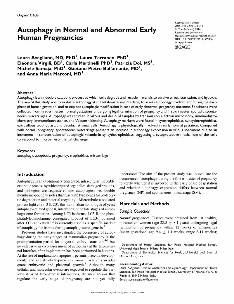
Figure 1. Localization of autophagy in normal pregnancy. A and B, negative controls. C-H, Localization of autophagy marker (LC3) by immu-nohistochemical staining. (C, E, and G), LC3 staining (autophagy marker); (D and F) cytokeratin 7 staining (CK7—marker of trophoblastic cells);(H) HLA-G staining (marker of endovascular trophoblast). Signs of autophagy are detectable in villous trophoblast (C and E) in decidual samples(E and G) and in endovascular trophoblast (G). Positivity are detectable not only in trophoblastic cells but also in some stromal cells both invillous and in decidual tissues. Original magnification 40� in (A), (B), (C), (D), (G), and (H); 20� in (E) and (F). I-L, Evaluation of autophagosomeby transmission electron microscopy. I, Villous sample: autophagosomes are clearly identifiable at higher magnification (inset) both in syncytio-trophoblast and cytotrophoblast. L, Decidual sample: double membrane autophagic compartment containing material in degradation is detect-able at higher magnification (inset). No Ab indicates no primary antibody control; C-, slice incubated with a matching concentration of nonspecificrabbit immunoglobulin; a, anchoring villous; ac, anchoring column; e.va.t, endovascular trophoblast; f, floating villous; gl, decidual gland; MMV,microvillous membrane; S, syncytiotrophoblast; C, cytotrophoblast; Fc, fetal capillary. LC3, microtubule-associated protein light chain 3.
Avagliano et al 841
by guest on June 24, 2015rsx.sagepub.comDownloaded from
apoptosis and hypoxia were increased in SM. Our finding of
increased apoptosis in first-trimester decidua of SM is in agree-
ment with a previous report where apoptosis was responsible
for the pregnancy loss.21 In our samples of SM, we found an
increased expression of autophagy in villous specimens but a
low expression of apoptosis; on the contrary, in decidua speci-
mens we found low levels of autophagy but an increased sus-
ceptibility to apoptosis. It is possible that the opposite trend
of expression of autophagy compared to hypoxia and apoptosis
markers in villi and decidua of SM might reflect the role of
autophagy in antagonizing hypoxia-induced apoptosis, but fur-
ther investigation is needed to clarify this issue.
This study has some limitations. First, samples obtained
from legal interruption of pregnancies include viable tissues,
whereas samples from SMs include tissues from nonviable
pregnancies; it is therefore not possible to determine whether
autophagy has been induced in vivo before or after fetal
demise. Second, samples were stored in liquid nitrogen before
the analysis, whereas a previous study suggested a role of cold
stress in the induction of autophagy,22 therefore our results
might be influenced by the freezing of samples before the anal-
ysis. Third, we could not exclude that the intersample variabil-
ity in villi affected our results: Autophagy is a dynamic and
continuous process and LC3-II is just a marker of one stage
of it. Different stages of the evolution during the maturation
from the phagophore through the autolysosome in our samples
could affect the presence or the level of LC3-II.
In conclusion, our study demonstrates the occurrence of
autophagy in vivo at the fetomaternal interface during the first
trimester of normal human gestation. A low level of autophagy
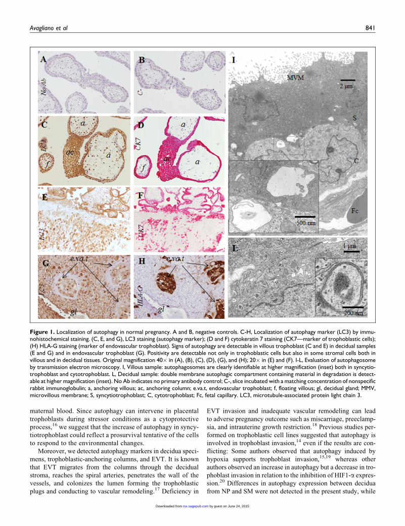
Figure 2. Autophagy expression in normal pregnancy (NP) and spontaneous miscarriage (SM). A, immunofluorescence staining (LC3 in green)in villi (v) and decidua (d). Arrows indicate syncytiotrophoblast. Note the increase number of fluorescent punta dots in syncytiotrophoblasticlayer of spontaneous miscarriage, well identifiable in the insets. White scale bar: 25 mm, Yellow scale bar: 50 mm. B, Representative Westernblotting bands of autophagy markers LC3-I and II (18 and 16 kDa, respectively) and b-actin (42 kDa, housekeeping gene for normalization).Despite the high intersample variability in villous samples, the amount of LC3-II normalized onto b-actin in NP was always lower than LC3-IIin SP, as shown in (C). C, Western blotting analysis showed that the level of LC3-II expression was higher in villi obtained from spontaneousmiscarriage. *P ¼ .02 villi SM versus villi NP (Mann-Whitney test); **P ¼ .004 villi SM versus decidua SM (Wilcoxon test). D, Autophagosomecontent in placental villi, expressed as the ratio between the sum of the areas of autophagosomes and the total area, evaluated in cytotrophoblastand syncytiotrophoblast by transmission electron microscopy. * P¼ .05 syncytiotrophoblast SM versus syncytiotrophoblast NP (Mann-Whitneytest). E, Autophagosome detection by transmission electron microscopy in a case of spontaneous miscarriage. Arrow heads indicate autopha-gosome vacuoles in syncytiotrophoblast and cytotrophoblast. c indicates cytotrophoblast; s, syncytiotrophoblast; MMV, microvillous membrane;Fc, fetal capillary. (The color version of this figure is available in the online version at http://rs.sagepub.com/.)
842 Reproductive Sciences 22(7)
by guest on June 24, 2015rsx.sagepub.comDownloaded from
at this site may represent a physiological event necessary to the
normal evolution of pregnancy. For the first time to our knowl-
edge, our study demonstrates that SM specimens show higher
autophagy expression in villi. Further studies are needed to
clarify the role of autophagy in SM and to distinguish whether
the autophagy expression changes represent the cause or the
consequence of the early adverse pregnancy outcome.
Authors’ Note
Laura Avagliano had the idea, collected and analyzed the data, and
wrote the manuscript. Laurea Terraneo and Eleonora Virgili per-
formed the immunoblotting analysis and approved the last version
of the manuscript. Carla Martinelli performed the electron micro-
scopic study and approved the last version of the manuscript. Patrizia
Doi performed the immunohistochemical staining and approved the
last version of the manuscript. Michele Samaja discussed the data,
revised the manuscript, and approved its last version. Gaetano Bulfa-
mante analyzed and discussed the data, revised the manuscript, and
approved its last version. Anna Maria Marconi revised and approved
the last version of the manuscript.
Declaration of Conflicting Interests
The author(s) declared no potential conflicts of interest with respect to
the research, authorship, and/or publication of this article.
Funding
The author(s) received no financial support for the research, author-
ship, and/or publication of this article.
References
1. Klionsky DJ, Emr SD. Autophagy as a regulated pathway of
cellular degradation. Science. 2000;290(5497):1717-1721.
2. Noda T, Fujita N, Yoshimori T. The late stages of autophagy: how
does the end begin? Cell Death Differ. 2009;16(7):984-990.
3. He C, Klionsky DJ. Regulation mechanisms and signaling path-
ways of autophagy. Annu Rev Genet. 2009;43:67-93.
4. Mizushima N, Levine B. Autophagy in mammalian development
and differentiation. Nat Cell Biol. 2010;12:823-830.
5. Tsukamoto S, Kuma A, Murakami M, Kishi C, Yamamoto A,
Mizushima N. Autophagy is essential for preimplantation deve-
lopment of mouse embryos. Science. 2008;321:117-120.
Figure 3. Evaluation of apoptosis and hypoxia by Western blotting analysis. A, Western blotting analysis showed that HIF-1a expression(hypoxia marker) was high in decidual samples obtained from spontaneous miscarriage (SM). *P ¼ .0002 decidua SM versus decidua normalpregnancy (NP; Mann-Whitney test); on the right, representative bands of HIF-1a (120 kDa) and b-actin (42 kDa, housekeeping gene for normal-ization) are shown. B, Western blotting analysis showed an increased trend in the Bax/Bcl2 ratio in decidua obtained from spontaneous mis-carriage. C, Western blotting analysis of cleaved caspase-3. The extent of the cleavage is comparable to the Bax/Bcl2 ratio, supportinggreater apoptosis in decidua SM compared to villi SM and all samples from NP. D, Immunohistochemical expression of cleaved caspase 3. Pos-itive cells are in brown. Note the increased number of positive cells in decidua from spontaneous miscarriage respect to decidua from normalpregnancy. HIF-1a indicates hypoxia-inducible factor. (The color version of this figure is available in the online version at http://rs.sagepub.com/.)
Avagliano et al 843
by guest on June 24, 2015rsx.sagepub.comDownloaded from
6. Tsukamoto S, Kuma A, Mizushima N. The role of autophagy during
the oocyte-to-embryo transition. Autophagy. 2008;4:1076-1078.
7. Jerzak M, Bischof P. Apoptosis in the first trimester human
placenta: The role in maintaining immune privilege at the
maternal-foetal interface and in the trophoblast remodelling.
Eur J Obstet Gynecol Reprod Biol. 2002;100(2):138-142.
8. Caniggia I, Winter J, Lye SJ, Post M. Oxygen and placental deve-
lopment during the first trimester: implications for the pathophy-
siology of pre-eclampsia. Placenta. 2000;21(suppl A):S25-S30.
9. Garzia E, Clauser R, Persani L, et al. Prolactin and proinflamma-
tory cytokine expression at the fetomaternal interface in first
trimester miscarriage. Fertil Steril. 2013;100(1):108-15.e1-e2.
10. Signorelli P, Avagliano L, Virgili E, et al. Autophagy in term nor-
mal human placentas. Placenta. 2011;32(6):482-485.
11. Avagliano L, Virgili E, Garo C, et al. Autophagy and human
parturition: evaluation of LC3 expression in placenta from spon-
taneous or medically induced onset of labor. BioMed Res Int.
2013. Web site. www.hindawi.com/journals/bmri/2013/689768/.
Published 2013. Accessed December 16, 2014.
12. Avagliano L, Danti L, Doi P, et al. Autophagy in placentas from
acidotic newborns: an immunohistochemical study of LC3
expression. Placenta. 2013;34(11):1091-1094.
13. Chifenti B, Locci MT, Lazzeri G, et al. Autophagy-related protein
LC3 and Beclin-1 in the first trimester of pregnancy. Clin Exp
Reprod Med. 2013;40(1):33-37.
14. Hung TH, Hsieh TT, Chen SF, Li MJ, Yeh YL. Autophagy in the
human placenta throughout gestation. PLoS One. 2013;8(12):
e83475.
15. Nakashima A, Yamanaka-Tatematsu M, Fujita N, et al. Impaired
autophagy by soluble endoglin, under physiological hypoxia in
early pregnant period, is involved in poor placentation in pree-
clampsia. Autophagy. 2013;9(3):303-316.
16. Chen B, Longtine MS, Nelson DM. Hypoxia Induces Autophagy
in Primary Human Trophoblasts. Endocrinology. 2012;153(10):
4946-4954.
17. Huppertz B, Berghold VM, Kawaguchi R, Gauster M. A variety
of opportunities for immune interactions during trophoblast
development and invasion. Am J Reprod Immunol. 2012;67(5):
349-357.
18. Brosens I, Pijnenborg R, Vercruysse L, Romero R. The ‘‘Great
Obstetrical Syndromes’’ are associated with disorders of deep
placentation. Am J Obstet Gynecol. 2011;204(3):193-201.
19. Yamanaka-Tatematsu M, Nakashima A, Fujita N, Shima T,
Yoshimori T, Saito S. Autophagy induced by HIF1a overexpres-
sion supports trophoblast invasion by supplying cellular energy.
PLoS One. 2013;8(10):e76605.
20. Choi JH, Lee HJ, Yang TH, Kim GJ. Effects of hypoxia inducible
factors-1a on autophagy and invasion of trophoblasts. Clin Exp
Reprod Med. 2012;39(2):73-80.
21. Kokawa T, Shikone T, Nakano R. Apoptosis in human chorionic
villi and decidua during normal embryonic development and
spontaneous abortion in the first trimester. Placenta. 1998;
19(1):21-26.
22. Bang S, Shin H, Song H, Suh CS, Lim HJ. Autophagic activation
in vitrified-warmed mouse oocytes. Reproduction. 2014;148(1):
11-19.
844 Reproductive Sciences 22(7)
by guest on June 24, 2015rsx.sagepub.comDownloaded from