46
REPRODUCTIVE SYSTEM By Dr.Ahmed Salman Assistant Professor of Anatomy &embryology Anatomy Department The University Of Jordan Faculty Of Medicine

REPRODUCTIVE SYSTEM
By
Dr.Ahmed SalmanAssistant Professor of Anatomy &embryology
Anatomy Department
The University Of Jordan Faculty Of Medicine

Perineum
It is the diamond-shaped lower end of the trunk

Glossary : peri : around, ineo - discharge, evacuate
Location : it lies below the pelvic diaphragm, between the upper parts of the thighs.
Boundaries :
Anteriorly : Inferior margin of symphysis pubis.
Posteriorly : Tip of coccyx.
Anterolateral : Fused rami of pubis and ischium and ischial tuberosity.
Posterolateral : Sacrotuberous ligaments.
Dr. Ahmed Salman

Divisions of the Perineum :
By a line joining the anterior parts of the ischial tuberosities, the
perineum is divided into two triangles :
Anteriorly :Urogenital triangle
Posteriorly : Anal triangle
Dr. Ahmed Salman

Perineum
Urogenital triangle
Contains
1-External genitalia in male or female
2-Superficial perineal pouch
3-Deep perineal pouch
Anal triangle
Contains
1-Anal Canal in the median plane
2-Ischiorectal fossa on either side of anal canal
3- pudendal canal in side wall of
ischiorectal fossaDr. Ahmed Salman
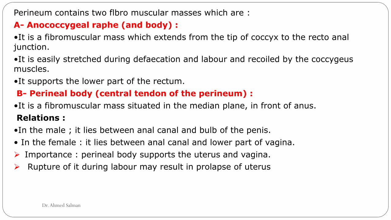
Perineum contains two flbro muscular masses which are :
A- Anococcygeal raphe (and body) :
•It is a fibromuscular mass which extends from the tip of coccyx to the recto anal junction.
•It is easily stretched during defaecation and labour and recoiled by the coccygeusmuscles.
•It supports the lower part of the rectum.
B- Perineal body (central tendon of the perineum) :
•It is a fibromuscular mass situated in the median plane, in front of anus.
Relations :
•In the male ; it lies between anal canal and bulb of the penis.
• In the female : it lies between anal canal and lower part of vagina.
Importance : perineal body supports the uterus and vagina.
Rupture of it during labour may result in prolapse of uterus
Dr. Ahmed Salman

Muscles attached to the perineal body : 3 paired and 3 single muscles :
3 paired muscles 3 single muscles
1. Superficial transversus perinei.
2. Deep transversus perinei.
3. Levator prostate or sphincter vaginae part
of levator ani.
1. Bulbospongiosus.
2. Superficial part of external urethral
sphincter (sphincter urethrae).
3. Superficial part of external anal sphincter
Dr. Ahmed Salman

The perineal fascia
consists of superficial and deep layers
Superficial perineal fascia, consists of a superficial fatty layer and a deep membranous
layer (Colles fascia).
The superficial fatty layer
In females, It makes up the substance of the labia majora and mons pubis
It is continuous with the fatty layer of subcutaneous tissue of the abdomen (Camper
fascia)
In males, the fatty layer is replaced with smooth (dartos) muscle.
It is continuous between with the fatty layer of subcutaneous tissue of the abdomen .
In both sexes, it is continuous posteriorly with the ischio-anal fat pad in the anal region .
Dr. Ahmed Salman

The membranous layer (Colles fascia).
is attached To
Posteriorly : The posterior margin of the perineal membrane and the perineal body
Laterally, the fascia lata (deep fascia) of the upper medial aspect of the thigh.
Anteriorly, in the male, the membranous layer of subcutaneous tissue is continuous with
the dartos fascia of the penis and scrotum
On each side of and anterior to the scrotum, the membranous layer becomes continuous
with the membranous layer of subcutaneous tissue of the abdomen (Scarpa fascia) .
In females, the membranous layer passes superior to the fatty layer forming the labia
majora and becomes continuous with the membranous layer of the subcutaneous tissue of
the abdomen .
Dr. Ahmed Salman

The perineal fascia (deep perineal, investing, or Gallaudet fascia)
Invests the ischiocavernosus, bulbospongiosus, and superficial transverse perineal
muscles.
It is also attached laterally to the ischiopubic rami.
Anteriorly, it is fused to the suspensory ligament of the penis
Dr. Ahmed Salman

The perineal fascia
Deep perineal (Gallaudet fascia)Superficial perineal fascia
The
superficial
fatty layer
It is continuous
with (Camper
fascia)
The membranous layer
(Colles fascia)
It is attached to fascia lata
It is continuous with
(Scarpa fascia)
Dr. Ahmed Salman

Urogenital triangle:
The perineal membrane
It is a triangular fibrous sheet which lies across the pubic arch• Forms the floor of the deep perineal pouch.• Forms the roof of the superficial perineal pouch
.
Dr. Ahmed Salman

Structures piercing perineal membrane in the male and the female
Male Female
A-Genitourinary
structures
Urethra.
Ducts of bulbo-urethral glands.
• Urethra.
• Vagina
B- Vessels • Internal pudendal A.
• Artery of the bulb
• Urethral A.
• Internal pudendal A.
• Artery of the bulb of
vestibule.
C- Nerves Dorsal nerve of penis. Dorsal nerve of clitoris.
Dr. Ahmed Salman

Dr. Ahmed Salman

Dr. Ahmed Salman
Urogenital Diaphragm
It is a triangular musculofascial diaphragm situated in the anterior part of the perineum
It fills the gap of the pubic arch
It is formed by the sphincter urethrae and the deep transverse perineal muscles
These two muscles are enclosed between a superior and an inferior layer of fascia of the urogenital diaphragm.
The inferior layer of fascia is the perineal membrane.

Perineal pouches
Dr. Ahmed Salman

Dr. Ahmed Salman

Dr. Ahmed Salman

Deep Perineal pouch :
Boundaries :
Floor : Perineal membrane (inferior fascia of urogenital diaphragm)
Roof: Inferior fascia of pelvic diaphragm (levator ani)
On either side : related to obturator fascia.
Posteriorly : the pouch is closed by union of roof and floor.
Anteriorly : The pouch is closed by union of roof and floor below symphysis pubis to form
the transverse perineal ligament.
The transverse perineal ligament
Is separated from the symphysis pubis by an oval opening. Through this opening
In male the deep dorsal vein of penis enters the pelvis to join the prostatic venous plexus.
In female, the deep dorsal vein of clitoris joins the vesical venous plexusDr. Ahmed Salman

Contents
Male Female
Urogenital Structures
• Membranous urethra. • Bulbourethral glands,
• Part of the urethra.• Part of vagina
Muscles • Sphincter urethrae. • Deep transversus perineimuscles(These two muscles form the urogenital diaphragm.)
• Sphincter urethrae.• Deep transversus perineimuscles. (These two muscles form the urogenital diaphragm)
Vessels • Internal pudendal A.• Artery of bulb. • Urethral A.
• Internal pudendal A. • Artery of bulb of vestibule.
Nerves • Dorsal N. of penis. • Dorsal N. of clitoris.
N.B. : the greater vestibular glands of the female lie in the superficial perineal pouch, the bulbourethral glands of the male lie in the deep perineal pouch

Dr. Ahmed Salman

Deep perineal pouch in male
Dr. Ahmed Salman

Deep perineal pouch in female
Dr. Ahmed Salman

Dr. Ahmed Salman

Superficial Perineal Pouch:Boundaries :Floor : membranous layer of the superficial fascia of the perineum (Colles fascia).Roof : perineal membrane.On either side : both roof and floor are attached to the side of the pubic arch .Posteriorly : the pouch is closed by union of the roof and floor.Anteriorly : the pouch is opened and continuous with the interval between the membranous layer of anterior abdominal wall and the external oblique aponeurosis
Dr. Ahmed Salman

Contents
Male Female
Urogenital
Structures
• Root of penis (2 crura)
• Penile urethra in corpus
spongiosum
• Root of clitoris (2 crura)
• Two bulbs of vestibule
• Greater vestibular glands.
Muscles •Two ischiocavernosus muscles
cover the 2 crura
• Bulbospongiosus muscle covers
bulb of penis.
• Two superficial transversus
perinei muscles.
• Two ischiocavernosus
• Bulbospongiosus muscle
• Two superficial transversus perinei
muscles.
Vessels • Dorsal A. of penis.
• Deep A. of penis.
• Two scrotal arteries
• Dorsal A. of clitoris.
• Deep A. of clitoris.
• Two labial arteries.
Nerves • Dorsal N. of clitoris.
• Two labial nerves
Dorsal N. of penis.
Two scrotal nervesDr. Ahmed Salman
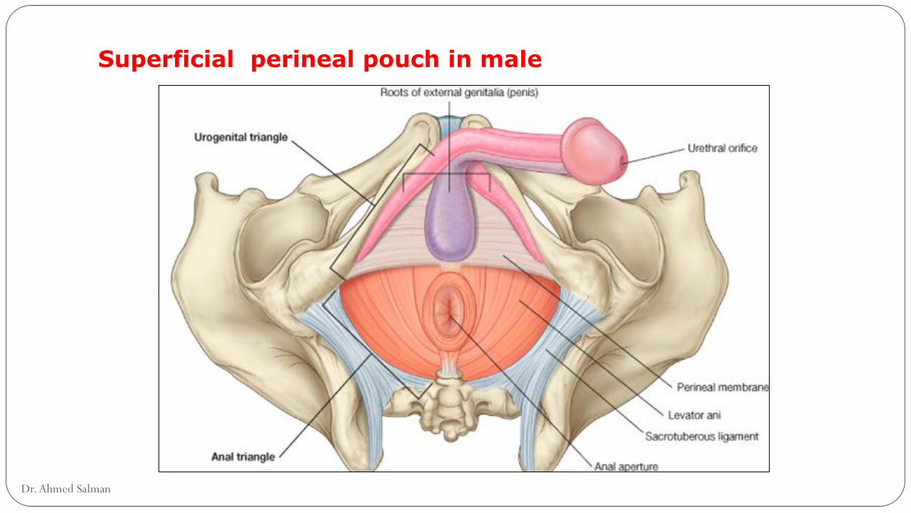
Superficial perineal pouch in male
Dr. Ahmed Salman

Superficial perineal pouch in male (muscles)
Dr. Ahmed Salman

Dr. Ahmed Salman

Superficial perineal pouch in female
Dr. Ahmed Salman

Superficial perineal pouch in female (muscles)
Dr. Ahmed Salman

Muscles of Superficial Perineal Pouch :
Nerve Supply : All muscles in superficial and deep perineal pouches are supplied byperineal branch of pudendal nerve
1- Ischiocavernosus :
Site : Each covers the crus penis.
Origin : Arise from medial side of ischial ramus.
Insertion : into sides and inferior surface of crus penis.
Action : it compresses crus penis to maintain erection of penis.
2- Bulbospongiosus :
Site : covers bulb of penis. In female, it is splitted into two parts to cover bulbs ofvestibule.
Origin : from perineal body.
Insertion : the muscle fibres encircle the bulb and corpus spongiosum to be insertedon the upper surface of the penis.
Action : Assist in erection of penis and eject last drops of urine during micturition.
Dr. Ahmed Salman

3- Superficial transverse perineal:
Site : on posterior edge of perineal membrane in front of anus.
Origin : from medial surface of ischial tuberosity.
Insertion : into the perineal body.
Action : both muscles fix the perineal body.
Muscles of the Deep perineal Pouch : These muscles form the urogenital diaphragm.
1- Sphincter urethrae :
Site : it surrounds membranous urethra.
Attachments : it is formed of two parts :
Inferior (or superficial) part : its muscle fibres extend from the transverse perineal ligament and pass backwards encircling the urethra to converge on the perineal body.
Superior (or deep) part : its muscle fibres arise from the medial side of pubic ramus and pass medially to surround the membranous urethra.
Action : It represents the voluntary control of urethra.
Dr. Ahmed Salman

2- Deep transverse perineal
Site : on deep surface of posterior border of the perineal membrane.
Origin : from medial side of ischial ramus.
Insertion : into perineal body.
Action : fixation of perineal body.
Applied Anatomy :
•Rupture of the membranous urethra leads to accumulation of urine in the deep perineal
pouch which is closed all around.
•Rupture of penile urethra leads to escape of urine into the superficial perineal pouch.
Urine escapes into the scrotum, penis and ascends into the interval between
membranous layer of superficial fascia of anterior abdominal wall and external oblique
aponeurosis.
Urine does not descend into the thigh due to fusion of Scarpa's fascia of the anterior
abdominal wall to the fascia lata of the thigh below the inguinal ligament. Dr. Ahmed Salman

Anal triangleIschiorectal fossa
Location : It is wedge shaped space on either side of the anal canal
Ischiorectal in Female Ischiorectal in MaleDr. Ahmed Salman

Boundaries : It has
1. Apex : it is the origin of levator ani from the lateral pelvic wall
2. Base : skin on either side of the anal orifice (skin of the base is supplied by inferior rectal N.).
3. Anteriorly : the posterior border of the perineal membrane
4. Posteriorly : sacrotuberous ligament covered by lower border of gluteus maximusmuscle.
5. Lateral wall : is vertical formed by :
Lower part of obturator internus muscle and lower part of obturator fascia splitting to form pudendal canal.
Medial surface of ischial tuberosity.
6. Medial wall : formed by :
Levator ani muscle (lower surface).
External anal sphincter.
Dr. Ahmed Salman

Dr. Ahmed Salman

Contents of Ischiorectal fossa :1. Pudendal nerve.2. Internal pudendal vessels. 3. Inferior rectal nerve.4. Inferior rectal vessels. 5. Posterior scrotal nerves.6. Posterior scrotal vessels.7. Perineal branch of the 4th sacral nerve.8. Perforating cutaneous N. (S2, S3) .9. Pad of Fat : It is rich in fibroelastic fibres and has two functions :
• It acts as a cushion support for rectum and anal canal.• It allows distention of the rectum and anal canal during defaecation,
Then compress them after termination of the act.
Dr. Ahmed Salman

Dr. Ahmed Salman

Applied Anatomy:Infections in the ischiorectal pad of fat is common and lead to abscess formation.The abscess may rupture medially into the anal canal or downwards into the skin at the fossa. This may leads to anal fistula
Dr. Ahmed Salman

Pudendal canal:Location : Fascial canal in lower part of the lateral wall of ischiorectal fossaExtend: from the lesser sciatic foramen to the posterior border of the perineal membrane.contents: 1- pudendal nerve 2- internal pudendal vessels
Dr. Ahmed Salman

Pudendal Nerve :
It is the somatic nerve of the perineum and external genitalia.
It arises from the sacral plexus;S2,3,4
Course and Relations :
The nerve leaves the pelvis through the greater sciatic foramen below the piriformis
to enter the gluteal region.
In the gluteal region, the nerve crosses the sacrospinous ligament medial to the
internal pudendal vessels which cross the ischial spine.
The nerve and the vessels pass through the lesser sciatic foramen to enter the
pudendal canal in side wall of ischiorectal fossa.
In the posterior part of pudendal canal, the nerve gives inferior rectal N. and then
divides into 2 terminal branches which are the perineal N. and dorsal nerve of penis
(or clitoris).
Dr. Ahmed Salman

Branches and Distribution :
1- Inferior rectal N. (mixed nerve)
• Motor to external canal sphincter and levator ani.
• Sensory to anal canal below pectinate line, skin around the anus and lower inch of
vagina.
2- Perineal nerve (mixed) : it gives :
• Motor : to all muscles in the superficial and deep perineal pouches.
• Sensory : It gives 2 scrotal (labial) nerves,
they pierce perineal membrane to supply posterior 2/3 of scrotum (or labium majus).
3- Dorsal nerve of penis or clitoris (sensory) :
- It enters the deep perineal pouch, then pierces the perineal membrane to enter the
superficial perineal pouch.
- Then it runs on dorsum of penis, supplying its skin and glans.
Dr. Ahmed Salman

Internal Pudenda Artery:
It is the artery of perineum and external genital organs.
It is one of the two terminal branches of anterior division of internal iliac artery.
Course and Relations :
In the pelvis : it descends in front of sacral plexus and piriformis and leaves the pelvis
(with pudendal N.) below the piriformis to enter the gluteal region.
In gluteal region: The artery crosses the tip of the ischial spine and passes through the
lesser sciatic foramen to enter the pudendal canal in side wall of ischiorectal fossa.
In the pudendal canal and perineum :
At the end of the pudendal canal, it enters the deep perineal pouch then pierces the
perineal membrane to enter the superficial pouch to run close to the side of pubic arch
It ends by dividing into dorsal and deep arteries of the penis
Dr. Ahmed Salman

Branches and Distribution :
two in the pudendal canal, two in the deep perineal pouch and two terminal in thesuperficial perineal pouch.
A. In the pudendal canal :
1-Inferior rectal A. : It supplies anal canal below the pectinate line.
2-Perineal A. : it gives
Two scrotal (or libial) arteries to scrotum (or labium majus)
Transverse perineal A. which anastomoses with its corresponding one .
B- In the deep perineal pouch :
3-Artery of bulb : supplies bulbourethral glands and pierces perineal membrane to supplybulb of root of penis (or bulb of vestibule).
4-urethral A. : pierces perineal membrane to enter superficial perineal pouch to supplycorpus spongiosum and urethra.
C-Superficial perineal pouch :
5-Dorsal A. of penis : runs on dorsum of penis, supplying its skin, fascia and glans of penis.
6-Deep A. of penis : runs in corpus cavernosum, supplying its cavernous tissue.
Dr. Ahmed Salman

Dr. Ahmed Salman


![Working Length Determination[Lecture by Dr.Ahmed Labib @AmCoFam]](https://static.documents.pub/doc/80x56/547aeff4b47959a4098b4c97/working-length-determinationlecture-by-drahmed-labib-amcofam.jpg)


