Reseach_Case-Study-Series case Id (autoincr) encrypted institutionRecordId rel Institution Id rel Ethnicity Id (study entry form) Gender (0010 0040) relSmoking History (from subject entry form) pack years 1 agj(&@#... 5 1 F 1 0 2 is^#&#... 5 4 M 2 52 3 ^&*^^^Kjj6... 5 5 M 4 300 … Smoking history and pack years are required elements for data gathered prospectively Tables/columns underlined with a black bar are necessary elements that must be stored (and retreivable) in a rudimentary database when the LIDC begins gathering preliminary data on/after Aug. 1. 1 %$^&(*&(9 1 20020717 1 1 LSUltra Lung 55 2 $#!$#$*)0*(*& 1 20030123 1 1 LSUltra Lung 31 3 %$&#@&88*()^ 2 20030405 4 4 Emotion Duo PET-CT Thorax 43 … min max min max 1 )(*&%$%^%$ 1 1 1 1 n 2 &*^*%$ 1 1 3 1 n 3 )(**^$#FHHG 1 1 10 1 y, NLST … … … … … … … … … … … … … … … … … … … … … … … … … … 990 *(&HGFDT$R 2 2 2 1 y, dev ... ... ... ... ... ... ... ... ... ... ... ... ... ... ... ... ... ... ... ... ... ... ... ... ... ... 4558 _)*GDXDS$$^ 3 4 Note: all of the acquition/image/offset parameters are still in each of the stored DICOM headers because they have NOT been scrubbed; we are simply adding the above parameters to the database tables to make these values available for searches… Tube Current (0018 1151) Exposur e (0018 1152) Extremet y Filter Type (0018 1160) Focal Spot Size (0018 1190) research case Although the reseach case , study and series tables have encrypted patient/subject fields associated with them, in fact these encrypted fields are physically contained in separate tables that are unavailable for user searches/retreival. These encrypted tables are available to compiled system code to check for pre-existing data values to support grouping of new incoming data belonging to the same subject, study, and series, respectively, in the database tables. Reconstr uction Kernel (0018 1210) Subject Position (0018 5100) Data Collecion Diameter (0018 0090) Software Version (0018 1020) Contrast/ Bolus Route (0018 1040) Reconstru ction Diameter (0018 1100) encrypted Series Instance UID (0020 000e) The Study Instance UID defines a unique study, i.e. combination of acquired series. For each incoming image (DICOM image object), the Study Instance UID is extracted from the header and the study table is searched for the prior existance of the same Study Instance UID as stored in the encrypted Study Instance UID field. If the Study Instance UID already exists in the table, no further action is required. If not, then a new row is added to the study table and the indicated values are entered. The same studyId number is also entered in the same object's row in the series table below in the relStudyId field. The study date, not time, is preserved for calculation of intervals between multiple exams. studyId (auto incr) rel Case Id rel Modality Id (0008 0060) study Descriptive Text encrypted Study Instance UID (0020 000d) study lung field: 1: full 2: partial (radiologist definition session entry) acq&ReconParams relSeries Description Id (radiologist definition session entry) relStorageDire ctory Id (identifies loci of stored series' image files) relStudyId KVP (001 8 0060 ) Source- subject Distance (0018 1111) Gantry Tilt (0018 1120) Source- Detecto r Distanc e (0018 1110) Exposur e Time (0018 1150) subject Age (0010 1010) relManu- facturer Id (0008 0070) model Name (0008 1090) Although organizationally and logically the study table precedes the series table here, in fact the series table may be built first since functionally all of the "objects", i.e. image files, come from the scanner or archive as members of a series and study. When the series table is built first, its relStudyId column is left blank until a later sorting of the series occurs in order to create the study table and then the appropriate studyId number is carried into the series table's relStudyId column. series Series Shared Outside of LIDC? (Y/N) Likewise the Series Instance UID for each incoming DICOM object (i.e. image file) defines a unique series, i.e. subject volume scan. For each incoming image (DICOM image object), the Series Instance UID is extracted from the header and the series table is searched for the prior existance of the same Series Instance UID as stored in the encrypted Series Instance UID field . If the Series Instance UID exists in the table, the object is stored in the storageDirectory specified in the column entry of the same name. If the Series Instance UID does not exist, a new row and seriesID is added to the series table and the object is stored in the new storageDirectory entry (how this is defined is left to each user institution). Data from the specified DICOM elements in the object header is used to fill out most of the remaining series table entries. Study Date: YYYYMMDD (0008: 0020) seriesId (auto incr) slice thickness (0018 0050) Spacing between slices (0018 0088) Scan Option s, e.g. Helical, etc. (0018 0022) Contrast / Bolus Agent (0018 0010) Page 1 of 1
Transcript
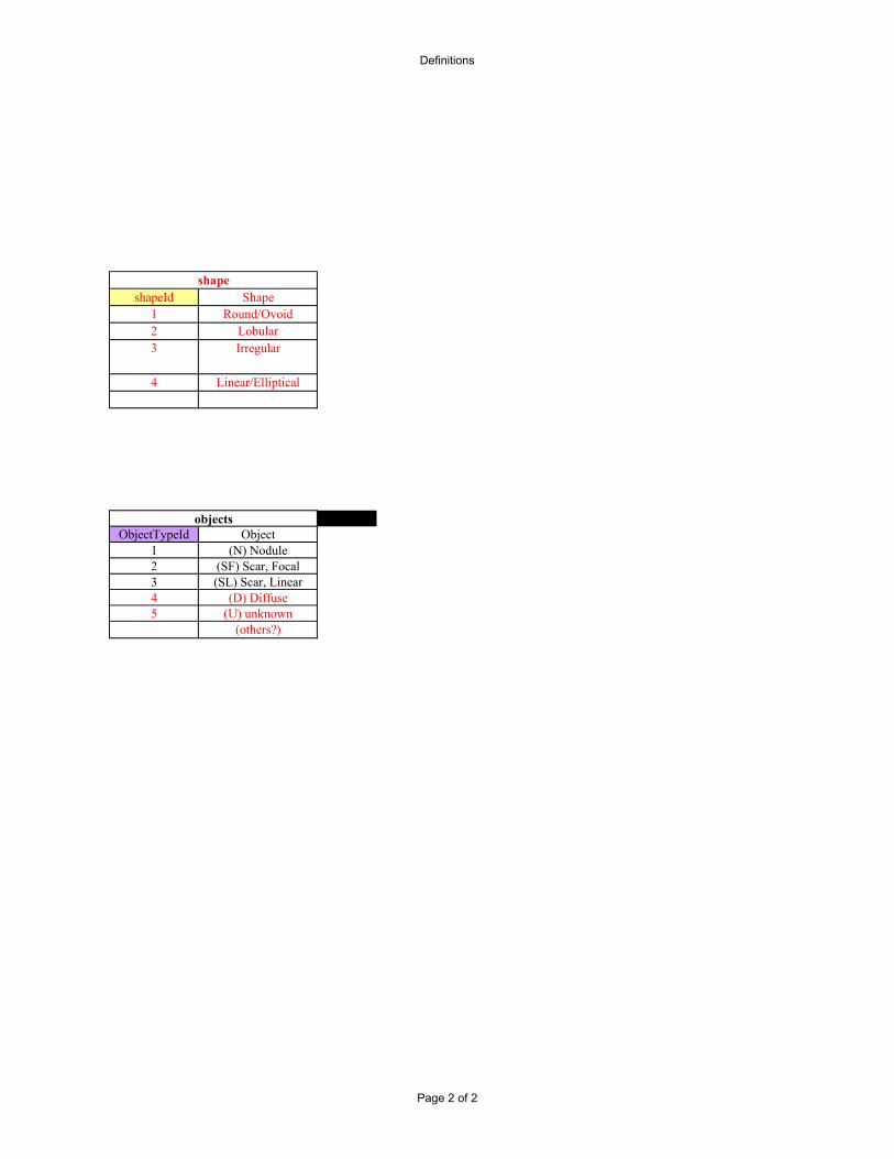
Reseach_Case-Study-Series
case Id (autoincr)
encrypted institutionRecordId
rel Institution Idrel Ethnicity Id
(study entry form)
Gender (0010 0040)
relSmoking History (from subject entry
form)
pack years
1 agj(&@#... 5 1 F 1 02 is^#&#... 5 4 M 2 523 ^&*^^^Kjj6... 5 5 M 4 300… Smoking history and pack years are required elements for data gathered prospectively
Tables/columns underlined with a black bar are necessary elements that must be stored (and retreivable) in a rudimentary database when the LIDC begins gathering preliminary data on/after Aug. 1.
4558 _)*GDXDS$$^ 3 4Note: all of the acquition/image/offset parameters are still in each of the stored DICOM headers because they have NOT been scrubbed; we are simply adding the above parameters to the database tables to make these values available for searches…
Tube Current (0018 1151)
Exposure (0018 1152)
Extremety Filter Type (0018 1160)
Focal Spot Size (0018
1190)
research caseAlthough the reseach case , study and series tables have encrypted patient/subject fields associated with them, in fact these encrypted fields are physically contained in separate tables that are unavailable for user searches/retreival. These encrypted tables are available to compiled system code to check for pre-existing data values to support grouping of new incoming data belonging to the same subject, study, and series, respectively, in the database tables.
Reconstruction Kernel (0018 1210)
Subject Position (0018 5100)
Data Collecion Diameter
(0018 0090)
Software Version (0018 1020)
Contrast/Bolus Route (0018 1040)
Reconstruction
Diameter (0018 1100)
encrypted Series Instance UID
(0020 000e)
The Study Instance UID defines a unique study, i.e. combination of acquired series. For each incoming image (DICOM image object), the Study Instance UID is extracted from the header and the study table is searched for the prior existance of the same Study Instance UID as stored in the encrypted Study Instance UID field. If the Study Instance UID already exists in the table, no further action is required. If not, then a new row is added to the study table and the indicated values are entered. The same studyId number is also entered in the same object's row in the series table below in the relStudyId field. The study date, not time, is preserved for calculation of intervals between multiple exams.
studyId (auto incr)
rel Case Idrel Modality Id
(0008 0060)
study Descriptive
Text
encrypted Study Instance UID
(0020 000d)
study
lung field: 1: full 2:
partial (radiologist definition
session entry)
acq&ReconParams
relSeries Description Id
(radiologist definition
session entry)
relStorageDirectory Id
(identifies loci of stored series'
image files)
relStudyId KVP (001
8 0060
)
Source-subject
Distance (0018 1111)
Gantry Tilt (0018 1120)
Source-Detecto
r Distance (0018 1110)
Exposure Time (0018 1150)
subject Age
(0010 1010)
relManu-facturer Id
(0008 0070)
model Name (0008 1090)
Although organizationally and logically the study tableprecedes the series table here, in fact the series table may bebuilt first since functionally all of the "objects", i.e. imagefiles, come from the scanner or archive as members of aseries and study. When the series table is built first, itsrelStudyId column is left blank until a later sorting of theseries occurs in order to create the study table and then theappropriate studyId number is carried into the series table'srelStudyId column.
series
Series Shared Outside of
LIDC? (Y/N)
Likewise the Series Instance UID for each incoming DICOM object (i.e. image file) defines a unique series, i.e. subject volume scan. For each incoming image (DICOM image object), the Series Instance UID is extracted from the header and the series table is searched for the prior existance of the same Series Instance UID as stored in the encrypted Series Instance UID field . If the Series Instance UID exists in the table, the object is stored in the storageDirectory specified in the column entry of the same name. If the Series Instance UID does not exist, a new row and seriesID is added to the series table and the object is stored in the new storageDirectory entry (how this is defined is left to each user institution). Data from the specified DICOM elements in the object header is used to fill out most of the remaining series table entries.
series Nodule NumberId (autoincr within same, most
recent series)
mostRecent nodule Cyto/Path (update here triggered by entry in relNoduleCytoPath table below)
1 990 1 1472 2844 13 2844 2... … … …
Date (these entries will come from the general Pathology & Cytology definition table when finished)
relShapeId
Data for this table comes from the first review of newly submitted cases by the local institution's LIDC expert. Identifying an object as a nodule (N) in this table generates a "to do" entry in the nodule definition worklist for 4 experts.
do we want to solicit nodule class, margin, shape, subtley, & actionable parameters on first read?
computedQuantitativeParams
relNodule Class
definitionFileId (XML format, D:expert def; P: prob def)
relNoduleId
objects identified in a series
relSeriesIdseriesObjectId
(auto incr)approxObjectLoc
(x,y,z pixel coords)
approxTextualLociDescription (Text String:
LUL, LLL, RUL, RML, RLL)relObjectType
This simple, but important table contains the one-to-many mapping that tracks nodules in the above table over exams for the same research caseId. The entry of a new nodule into this tableoccurs at the time of the expert's Redraw session (or initial Draw session?). Current process model specifies that each of 4 experts will draw nodule outlines at the initial setting and then redrawnodules at a second sitting while observing the resultant drawings of all experts from the intial sitting. Existing nodules are identified by an expert's viewing of the most recent CT series wherepreviously identified nodules in the series have been numbered. A new nodule is one that is not identified/numbered on the previous series and must be entered into this table. As numberingnodules will define existing nodules before reading the next CT series acquired, and assuming experts across institutions will identify nodules using an interface to their own local database,sequential numbering of all newly identified nodules within the same caseId must be consistent across participating institutions!
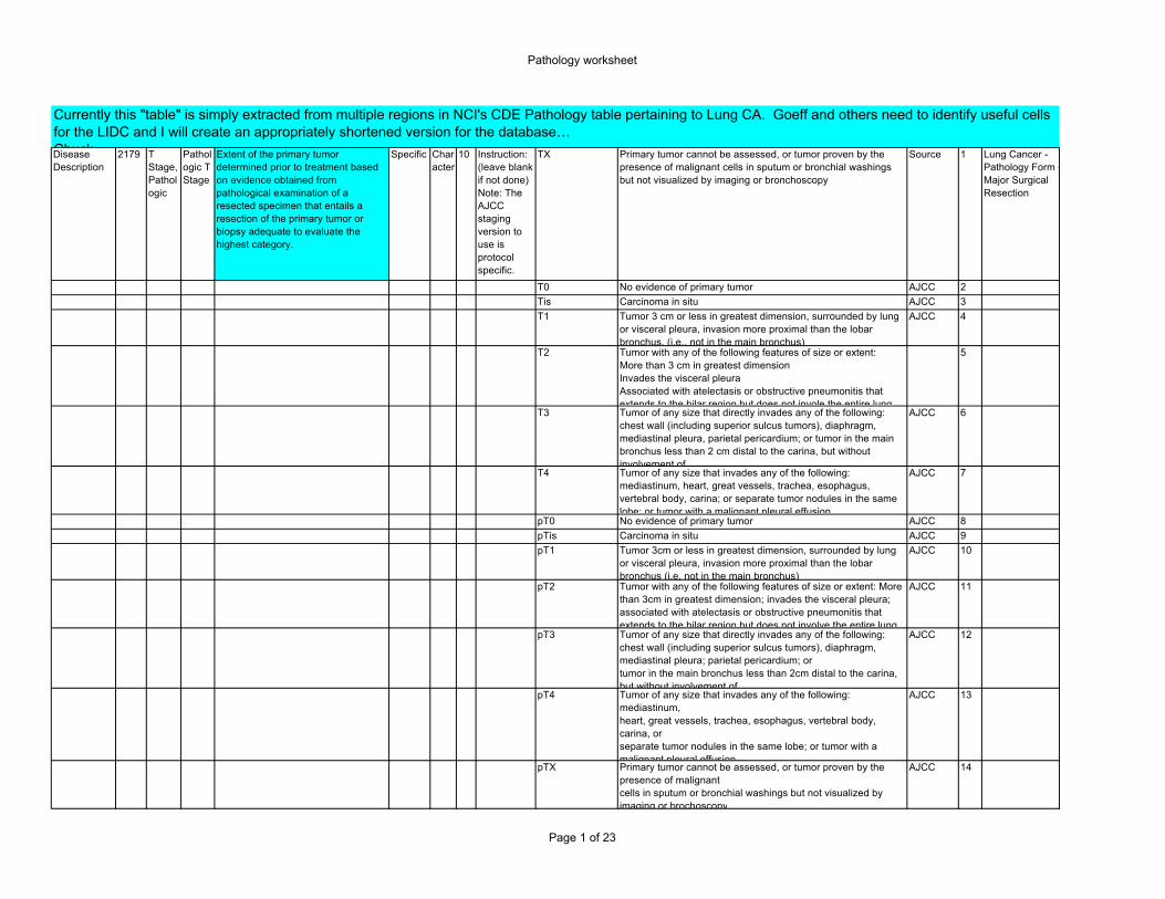
Extent of the primary tumor determined prior to treatment based on evidence obtained from pathological examination of a resected specimen that entails a resection of the primary tumor or biopsy adequate to evaluate the highest category.
Specific Character
10 Instruction: (leave blank if not done) Note: The AJCC staging version to use is protocol specific.
TX Primary tumor cannot be assessed, or tumor proven by the presence of malignant cells in sputum or bronchial washings but not visualized by imaging or bronchoscopy
Source 1 Lung Cancer - Pathology Form - Major Surgical Resection
T0 No evidence of primary tumor AJCC 2Tis Carcinoma in situ AJCC 3T1 Tumor 3 cm or less in greatest dimension, surrounded by lung
or visceral pleura, invasion more proximal than the lobar bronchus, (i.e., not in the main bronchus)
AJCC 4
T2 Tumor with any of the following features of size or extent: More than 3 cm in greatest dimension Invades the visceral pleura Associated with atelectasis or obstructive pneumonitis that extends to the hilar region but does not invole the entire lung
5
T3 Tumor of any size that directly invades any of the following: chest wall (including superior sulcus tumors), diaphragm, mediastinal pleura, parietal pericardium; or tumor in the main bronchus less than 2 cm distal to the carina, but without involvement of
AJCC 6
T4 Tumor of any size that invades any of the following: mediastinum, heart, great vessels, trachea, esophagus, vertebral body, carina; or separate tumor nodules in the same lobe; or tumor with a malignant pleural effusion
AJCC 7
pT0 No evidence of primary tumor AJCC 8pTis Carcinoma in situ AJCC 9pT1 Tumor 3cm or less in greatest dimension, surrounded by lung
or visceral pleura, invasion more proximal than the lobar bronchus (i.e. not in the main bronchus)
AJCC 10
pT2 Tumor with any of the following features of size or extent: More than 3cm in greatest dimension; invades the visceral pleura; associated with atelectasis or obstructive pneumonitis that extends to the hilar region but does not involve the entire lung
AJCC 11
pT3 Tumor of any size that directly invades any of the following: chest wall (including superior sulcus tumors), diaphragm, mediastinal pleura; parietal pericardium; or tumor in the main bronchus less than 2cm distal to the carina, but without involvement of
AJCC 12
pT4 Tumor of any size that invades any of the following: mediastinum, heart, great vessels, trachea, esophagus, vertebral body, carina, or separate tumor nodules in the same lobe; or tumor with a malignant pleural effusion
AJCC 13
pTX Primary tumor cannot be assessed, or tumor proven by the presence of malignantcells in sputum or bronchial washings but not visualized by imaging or brochoscopy
AJCC 14
Currently this "table" is simply extracted from multiple regions in NCI's CDE Pathology table pertaining to Lung CA. Goeff and others need to identify useful cells for the LIDC and I will create an appropriately shortened version for the database… Chuck
Page 1 of 23
Pathology worksheet
2181 N Stage, Pathologic
Pathologic N Stage
Extent of nodal involvement determined prior to treatment based on evidence obtained from removal of the regional lymph nodes adequate to validate the absence of regional lymph node metastasis and sufficient to evaluate the highest category.
Specific Character
10 (leave blank if not done)
NX Regional lymph nodes cannot be assessed AJCC 1 Lung Cancer - Pathology Form - Major Surgical Resection
N0 No regional lymph node metastasis AJCC 2N1 Metastasis to ipsilateral peribronchial and/or ipsilateral hilar
lymph nodes, and intrapulmonary nodes including involvement by direct extension of the primary tumor
AJCC 3
N2 Metastasis to ipsilateral mediastinal and/or subcarinal lymph node(s)
AJCC 4
N3 Metastasis to contralateral mediastinal, contralateral hilar, ipsilateral or contralateral scalene, or supraclavicular lymph node(s)
AJCC 5
pN0 No lymph node metastasis AJCC 6pN1 Metastasis to ipsilateral peribronchial and/or ipsilateral hilar
lymph nodes, and intrapulmonary nodes including involvement by direct extension of the primary tumor
AJCC 7
pN2 Metastasis to ipsilateral mediastinal and/or subcarinal lymph node(s)
AJCC 8
pN3 Metastasis to contralateral mediastinal, contralateral hilar, ipsilateral or contralateral scalene, or supraclavicular lymph node(s)
AJCC 9
pNX Regional lymph nodes cannot be assessed AJCC 10NX Regional lymph nodes cannot be assessed AJCC 1 Lung Cancer -
Surgery FormN0 No regional lymph node metastasis AJCC 2N1 Metastasis to ipsilateral peribronchial and/or ipsilateral hilar
lymph nodes, and intrapulmonary nodes including involvement by direct extension of the primary tumor
AJCC 3
N2 Metastasis to ipsilateral mediastinal and/or subcarinal lymph node(s)
AJCC 4
N3 Metastasis to contralateral mediastinal, contralateral hilar, ipsilateral or contralateral scalene, or supraclavicular lymph node(s)
AJCC 5
pN0 No lymph node metastasis AJCC 6pN1 Metastasis to ipsilateral peribronchial and/or ipsilateral hilar
lymph nodes, and intrapulmonary nodes including involvement by direct extension of the primary tumor
AJCC 7
pN2 Metastasis to ipsilateral mediastinal and/or subcarinal lymph node(s)
AJCC 8
pN3 Metastasis to contralateral mediastinal, contralateral hilar, ipsilateral or contralateral scalene, or supraclavicular lymph node(s)
AJCC 9
pNX Regional lymph nodes cannot be assessed AJCC 102183 M
Stage, Pathologic
Pathologic M Stage
Absence or presence of distant metastasis as determined prior to treatment based on evidence of tumor in microscopic examination of distant lesions
Specific Character
2 (leave blank if not done)
pM0 No distant metastasis AJCC 1 Lung Cancer - Pathology Form - Major Surgical Resection
Right lobar (12R) Right lobar node Cooperative Group-Forms Committee
15
Left lobar (12L) Left lobar node Cooperative Group-Forms Committee
16
Right segmental (13R)
Right segmental node Cooperative Group-Forms Committee
17
Left segmental (13L)
Left segmental node Cooperative Group-Forms Committee
18
Right subsegmental (14R)
Right subsegmental node Cooperative Group-Forms Committee
19
Left subsegmental (14L)
Left subsegmental node Cooperative Group-Forms Committee
20
#### Associated Pre-Malignant Histologic Changes
Associated Pre-Malignant Histologic Changes
The histologic changes in the specimen which are associated with pre-malignant conditions.
Specific Character
39 Add 2 instructions to form: If Pre-Malignant changes Observed, AND (check all that apply)
Basal cell hyperplasia
Basal cell hyperplasia CDE Committee
1 Lung Cancer - Pathology Form - Bronchoscopy
Page 7 of 23
Pathology worksheet
Basal cell hyperplasia with ASD changes
Basal cell hyperplasia with Angiogenic Squamous Dysplasia changes
CDE Committee
2
Squamous metaplasia
Squamous metaplasia CDE Committee
3
Squamous metaplasia with ASD changes
Squamous metaplasia with Angiogenic Squamous Dysplasia changes
CDE Committee
4
Carcinoma in situ Carcinoma in situ CDE Committee
5
Atypical adenomatous hyperplasia
Atypical adenomatous hyperplasia CDE Committee
6
Mild dysplasia Mild dysplasia CDE Committee
7
Mild dysplasia with ASD changes
Mild dyplasia with Angiogenic Squamous Dysplasia changes CDE Committee
8
Moderate dysplasia
Moderate dysplasia CDE Committee
9
Moderate dysplasia with ASD changes
Moderate dysplasia with Angiogenic Squamous Dysplasia changes
CDE Committee
10
Severe dysplasia Severe dysplasia CDE Committee
11
Severe dysplasia with ASD changes
Severe dysplasia with Angiogenic Squamous Dysplasia changes
CDE Committee
12
Other, specify Other, specify CDE Committee
13
Basal cell hyperplasia
Basal cell hyperplasia CDE Committee
1 Lung Cancer - Pathology Form - Cytology
Basal cell hyperplasia with ASD changes
Basal cell hyperplasia with Angiogenic Squamous Dysplasia changes
CDE Committee
2
Squamous metaplasia
Squamous metaplasia CDE Committee
3
Squamous metaplasia with ASD changes
Squamous metaplasia with Angiogenic Squamous Dysplasia changes
CDE Committee
4
Carcinoma in situ Carcinoma in situ CDE Committee
5
Atypical adenomatous hyperplasia
Atypical adenomatous hyperplasia CDE Committee
6
Mild dysplasia Mild dysplasia CDE Committee
7
Page 8 of 23
Pathology worksheet
Mild dysplasia with ASD changes
Mild dyplasia with Angiogenic Squamous Dysplasia changes CDE Committee
8
Moderate dysplasia
Moderate dysplasia CDE Committee
9
Moderate dysplasia with ASD changes
Moderate dysplasia with Angiogenic Squamous Dysplasia changes
CDE Committee
10
Severe dysplasia Severe dysplasia CDE Committee
11
Severe dysplasia with ASD changes
Severe dysplasia with Angiogenic Squamous Dysplasia changes
CDE Committee
12
Other, specify Other, specify CDE Committee
13
Basal cell hyperplasia
Basal cell hyperplasia CDE Committee
1 Lung Cancer - Pathology Form - Major Surgical Resection
Basal cell hyperplasia with ASD changes
Basal cell hyperplasia with Angiogenic Squamous Dysplasia changes
CDE Committee
2
Squamous metaplasia
Squamous metaplasia CDE Committee
3
Squamous metaplasia with ASD changes
Squamous metaplasia with Angiogenic Squamous Dysplasia changes
CDE Committee
4
Carcinoma in situ Carcinoma in situ CDE Committee
5
Atypical adenomatous hyperplasia
Atypical adenomatous hyperplasia CDE Committee
6
Mild dysplasia Mild dysplasia CDE Committee
7
Mild dysplasia with ASD changes
Mild dyplasia with Angiogenic Squamous Dysplasia changes CDE Committee
8
Moderate dysplasia
Moderate dysplasia CDE Committee
9
Moderate dysplasia with ASD changes
Moderate dysplasia with Angiogenic Squamous Dysplasia changes
CDE Committee
10
Severe dysplasia Severe dysplasia CDE Committee
11
Severe dysplasia with ASD changes
Severe dysplasia with Angiogenic Squamous Dysplasia changes
CDE Committee
12
Other, specify Other, specify CDE Committee
13
Page 9 of 23
Pathology worksheet
#### Bronchial Margin Involved Ind
Is the bronchial margin involved
A yes/no indicator to ask if the margins of the bronchi were involved or infiltrated by tumor.
Specific Character
7 Yes Yes CDE Committee
1 Lung Cancer - Pathology Form - Major Surgical Resection
No No CDE Committee
2
Unknown Unknown CDE Committee
3
no #### Bronchial Site
Bronchial Site
The site or area of the brochus where the sample was obtained.
Specific Character
36 Note: This is only a limited selection of valid values. The complete list can be viewed on the CDE website. Add 2 instructions to form: (check all that apply) AND Note: Only list values needed for
Carina, NOS Carina, not otherwise specified CDE Committee
1 Lung Cancer - Pathology Form - Bronchoscopy
Carina between RB1 and RB2, RB1/2
Carina between right bronchus 1 and right bronchus 2, right bronchus 1/2
CDE Committee
2
Carina between RB1 A and B, RB1A/B
Carina between right bronchus 1 A and B, right bronchus 1A/B CDE Committee
3
Carina between RB1 and RB3, RB1/3
Carina between right bronchus 1 and right bronchus 3, right bronchus 1/3
CDE Committee
4
Carina between RB2 and RB3, RB2/3
Carina between right bronchus 2 and right bronchus 3, right bronchus 2/3
CDE Committee
5
Carina between RB2 A and B, RB2A/B
Carina between right bronchus 2 A and B, right bronchus 2A/B CDE Committee
6
Carina between RB3 A and B, RB3A/B
Carina between right bronchus 3 A and B, right bronchus 3A/B CDE Committee
7
Carina between RB4 and RB5, RB4/5
Carina between right bronchus 4 and right bronchus 5, right bronchus 4/5
CDE Committee
8
Carina between RB4 A and B, RB4A/B
Carina between right bronchus 4 A and B, right bronchus 4A/B CDE Committee
9
Page 10 of 23
Pathology worksheet
Carina between RB5 A and B, RB5A/B
Carina between right bronchus 5 A and B, right bronchus 5A/B CDE Committee
10
Carina between RB6 A and B, RB6A/B
Carina between right bronchus 6 A and B, right bronchus 6A/B CDE Committee
11
Carina between RB6 A and C, RB6A/C
Carina between right bronchus 6 A and C, right bronchus 6A/C CDE Committee
12
Carina between RB6 B and C, RB6B/C
Carina between right bronchus 6 B and C, right bronchus 6B/C CDE Committee
13
Carina between RB7 A and B, RB7A/B
Carina between right bronchus 7 A and B, right bronchus 7A/B CDE Committee
14
Carina between RB8 and RB9, RB8/9
Carina between right bronchus 8 and right bronchus 9, right bronchus 8/9
CDE Committee
15
Carina between RB8 A and B, RB8A/B
Carina between right bronchus 8 A and B, right bronchus 8A/B CDE Committee
16
Carina between RB9 and RB10, RB9/10
Carina between right bronchus 9 and right bronchus 10, right bronchus 9/10
CDE Committee
17
Carina between RB 9 A and B, RB9A/B
Carina between right bronchus 9 A and B, right bronchus 9A/B CDE Committee
18
Carina between RB10 A and B, RB10A/B
Carina between right bronchus 10 A and B, right bronchus 10A/B
CDE Committee
19
Carina between RB10 A and C, RB10A/C
Carina between right bronchus 10 A and C, right bronchus 10A/C
CDE Committee
20
Carina between RB10 B and C, RB10B/C
Carina between right bronchus 10 B and C, right bronchus 10B/C
CDE Committee
21
Carina between LB1 and LB2, LB1/2
Carina between left bronchus 1 and left bronchus 2, left bronchus 1/2
CDE Committee
22
Carina between LB1 A and B, LB1A/B
Carina between left bronchus 1 A and B, left bronchus 1A/B CDE Committee
23
Carina between LB1 A and C, LB1A/C
Carina between left bronchus 1 A and C, left bronchus 1A/C CDE Committee
24
Carina between LB1 B and C, LB1B/C
Carina between left bronchus 1 B and C, left bronchus 1B/C CDE Committee
25
Carina between LB2 A and B, LB2A/B
Carina between left bronchus 2 A and B, left bronchus 2A/B CDE Committee
26
Carina between LB2 A and C, LB2A/C
Carina between left bronchus 2 A and C, left bronchus 2A/C CDE Committee
27
Carina between LB2 B and C, LB2B/C
Carina between left bronchus 2 B and C, left bronchus 2B/C CDE Committee
28
Page 11 of 23
Pathology worksheet
Carina between LB1+2 and LB3, LB1+2/3
Carina between left bronchus 1+2 and left bronchus 3, left bronchus 1+2/3
CDE Committee
29
Carina between LB3 A and B, LB3A/B
Carina between left bronchus 3 A and B, left bronchus 3A/B CDE Committee
30
Carina between LB4 and LB5, LB4/5
Carina between left bronchus 4 and left bronchus 5, left bronchus 4/5
CDE Committee
31
Carina between LB4 A and B, LB4A/B
Carina between left bronchus 4 A and B, left bronchus 4A/B CDE Committee
32
Carina between LB5 A and B, LB5A/B
Carina between left bronchus 5 A and B, left bronchus 5A/B CDE Committee
33
Carina between LB6 A and B, LB6A/B
Carina between left bronchus 6 A and B, left bronchus 6A/B CDE Committee
34
Carina between LB6 A and C, LB6A/C
Carina between left bronchus 6 A and C, left bronchus 6A/C CDE Committee
35
Carina between LB6 B and C, LB6B/C
Carina between left bronchus 6 B and C, left bronchus 6B/C CDE Committee
36
Carina between LB8 and LB9, LB8/9
Carina between left bronchus 8 and left bronchus 9, left bronchus 8/9
CDE Committee
37
Carina between LB8 A and B, LB8A/B
Carina between left bronchus 8 A and B, left bronchus 8A/B CDE Committee
38
Carina between LB9 and LB10, LB9/10
Carina between left bronchus 9 and left bronchus 10, left bronchus 9/10
CDE Committee
39
Carina between LB9 A and B, LB9A/B
Carina between left bronchus 9 A and B, left bronchus 9A/B CDE Committee
40
Carina between LB10 A and B, LB10A/B
Carina between left bronchus 10 A and B, left bronchus 10A/B CDE Committee
41
Carina between LB10 A and C, LB10A/C
Carina between left bronchus 10 A and C, left bronchus 10A/C CDE Committee
42
Carina between LB10 B and C, LB10B/C
Carina between left bronchus 10 B and C, left bronchus 10B/C CDE Committee
43
no #### Bronchoscopy Type
Type of Bronchoscopy
The type or method of bronchoscopic procedure utilized.
Specific Character
12 Add instruction to form: (check all that apply)
Fluorescence Fluorescence CDE Committee
1 Lung Cancer - Pathology Form - Bronchoscopy
Rigid Rigid CDE Committee
2
White light White light CDE Committee
3
Other, specify Other, specify CDE Committee
4
Page 12 of 23
Pathology worksheet
Yes Yes CDE Committee
1 Lung Cancer - Pathology Form - Major Surgical Resection
No No CDE Committee
2
Unknown Unknown CDE Committee
3
#### Parietal Pleural Margin Involved Ind
Is the parietal pleural margin involved
A yes/no indicator to ask if the margins of the parietal pleura were involved or infiltrated by tumor.
Specific Character
7 Yes Yes CDE Committee
1 Lung Cancer - Pathology Form - Major Surgical Resection
No No CDE Committee
2
Unknown Unknown CDE Committee
3
#### Perineural Involvement Ind
Perineural tumor infiltration or invasion
A yes/no indicator to ask if perineural infiltration or invasion of the tumor is present.
Specific Character
7 Yes Yes CDE Committee
1 Lung Cancer - Pathology Form - Major Surgical Resection
No No CDE Committee
2
Unknown Unknown CDE Committee
3
#### Pleura Involved Ind
Does the tumor involve the pleura
A yes/no indicator to ask if the pleura is involved or infiltrated by the tumor.
Specific Character
7 Yes Yes CDE Committee
1 Lung Cancer - Pathology Form - Major Surgical Resection
No No CDE Committee
2
Unknown Unknown CDE Committee
3
#### Pleural Margin Involved Ind
Is the pleural margin involved
A yes/no indicator to ask if the margins of the pleura (visceral and/or parietal) were involved or infiltrated by tumor.
Specific Character
7 Yes Yes CDE Committee
1 Lung Cancer - Pathology Form - Major Surgical Resection
No No CDE Committee
2
Page 13 of 23
Pathology worksheet
Unknown Unknown CDE Committee
3
#### Primary Tumor Multiple Ind
Are there multiple primary tumors
A yes/no indicator to ask if multiple primary tumors were identified.
Specific Character
7 Yes Yes CDE Committee
1 Lung Cancer - Pathology Form - Cytology
No No CDE Committee
2
Unknown Unknown CDE Committee
3
Yes Yes CDE Committee
1 Lung Cancer - Pathology Form - Major Surgical Resection
No No CDE Committee
2
Unknown Unknown CDE Committee
3
#### Residual Disease Site, Gross
Sites of Gross Residual Disease
The sites of residual macroscopic or gross disease.
Specific Character
58 Add 2 instructions to form: (check all that apply) AND Surgeon's assessment or from operative report
Lung, at original tumor site
Lung, at original tumor site CDE Committee
1 Lung Cancer - Pathology Form - Major Surgical Resection
Blood vessels (main pulmonary artery or vein)
Blood vessels (main pulmonary artery or vein) CDE Committee
2
Lung, elsewhere Lung, elsewhere CDE Committee
3
Ipsilateral mediastinal nodes
Ipsilateral mediastinal nodes CDE Committee
4
Bronchial resection margin
Bronchial resection margin CDE Committee
5
Contralateral mediastinal nodes
Contralateral mediastinal nodes CDE Committee
6
Vascular resection margin
Vascular resection margin CDE Committee
7
Mediastinal structures (other than nodes)
Mediastinal structures (other than nodes) CDE Committee
Blood vessels (aorta, innominate vein, superior vena cava)
Blood vessels (aorta, innominate vein, superior vena cava) CDE Committee
11
Contralateral hilar nodes
Contralateral hilar nodes CDE Committee
12
Page 15 of 23
Pathology worksheet
#### Stage Grouping, Pathologic
Stage Grouping, Pathologic
The pathologic stage grouping. Specific Character
4 0 0, according to current AJCC guidelines AJCC 1 Lung Cancer - Pathology Form - Major Surgical Resection
IA IA, according to current AJCC guidelines AJCC 2IB IB, according to current AJCC guidelines AJCC 3IIA IIA, according to current AJCC guidelines AJCC 4IIB IIB, according to current AJCC guidelines AJCC 5IIIA IIIA, according to current AJCC guidelines AJCC 6IIIB IIIB, according to current AJCC guidelines AJCC 7IV IV, according to current AJCC guidelines AJCC 8
#### Surgical Margin Involved Ind
Does the tumor involve the surgical margin
A yes/no indicator to ask if the margins of surgical resection are involved or infiltrated by the tumor.
Specific Character
7 Yes Yes CDE Committee
1 Lung Cancer - Pathology Form - Major Surgical Resection
No No CDE Committee
2
Unknown Unknown CDE Committee
3
#### Surgical Margin Involved Microscopically Ind
Are surgical margins microscopically involved
A yes/no indicator to ask if the margins of surgical resection were determined to be involved or infiltrated by the tumor via microscopic evaluation.
Specific Character
7 Yes Yes CDE Committee
1 Lung Cancer - Pathology Form - Major Surgical Resection
No No CDE Committee
2
Unknown Unknown CDE Committee
3
#### Tumor Extension
Tumor Extension
A field to indicate the organs and structures to which the tumor has become adherent or has invaded.
Specific Character
56 Add instruction to form: (check all that apply)
Chest wall Chest wall CDE Committee
1
Mediastinum Mediastinum CDE Committee
2
Esophagus Esophagus CDE Committee
3
Pericardium Pericardium CDE Committee
4
Page 16 of 23
Pathology worksheet
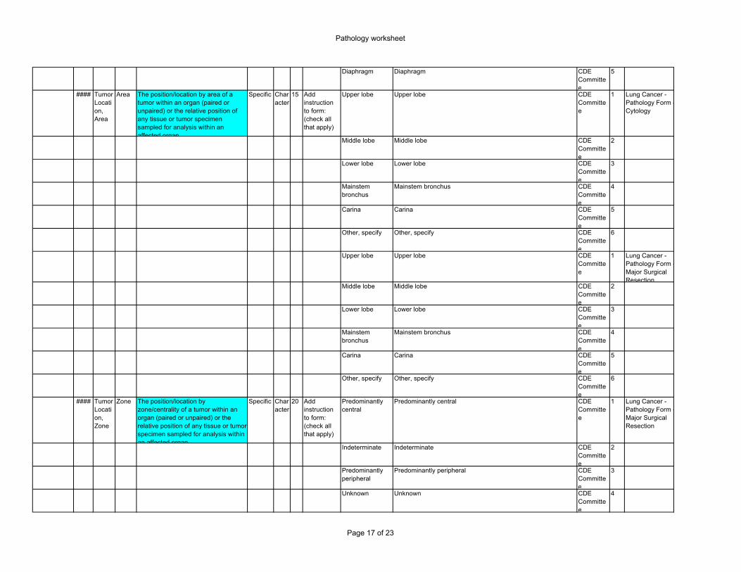
Diaphragm Diaphragm CDE Committee
5
#### Tumor Location, Area
Area The position/location by area of a tumor within an organ (paired or unpaired) or the relative position of any tissue or tumor specimen sampled for analysis within an affected organ.
Specific Character
15 Add instruction to form: (check all that apply)
Upper lobe Upper lobe CDE Committee
1 Lung Cancer - Pathology Form - Cytology
Middle lobe Middle lobe CDE Committee
2
Lower lobe Lower lobe CDE Committee
3
Mainstem bronchus
Mainstem bronchus CDE Committee
4
Carina Carina CDE Committee
5
Other, specify Other, specify CDE Committee
6
Upper lobe Upper lobe CDE Committee
1 Lung Cancer - Pathology Form - Major Surgical Resection
Middle lobe Middle lobe CDE Committee
2
Lower lobe Lower lobe CDE Committee
3
Mainstem bronchus
Mainstem bronchus CDE Committee
4
Carina Carina CDE Committee
5
Other, specify Other, specify CDE Committee
6
#### Tumor Location, Zone
Zone The position/location by zone/centrality of a tumor within an organ (paired or unpaired) or the relative position of any tissue or tumor specimen sampled for analysis within an affected organ.
Specific Character
20 Add instruction to form: (check all that apply)
Predominantly central
Predominantly central CDE Committee
1 Lung Cancer - Pathology Form - Major Surgical Resection
Indeterminate Indeterminate CDE Committee
2
Predominantly peripheral
Predominantly peripheral CDE Committee
3
Unknown Unknown CDE Committee
4
Page 17 of 23
Pathology worksheet
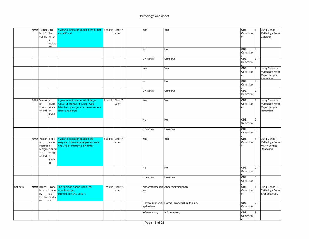
#### Tumor Multifocal Ind
Are the tumors multifocal
A yes/no indicator to ask if the tumor is multifocal.
Specific Character
7 Yes Yes CDE Committee
1 Lung Cancer - Pathology Form - Cytology
No No CDE Committee
2
Unknown Unknown CDE Committee
3
Yes Yes CDE Committee
1 Lung Cancer - Pathology Form - Major Surgical Resection
No No CDE Committee
2
Unknown Unknown CDE Committee
3
#### Vascular Invasion Ind
Is there vascular invasion
A yes/no indicator to ask if large vessel or venous invasion was detected by surgery or presence in a tumor specimen.
Specific Character
7 Yes Yes CDE Committee
1 Lung Cancer - Pathology Form - Major Surgical Resection
No No CDE Committee
2
Unknown Unknown CDE Committee
3
#### Visceral Pleural Margin Involved Ind
Is the visceral pleural margin involved
A yes/no indicator to ask if the margins of the visceral pleura were involved or infiltrated by tumor.
Specific Character
7 Yes Yes CDE Committee
1 Lung Cancer - Pathology Form - Major Surgical Resection
No No CDE Committee
2
Unknown Unknown CDE Committee
3
not path #### Bronchoscopy Findings
Bronchoscopic Findings
The findings based upon the bronchoscopic examination/evaluation.
Specific Character
27 Abnormal/malignant
Abnormal/malignant CDE Committee
1 Lung Cancer - Pathology Form - Bronchoscopy
Normal bronchial epithelium
Normal bronchial epithelium CDE Committee
2
Inflammatory Inflammatory CDE Committee
3
Page 18 of 23
Pathology worksheet
Unknown Unknown CDE Committee
4
Other, specify Other, specify CDE Committee
5
not path #### Inflammatory Change
Inflammatory Change
The amount of inflammatory change present in the specimen.
Specific Character
25 <10% inflammatory cells
specimen or sample is comprised of less than 10% inflammatory cells
CDE Committee
1 Lung Cancer - Pathology Form - Bronchoscopy
10-75% inflammatory cells
specimen or sample is comprised of 10% to 75% (inclusive) inflammatory cells
CDE Committee
2
>75% inflammatory cells
specimen or sample is comprised of more than 75% inflammatory cells
CDE Committee
3
not path #### Sample Period
Sample Period
The timeframe of specimen collection relative to treatment and diagnosis.
Specific Character
14 Pre-treatment sample or specimen collected before patient received treatment for cancer
CDE Committee
1 Lung Cancer - Pathology Form - Cytology
Pre-diagnosis sample or specimen collected before patient was diagnosed with cancer
CDE Committee
2
Post-treatment sample or specimen collected after patient received treatment for cancer
CDE Committee
3
Unknown unknown CDE Committee
4
Other, specify other, specify CDE Committee
5
Pre-treatment sample or specimen collected before patient received treatment for cancer
CDE Committee
1 Lung Cancer - Pathology Form - Major Surgical Resection
Pre-diagnosis sample or specimen collected before patient was diagnosed with cancer
CDE Committee
2
Post-treatment sample or specimen collected after patient received treatment for cancer
CDE Committee
3
Unknown unknown CDE Committee
4
Other, specify other, specify CDE Committee
5
don’t use #### Specimen Cell Source
Source of Cells used for Analysis
A field to describe the type of sample cells used for analysis for pathology.
Specific Character
25 Non-malignant lung
Non-malignant lung CDE Committee
1 Lung Cancer - Pathology Form - Bronchoscopy
Primary tumor Primary tumor CDE Committee
2
Page 19 of 23
Pathology worksheet
Lymph node Lymph node CDE Committee
3
Bronchus, specify
Bronchus CDE Committee
4
Bronchial tissue, suspicious for abnormality
Bronchial tissue, suspicious for abnormality CDE Committee
5
Dysplastic site Dysplastic site CDE Committee
6
Normal tissue Normal tissue CDE Committee
7
Other, specify CDE Committee
8
Non-malignant lung
Non-malignant lung CDE Committee
1 Lung Cancer - Pathology Form - Cytology
Primary tumor Primary tumor CDE Committee
2
Lymph node Lymph node CDE Committee
3
Bronchus, specify
Bronchus CDE Committee
4
Bronchial tissue, suspicious for abnormality
Bronchial tissue, suspicious for abnormality CDE Committee
5
Dysplastic site Dysplastic site CDE Committee
6
Normal tissue Normal tissue CDE Committee
7
Other, specify CDE Committee
8
Non-malignant lung
Non-malignant lung CDE Committee
1 Lung Cancer - Pathology Form - Major Surgical Resection
Primary tumor Primary tumor CDE Committee
2
Lymph node Lymph node CDE Committee
3
Bronchus, specify
Bronchus CDE Committee
4
Bronchial tissue, suspicious for abnormality
Bronchial tissue, suspicious for abnormality CDE Committee
5
Page 20 of 23
Pathology worksheet
Dysplastic site Dysplastic site CDE Committee
6
Normal tissue Normal tissue CDE Committee
7
Other, specify CDE Committee
8
use at top of path form
#### Specimen Collection Method
How was the specimen obtained
The type of procedure or method used to collect the specimen.
Specific Character
31 Biopsy sample or specimen collected via biopsy CDE Committee
1 Lung Cancer - Pathology Form - Cytology
Sputum, spontaneous
sample or specimen collected via spontaneous sputum CDE Committee
2
Sputum, induced sample or specimen collected via induced sputum CDE Committee
3
Pleural effusion sample or specimen collected via pleural effusion CDE Committee
4
Pericardial effusion
sample or specimen collected via pericardial effusion CDE Committee
5
Abdominal/ascites effusion
sample or specimen collected via abdominal effusion or ascites CDE Committee
6
Fine needle aspiration, specify site
sample or specimen collected via fine needle aspiration CDE Committee
7
Bronchial alveolar lavage (BAL)
sample or specimen collected via bronchial alveolar lavage CDE Committee
8
Pleural lavage sample or specimen collected via pleural lavage CDE Committee
9
Bronchial brushing/washing
sample or specimen collected via bronchial brushing or washing
CDE Committee
10
Mediastinoscopy sample or specimen collected via mediastinoscopy CDE Committee
11
Other, specify other, specify CDE Committee
12
#### Specimen Condition
Condition of Specimen
The condition or adequacy of the specimen as received.
Specific Character
12 Satisfactory Satisfactory CDE Committee
1 Lung Cancer - Pathology Form - Bronchoscopy
Suboptimal Suboptimal CDE Committee
2
Inadequate Inadequate CDE Committee
3
Page 21 of 23
Pathology worksheet
Patient Characteristics
2572 Surgical Approach
Surgical Approach
The surgical technique used to acquire tissue for a definitive diagnosis.
Specific Character
70 thoracotomy CDE Committee
1 Lung Cancer - Pathology Form - Major Surgical Resection
thoracoscopy CDE Committee
2
thoracoscopy / video-assisted (VATS), with conversion to thoracotomy
CDE Committee
3
thoracoscopy / video-assisted (VATS)
4
thoracotomy CDE Committee
1 Lung Cancer - Surgery Form
thoracoscopy CDE Committee
2
thoracoscopy / video-assisted (VATS), with conversion to thoracotomy
CDE Committee
3
thoracoscopy / video-assisted (VATS)
4
thoracotomy CDE Committee
1 Lung Cancer NSCLC Stage I-III On-Study Form
thoracoscopy CDE Committee
2
thoracoscopy / video-assisted (VATS), with conversion to thoracotomy
CDE Committee
3
thoracoscopy / video-assisted (VATS)
4
bottom form #### Associated Diseases
Diseases Associated with Cancer
A field to identify other diseases associated with the cancer and affecting the same organs and structures being treated.
Specific Character
25 Add instruction to form: (check all that apply)
Pneumonia Pneumonia CDE Committee
1 Lung Cancer - Pathology Form - Major Surgical Resection
A field to identify other diseases related to granulomatous disease associated with the cancer and affecting the same organs and structures being treated.
Specific Character
12 Tuberculosis Tuberculosis CDE Committee
1 Lung Cancer - Pathology Form - Major Surgical Resection
Sarcoid Sarcoid CDE Committee
2
Fungal Fungal CDE Committee
3
#### Associated Diseases, Pneumoconiosis
If pneumoconiosis, specify
A field to identify other diseases related with pneumoconiosis associated with the cancer and affecting the same organs and structures being treated.
Specific Character
10 Asbestosis Asbestosis CDE Committee
1 Lung Cancer - Pathology Form - Major Surgical Resection