Research ArticleComparison of the Biological Characteristics of MesenchymalStem Cells Derived from Bone Marrow and Skin
Ruifeng Liu,1 Wenjuan Chang,1 Hong Wei,2 and Kaiming Zhang1
1 Institute of Dermatology, Taiyuan City Centre Hospital, Shanxi Provincial Key Laboratory of Immunological Skin Diseases,No. 1 Dong San Dao Xiang, Taiyuan, Shanxi 030009, China2Department of Dermatology, Zibo City First Hospital, Shandong 255200, China
Correspondence should be addressed to Kaiming Zhang; [email protected]
Received 13 January 2016; Revised 18 March 2016; Accepted 6 April 2016
Mesenchymal stem cells (MSCs) exhibit high proliferation and self-renewal capabilities and are critical for tissue repair andregeneration during ontogenesis. They also play a role in immunomodulation. MSCs can be isolated from a variety of tissuesand have many potential applications in the clinical setting. However, MSCs of different origins may possess different biologicalcharacteristics. In this study, we performed a comprehensive comparison of MSCs isolated from bone marrow and skin (BMMSCsand SMSCs, resp.), including analysis of the skin sampling area, separationmethod, culture conditions, primary and passage culturetimes, cell surface markers, multipotency, cytokine secretion, gene expression, and fibroblast-like features. The results showed thatthe MSCs from both sources had similar cell morphologies, surface markers, and differentiation capacities. However, the two celltypes exhibited major differences in growth characteristics; the primary culture time of BMMSCs was significantly shorter thanthat of SMSCs, whereas the growth rate of BMMSCs was lower than that of SMSCs after passaging. Moreover, differences in geneexpression and cytokine secretion profiles were observed. For example, secretion of proliferative cytokines was significantly higherfor SMSCs than for BMMSCs. Our findings provide insights into the different biological functions of both cell types.
1. Introduction
Mesenchymal stem cells (MSCs) are adherent stromal cellsthat were first isolated from the bone marrow [1] and arecharacterized by their ability to differentiate into mesenchy-mal tissues such as bone, cartilage, and fat. In addition,MSCs have been shown to suppress immune responses [2–5]. Because of these properties, MSCs have recently gainedincreasing attention from researchers and have now beenshown to be present in a variety of tissues, including theumbilical cord, placenta, adipose tissues, and skin [6–11].MSCs derived from different tissues may have some uniquebiological characteristics.
In a previous study, we found that the biological behaviorsof bone marrow MSCs (BMMSCs) in patients with psoriasiswere abnormal [12, 13]. Because psoriasis is a type of skindisease associated with immune abnormalities, the biologicalcharacteristics of MSCs from psoriatic skin lesions maymoreaccurately reflect the features of psoriasis. Indeed, analysis of
MSCs from psoriatic lesions showed that these cells exhibitabnormalities in gene expression, cytokine secretion, andimmune properties [14–16]. Moreover, BMMSCs and MSCsisolated from skin (SMSCs) have been shown to have differentproperties. Although themethods for isolation and culture ofBMMSCs have been extensively studied, culture methods forSMSCs are not yet optimized, and some researchers believethat SMSCs may actually be fibroblasts [17].
Therefore, in the current study, we performed a compre-hensive comparison ofMSCs from the two sources, includinganalysis of the skin sampling area, separationmethod, cultureconditions, primary and passage culture times, cell surfacemarkers, multipotency, cytokine secretion, gene expression,and fibroblast-like features.
2. Material and Methods
2.1. Participants. All volunteers provided informed consentfor their participation in the study. The protocol involving
human subjects was approved by the Medical Ethics Com-mittee of Taiyuan City Centre Hospital and was performed inaccordance with the 1964 Declaration of Helsinki and its lateramendments or comparable ethical standards.
Twenty bone marrow samples were from normal bonemarrow donors, and 20 sex- and age-matched volunteersfrom the Urology and Plastic Surgery Department, TaiyuanCity Centre Hospital, were enrolled in this study.
2.2. Reagents. Cell culture plates and plastic flasks werepurchased from Corning Incorporated (Corning, NY, USA).Dulbecco’s modified Eagle’s medium (DMEM)/F12 medium,B-27 supplement, fetal bovine serum (FBS), and Per-coll were purchased from Invitrogen (Grand Island, NY,USA). Trypsin, dispase enzyme II, recombinant humanbasic fibroblast growth factor (bFGF), and toluidine bluewere purchased from Sigma-Aldrich (St. Louis, MO, USA).Mouse monoclonal antibodies against human stem cellfactor (SCF), granulocyte colony-stimulating factor (G-CSF), granulocyte-macrophage colony-stimulating factor(GM-CSF), macrophage colony-stimulating factor (M-CSF),interleukin-1 (IL-1), IL-3, IL-6, IL-7, IL-8, IL-11, epider-mal growth factor (EGF), vascular endothelial growth fac-tor (VEGF), tumor necrosis factor-𝛼 (TNF-𝛼), leukemiainhibitory factor (LIF), hepatocyte growth factor (HGF),and transforming growth factor-𝛽1 (TGF-𝛽1), as well ashorseradish peroxidase- (HRP-) labeled rabbit antibodiesagainst mouse IgG, were obtained from Abcam (Cambridge,UK). Phycoerythrin- (PE-) or fluorescein isothiocyanate-(FITC-) labeled mouse antibodies against human CD29,CD44, CD73, CD90, CD105, CD14, CD34, CD45, and humanleukocyte antigen- (HLA-) DR were purchased from BDBiosciences (San Jose, CA, USA). The IMT2 inverted phase-contrast microscope was obtained from Olympus (Tokyo,Japan). Type 352 automatic microplate reader was obtainedfrom Labsystems (Helsinki, Finland), and the EPICS-XLFlow Cytometer FACSCalibur was obtained from BeckmanCoulter (Los Angeles, CA, USA).
2.3. MSC Separation and Cultivation and Measurement of theCulture Time. Human BMMSCs were grown from aspiratestaken from the posterior superior iliac spine of healthy volun-teers. Five milliliters of heparinized aspirate was diluted 1 : 2with DMEM/F12 medium and centrifuged through a Percolldensity gradient at 700×g for 20min. The mononuclear cellsat the interface were collected, washed twice withDMEM/F12medium, resuspended at a concentration of 1 × 10⁶ cells/mLin complete medium (DMEM/F12 supplemented with 10%FBS, 100U/mL penicillin, and 100 𝜇g/mL streptomycin), andplated at 1 × 10⁶ cells/well in 24-well plates. The cells wereincubated at 37∘C in a humidified atmosphere supplementedwith 5%CO
2. Twodays later, nonadherent cells were removed
by replacing the medium. Half of the medium was thenchanged every 4 days. At 90% confluency, the cells weredetached by incubation with 0.25% trypsin, diluted 1 : 2 withcomplete medium, and then at 5 × 10⁴ cells/well in 24-wellplates. Cell growth was observed daily under an invertedphase-contrast microscope; growth morphology, the level
of confluence, and the culture time were recorded. Theculture time for primary cells was defined as the time frominoculation of mononuclear cells to 90% confluence, theculture time for cells of passage 1 was defined as the time frominoculation of passage 1 cells to 90% confluence, and so forth.
For isolation of SMSCs, skin specimens were cut into1mm3 tissue blocks under sterile conditions and thendigested with 0.25% dispase enzyme II at 37∘C for 2–4 h.The epidermis and dermis were separated mechanically; thedermis was collected and finelyminced. DMEM/F12mediumcontaining 10% FBS was added to the minced dermis, andthe cells were separated by pipetting. After filtering througha 40 𝜇m aperture sieve, the filtrate was allowed to stand onice for 20–30min, after which it was centrifuged at 200×gfor 5min. The supernatant was discarded and the filtrate wasadded to culture medium to resuspend the cells. The cellswere then cultured in DMEM/F12 medium supplementedwith 10% FBS, 10 ng/mL bFGF, 20𝜇L/mL B27 supplement,100U/mL penicillin, and 100 𝜇g/mL streptomycin. The cul-ture was inoculated into T25 plastic flasks at a density of1 × 105 cells/cm2 and kept in an incubator at 37∘C, with 5%CO2and saturated humidity. After 72 h, the medium was
removed, and all suspended cells were discarded. Freshlypreparedmedium, as described above, was added to continuethe cultivation of the adherent cells. Half of the medium wasreplaced every fifth day. When cells had grown to nearly90% confluence, they were digested with 0.25% trypsin andtransferred to subcultures at 5 × 10⁴ cells/well in 24-wellplates. After passaging, the cells were cultured in the mediumas described above but without bFGF. Cell growth wasobserved daily under an inverted phase-contrast microscope;growth morphology, the level of confluence, and the culturetime were recorded. The culture time for primary cells wasdefined as the time from cell inoculation to 90% confluence,the culture time for cells of passage 1 was defined as the timefrom inoculation of passage 1 cells to 90% confluence, and soforth.
2.4. Cultivation of Skin Fibroblasts. To differentiate betweenSMSCs and skin fibroblasts and to ascertain whether the cul-tivated cells were MSCs rather than fibroblasts, we culturedskin fibroblasts using explant culture techniques [18]. Skinspecimens were cut into 2mm3 blocks and washed twicewith phosphate-buffered saline (PBS) containing antibiotics.After seeding the specimen blocks into T25 flasks using anaspirator, they were distributed evenly on the bottom ofthe flasks. The optimal distance between each seeded blockwas determined to be 0.5 cm, and each flask contained 20specimen blocks.The flasks were tilted and filled with 0.5mLof DMEM/F12 containing 10% FBS and then kept in anincubator at 37∘C with 5% CO
2and saturated humidity for
4 h. After the tissues adhered to the surface, an additional4mL of DMEM/F12 containing 10% FBS was added carefullyto prevent resuspension of the tissue blocks. The flasks werereturned to the incubator, and the culture medium waschanged every 3-4 days. After the seeded cells had grown to90% confluence, they were passaged and subcultured into 24-well culture plates.
Stem Cells International 3
2.5. Identification of Cell Purity and Collection of CellMedium.BMMSCs at passage 3 or SMSCs at passage 5 and theirculture supernatants were collected from each of the 20wells. Culture supernatants were stored in sterile tubes at−20∘C after filtering through a 0.45𝜇m filter for enzyme-linked immunosorbent assays (ELISAs) to determine thecytokine contents. Cells (BMMSCs at passage 3, SMSCs andskin fibroblasts at passage 5) were detached with 0.25%trypsin, washed, and resuspended in PBS. Cells (2 × 10⁵)were incubated in the dark with PE- or FITC-labeled mouseantibodies against the human cell surface markers CD29,CD44, CD73, CD90, CD105, CD14, CD34, CD45, and HLA-DR for 30min. After washing with PBS, the cells weresubjected to two-color flow-cytometric analysis to examinethe proportion of cells positive for the respective antigens.
2.6. Multipotent Differentiation of MSCs and Identificationof the Differentiated Cells. BMMSCs at passage 3 or SMSCsat passage 5 were induced to differentiate into lipocytes,osteoblasts, or chondrocytes.The specific induction methodswere described previously [15]. After adipogenic differenti-ation for 10 days, the cells were fixed with 10% formalin,washed with 60% isopropyl alcohol, and stained with oilred O. After osteogenic differentiation for 3 weeks, thecells were fixed with 10% formalin and stained with 2%alizarin red solution. After chondrogenic differentiation for21 days, micromasses were fixed with 4% paraformaldehyde,embedded in optimal cutting temperature compound, cutinto 5 𝜇m sections, and stained with toluidine blue. Nega-tive controls, for which differentiation-inducing supplementswere omitted from the culture medium, were included foreach differentiation assay.
The fifth-passage fibroblasts were induced to differentiateinto fat, bone, and cartilage as described above.
2.7. Quantification of Cytokines Secreted into the MSCMedium. Cytokine content was measured based on directELISA. A 96-well plate was coated overnight at 4∘C with50𝜇L medium from BMMSC and SMSC cultures per well.After washing of the plate, 200 𝜇L of 0.25% gelatin wasadded per well, and the plate was incubated for 2 h atroom temperature (RT). Primary antibodies (50 𝜇L, diluted1 : 100) were introduced into the wells, and the plates wereincubated for 1 h at RT. After washing away excess primaryantibody, 50 𝜇L of HRP-labeled secondary antibody (diluted1 : 1000) was added to the wells and incubated for 45min at37∘C. Finally, after washing off excess labeled antibody, HRPenzyme activity was determined by the o-phenylenediaminedihydrochloride reaction, which was terminated by adding1M H
2SO4after incubation for 10min at RT. The concentra-
tion of each cytokine was calculated using CurveExpert Basic1.40 software (https://www.curveexpert.net/).
2.8. Microarray Analysis. Total RNA was purified from eachsample (𝑛 = 8) using an RNeasy mini kit (Qiagen Valencia,CA, USA) according to themanufacturer’s instructions. RNAintegrity was assessed using standard denaturing agarose gelelectrophoresis. RNA quantity and quality were evaluated
using NanoDrop ND-1000 spectrophotometer (NanoDropTechnologies, Wilmington, DE, USA).
The RNA was amplified and labeled using an AgilentLow Input Quick Amp Labeling Kit (Agilent Technologies,Waldbronn, Germany) and then hybridized to the Agi-lent Whole Human Genome Oligo Microarray. The arraydata were extracted using the Agilent Feature Extractionsoftware (version 10.7.3.1). Global mean normalization wasperformed, and the probes with a signal intensity <800 orcoefficient of variation of intensity of <20% in all samples,which represent the low-abundance and housekeeping genes,respectively, were selected for further analysis. Unsupervisedhierarchical cluster analysis was performed using Cluster3.0 software (http://www.falw.vu/∼huik/cluster.htm). Differ-entially expressed genes (fold change, >2.0) with statisticalsignificance (𝑝 < 0.05) were identified using volcano plotfiltering. Significant enrichment of gene ontology (GO) termswas analyzed using the hypergeometric distribution in the Rlanguage package software (https://cran.r-project.org/), withstatistical thresholds of 𝑝 < 0.05 and false discovery rate(FDR) < 0.05.
2.9. Statistical Analysis. Data were expressed as the mean ±SD. Independent sample 𝑡-tests were used to compare themean values of samples from bone marrow and skin inSPSS16.0 software (SPSS Inc., Chicago, IL, USA). Differenceswith 𝑝 values of less than 0.05 were considered statisticallysignificant.
3. Results
3.1. Morphological Features and Culture Times of BMMSCsand SMSCs. The cell morphologies of BMMSCs and SMSCswere similar. Isolated BMMSCs attached to the bottomsof the plates after incubation for 24 h. On days 7–10, thecells showed obvious enlargement and proliferation, formingsmall colonies with several to tens of fusocellular, triangular,and polygonal cells. The cells displayed typical fibroblastmorphology with multilayered flat cell bodies having shortcell processes connected to adjacent cells (Figure 1(a)). Atapproximately day 16, the cells reached 90% confluence(Figure 1(b)).When treated with trypsin, they became round;after reattachment to the plate and incubation for 24 h, thecell morphology reverted to the primary BMMSC shape.Thecells reached 90% confluence after incubation for an averageof 12 days.
A small number of adherent cells appeared 72 h afterprimary SMSCs were seeded; these cells then graduallyincreased in number and became significantly larger. Afew cells were triangular or polygonal in shape; however,most were basically short or long spindle-shaped and hada fibroblast-like morphology. Cell bodies were enlarged andhad cytoplasmic projections of various lengths and sizes,which were interconnected; the cells overlapped and pro-liferated in a stratified fashion, as shown in Figure 1(c).The time required for cultivation of the primary cell cultureto 90% confluence was 29 days (Figure 1(d)). Subculturedcells had rounded shapes after digestion with 0.25% trypsinbut returned to their original shapes after 24 h, showing
4 Stem Cells International
(a) (b)
(c) (d)
Figure 1: Morphological characteristics of bone marrow mesenchymal stem cells (BMMSCs) and skin mesenchymal stem cells (SMSCs). (a)BMMSCs cultured for 7 days. (b) BMMSCs cultured for 16 days. (c) SMSCs cultured for 16 days. (d) SMSCs cultured for 29 days. Scale bar:10𝜇m.
adherence and proliferation, while retaining morphologiessimilar to those of the primary cells. The required level ofconfluence was reached within 3-4 days.
Table 1 shows the culture times of BMMSCs and SMSCs.The primary culture time of BMMSCs was significantlyshorter than that of SMSCs (16.35 ± 4.38 versus 28.85 ± 5.52days, resp.; 𝑝 < 0.001). However, the growth rate of BMMSCswas lower than that of SMSCs after passage (time of passage1, 12.25 ± 4.49 versus 3.85 ± 1.09 days; 𝑝 < 0.001).
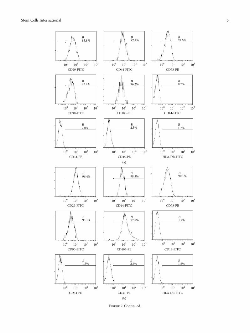
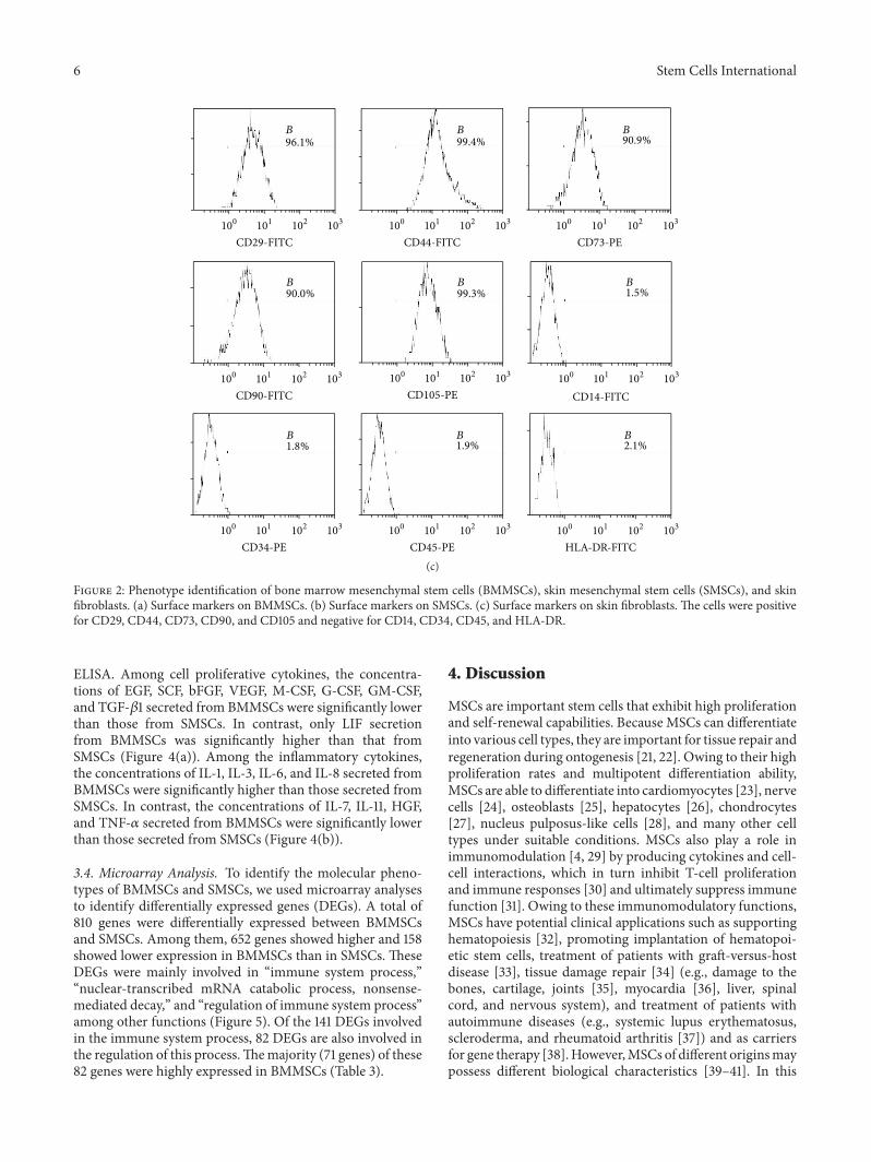
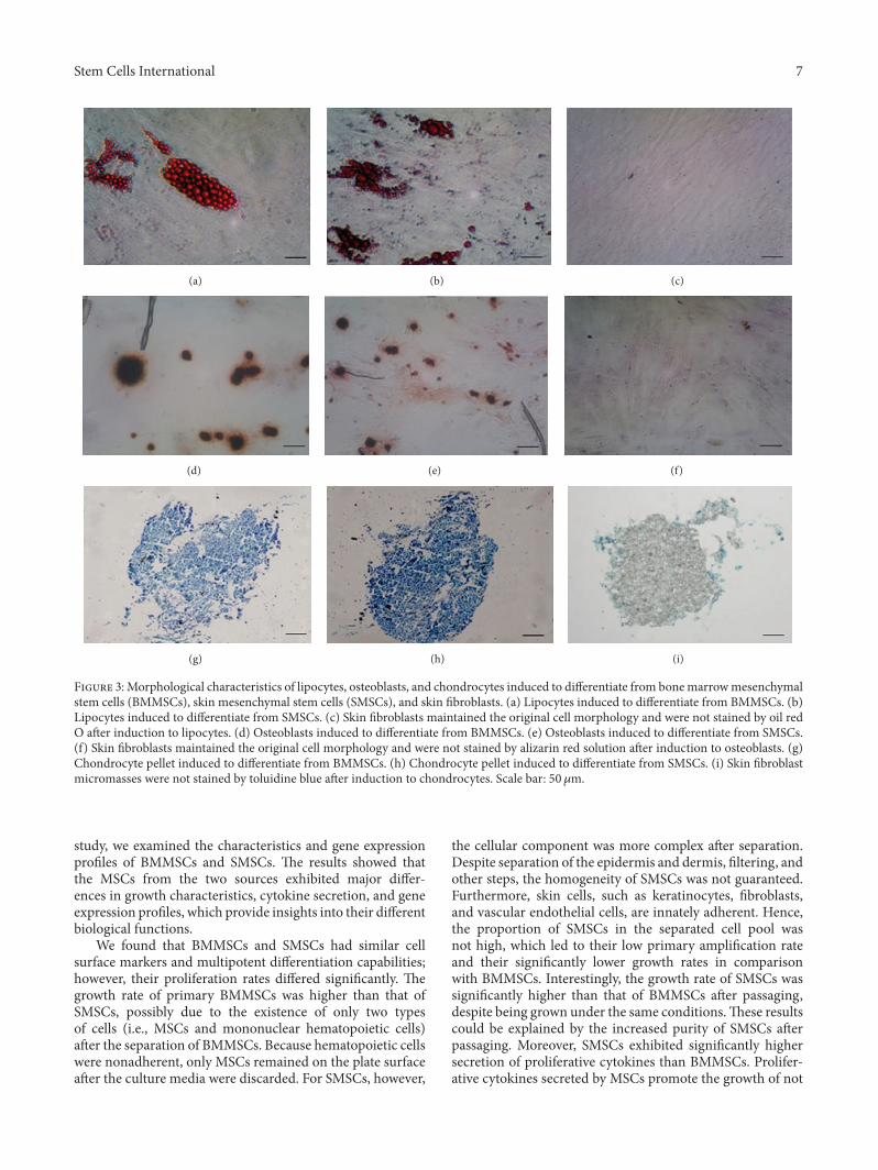
3.2. Identification of BMMSCs, SMSCs, and Fibroblasts. Flowcytometry results showed that the purity of BMMSCs atpassage 3 reached up to 90%, whereas that of SMSCs reachedonly 70%. The purity of SMSCs was more than 90% afterthe fifth passage. Expansion of BMMSCs results in gradualloss of osteogenic potential after passages 5-6 [19]. Therefore,in order to guarantee the purity of the cells and avoid theloss of cell biological characteristics as a result of passaging,we used BMMSCs at passage 3 and SMSCs at passage 5 forfollow-up experiments. Flow-cytometric analysis of surfaceantigens of both groups of MSCs and fibroblasts showed highexpression levels of CD29, CD44, CD73, CD90, and CD105and negative expression of CD14, CD34, CD45, andHLA-DRin all three cell types (Figure 2). The MSCs all differentiatedinto the relevant cells and tissues after adipogenic, osteogenic,and chondrogenic induction (Figures 3(a), 3(b), 3(d), 3(e),
Table 1: Culture times for MSCs isolated from bone marrow andskin (mean ± SD, days).
3(g), and 3(h)), indicating that the isolated and cultured cellsmet the identification criteria for MSCs [20]. Control cellsdid not show these important stem cell characteristics (datanot presented). However, the skin fibroblasts of passage 5maintained the original cell morphology after induction, andno positive results were observed after staining (Figures 3(c),3(f), and 3(i)). These data suggested that fibroblasts were notdifferentiated into fat, bone, and cartilage.
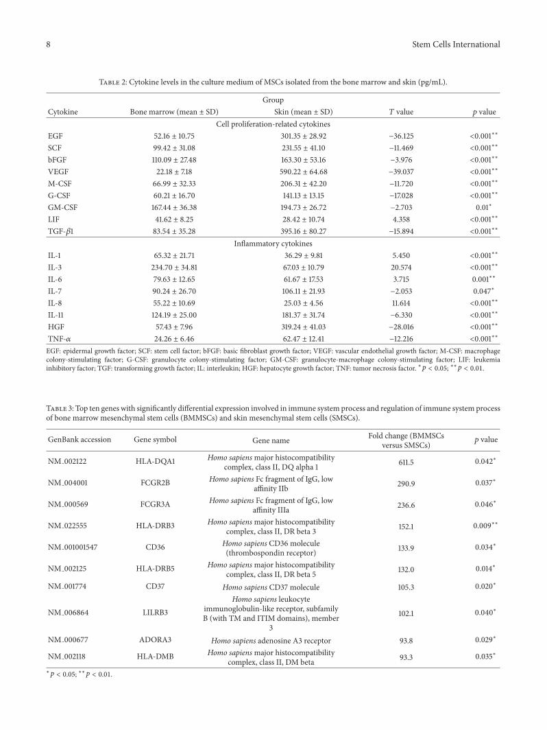
3.3. Differential Secretion of Cytokines from BMMSCs andSMSCs. Table 2 shows the cytokine contents in culturemediafrom BMMSC and SMSC cultures, as measured by direct
Stem Cells International 5
92.4%
101 102 103100
CD90-FITC
96.2%
101 102 103100
CD105-PE
0.7%
101 102 103100
CD14-FITC
2.3%
103100 101 102
CD45-PE
1.7%
101 102 103100
HLA-DR-FITC
2.0%
101 102 103100
CD34-PE
91.6%
101 102 103100
CD73-PE
97.7%
101 102 103100
CD44-FITC
95.8%
100 102 103101
CD29-FITC
B B B
B B B
B B B
(a)
96.4%
101 102 103100
CD29-FITC
90.1%
101 102 103100
CD73-PE
98.5%
101 102 103100
CD44-FITC
93.1%
101 102 103100
CD90-FITC
97.9%
101 102 103100
CD105-PE
1.2%
101 102 103100
CD14-FITC
2.6%
103100 101 102
CD45-PE
1.6%
101 102 103100
HLA-DR-FITC
1.5%
101 102 103100
CD34-PE
B B B
B B B
B B B
(b)
Figure 2: Continued.
6 Stem Cells International
96.1%
101 102 103100
CD29-FITC
90.9%
101 102 103100
CD73-PE
99.4%
101 102 103100
CD44-FITC
90.0%
101 102 103100
CD90-FITC
99.3%
101 102 103100
CD105-PE
1.5%
101 102 103100
CD14-FITC
1.9%
103100 101 102
CD45-PE
2.1%
101 102 103100
HLA-DR-FITC
1.8%
101 102 103100
CD34-PE
B B B
B B B
B B B
(c)
Figure 2: Phenotype identification of bone marrow mesenchymal stem cells (BMMSCs), skin mesenchymal stem cells (SMSCs), and skinfibroblasts. (a) Surface markers on BMMSCs. (b) Surface markers on SMSCs. (c) Surface markers on skin fibroblasts. The cells were positivefor CD29, CD44, CD73, CD90, and CD105 and negative for CD14, CD34, CD45, and HLA-DR.
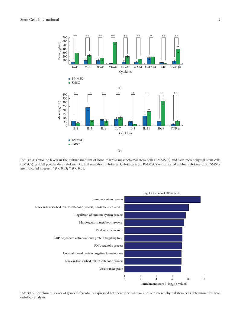
ELISA. Among cell proliferative cytokines, the concentra-tions of EGF, SCF, bFGF, VEGF, M-CSF, G-CSF, GM-CSF,and TGF-𝛽1 secreted from BMMSCs were significantly lowerthan those from SMSCs. In contrast, only LIF secretionfrom BMMSCs was significantly higher than that fromSMSCs (Figure 4(a)). Among the inflammatory cytokines,the concentrations of IL-1, IL-3, IL-6, and IL-8 secreted fromBMMSCs were significantly higher than those secreted fromSMSCs. In contrast, the concentrations of IL-7, IL-11, HGF,and TNF-𝛼 secreted from BMMSCs were significantly lowerthan those secreted from SMSCs (Figure 4(b)).
3.4. Microarray Analysis. To identify the molecular pheno-types of BMMSCs and SMSCs, we used microarray analysesto identify differentially expressed genes (DEGs). A total of810 genes were differentially expressed between BMMSCsand SMSCs. Among them, 652 genes showed higher and 158showed lower expression in BMMSCs than in SMSCs. TheseDEGs were mainly involved in “immune system process,”“nuclear-transcribed mRNA catabolic process, nonsense-mediated decay,” and “regulation of immune system process”among other functions (Figure 5). Of the 141 DEGs involvedin the immune system process, 82 DEGs are also involved inthe regulation of this process.Themajority (71 genes) of these82 genes were highly expressed in BMMSCs (Table 3).
4. Discussion
MSCs are important stem cells that exhibit high proliferationand self-renewal capabilities. Because MSCs can differentiateinto various cell types, they are important for tissue repair andregeneration during ontogenesis [21, 22]. Owing to their highproliferation rates and multipotent differentiation ability,MSCs are able to differentiate into cardiomyocytes [23], nervecells [24], osteoblasts [25], hepatocytes [26], chondrocytes[27], nucleus pulposus-like cells [28], and many other celltypes under suitable conditions. MSCs also play a role inimmunomodulation [4, 29] by producing cytokines and cell-cell interactions, which in turn inhibit T-cell proliferationand immune responses [30] and ultimately suppress immunefunction [31]. Owing to these immunomodulatory functions,MSCs have potential clinical applications such as supportinghematopoiesis [32], promoting implantation of hematopoi-etic stem cells, treatment of patients with graft-versus-hostdisease [33], tissue damage repair [34] (e.g., damage to thebones, cartilage, joints [35], myocardia [36], liver, spinalcord, and nervous system), and treatment of patients withautoimmune diseases (e.g., systemic lupus erythematosus,scleroderma, and rheumatoid arthritis [37]) and as carriersfor gene therapy [38].However,MSCs of different originsmaypossess different biological characteristics [39–41]. In this
Stem Cells International 7
(a) (b) (c)
(d) (e) (f)
(g) (h) (i)
Figure 3:Morphological characteristics of lipocytes, osteoblasts, and chondrocytes induced to differentiate from bonemarrowmesenchymalstem cells (BMMSCs), skin mesenchymal stem cells (SMSCs), and skin fibroblasts. (a) Lipocytes induced to differentiate from BMMSCs. (b)Lipocytes induced to differentiate from SMSCs. (c) Skin fibroblasts maintained the original cell morphology and were not stained by oil redO after induction to lipocytes. (d) Osteoblasts induced to differentiate from BMMSCs. (e) Osteoblasts induced to differentiate from SMSCs.(f) Skin fibroblasts maintained the original cell morphology and were not stained by alizarin red solution after induction to osteoblasts. (g)Chondrocyte pellet induced to differentiate from BMMSCs. (h) Chondrocyte pellet induced to differentiate from SMSCs. (i) Skin fibroblastmicromasses were not stained by toluidine blue after induction to chondrocytes. Scale bar: 50𝜇m.
study, we examined the characteristics and gene expressionprofiles of BMMSCs and SMSCs. The results showed thatthe MSCs from the two sources exhibited major differ-ences in growth characteristics, cytokine secretion, and geneexpression profiles, which provide insights into their differentbiological functions.
We found that BMMSCs and SMSCs had similar cellsurface markers and multipotent differentiation capabilities;however, their proliferation rates differed significantly. Thegrowth rate of primary BMMSCs was higher than that ofSMSCs, possibly due to the existence of only two typesof cells (i.e., MSCs and mononuclear hematopoietic cells)after the separation of BMMSCs. Because hematopoietic cellswere nonadherent, only MSCs remained on the plate surfaceafter the culture media were discarded. For SMSCs, however,
the cellular component was more complex after separation.Despite separation of the epidermis and dermis, filtering, andother steps, the homogeneity of SMSCs was not guaranteed.Furthermore, skin cells, such as keratinocytes, fibroblasts,and vascular endothelial cells, are innately adherent. Hence,the proportion of SMSCs in the separated cell pool wasnot high, which led to their low primary amplification rateand their significantly lower growth rates in comparisonwith BMMSCs. Interestingly, the growth rate of SMSCs wassignificantly higher than that of BMMSCs after passaging,despite being grown under the same conditions.These resultscould be explained by the increased purity of SMSCs afterpassaging. Moreover, SMSCs exhibited significantly highersecretion of proliferative cytokines than BMMSCs. Prolifer-ative cytokines secreted by MSCs promote the growth of not
8 Stem Cells International
Table 2: Cytokine levels in the culture medium of MSCs isolated from the bone marrow and skin (pg/mL).
GroupCytokine Bone marrow (mean ± SD) Skin (mean ± SD) 𝑇 value 𝑝 value
Table 3: Top ten genes with significantly differential expression involved in immune system process and regulation of immune system processof bone marrow mesenchymal stem cells (BMMSCs) and skin mesenchymal stem cells (SMSCs).
GenBank accession Gene symbol Gene name Fold change (BMMSCsversus SMSCs)
𝑝 value
NM 002122 HLA-DQA1 Homo sapiensmajor histocompatibilitycomplex, class II, DQ alpha 1 611.5 0.042∗
NM 004001 FCGR2B Homo sapiens Fc fragment of IgG, lowaffinity IIb 290.9 0.037∗
NM 000569 FCGR3A Homo sapiens Fc fragment of IgG, lowaffinity IIIa 236.6 0.046∗
NM 022555 HLA-DRB3 Homo sapiensmajor histocompatibilitycomplex, class II, DR beta 3 152.1 0.009∗∗
NM 001001547 CD36 Homo sapiens CD36 molecule(thrombospondin receptor) 133.9 0.034∗
NM 002125 HLA-DRB5 Homo sapiensmajor histocompatibilitycomplex, class II, DR beta 5 132.0 0.014∗
NM 001774 CD37 Homo sapiens CD37 molecule 105.3 0.020∗
NM 006864 LILRB3Homo sapiens leukocyte
immunoglobulin-like receptor, subfamilyB (with TM and ITIM domains), member
3
102.1 0.040∗
NM 000677 ADORA3 Homo sapiens adenosine A3 receptor 93.8 0.029∗
NM 002118 HLA-DMB Homo sapiensmajor histocompatibilitycomplex, class II, DM beta 93.3 0.035∗
Figure 4: Cytokine levels in the culture medium of bone marrow mesenchymal stem cells (BMMSCs) and skin mesenchymal stem cells(SMSCs). (a) Cell proliferative cytokines. (b) Inflammatory cytokines. Cytokines from BMMSCs are indicated in blue; cytokines from SMSCsare indicated in green. ∗𝑝 < 0.05; ∗∗𝑝 < 0.01.
SRP-dependent cotranslational protein targeting to. . .
RNA catabolic process
Cotranslational protein targeting to membrane
Nuclear-transcribed mRNA catabolic process
Viral transcription
0 4 62 8 10
Sig. GO terms of DE gene-BP
Immune system process
Enrichment score (−log10(p value))
Figure 5: Enrichment scores of genes differentially expressed between bone marrow and skin mesenchymal stem cells determined by geneontology analysis.
10 Stem Cells International
only neighboring cells, but also their own. Thus, the growthrate of later generations of SMSCs was significantly higherthan that of BMMSCs.
In our study, the cultivation conditions of both typesof MSCs were different. Because the purity and growthrates of the original SMSCs were low, we supplemented thecultures with 10% FBS (similar to the culture medium usedfor BMMSCs), bFGF, and B27 additive to the DMEM/F12culture medium. bFGF is an important mitogenic factor thatpromotes cellular proliferative activity and thus promotesthe growth of the original SMSCs. The B27 additive caninhibit the growth of fibroblasts, particularly those found individed cell pools. Previously reported cultivation conditionsfor SMSCs have been variable. Some researchers have usedmedium containing no bFGF and B27 additives, only MSCgrowth medium alpha-modification (𝛼-MEM) plus 10% FBS[42], or MSC growth medium (MSCGM) plus 10% FBS [43].We also attempted to use culture medium without bFGF; allcultured SMSCs did not show any differences from thosegrown with bFGF in terms of their cellular morphologies,immunomodulatory responses, and differentiation capabili-ties, with the exception of the lower growth rate of the originalpool of MSCs.
Although the purity of SMSCs was markedly increasedafter passaging, it was still lower than that of BMMSCs. Thisstudy showed that the third generation of BMMSCs reached apurity of more than 90%, while SMSCs reached only around70% purity at the third generation and more than 90% purityat the fifth generation.This findingmay be associatedwith thecomplex cellular components of the separated SMSCs; thus,we used the third generation of BMMSCs and fifth generationof SMSCs for subsequent studies. Overall, primary SMSCsgrew slower than BMMSCs, while, after passaging, SMSCsgrew faster than BMMSCs. Both cell types required a similaramount of time to reach the same level of purity.
To distinguish SMSCs from skin fibroblasts and to con-firm that our cultured cells wereMSCs, we used tissue culturetechniques to cultivate skin fibroblasts. After passaging to thefifth generation and inducing differentiation, skin fibroblastsdid not have the same characteristics as the SMSCs anddid not differentiate into lipocytes, osteoblasts, and chon-drocytes. Thus, the SMSCs and fibroblasts were indeed twodifferent types of cells. In addition, we found that successfulculturing of SMSCs required a minimum sample coveragearea. If the area was too small, the number of MSCs after sep-aration was insufficient for effective growth, resulting in cul-ture failure. Fromour experience, the coverage area should beat least 2 cm2 for effective culturing of the desired type of cells.
The results of microarray analysis showed that BMMSCsand SMSCs displayed different gene expression. The DEGswere mainly related to the immune system and immuneregulation, indicating that the immune and immune regula-tion functions of BMMSCs and SMSCs are different. Amongthe 82 DEGs involved in immune regulation, 71 were highlyexpressed in BMMSCs. This result suggests that the functionof immune regulation is more active in BMMSCs than inSMSCs.This finding can provide guidance for the clinical useof different sources of MSCs.
In summary, in this study, we compared the morpho-logical and molecular features of BMMSCs and SMSCs.Our results showed that these two types of MSCs exhibitsome unique features. However, we have only compared twodifferent sources of MSCs and studied only a few aspects ofcell biology. Our study represents an initial investigation ofthe broad range of applications and sources of MSCs. Moreextensive studies are required to facilitate thewide applicationof MSCs.
Competing Interests
The authors declare that they have no competing interests.
Acknowledgments
This work was supported by the National Natural ScienceFoundation of China (NFSC Grants nos. 81271768 and81472888).
References
[1] A. J. Friedenstein, R. K. Chailakhyan, and U. V. Gerasimov,“Bone marrow osteogenic stem cells: in vitro cultivation andtransplantation in diffusion chambers,” Cell and Tissue Kinetics,vol. 20, no. 3, pp. 263–272, 1987.
[2] M. Di Nicola, C. Carlo-Stella, M. Magni et al., “Humanbonemarrow stromal cells suppress T-lymphocyte proliferationinduced by cellular or nonspecificmitogenic stimuli,”Blood, vol.99, no. 10, pp. 3838–3843, 2002.
[3] P. Batten, P. Sarathchandra, J. W. Antoniw et al., “Humanmesenchymal stem cells induce T cell anergy and downregulateT cell allo-responses via the TH2 pathway: relevance to tissueengineering human heart valves,”Tissue Engineering, vol. 12, no.8, pp. 2263–2273, 2006.
[4] F. Gieseke, J. Bohringer, R. Bussolari, M. Dominici, R. Hand-gretinger, and I. Muller, “Human multipotent mesenchymalstromal cells use galectin-1 to inhibit immune effector cells,”Blood, vol. 116, no. 19, pp. 3770–3779, 2010.
[5] K. Nemeth, A. Keane-Myers, J. M. Brown et al., “Bone marrowstromal cells use TGF-𝛽 to suppress allergic responses in amouse model of ragweed-induced asthma,” Proceedings of theNational Academy of Sciences of the United States of America,vol. 107, no. 12, pp. 5652–5657, 2010.
[6] A. J. Cutler, V. Limbani, J. Girdlestone, and C. V. Navarrete,“Umbilical cord-derived mesenchymal stromal cells modulatemonocyte function to suppress T cell proliferation,”The Journalof Immunology, vol. 185, no. 11, pp. 6617–6623, 2010.
[7] Z. Miao, J. Jin, L. Chen et al., “Isolation of mesenchymalstem cells from human placenta: comparison with human bonemarrow mesenchymal stem cells,” Cell Biology International,vol. 30, no. 9, pp. 681–687, 2006.
[8] W.-C. Son, J.-W. Yun, and B.-H. Kim, “Adipose-derived mes-enchymal stem cells reduceMMP-1 expression inUV-irradiatedhuman dermal fibroblasts: therapeutic potential in skin wrin-kling,” Bioscience, Biotechnology and Biochemistry, vol. 79, no.6, pp. 919–925, 2015.
[9] C. Vaculik, C. Schuster, W. Bauer et al., “Human dermisharbors distinct mesenchymal stromal cell subsets,” Journal ofInvestigative Dermatology, vol. 132, no. 3, part 1, pp. 563–574,2012.
Stem Cells International 11
[10] A. Campanati, M. Orciani, S. Gorbi, F. Regoli, R. Di Primio,and A. Offidani, “Effect of biologic therapies targeting tumournecrosis factor-𝛼 on cutaneous mesenchymal stem cells inpsoriasis,” British Journal of Dermatology, vol. 167, no. 1, pp. 68–76, 2012.
[11] M. Orciani, S. Gorbi, M. Benedetti et al., “Oxidative stressdefense in human-skin-derived mesenchymal stem cells versushuman keratinocytes: different mechanisms of protection andcell selection,” Free Radical Biology and Medicine, vol. 49, no. 5,pp. 830–838, 2010.
[12] K. Zhang, R. Liu, G. Yin, X. Li, J. Li, and J. Zhang, “Differentialcytokine secretion of cultured bone marrow stromal cellsfrom patients with psoriasis and healthy volunteers,” EuropeanJournal of Dermatology, vol. 20, no. 1, pp. 49–53, 2010.
[13] R. Hou, R. Liu, X. Niu et al., “Biological characteristics and geneexpression pattern of bone marrow mesenchymal stem cells inpatients with psoriasis,” Experimental Dermatology, vol. 23, no.7, pp. 521–523, 2014.
[14] R. Hou, G. Yin, P. An et al., “DNAmethylation of dermal MSCsin psoriasis: identification of epigenetically dysregulated genes,”Journal of Dermatological Science, vol. 72, no. 2, pp. 103–109,2013.
[15] R. Liu, Y. Yang, X. Yan, and K. Zhang, “Abnormalities incytokine secretion from mesenchymal stem cells in psoriaticskin lesions,” European Journal of Dermatology, vol. 23, no. 5,pp. 600–607, 2013.
[16] R. Liu, Y. Wang, X. Zhao, Y. Yang, and K. Zhang, “Lymphocyteinhibition is compromised in mesenchymal stem cells frompsoriatic skin,” European Journal of Dermatology, vol. 24, no. 5,pp. 560–567, 2014.
[17] M. A. Haniffa, M. P. Collin, C. D. Buckley, and F. Dazzi,“Mesenchymal stem cells: the fibroblasts’ new clothes?”Haema-tologica, vol. 94, no. 2, pp. 258–263, 2009.
[18] C. Bogdan, N. Donhauser, R. Doring, M. Rollinghoff, A.Diefenbach, and M. G. Rittig, “Fibroblasts as host cells in latentleishmaniosis,” Journal of ExperimentalMedicine, vol. 191, no. 12,pp. 2121–2129, 2000.
[19] S. Halfon, N. Abramov, B. Grinblat, and I. Ginis, “Markersdistinguishing mesenchymal stem cells from fibroblasts aredownregulated with passaging,” Stem Cells and Development,vol. 20, no. 1, pp. 53–66, 2011.
[20] M. Dominici, K. Le Blanc, I. Mueller et al., “Minimal crite-ria for defining multipotent mesenchymal stromal cells. TheInternational Society for Cellular Therapy position Statement,”Cytotherapy, vol. 8, no. 4, pp. 315–317, 2006.
[21] Y.Chen, Y. Yu, L. Chen et al., “Humanumbilical cordmesenchy-mal stem cells: a new therapeutic option for tooth regeneration,”Stem Cells International, vol. 2015, Article ID 549432, 11 pages,2015.
[22] C. Zhang, H. Yuan, H. Liu et al., “Well-aligned chitosan-based ultrafine fibers committed teno-lineage differentiationof human induced pluripotent stem cells for Achilles tendonregeneration,” Biomaterials, vol. 53, pp. 716–730, 2015.
[23] J. Park, S. Park, S. Ryu et al., “Graphene-regulated cardiomyo-genic differentiation process of mesenchymal stem cells byenhancing the expression of extracellular matrix proteins andcell signaling molecules,”Advanced Healthcare Materials, vol. 3,no. 2, pp. 176–181, 2014.
[24] Q. Liu, G. Cheng, Z. Wang, S. Zhan, B. Xiong, and X. Zhao,“Bone marrow-derived mesenchymal stem cells differentiate
into nerve-like cells in vitro after transfection with brain-derived neurotrophic factor gene,” In Vitro Cellular & Develop-mental Biology—Animal, vol. 51, no. 3, pp. 319–327, 2015.
[25] G. M. Policastro, F. Lin, L. A. Smith Callahan et al., “OGP func-tionalized phenylalanine-based poly(ester urea) for enhancingosteoinductive potential of human mesenchymal stem cells,”Biomacromolecules, vol. 16, no. 4, pp. 1358–1371, 2015.
[26] T. Talaei-Khozani, M. Borhani-Haghighi, M. Ayatollahi, and Z.Vojdani, “An in vitro model for hepatocyte-like cell differenti-ation from wharton’s jelly derived-mesenchymal stem cells bycell-base aggregates,”Gastroenterology andHepatology fromBedto Bench, vol. 8, no. 3, pp. 188–198, 2015.
[27] K. Zhang, S. Yan, G. Li, L. Cui, and J. Yin, “In-situ birth ofMSCsmulticellular spheroids in poly (L-glutamic acid)/chitosan scaf-fold for hyaline-like cartilage regeneration,” Biomaterials, vol.71, pp. 24–34, 2015.
[28] X. Zhou, Y. Tao, C. Liang, Y. Zhang, H. Li, and Q. Chen, “BMP3alone and together with TGF-𝛽 promote the differentiation ofhuman mesenchymal stem cells into a nucleus pulposus-likephenotype,” International Journal of Molecular Sciences, vol. 16,no. 9, pp. 20344–20359, 2015.
[29] Y.-H. Chao, H.-P. Wu, K.-H. Wu et al., “An increase inCD3+CD4+CD25+ regulatory T cells after administration ofumbilical cord-derived mesenchymal stem cells during sepsise110338,” PLoS ONE, vol. 9, no. 10, Article ID e110338, 2014.
[30] M. D. Nicola, C. Carlo-Stella, M. Magni et al., “Humanbonemarrow stromal cells suppress T-lymphocyte proliferationinduced by cellular or nonspecificmitogenic stimuli,”Blood, vol.99, no. 10, pp. 3838–3843, 2002.
[31] A. Uccelli, L. Moretta, and V. Pistoia, “Mesenchymal stem cellsin health and disease,” Nature Reviews Immunology, vol. 8, no.9, pp. 726–736, 2008.
[32] S. Nishiwaki, T. Nakayama, S. Saito et al., “Efficacy and safetyof human adipose tissue-derived mesenchymal stem cells forsupporting hematopoiesis,” International Journal of Hematol-ogy, vol. 96, no. 3, pp. 295–300, 2012.
[33] X.-H. Li, C.-J. Gao, W.-M. Da et al., “Reduced inten-sity conditioning, combined transplantation of haploidenti-cal hematopoietic stem cells and mesenchymal stem cells inpatients with severe aplastic anemia,” PLoS ONE, vol. 9, no. 3,Article ID e89666, 2014.
[34] N. H. Nicolay, R. Lopez Perez, J. Debus, and P. E. Huber, “Mes-enchymal stem cells—a new hope for radiotherapy-inducedtissue damage?”Cancer Letters, vol. 366, no. 2, pp. 133–140, 2015.
[35] C. Pipino, P. Di Tomo, D. Mandatori et al., “Calcium sensingreceptor activation by calcimimetic R-568 in human amnioticfluid mesenchymal stem cells: correlation with osteogenicdifferentiation,” Stem Cells and Development, vol. 23, no. 24, pp.2959–2971, 2014.
[36] P. J. Kim, M. Mahmoudi, X. Ge et al., “Direct evaluation ofmyocardial viability and stem cell engraftment demonstratessalvage of the injured myocardium,” Circulation Research, vol.116, no. 7, pp. e40–e50, 2015.
[37] K. Sonomoto, K. Yamaoka, andY. Tanaka, “An approach to boneand cartilage repair of rheumatoid arthritis by mesenchymalstem cells,” Journal of UOEH, vol. 36, no. 2, pp. 141–146, 2014.
[38] Y.-L. Hu, B. Huang, T.-Y. Zhang et al., “Mesenchymal stem cellsas a novel carrier for targeted delivery of gene in cancer therapybased on nonviral transfection,” Molecular Pharmaceutics, vol.9, no. 9, pp. 2698–2709, 2012.
[39] M. Al-Nbaheen, R. vishnubalaji, D. Ali et al., “Human stromal(mesenchymal) stem cells from bone marrow, adipose tissue
12 Stem Cells International
and skin exhibit differences in molecular phenotype and differ-entiation potential,” Stem Cell Reviews and Reports, vol. 9, no. 1,pp. 32–43, 2013.
[40] M. Mattioli-Belmonte, G. Teti, V. Salvatore et al., “Stem cellorigin differently affects bone tissue engineering strategies,”Frontiers in Physiology, vol. 6, article 266, 2015.
[41] C. Ferretti, G. Vozzi, M. Falconi et al., “Role of IGF1 andIGF1/VEGF on human mesenchymal stromal cells in bonehealing: two sources and two fates,” Tissue Engineering, Part A,vol. 20, no. 17-18, pp. 2473–2482, 2014.
[42] C. Vaculik, C. Schuster, W. Bauer et al., “Human dermisharbors distinct mesenchymal stromal cell subsets,” Journal ofInvestigative Dermatology, vol. 132, no. 3, pp. 563–574, 2012.
[43] D. Foudah,M.Monfrini, E.Donzelli et al., “Expression of neuralmarkers by undifferentiated mesenchymal-like stem cells fromdifferent sources,” Journal of Immunology Research, vol. 2014,Article ID 987678, 16 pages, 2014.