Hindawi Publishing CorporationISRN VirologyVolume 2013, Article ID 751904, 9 pageshttp://dx.doi.org/10.5402/2013/751904
Research ArticleDetermination of Infectious Bovine Viral Diarrhea Virus inBovine Lung Lavages by a Combination of Virus Propagation inCell Culture and Quantitative Real-Time PCR
Benjamin Zeitler and Ingrid Rapp
Labor Dr. Merk & Kollegen GmbH, Beim Braunland 1, 88416 Ochsenhausen, Germany
Correspondence should be addressed to Benjamin Zeitler; [email protected]
Received 15 May 2013; Accepted 4 June 2013
Academic Editors: A. Doglio, A. Kfutwah, B. Kim, A. Mastino, and C. Risco
Material of bovine origin is often used in biotechnological applications. Bovine viral diarrhea virus (BVDV) is one of the majorviral contaminants, and not only detection and inactivation but also quantification of the viral load in bovine starting material isrequired by the regulatory agencies. Here, we investigated combined virus propagation in cell culture and quantitative real-timePCR (qRT-PCR) for the applicability to detect and estimate lowBVDV titers in bovine lung lavages, the sourcematerial formanufac-turing pulmonary surfactant. qRT-PCR analyses of the crude lung lavages were performed and qRT-PCR calibration curves basedon infective viral doses (TCID
50/mL) were generated with a detection limit of 100 TCID
50/mL. Lung lavages were inoculated on
susceptibleMDBK cells and cell culture samples were again analyzed by qRT-PCR. Immunofluorescence staining was performed toprove qRT-PCR results. Interestingly, initial BVDV contaminations in lung lavages were below qRT-PCR detection limit. An ampli-fication step in cell culture enabled BVDV propagation to levels detectable by qRT-PCR. In comparison with the qRT-PCR calibra-tion curve and control experiments with defined inoculation doses, the estimation of minor BVDV contaminations in lung lavageswas possible. Both techniques can be successfully combined to estimate the viral load in dilute sample material.
1. Introduction
The genus Pestivirus of the Flaviviridae family includes thethree important animal viruses classical swine fever virus(CSFV), border disease virus (BDV), and bovine viral diar-rhea virus (BVDV) [1]. The latter is one of the most wide-spread cattle pathogens worldwide and two genotypes, eachwith a noncytopathic and cytopathic biotype, are known[2, 3]. Infection of nonpregnant immunocompetent animalsgenerally causes mild indisposition like ulceration of nose,mouth, or gastrointestinal mucosa resulting in continuoussalivation, coughing, and diarrhea [4]. However, when preg-nant animals become infected with BVDV, then abortion ormalformation of the calf may occur. When infection takesplace before the fourth month of gestation, persistentlyinfected (PI) calves may be born.These animals are immuno-tolerant and develop normally in the absence of serious clin-ical symptoms of BVDV infection and may even be selected
for breeding [5, 6]. Severe mucosal disease, leading to deathwithin weeks after breakout, can develop in PI animals aftermutation of the noncytopathic BVDV into the cytopathicform. For healthy animals, PI animals represent an enormousrisk of infection as they release large amounts of virus duringtheir lifetime. In this way, BVDV guarantees preservation inthe host population [5]. About 1% of all cattle in BVDVcontaminated regions are persistently infected. Furthermore,this virus is transmitted easily to other mammals, includingsheep and goat [4, 7]. Hence, a permanent risk of infection ispresent and efficient strategies to control BVDV infestation inprone regions have to be applied [8]. Particularly in countriesand states where control-and-eradication campaigns do notrely on vaccination, also sensitive and simple test methodsmust be available to detect the virus in sample material ofdiverse origin [9].
Virus isolation and inoculation of cultured cells, followedby identification of the viral isolate by immunofluorescence
2 ISRN Virology
or immunoperoxidase monolayer assay, are considered to bethe gold standard for detection of BVDV [10]. If titers in thesamples material are high enough, it is even possible to quan-tify the amount of virus by titration in susceptible cell lines[9]. Also, RT-PCR and antigen capture-ELISA are commontechniques to identify virus infection directly. In particular,nucleic acid-based methods can be applied easily to quantifyviral genomes in sample material. Nevertheless, the discrim-ination between uninfectious viral RNA and infectious viralparticles is not possible as also free and defective genomes canserve as templates during the amplification reaction. Virusneutralization tests and anti-BVDV ELISA can be used todemonstrate BVDV infection indirectly by the detection ofantibodies raised against the virus [11, 12]. Of course, the testmethodhas to be adapted to the available samplematerial, butindependent of the technique these analyses cause costs andare often time consuming. For screening purposes and rou-tinely inspection of entire herds or huge numbers of samples,sample pooling may be applied to reduce expenses. However,care must be taken, as with increasing pool size the dangerof false negative results increases when assay sensitivity andspecificity are too low [13]. On the other hand, pooling toofew samples unnecessarily keeps the examination costs high[10, 14].
In addition to monitoring BVDV prevalence in cattleherds to control virus spreading, bovine raw material withintended use in industrial or pharmaceutical applications hasto be analyzed for freedomof BVDV.According to theGuide-lines of the International Conference on Harmonisation(ICH) or the EuropeanMedicines Agency (EMEA), pharma-ceutical manufacturing processes must be able to remove orinactivate viral contaminations. Furthermore, independent ofthe production process the unprocessed bulk material hasto be tested for viruses and the viral load has to be deter-mined. Hence, not only the confirmation of presence orabsence of viral nucleic acids, but also the identification andquantification of infectious particles are of special interest.
The present study was aimed at determining the viral loadof BVDV in lung lavages of bovine origin. A combinationalapproach of virus propagation in cell culture and quantitativereal-time PCR (qRT-PCR) was applied. Correctness of resultswas confirmed by an immunofluorescence test (IFT). Lungsof 80–100 slaughtered cattle were washed each with 80–100liters of a saline solution to obtain a lung lavage batch ofabout 8,000 liters that serves as source material for the pro-duction of pulmonary surfactant. By reduction of the surfacetension, surfactants may prevent the collapse of alveoli ofpremature infants and allow the lung to inflate much moreeasily. The survival rate of premature infants suffering frominfant respiratory distress syndrome can be increased dra-matically by the application of pulmonary surfactants [15, 16].Commonly, bovine lung lavage batches are analyzed forvirus contaminations by inoculation of susceptible cells, fol-lowed by staining with specific FITC-coupled antibodies.Thepharmaceutical application of pulmonary surfactant impliesintensive testing of raw materials for freedom of viruses. Inparticular, unidentified PI animals represent a source of viruscontamination, and one single asymptomatic BVDV infectedanimal is sufficient to contaminate the entire batch. Cell
culture-based assays take several days to weeks and are laborand cost intensive. Furthermore, to prevent degradation,crude lung lavages must be brought into a laborious manu-facturing process immediately after extraction. At this point,results of the virus safety testing are not yet available, but iftested positive for pestiviruses, the whole pool has to be dis-carded. Albeit costs for lung lavage processing and by block-ing the manufacturing line were unnecessarily generated.
Hence, we evaluated whether qRT-PCR might be usedas cheap and fast alternative method to detect BVDV inbovine lung lavages. Regulations indicate the investigation ofpharmaceutical raw material to determine the extent of viralburden. Thus, special emphasis was placed on the quantifi-cation of infectious BVDV particles, rather than solely onthe detection of viral RNA in the sample material. For thatpurpose, we generated qRT-PCR calibration curves basedon the TCID
50/mL (tissue culture infective dose) by spiking
BVDV strainNADL in previously BVDVnegative tested lunglavages aswell as inwater to identify potential PCR inhibitors.The inclusion of defined calibrator samples in all analysesrevealed validity and precise reproducibility of the calibrationmeasurements. The limit of detection (LOD) was found tobe 100 TCID
50/mL. Interestingly, initial qRT-PCR results of
three IFT BVDV positive lung lavages were negative. Thisindicated that viral titers in contaminated lung lavages werebelow qRT-PCR detection limit and terminated the originalidea to replace the cell culture assay with qRT-PCR alreadyat this point. However, another task was the viral load deter-mination and we employed an amplification step in cell cul-ture to propagate minimal BVDV contaminations to levelsdetectable by qRT-PCR. We monitored the increasing virusconcentrations in lung lavage inoculated MDBK cells duringtwo subcultivation steps. By comparison with the calibrationcurve, it was found that titers exceeded 106.0 TCID
50/mL after
two propagation steps, and results were confirmed by im-munofluorescence staining. When cells were inoculated witha low BVDV strain NADL dose of 10 TCID
50/mL the mea-
sured Ct (Cycle threshold) values were only slightly abovedetection limit and, hence, the titerwas far below titers in lunglavage inoculated MDBK cell cultures. Based on these exper-iments the titer of replication competent BVDV in analyzedlung washes can be estimated to range between 10 and100 TCID
50/mL. In all, these results highlight the sensitivity
of cell culture-based BVDV detection and demonstrate theimportance of accurate pool size determination to avoid falsenegatives in consequence of too dilute sample material. Fur-thermore, both techniques can be successfully combined toreceive an impression of the viral load in dilute sample mate-rial representing a large sample pool. Thus, qRT-PCR cancomplement the qualitative IFT. Even approximate quantifi-cation of BVDV in unusual sample material like wash liquidof bovine lungs is conceivable when bothmethods are appliedin combination.
2. Material and Methods
2.1. Cells and Virus. MDBK cells (CCL-22) were obtainedfromATCC (Rockville, USA) and grown at 37∘C and 5%CO
2
ISRN Virology 3
in Dulbecco’s modified Eagle’s medium (DMEM) sup-plemented with 7.5% (v/v) fetal bovine serum (FBS),100 units/mL penicillin, and 130𝜇g/mL streptomycin. Cellsat 80% confluency were infected with BVDV type 1 (strainNADL) and virus was propagated at 37∘C and 5% CO
2
in maintenance medium (DMEM supplemented with 2.0%(v/v) FBS, 100 units/mL penicillin, and 130 𝜇g/mL strepto-mycin) for 3–5 d. Virus was released from infected cells bytwo freeze-thaw cycles and cell debris was removed fromBVDVcontaining supernatant by centrifugation.MDBKcellsin 96-well plates (Nunc, Germany) were inoculated with adilution series from 10−1 to 10−8 with seven wells for eachdilution and the titer of the 50% tissue culture infective doseof virus (TCID
50) was calculated according to the Spearman-
Karber method [17, 18].
2.2. Bovine Lung Lavages. Lungs of slaughtered cattle wereremoved and each washed out with 80–100 liters of 0.9%NaCl. After filtration to remove coarse impurities, CaCl
2
(7.9 g/L) was added and the liquid was cooled to 5∘C untilfurther processing. Washing of 80–100 lungs of healthy andveterinary controlled cattle resulted in approximately 8,000liters of bovine lung lavage. Samples of 500mL were drawnand stored below−20∘Cuntil putative BVDV contaminationswere analyzed by IFT and qRT-PCR. In this study, four lunglavage samples of the different production lots 1102 (BVDV-negative), 1104, 1108, and 1111 were investigated.
2.3. BVDV Detection in Bovine Lung Lavages by IFT. Frozenlung lavage samples were thawed to room temperature andfiltered consecutively through 0.45 𝜇m and 0.22𝜇m ster-ile syringe filters to remove crude particles and microbialcontaminations. When MDBK cells in cell culture flasksreached 80% confluency, the medium was removed and cellswere overlaid with 0.075mL/cm2 of the sterile filtrated lunglavages. Flasks were incubated at 37∘C for 3 h before lunglavageswere replacedwithmaintenancemedium and incuba-tion continued at 37∘C and 5% CO
2. Medium was exchanged
every 3-4 d and cells were subcultivated 7 dpi and 14 dpi.Additionally, at each subcultivation step, cell cultivation onLab-Tec chamber slides (Nunc, Germany) was initiated. After7 d, medium and upper structures were removed from thechamber slides; cells were washed in PBS and fixed in acetonefor 10min at room temperature. Air-dried microscope slideswere incubated with BVDV type 1 and 2-reactive BVDV-FITC-conjugate (VMRD, Inc., USA) for 30min at 37∘C in thedark, washed in carbonate buffer, and sealed with glycerol.Microscopic examination was performed under fluorescentlight at 100–200xmagnification with an Eclipse E400 fluores-cence microscope (Nikon, Germany) and photographs weretaken using the AxioCam MRC (Zeiss, Germany). Positiveand negative controls were treated exactly the same way butwere inoculated with either PBS or a dilute solution of BVDVstrain NADL (100 TCID
50/mL).
2.4. RNA Isolation and qRT-PCR. Samples for PCR from cellculture or lung lavages were subjected to one freeze-thawcycle to improve cell lysis, and RNA was extracted using
the Ambion MagMAX-96 Blood RNA isolation kit (AppliedBiosystems, Germany) and quantified with RiboGreen. Aninternal control emitting an additional signal during sub-sequent qRT-PCR was included during the RNA isolationprocedure to identify potential PCR inhibitors and to assureequal sample processing. RNA concentration was adjusted to15 ng/𝜇L and qRT-PCR analyses were performed using thecador BVDV RT-PCR Kit (Qiagen, Germany) and the Rotor-gene 6000 RT-PCR cycler (Corbett, USA). 4 𝜇L samples(60 ng RNA) were used for PCR reactions and samples weremeasured in triplicate over 45 cycles. To avoid virus degra-dation, samples were always kept on ice and analyzed on thesame day. Samples with no fluorescence signal (green chan-nel) within 40 cycles were defined as BVDV-negative. Onlymeasurements with the internal control (yellow channel)detected at Ct values around 30 were rated valid.
2.5. Generation of qRT-PCR Calibration Curves and BVDVQuantification. Calibration curves were generated by spikingdefined amounts of BVDV strain NADL in water and, todetect putative PCR inhibiting matrix effects, in lung lavagesamples that were previously tested BVDVnegative (lot 1102).Thefinal BVDV titers in sampleswere 0, 101.0, 102.0, 102.5, 104.0,105.5, and 107.0 TCID
50/mL and each sample was measured in
triplicate.The calibration curves were constructed by plottingobtained Ct values over titers of infectious BVDV in the sam-ples. BVDV contents in unknown samples were estimated byassigning measured Ct values with the respective virus titers.
2.6. BVDV Detection in Bovine Lung Lavages and Cell CultureSamples by RT-PCR. Unprocessed lung lavage samples wereanalyzed for the presence of BVDV as described in the RNAisolation and qRT-PCR section.
To estimate BVDV titers in lung lavage inoculated cellcultures, MDBK cells were treated as described for IFT andsamples were taken at the first and second subcultivationsteps 7 dpi and 14 dpi, respectively. For that purpose, over-laying medium was removed cells were washed once withPBS and released by trypsin-EDTA treatment. Reaction wasstopped by the addition of maintenance medium (0.125mL/cm2) before samples were drawn. RNA isolation, qRT-PCR,and BVDV quantification, in both medium supernatant andcell containing fraction, were performed as described in theRNA isolation and qRT-PCR section.
Control experimentswere performedwith defined inocu-lation doses of BVDV strainNADL.Themediumof 80% con-fluent MDBK cells was removed and cells were overlaid with0.0125mL/cm2 of the BVDV inoculum adjusted to 10 and1000 TCID
50/mL. After 1 h of incubation at 37∘C, mainte-
nancemediumwas added and virus was allowed to propagatefor 7 d. Sampling and adjacent steps were performed as des-cribed for lung lavage treated cells.
2.7. Validation of Reproducibility of qRT-PCR Measurements.Intra-assay variability was assessed bymeasuring each samplein triplicate.The coefficient of variation (CV) for each samplewas calculated by dividing the standard deviation (SD) ofresults 1 to 3 by the triplicate mean and multiplying by 100.
4 ISRN Virology
The average of individual CVs displays the intra-assay CV[%].
To prove the validity of the calibration curve and toensure reproducible sample processing, low and high valuecalibrator samples (102.5 and 105.5 TCID
50/mL) were included
in the measurements when unknown samples were analyzed.In addition to the calibration curve generation, qRT-PCRanalyseswere performed at three different days and interassayvariability was determined as follows. Individual means oflow and high value samples, tested in total at four differentdays and in each case in triplicate, were used to calculate theoverall means, SDs and CVs. The average of overall low andhigh value CVs reflects the interassay CV [%].
3. Results
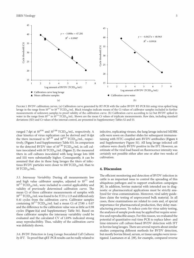
3.1. Generation of BVDV Calibration Curves. The possibilityto replace cell culture-based BVDV detection in bovinelung lavages with qRT-PCR was evaluated during this study.Hence, in a first attempt, storage samples of the three previ-ously BVDV-positive tested lung lavage lots 1104, 1108, and1111 (data not shown) were analyzed for presence of BVDVby qRT-PCR using a commercial RT-PCR kit. Interestingly,although lung lavages were clearly identified BVDV-positiveby immunofluorescence staining of inoculated MDBK cells,no BVDV specific signal was detected by qRT-PCR (seeSupplementary Table S1, available online at http://dx.doi.org/10.5402/2013/751904). Nevertheless, as certainly infec-tive BVDV particles must be present in the analyzed samples,the detection limit of the qRT-PCR was determined. Ofspecial interest was the actual amount of viruses able to infectand propagate in cell culture. Thus, calibration samples ofBVDV strain NADL were referred to the TCID
50rather than
to free RNA. To detect putative PCR inhibitors present in thelung lavage, calibration curves were generated not only withBVDV spiked in negative lung lavage, but also with BVDVspiked in solely water. In addition, an internal control wasincluded in the qRT-PCRkit by default to proof accurate assayperformance. By plotting the obtained Ct values over corre-sponding BVDV strain NADL dilutions in lung lavage rang-ing from 101.0 to 107.0 TCID
50/mL, a calibration curve with
high linearity over 5 log10units could be constructed. In this
experimental setup a detection limit of 100 TCID50/mL was
identified (Figure 1(a) and Supplementary Table S2).The cor-relation coefficient 𝑅2 = 0.9987 indicated strong linearity ofthe calibration curve with the linear equation 𝑦 = −3.808𝑥 +47.281. Control experiments with BVDV spikes rangingfrom 102.5 to 107.0 TCID
50/mL in water, to exclude putative
PCR-inhibitingmatrix effects, resulted in a similar graphwiththe linear equation 𝑦 = −4.0427𝑥 + 48.963, 𝑅2 = 0.9918(Figure 1(b) and Supplementary Table S3). The Δ slope wasas little as 0.23.
3.2. Intra-Assay Variability. Data of the calibration mea-surements were used to assess the intra-assay variability.Calculation of the coefficient of variation of both the pooledBVDV-spiked lung lavage and water samples as well as of thepooled internal controls revealed CVs of 0.66% and 0.41%,
respectively. This demonstrates high precision and accuracyof test results.
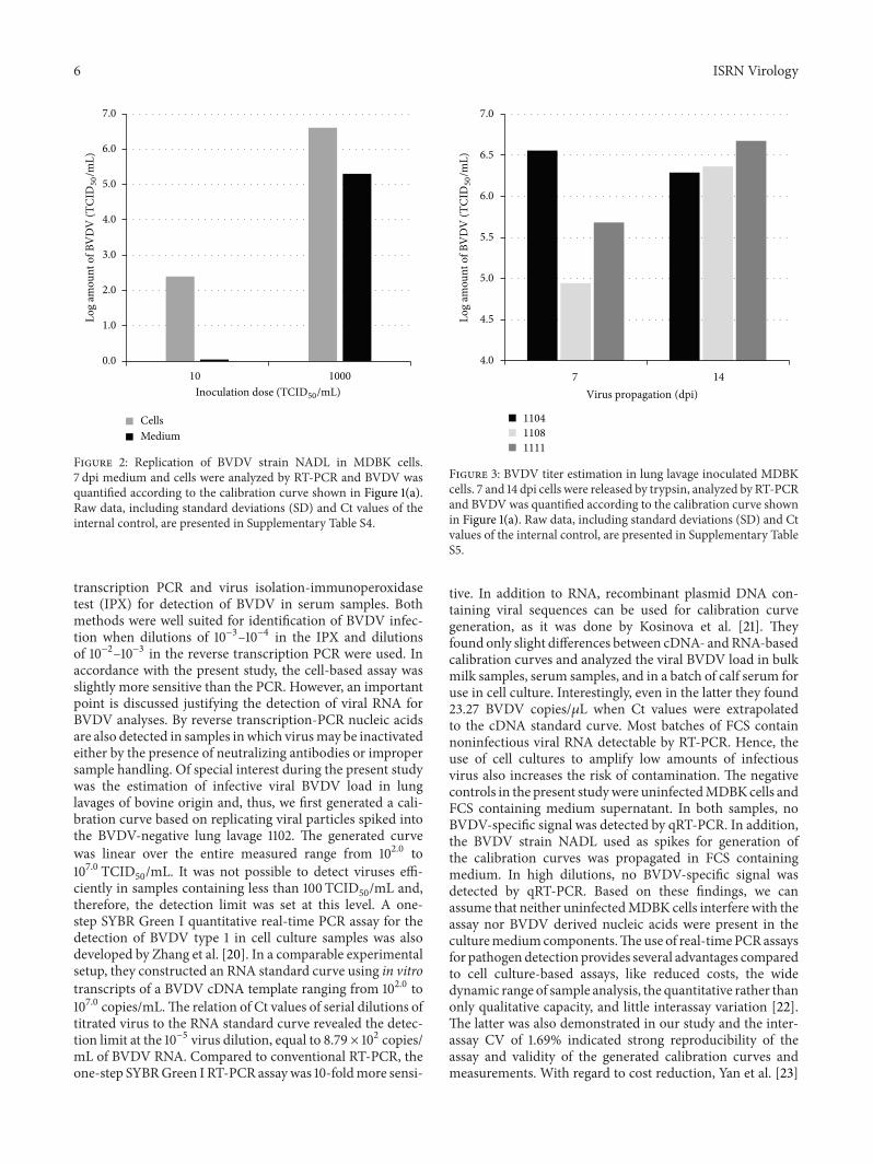
3.3. Replication of BVDV Strain NADL inMDBKCells. Whensusceptible MDBK cells were inoculated with lung lavages1104, 1108, and 1111, the contaminating BVDV could be pro-pagated to levels allowing detection by IFT. But the failureto detect BVDV by qRT-PCR, although the LOD of100 TCID
50/mL confirmed high sensitivity of the method,
implied very low virus titers in the lavage liquids. To comparethe degree of virus contamination, we monitored the virusreplication in cell culture after infection with a low BVDVinoculum.MDBK cells were exposed to 10 TCID
50, similar to
lung lavage inoculation, and viruses were allowed to multiplyfor 7 d before cells and medium were analyzed by qRT-PCR.Infection of cells with 1000TCID
50was performed in parallel.
Inoculation with 10 TCID50was sufficient to permit BVDV to
multiply in cell culture to detectable levels within one week.Based on previously generated qRT-PCR calibration curve, avirus titer of 102.40 TCID
50/mL was measured in the cellular
fraction. The amount of virus released into the mediumsupernatant was below LOD. Contrary, cells inoculated with1000 TCID
50were highly infected, displayed by a calcu-
lated titer of 106.61 TCID50/mL, and the measured titer of
105.30 TCID50/mL in the culture supernatant implied that
even high amount of virus was released from infected cells(Figure 2).
3.4. BVDV Propagation in Lung Lavage Inoculated Cell Cul-tures and Titer Estimation by qRT-PCR. Theabovementionedresults suggest that viral load in bovine lung lavages wasbelow 100 TCID
50/mL, as qRT-PCR did not produce any
signal, but above 10 TCID50/mL, as this titer was sufficient
to infect cell cultures and to allow BVDV to propagate todetectable levels within one week.
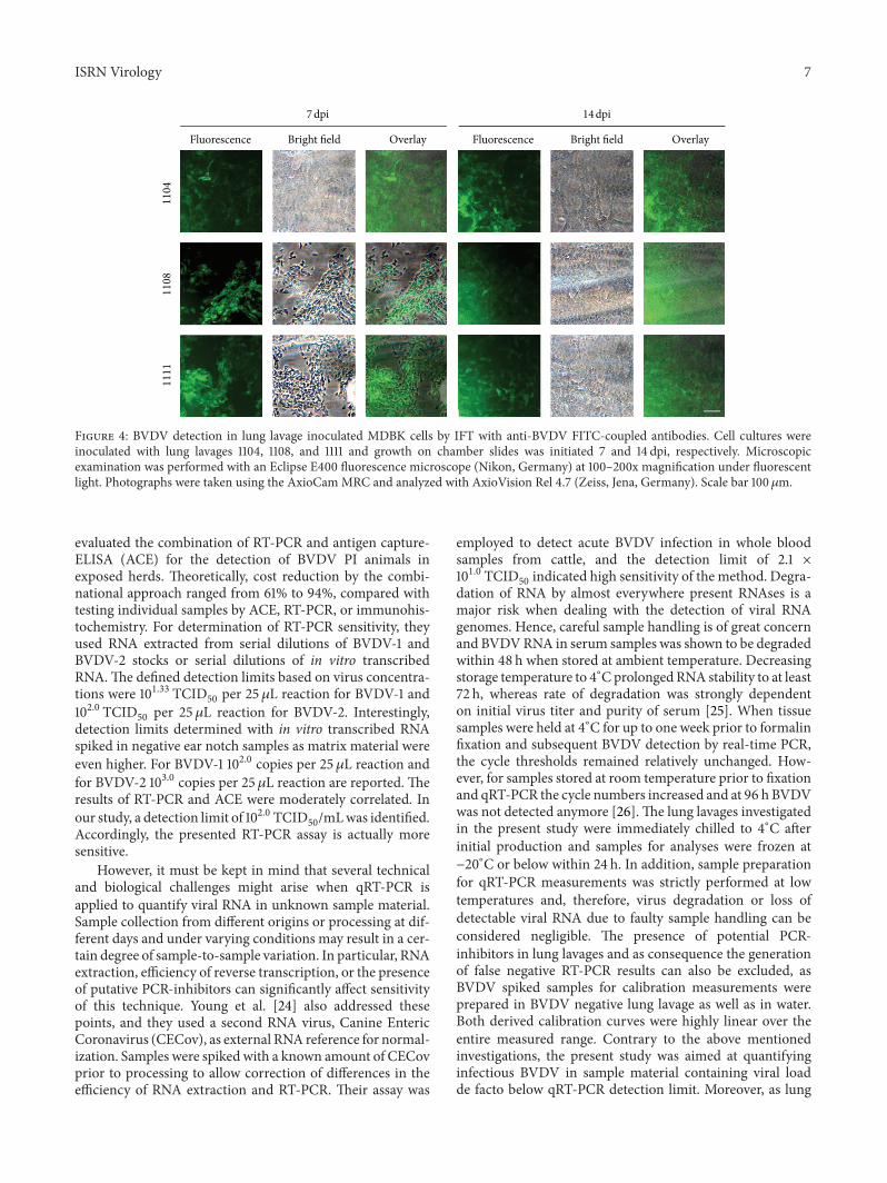
To gain further insights into the extent of BVDV con-tamination, we infected MDBK cells with the lung lavages.7 and 14 dpi samples were drawn and again analyzed byqRT-PCR and, in addition, cultivation on chamber slideswas initiated for IFT. Since BVDV titers were higher in cellcontaining samples than in medium supernatant (Figure 2),qRT-PCR was only performed with detached cells. Notably,already after one week lot 1104 inoculated cell culture washighly BVDV-positive, reflected by the calculated titer of106.56 TCID
50/mL (Figure 3). Probably, BVDV content in the
lung lavage was only slightly below qRT-PCR detection limitof 100 TCID
50/mL and, thus, favored BVDV propagation.
This finding was in accordance with the primary qRT-PCRanalysis of the lung lavage lot 1104. Although in sum the initialmeasurement was rated BVDV-negative, in one of the threereplicates a Ct value of 40.23 indicated the presence of BVDV(Supplementary Table S1). After another week of virus mul-tiplication, the BVDV titer in lot 1104 inoculated cell culturewas 106.29 TCID
50/mL and, hence, remainedmore or less con-
stant (Supplementary Table S5). Possibly, virus propagationreached a plateau reflecting maximal BVDV multiplicationrate in cell culture. In lot 1108 and lot 1111 inoculated cellcultures the initial BVDV titers were apparently lower and
Figure 1: BVDV calibration curves. (a) Calibration curve generated by RT-PCR with the cador BVDV RT-PCR Kit using virus spiked lunglavage in the range from 102.0 to 107.0 TCID
50/mL. Black triangles indicate means of the Ct values of calibrator samples included in further
measurements of unknown samples to proof validity of the calibration curve. (b) Calibration curve according to (a) but BVDV spiked inwater in the range from 102.5 to 107.0 TCID
50/mL. Shown are the mean Ct values of triplicate measurements. Raw data, including standard
deviations (SD) and Ct values of the internal control, are presented in Supplementary Tables S2 and S3.
ranged 7 dpi at 104.94 and 105.68 TCID50/mL, respectively. A
clear kinetics of virus replication can be derived and 14 dpithe titers increased to 106.36 and 106.67 TCID
50/mL, respec-
tively, (Figure 3 and Supplementary Table S5). In comparisonto the detected BVDV titer of 102.40 TCID
50/mL in cell cul-
ture inoculated with 10 TCID50/mL (Figure 2), the measured
titers in cell cultures inoculated with lung lavage lots 1108and 1111 were substantially higher. Consequently, it can beassumed that also in these lung lavages the titers of infec-tious BVDV particles were closer to 100 TCID
50/mL than to
10 TCID50/mL.
3.5. Interassay Variability. During all measurements lowand high value calibrator samples, adjusted to 102.5 and105.5 TCID
50/mL, were included to control applicability and
validity of previously determined calibration curve. Themean Ct of three calibrator measurements of samples with102.5 TCID
50/mLwas found to be 37.33± 1.13 and differed only
0.41 cycles from the calibration curve. Calibrator samplescontaining 105.5 TCID
50/mL had a mean Ct of 27.80 ± 0.07
and the difference to the calibration value was as little as 0.98cycles (Figure 1(a) and Supplementary Table S6). Based onthese calibrator samples the interassay variability could beevaluated and the calculated CV of 1.69% indicated strongassay reproducibility. Thus, validity of the calibration curvewas definitely shown.
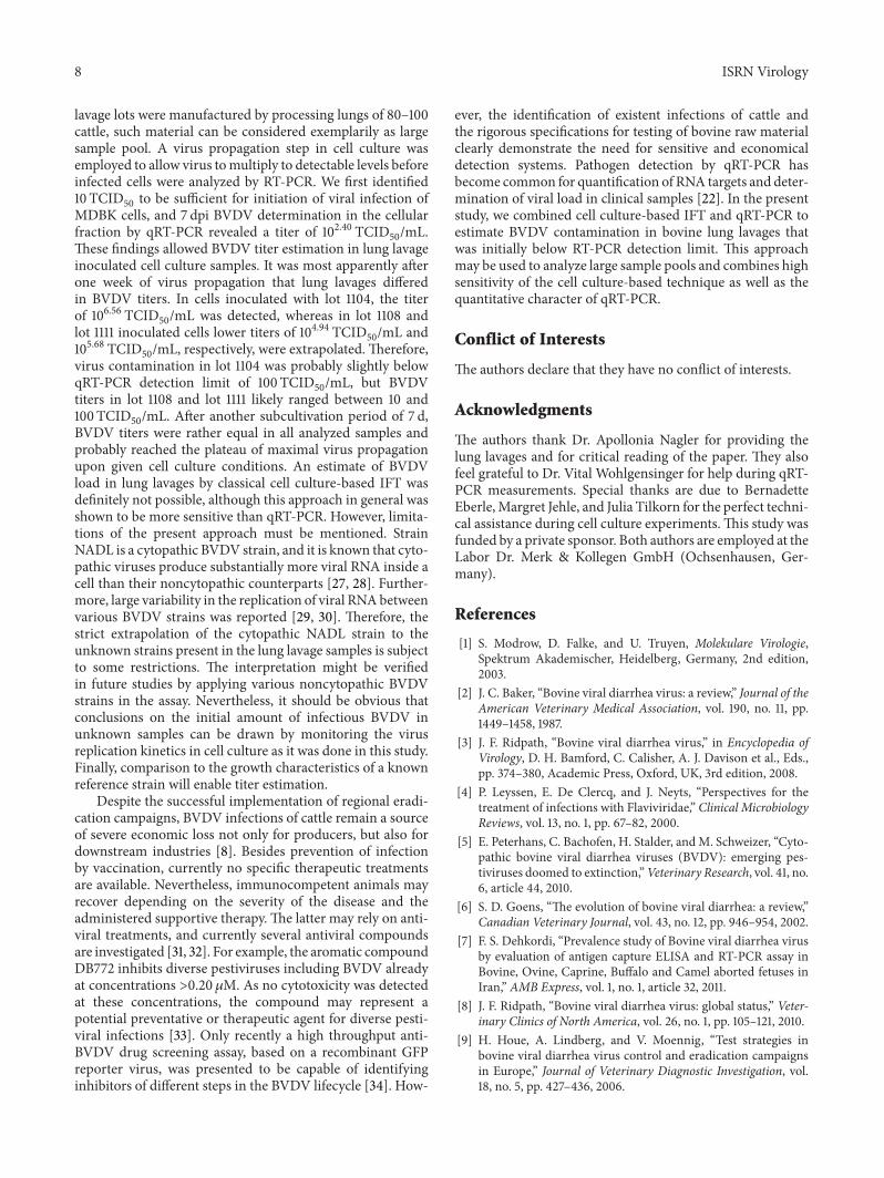
3.6. BVDV Detection in Lung Lavage Inoculated Cell Culturesby IFT. To proof that qRT-PCR results can be really related to
infective, replicating viruses, the lung lavage infected MDBKcells were sown on chamber slides for subsequent immunos-taining with FITC-coupled anti-BVDV-antibodies (Figure 4and Supplementary Figure S1). All lung lavage infected cellcultures were clearly BVDV-positive in the IFT. However, anestimate of the viral load based on fluorescence intensity wascertainly not possible either after one or after two weeks ofcultivation.
4. Discussion
The efficient monitoring and detection of BVDV infection incattle is an important issue to control the spreading of thisubiquitous pathogen and to support eradication campaigns[8]. In addition, bovine material with intended use in diag-nostic or pharmaceutical applications must be strictly ana-lyzed for virus contaminations. Moreover, viral safety guide-lines claim the testing of unprocessed bulk material. In allcases, these examinations are related to costs and, of specialimportance for pharmaceutical production, they delay man-ufacturing processes. To reduce costs for virus safety testing,the analyses of sample poolsmay be applied but require sensi-tive and reproducible assays. For this reason, we evaluated thepotential of quantitative real-time PCR to replace labor- andtime-intensive cell culture-based BVDV detection with IFTin bovine lung lavages.There are several reports about similarstudies comparing different methods for BVDV detection,butmostly bovine blood, serum, or tissue samples were inves-tigated. Laamanen et al. [19], for example, compared reverse
6 ISRN Virology
0.0
1.0
2.0
3.0
4.0
5.0
6.0
7.0
10 1000
CellsMedium
Log
amou
nt o
f BV
DV
(TCI
D50/m
L)
Inoculation dose (TCID50/mL)
Figure 2: Replication of BVDV strain NADL in MDBK cells.7 dpi medium and cells were analyzed by RT-PCR and BVDV wasquantified according to the calibration curve shown in Figure 1(a).Raw data, including standard deviations (SD) and Ct values of theinternal control, are presented in Supplementary Table S4.
transcription PCR and virus isolation-immunoperoxidasetest (IPX) for detection of BVDV in serum samples. Bothmethods were well suited for identification of BVDV infec-tion when dilutions of 10−3–10−4 in the IPX and dilutionsof 10−2–10−3 in the reverse transcription PCR were used. Inaccordance with the present study, the cell-based assay wasslightly more sensitive than the PCR. However, an importantpoint is discussed justifying the detection of viral RNA forBVDV analyses. By reverse transcription-PCR nucleic acidsare also detected in samples inwhich virusmay be inactivatedeither by the presence of neutralizing antibodies or impropersample handling. Of special interest during the present studywas the estimation of infective viral BVDV load in lunglavages of bovine origin and, thus, we first generated a cali-bration curve based on replicating viral particles spiked intothe BVDV-negative lung lavage 1102. The generated curvewas linear over the entire measured range from 102.0 to107.0 TCID
50/mL. It was not possible to detect viruses effi-
ciently in samples containing less than 100 TCID50/mL and,
therefore, the detection limit was set at this level. A one-step SYBR Green I quantitative real-time PCR assay for thedetection of BVDV type 1 in cell culture samples was alsodeveloped by Zhang et al. [20]. In a comparable experimentalsetup, they constructed an RNA standard curve using in vitrotranscripts of a BVDV cDNA template ranging from 102.0 to107.0 copies/mL.The relation of Ct values of serial dilutions oftitrated virus to the RNA standard curve revealed the detec-tion limit at the 10−5 virus dilution, equal to 8.79 × 10² copies/mL of BVDV RNA. Compared to conventional RT-PCR, theone-step SYBRGreen I RT-PCR assaywas 10-foldmore sensi-
4.0
4.5
5.0
5.5
6.0
6.5
7.0
7 14Virus propagation (dpi)
110411081111
Log
amou
nt o
f BV
DV
(TCI
D50/m
L)Figure 3: BVDV titer estimation in lung lavage inoculated MDBKcells. 7 and 14 dpi cells were released by trypsin, analyzed by RT-PCRand BVDVwas quantified according to the calibration curve shownin Figure 1(a). Raw data, including standard deviations (SD) and Ctvalues of the internal control, are presented in Supplementary TableS5.
tive. In addition to RNA, recombinant plasmid DNA con-taining viral sequences can be used for calibration curvegeneration, as it was done by Kosinova et al. [21]. Theyfound only slight differences between cDNA- andRNA-basedcalibration curves and analyzed the viral BVDV load in bulkmilk samples, serum samples, and in a batch of calf serum foruse in cell culture. Interestingly, even in the latter they found23.27 BVDV copies/𝜇L when Ct values were extrapolatedto the cDNA standard curve. Most batches of FCS containnoninfectious viral RNA detectable by RT-PCR. Hence, theuse of cell cultures to amplify low amounts of infectiousvirus also increases the risk of contamination. The negativecontrols in the present studywere uninfectedMDBKcells andFCS containing medium supernatant. In both samples, noBVDV-specific signal was detected by qRT-PCR. In addition,the BVDV strain NADL used as spikes for generation ofthe calibration curves was propagated in FCS containingmedium. In high dilutions, no BVDV-specific signal wasdetected by qRT-PCR. Based on these findings, we canassume that neither uninfectedMDBK cells interfere with theassay nor BVDV derived nucleic acids were present in theculturemediumcomponents.Theuse of real-timePCRassaysfor pathogen detection provides several advantages comparedto cell culture-based assays, like reduced costs, the widedynamic range of sample analysis, the quantitative rather thanonly qualitative capacity, and little interassay variation [22].The latter was also demonstrated in our study and the inter-assay CV of 1.69% indicated strong reproducibility of theassay and validity of the generated calibration curves andmeasurements. With regard to cost reduction, Yan et al. [23]
ISRN Virology 7
Fluorescence Bright field Overlay Fluorescence Bright field Overlay
1104
1108
1111
7 dpi 14 dpi
Figure 4: BVDV detection in lung lavage inoculated MDBK cells by IFT with anti-BVDV FITC-coupled antibodies. Cell cultures wereinoculated with lung lavages 1104, 1108, and 1111 and growth on chamber slides was initiated 7 and 14 dpi, respectively. Microscopicexamination was performed with an Eclipse E400 fluorescence microscope (Nikon, Germany) at 100–200x magnification under fluorescentlight. Photographs were taken using the AxioCamMRC and analyzed with AxioVision Rel 4.7 (Zeiss, Jena, Germany). Scale bar 100𝜇m.
evaluated the combination of RT-PCR and antigen capture-ELISA (ACE) for the detection of BVDV PI animals inexposed herds. Theoretically, cost reduction by the combi-national approach ranged from 61% to 94%, compared withtesting individual samples by ACE, RT-PCR, or immunohis-tochemistry. For determination of RT-PCR sensitivity, theyused RNA extracted from serial dilutions of BVDV-1 andBVDV-2 stocks or serial dilutions of in vitro transcribedRNA.The defined detection limits based on virus concentra-tions were 101.33 TCID
50per 25 𝜇L reaction for BVDV-1 and
102.0 TCID50
per 25 𝜇L reaction for BVDV-2. Interestingly,detection limits determined with in vitro transcribed RNAspiked in negative ear notch samples as matrix material wereeven higher. For BVDV-1 102.0 copies per 25 𝜇L reaction andfor BVDV-2 103.0 copies per 25 𝜇L reaction are reported. Theresults of RT-PCR and ACE were moderately correlated. Inour study, a detection limit of 102.0 TCID
50/mLwas identified.
Accordingly, the presented RT-PCR assay is actually moresensitive.
However, it must be kept in mind that several technicaland biological challenges might arise when qRT-PCR isapplied to quantify viral RNA in unknown sample material.Sample collection from different origins or processing at dif-ferent days and under varying conditions may result in a cer-tain degree of sample-to-sample variation. In particular, RNAextraction, efficiency of reverse transcription, or the presenceof putative PCR-inhibitors can significantly affect sensitivityof this technique. Young et al. [24] also addressed thesepoints, and they used a second RNA virus, Canine EntericCoronavirus (CECov), as external RNA reference for normal-ization. Samples were spiked with a known amount of CECovprior to processing to allow correction of differences in theefficiency of RNA extraction and RT-PCR. Their assay was
employed to detect acute BVDV infection in whole bloodsamples from cattle, and the detection limit of 2.1 ×101.0 TCID
50indicated high sensitivity of the method. Degra-
dation of RNA by almost everywhere present RNAses is amajor risk when dealing with the detection of viral RNAgenomes. Hence, careful sample handling is of great concernand BVDVRNA in serum samples was shown to be degradedwithin 48 h when stored at ambient temperature. Decreasingstorage temperature to 4∘CprolongedRNA stability to at least72 h, whereas rate of degradation was strongly dependenton initial virus titer and purity of serum [25]. When tissuesamples were held at 4∘C for up to one week prior to formalinfixation and subsequent BVDV detection by real-time PCR,the cycle thresholds remained relatively unchanged. How-ever, for samples stored at room temperature prior to fixationand qRT-PCR the cycle numbers increased and at 96 h BVDVwas not detected anymore [26].The lung lavages investigatedin the present study were immediately chilled to 4∘C afterinitial production and samples for analyses were frozen at−20∘C or below within 24 h. In addition, sample preparationfor qRT-PCR measurements was strictly performed at lowtemperatures and, therefore, virus degradation or loss ofdetectable viral RNA due to faulty sample handling can beconsidered negligible. The presence of potential PCR-inhibitors in lung lavages and as consequence the generationof false negative RT-PCR results can also be excluded, asBVDV spiked samples for calibration measurements wereprepared in BVDV negative lung lavage as well as in water.Both derived calibration curves were highly linear over theentire measured range. Contrary to the above mentionedinvestigations, the present study was aimed at quantifyinginfectious BVDV in sample material containing viral loadde facto below qRT-PCR detection limit. Moreover, as lung
8 ISRN Virology
lavage lots were manufactured by processing lungs of 80–100cattle, such material can be considered exemplarily as largesample pool. A virus propagation step in cell culture wasemployed to allow virus tomultiply to detectable levels beforeinfected cells were analyzed by RT-PCR. We first identified10 TCID
50to be sufficient for initiation of viral infection of
MDBK cells, and 7 dpi BVDV determination in the cellularfraction by qRT-PCR revealed a titer of 102.40 TCID
50/mL.
These findings allowed BVDV titer estimation in lung lavageinoculated cell culture samples. It was most apparently afterone week of virus propagation that lung lavages differedin BVDV titers. In cells inoculated with lot 1104, the titerof 106.56 TCID
50/mL was detected, whereas in lot 1108 and
lot 1111 inoculated cells lower titers of 104.94 TCID50/mL and
105.68 TCID50/mL, respectively, were extrapolated. Therefore,
virus contamination in lot 1104 was probably slightly belowqRT-PCR detection limit of 100 TCID
50/mL, but BVDV
titers in lot 1108 and lot 1111 likely ranged between 10 and100 TCID
50/mL. After another subcultivation period of 7 d,
BVDV titers were rather equal in all analyzed samples andprobably reached the plateau of maximal virus propagationupon given cell culture conditions. An estimate of BVDVload in lung lavages by classical cell culture-based IFT wasdefinitely not possible, although this approach in general wasshown to be more sensitive than qRT-PCR. However, limita-tions of the present approach must be mentioned. StrainNADL is a cytopathic BVDV strain, and it is known that cyto-pathic viruses produce substantially more viral RNA inside acell than their noncytopathic counterparts [27, 28]. Further-more, large variability in the replication of viral RNAbetweenvarious BVDV strains was reported [29, 30]. Therefore, thestrict extrapolation of the cytopathic NADL strain to theunknown strains present in the lung lavage samples is subjectto some restrictions. The interpretation might be verifiedin future studies by applying various noncytopathic BVDVstrains in the assay. Nevertheless, it should be obvious thatconclusions on the initial amount of infectious BVDV inunknown samples can be drawn by monitoring the virusreplication kinetics in cell culture as it was done in this study.Finally, comparison to the growth characteristics of a knownreference strain will enable titer estimation.
Despite the successful implementation of regional eradi-cation campaigns, BVDV infections of cattle remain a sourceof severe economic loss not only for producers, but also fordownstream industries [8]. Besides prevention of infectionby vaccination, currently no specific therapeutic treatmentsare available. Nevertheless, immunocompetent animals mayrecover depending on the severity of the disease and theadministered supportive therapy. The latter may rely on anti-viral treatments, and currently several antiviral compoundsare investigated [31, 32]. For example, the aromatic compoundDB772 inhibits diverse pestiviruses including BVDV alreadyat concentrations >0.20𝜇M. As no cytotoxicity was detectedat these concentrations, the compound may represent apotential preventative or therapeutic agent for diverse pesti-viral infections [33]. Only recently a high throughput anti-BVDV drug screening assay, based on a recombinant GFPreporter virus, was presented to be capable of identifyinginhibitors of different steps in the BVDV lifecycle [34]. How-
ever, the identification of existent infections of cattle andthe rigorous specifications for testing of bovine raw materialclearly demonstrate the need for sensitive and economicaldetection systems. Pathogen detection by qRT-PCR hasbecome common for quantification of RNA targets and deter-mination of viral load in clinical samples [22]. In the presentstudy, we combined cell culture-based IFT and qRT-PCR toestimate BVDV contamination in bovine lung lavages thatwas initially below RT-PCR detection limit. This approachmay be used to analyze large sample pools and combines highsensitivity of the cell culture-based technique as well as thequantitative character of qRT-PCR.
Conflict of Interests
The authors declare that they have no conflict of interests.
Acknowledgments
The authors thank Dr. Apollonia Nagler for providing thelung lavages and for critical reading of the paper. They alsofeel grateful to Dr. Vital Wohlgensinger for help during qRT-PCR measurements. Special thanks are due to BernadetteEberle,Margret Jehle, and Julia Tilkorn for the perfect techni-cal assistance during cell culture experiments.This study wasfunded by a private sponsor. Both authors are employed at theLabor Dr. Merk & Kollegen GmbH (Ochsenhausen, Ger-many).
References
[1] S. Modrow, D. Falke, and U. Truyen, Molekulare Virologie,Spektrum Akademischer, Heidelberg, Germany, 2nd edition,2003.
[2] J. C. Baker, “Bovine viral diarrhea virus: a review,” Journal of theAmerican Veterinary Medical Association, vol. 190, no. 11, pp.1449–1458, 1987.
[3] J. F. Ridpath, “Bovine viral diarrhea virus,” in Encyclopedia ofVirology, D. H. Bamford, C. Calisher, A. J. Davison et al., Eds.,pp. 374–380, Academic Press, Oxford, UK, 3rd edition, 2008.
[4] P. Leyssen, E. De Clercq, and J. Neyts, “Perspectives for thetreatment of infections with Flaviviridae,” Clinical MicrobiologyReviews, vol. 13, no. 1, pp. 67–82, 2000.
[5] E. Peterhans, C. Bachofen, H. Stalder, andM. Schweizer, “Cyto-pathic bovine viral diarrhea viruses (BVDV): emerging pes-tiviruses doomed to extinction,”Veterinary Research, vol. 41, no.6, article 44, 2010.
[6] S. D. Goens, “The evolution of bovine viral diarrhea: a review,”Canadian Veterinary Journal, vol. 43, no. 12, pp. 946–954, 2002.
[7] F. S. Dehkordi, “Prevalence study of Bovine viral diarrhea virusby evaluation of antigen capture ELISA and RT-PCR assay inBovine, Ovine, Caprine, Buffalo and Camel aborted fetuses inIran,” AMB Express, vol. 1, no. 1, article 32, 2011.
[8] J. F. Ridpath, “Bovine viral diarrhea virus: global status,” Veter-inary Clinics of North America, vol. 26, no. 1, pp. 105–121, 2010.
[9] H. Houe, A. Lindberg, and V. Moennig, “Test strategies inbovine viral diarrhea virus control and eradication campaignsin Europe,” Journal of Veterinary Diagnostic Investigation, vol.18, no. 5, pp. 427–436, 2006.
ISRN Virology 9
[10] M. A. Edmondson, M. D. Givens, P. H. Walz, J. A. Gard, D.A. Stringfellow, and R. L. Carson, “Comparison of tests fordetection of bovine viral diarrhea virus in diagnostic samples,”Journal of Veterinary Diagnostic Investigation, vol. 19, no. 4, pp.376–381, 2007.
[11] H. Houe, “Epidemiological features and economical impor-tance of bovine virus diarrhoea virus (BVDV) infections,”Veterinary Microbiology, vol. 64, no. 2-3, pp. 89–107, 1999.
[12] A. Ozkul, K. Yesilbag, and I. Burgu, “Comparison of four diag-nostic techniques for detecting Bovine Virus Diarrhoea Virus(BVDV) in buffy coat samples after long-term storage,” TurkishJournal of Veterinary and Animal Sciences, vol. 26, no. 5, pp.1043–1048, 2002.
[13] J. Laureyns, S. Ribbens, andA. deKruif, “Control of bovine virusdiarrhoea at the herd level: reducing the risk of false negatives inthe detection of persistently infected cattle,” Veterinary Journal,vol. 184, no. 1, pp. 21–26, 2010.
[14] K. Singh,M.M.Miller, L. J. Kohrt, G. Scherba, E. F. Garrett, andR. L. Fredrickson, “Development of a novel diagnostic test fordetection of bovine viral diarrhea persistently infected animalsusing hair,” Journal of Veterinary Science, vol. 12, no. 3, pp. 295–297, 2011.
[15] J. R.Glasser andR.K.Mallampalli, “Surfactant and its role in thepathobiology of pulmonary infection,” Microbes and Infection,vol. 14, no. 1, pp. 17–25, 2012.
[16] D. F. Willson and R. H. Notter, “The future of exogenous sur-factant therapy,” Respiratory Care, vol. 56, no. 9, pp. 1369–1386,2011.
[17] C. Spearman, “The method of “right and wrong cases” (“con-stant stimuli”) without Gauss’s formulae,” British Journal ofPsychology, vol. 2, no. 3, pp. 227–242, 1908.
[18] G. Karber, “Beitrag zur kollektiven Behandlung pharmakol-ogischer Reihenversuche,” Naunyn-Schmiedebergs Archiv furExperimentelle Pathologie und Pharmakologie, vol. 162, no. 4, pp.480–483, 1931.
[19] U. I. Laamanen, E. P. Neuvonen, E. M. Yliviuhkola, and P. M.-L.Veijalainen, “Comparison of RT-PCR assay and virus isolationin cell cultures for the detection of bovine viral diarrhoea virus(BVDV) in field samples,” Research in Veterinary Science, vol.63, no. 3, pp. 199–203, 1997.
[20] N. Zhang, Z. Liu, Q. Han et al., “Development of one-step SYBRGreen real-time RT-PCR for quantifying bovine viral diarrheavirus type-1 and its comparison with conventional RT-PCR,”Virology Journal, vol. 8, article 374, 2011.
[21] E. Kosinova, I. Psikal, B. Robesova, and K. Kovarcik, “Real-timePCR for quantitation of bovine viral diarrhea virus RNA usingSYBR Green I fluorimetry,” Veterinary Medicine, vol. 52, no. 6,pp. 253–261, 2007.
[22] S. A. Bustin and R. Mueller, “Real-time reverse transcriptionPCR (qRT-PCR) and its potential use in clinical diagnosis,” Cli-nical Science, vol. 109, no. 4, pp. 365–379, 2005.
[23] L. Yan, S. Zhang, L. Pace, F. Wilson, H. Wan, and M. Zhang,“Combination of reverse transcription real-time polymerasechain reaction and antigen capture enzyme-linked immunosor-bent assay for the detection of animals persistently infectedwithBovine viral diarrhea virus,” Journal of Veterinary DiagnosticInvestigation, vol. 23, no. 1, pp. 16–25, 2011.
[24] N. J. Young, C. J. Thomas, M. E. Collins, and J. Brownlie, “Real-time RT-PCR detection of Bovine Viral Diarrhoea virus inwhole blood using an external RNA reference,” Journal of Viro-logical Methods, vol. 138, no. 1-2, pp. 218–222, 2006.
[25] D.Weinstock, B. Bhudevi, and A. E. Castro, “Single-tube single-enzyme reverse transcriptase PCR assay for detection of bovineviral diarrhea virus in pooled bovine serum,” Journal of ClinicalMicrobiology, vol. 39, no. 1, pp. 343–346, 2001.
[26] B. Bhudevi and D. Weinstock, “Detection of bovine viral diar-rhea virus in formalin fixed paraffin embedded tissue sectionsby real time RT-PCR (Taqman),” Journal of Virological Methods,vol. 109, no. 1, pp. 25–30, 2003.
[27] Y. Li and J. McNally, “Characterization of RNA synthesis andtranslation of bovine viral diarrhea virus (BVDV),”Virus Genes,vol. 23, no. 2, pp. 149–155, 2001.
[28] V. B. Vassilev and R. O. Donis, “Bovine viral diarrhea virusinduced apoptosis correlates with increased intracellular viralRNA accumulation,” Virus Research, vol. 69, no. 2, pp. 95–107,2000.
[29] D. Yamane, K. Kato, Y. Tohya, and H. Akashi, “The double-stranded RNA-induced apoptosis pathway is involved in thecytopathogenicity of cytopathogenic Bovine viral diarrheavirus,” Journal of General Virology, vol. 87, no. 10, pp. 2961–2970,2006.
[30] S. A. La Rocca and T. Sandvik, “A short target real-time RT-PCRassay for detection of pestiviruses infecting cattle,” Journal ofVirological Methods, vol. 161, no. 1, pp. 122–127, 2009.
[31] B.W. Newcomer, M. S.Marley, P. K. Galik et al., “Antiviral treat-ment of calves persistently infected with bovine viral diarrhoeavirus,” Antiviral Chemistry & Chemotherapy, vol. 22, no. 4, pp.171–179, 2012.
[32] M. T. A. Salim, Y. Goto, T. Hamasaki et al., “Highly potent andselective inhibition of bovine viral diarrhea virus replication by𝛾-carboline derivatives,” Antiviral Research, vol. 88, no. 3, pp.263–268, 2010.
[33] B.W.Newcomer,M. S.Marley, J. F. Ridpath et al., “Efficacy of anantiviral compound to inhibit replication of multiple pestivirusspecies,” Antiviral Research, vol. 96, no. 2, pp. 127–129, 2012.
[34] Z.-C. Fan and R. C. Bird, “Development of a reporter bovineviral diarrhea virus and initial evaluation of its application forhigh throughput antiviral drug screening,” Journal of VirologicalMethods, vol. 180, no. 1-2, pp. 54–61, 2012.