1 Department of Radiology, Xuanwu Hospital of Capital Medical University, Beijing 100053, China2 Beijing Key Laboratory of Magnetic Resonance Imaging and Brain Informatics, Beijing 100053, China3Department of Neurology, Xuanwu Hospital of Capital Medical University, Beijing 100053, China4 State Key Laboratory for Cognitive Neuroscience and Learning, Beijing Normal University, Beijing 100875, China
The neuropsychological tests in patients with internal carotid artery (ICA) demonstrated cognitive deficits associated with frontallobe dysfunction, but the pathophysiological mechanism of memory impairment is not fully understood. This study evaluatedrelationship between degree of ICA stenosis and frontal activations induced by working memory (WM) task using fMRI. ThefMRI data of 21 patients with unilateral ICA stenosis (left/right, 11/10) and 21 controls were analyzed. In comparison with controls,ICA patients demonstrated significant activations in middle frontal gyrus (MFG) bilaterally, particularly in left MFG. In right ICAstenosis, there was slightly less MFG activation than that of controls. Importantly, lower MFG activity was associated with higherstenosis of ipsilateral ICA. For left ICA stenosis, weaker activation in left MFG was negatively correlated with degree of stenosis.Similarly, for right ICA stenosis, there was a significant negative correlation between right ICA stenosis and weaker activation ofright MFG. Cognitive impairments in ICA stenosis were associated with frontal lobe dysfunctions. Left ICA stenosis had worseWM impairments than right ICA stenosis, which was affected by the degree of stenosis.
1. Introduction
Cerebrovascular diseases are associated with cognitivedecline and dementia. Patients with occlusive diseases ofthe internal carotid artery (ICA) are at risk for cognitiveimpairment [1–3]. A systematic review of cognitive disordersin ICA patients finds subtle cognitive deficits in 70% ofthe studies reviewed [4]. Therefore, some patients may bein a preclinical stage of vascular dementia. Particularly,patients with carotid artery disease who have suffered atransient ischemic attack (TIA) can have lasting cognitiveimpairment, even without visible ischemic lesions on MRI[5, 6]. Similarly, neuropsychological tests show cognitivedeficits in working memory (WM), attention, reasoning,psychomotor speed, and executive functions; frontal lobedysfunction has been a consistent finding [4–7]. WMis the brain system that maintains a limited amount of
information for short periods of time and manipulates thatinformation [8]. The frontal cortex is involved in WM taskswith asymmetric activations in the left and right hemispheresduring verbal and nonverbal WM tasks [9, 10]. Furthermore,several studies reported worse memory impairment inpatients with left carotid artery disease than those with rightcarotid artery disease. The different patterns observed argueagainst that high-grade stenosis of ICA is simply a markerfor vascular disease and its risk factors [6, 11, 12]. To date, thecognitive functions of carotid occlusive disease have beenassessed using neuropsychological tests [4–7, 11, 13]. Variouscognitive functions have been linked to specific brain regions.However, previous neuropsychological tests are not able toprecisely reveal cognitive deficits in specific brain regionsinvolving particular tasks. Therefore, the pathophysiologicalmechanism of memory impairment is not fully understood.Recently, functional magnetic resonance imaging (fMRI)
Hindawi Publishing CorporationBioMed Research InternationalVolume 2014, Article ID 327270, 6 pageshttp://dx.doi.org/10.1155/2014/327270
2 BioMed Research International
has increasingly been used to study cognitive functionin humans. It has been explored for elucidating cognitiveimpairment mechanisms, especially WM impairment.
Wehypothesized that brain dysfunctionWMimpairmentin patients with symptomatic ICA disease; and specificallythat the differences in brain dysfunction between left andright ICA disease were associated with the degree of stenosisin ipsilateral ICA, and the functional differences betweenleft and right frontal lobes. Therefore, the purpose of thepresent study was as follows: (1) to investigate the abnormalfrontal activations of digit WM in patients with ICA steno-sis/occlusion and ipsilateral TIA and (2) to investigate therelationship between the activations in the frontal and thedegree of ICA stenosis using fMRI.
2. Subjects and Methods
2.1. Subjects. This study was comprised of 49 consecu-tive patients who were assessed for neurocognitive effectsof symptomatic carotid artery disease in the Departmentof Neurology. Symptomatic carotid stenosis is defined asstenosis having caused ischaemic events in the ipsilateraleye (transient monocular blindness) or cerebral hemisphere(transient ischaemic attack or stroke) in the past 6months [14,15].The study protocol was approved by the local InstitutionalReview Board and written informed consent was obtainedfrom all of subjects. 21 patients with symptoms of transientischemia (lasting <24 hours) were enrolled. As determinedby the use of a transcranial Doppler (TCD) or angiography,these patients demonstrated high-grade stenosis (70–99%)or unilateral internal carotid artery (ICA) occlusion. Allpatients had experienced at least one TIA and symptomshad occurred, at most, 6 months before inclusion in thestudy. All patients had normal intelligence; however, theirtotal memory scale scores (𝑀 = 72.54 ± 21.96) on theClinical Memory Scale (CMS) were lower than the healthysubjects (𝑀 = 88.54 ± 14.57) [16, 17]. The handedness ofthe subjects was assessed using the Edinburgh inventory [18].The exclusion criteria included left-handedness; contralateralICA occlusion or high-grade stenosis (≥70%); large infarctinfarction ormultiple lacunar infarctions (≥3) onMRI; severewhite matter lesions (≥ Grade 3) on MRI, especially lacunarinfarcts involved middle frontal gyrus and white matterlesions overstep the immediate subependymal region of theventricles; history of other brain diseases; deafness and/orblindness. The severity of leukoaraiosis (LA) was gradedusing the visual rating scale proposed by Sakakibara et al.[19]. These patients were divided into two groups: left ICAstenosis or occlusion (𝑛 = 11, age range from 39 to 75years, mean age 59.45 ± 11.72 years) and right ICA stenosisor occlusion (𝑛 = 10, age range from 38 to 70 years, mean age56.10 ± 10.86 years) based on the TCD or digital subtractionangiography (DSA) study. The control group consisted of21 healthy volunteers (age range was 33–69 years and meanage was 54.64 ± 11.85 years). These subjects were age- andeducation-matched to the patients (age: 𝑡 = 1.67, 𝑃 >0.05; education: 𝑡 = 1.74, 𝑃 > 0.05). In the controlgroup, carotid artery disease and intracerebral lesions were
excluded by TCD and MRI examinations. All subjects wereadministered a battery of neuropsychological tests involvingauditory digital memory and visual digital memory [20, 21].All patients were found to have impairedWMcomparedwiththe control group.The demographic data of the study subjectswere shown in Table 1.
2.2. MRI Data Acquisition. Scanning was performed ona 3.0-Tesla whole-body scanner (Trio Tim, Siemens). AT2-weighted (TR/TE: 3830ms/98ms; flip angle: 180∘; slicethickness 5mm; gap: 5mm; FOV: 230mm × 218mm; matrix:179 × 320) image was acquired for exclusion of intracraniallesions. High-resolution 3D magnetization prepared rapidgradient echo imaging (MPRAGE) and anatomical images(TR/TE: 1970ms/3.93ms; flip angle: 15∘; thickness 1.70mm;gap: 0.85mm; FOV: 250mm × 250mm; matrix: 448 × 512)of the entire brain were obtained before the functionalimages were acquired. A T2∗-weighted gradient-echo echo-planar imaging (EPI) sequencewas used to acquire functionalimages with 30 axial slices (TR/TE: 2000/30ms; flip angle:90∘, thickness: 5mm; gap: 0mm; FOV: 240× 240mm;matrix:64 × 64).
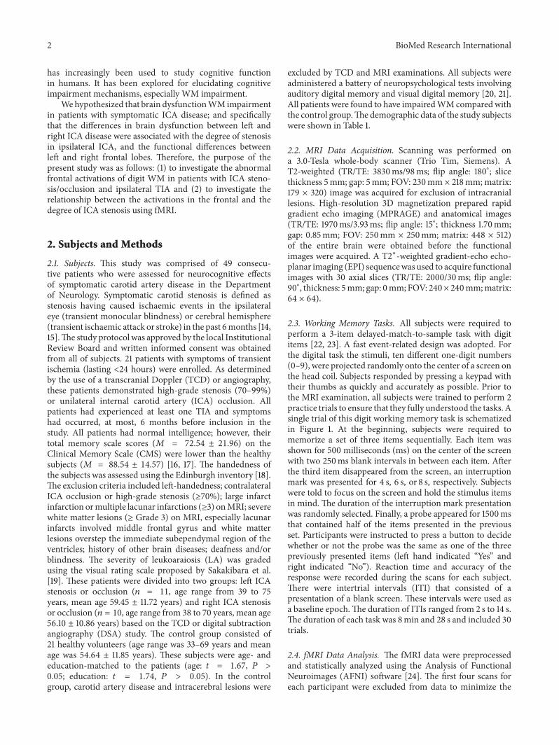
2.3. Working Memory Tasks. All subjects were required toperform a 3-item delayed-match-to-sample task with digititems [22, 23]. A fast event-related design was adopted. Forthe digital task the stimuli, ten different one-digit numbers(0–9), were projected randomly onto the center of a screen onthe head coil. Subjects responded by pressing a keypad withtheir thumbs as quickly and accurately as possible. Prior tothe MRI examination, all subjects were trained to perform 2practice trials to ensure that they fully understood the tasks. Asingle trial of this digit working memory task is schematizedin Figure 1. At the beginning, subjects were required tomemorize a set of three items sequentially. Each item wasshown for 500 milliseconds (ms) on the center of the screenwith two 250ms blank intervals in between each item. Afterthe third item disappeared from the screen, an interruptionmark was presented for 4 s, 6 s, or 8 s, respectively. Subjectswere told to focus on the screen and hold the stimulus itemsin mind. The duration of the interruption mark presentationwas randomly selected. Finally, a probe appeared for 1500msthat contained half of the items presented in the previousset. Participants were instructed to press a button to decidewhether or not the probe was the same as one of the threepreviously presented items (left hand indicated “Yes” andright indicated “No”). Reaction time and accuracy of theresponse were recorded during the scans for each subject.There were intertrial intervals (ITI) that consisted of apresentation of a blank screen. These intervals were used asa baseline epoch.The duration of ITIs ranged from 2 s to 14 s.The duration of each task was 8min and 28 s and included 30trials.
2.4. fMRI Data Analysis. The fMRI data were preprocessedand statistically analyzed using the Analysis of FunctionalNeuroimages (AFNI) software [24]. The first four scans foreach participant were excluded from data to minimize the
BioMed Research International 3
Table 1: Demographic characteristics of patients and controls.
Left ICA (𝑛 = 11) Right ICA (𝑛 = 10) Control (𝑛 = 21)Age, years (mean ± SD) 59.45 ± 11.72 56.10 ± 10.86 54.64 ± 11.85
transit effects of hemodynamic responses. Functional imageswere corrected for head motions by aligning all volumes tothe fifth volume using a six-parameter rigid-body transfor-mation. Statisticalmapswere spatially smoothedwith a 6mmfull width at half maximum (FWHM) Gaussian kernel.
We analyzed the brain activities of working memoryrelative to the baseline of the resting period during the ITI.Individual anatomical images and functional t-maps werecoregistered to the standard Talairach and Tournoux space[25]. All images were resized to 3mm × 3mm × 3mm voxel.At the group level, the threshold of group maps was set at avoxel level of 𝑡 > 2.040 (𝑃 < 0.005, number of voxels > 14)with a spatial extent correction.This threshold correspondedto an overall 𝛼 < 0.05 of family-wise error rate, as calculatedwith AlphaSim (http://afni.nimh.nih.gov) for all intracranialvoxels in the image volumes. Previous WM studies havefound that the bilateral frontal lobes play an important part inprocessingWM information [9, 10, 23, 26, 27]. For this reasonwe focused on the frontal lobes as our regions-of-interests(ROIs), to explore the relationship between the behavioraldata ofWM and the functional activation of ICA disease.TheROIs were defined functionally as spheres with a 6mm radiuson the basis of activation clusters in the bilateral frontal lobesfrom the group analysis (see Section 3). The peak activationcoordinates from the cluster of the contrast analysis wereselected as the center of each ROI. Then, these ROIs wereemployed as masks to extract the mean percent signal change
Table 2: Reaction time and accuracy of a digit workingmemory taskin all subjects.
Data are presented as mean ± SD. ∗𝑃 < 0.05 compared with controls.
(averaged over the ROI) in the blood oxygen level dependent(BOLD) response.
2.5. Statistical Analysis. Neuropsychological data were ana-lyzed using SPSS 11.0 computer software (SPSS Inc., Chicago,IL). The characteristics of patients and controls were com-pared using analysis of variance (ANOVA). The correlationsbetween response time (RT), response accuracy (RA) of digitWM task, activation intensity within the defined frontalROI, and the degree of ICA stenosis were analyzed using aPearson’s correlation.
3. Results
All subjects completed the fMRI digit task. The left andright ICA stenosis or occlusion patients showed significantlyweaker (left: 𝑃 = 0.032; right: 𝑃 = 0.041) and less accurateresponses (left: 𝑃 = 0.039; right: 𝑃 = 0.043) than those of thecontrol subjects (Table 2). Higher degrees of left ICA stenosiswere positively correlated with RT (𝑟 = 0.412, 𝑃 = 0.042), butnot with RA (𝑟 = −0.243, 𝑃 = 0.436). Higher degrees of rightICA stenosis were not correlated with either RT (𝑟 = 0.247,𝑃 = 0.424) or RA (𝑟 = −0.108, 𝑃 = 0.671).
The control group revealed a domain area of activationin the bilateral middle frontal gyrus (MFG), frontal gyrus,and supplementary motor area involving digit WM task. Thepeak of the activation was located in the bilateral MFG. ThedigitWM task induced significantly asymmetrical activationsin the MFG (peak coordinate: (−31, 31, 23), 𝑡 = 5.915)
4 BioMed Research International
Table 3: Medial frontal gyrus activations for a digit working memory task in subjects.
Left MFG Right MFGVoxels Peak (𝑥, 𝑦, 𝑧) 𝑡 value Voxels Peak (𝑥, 𝑦, 𝑧) 𝑡 value
and volume (475 voxels) in the left MFG. The left and rightICA groups showed similar activity clusters compared withthose of the control group. MFG activations for digit WMtask in subjects were listed in Table 3. A direct comparisonbetween left ICA patients and control subjects revealed thatthe patients had significantly less activations in the left MFGand slightly less activations in the right MFG (Figure 2(a)).Right ICApatients demonstrated slightly less activation in theright MFG than control subjects (Figure 2(b)).
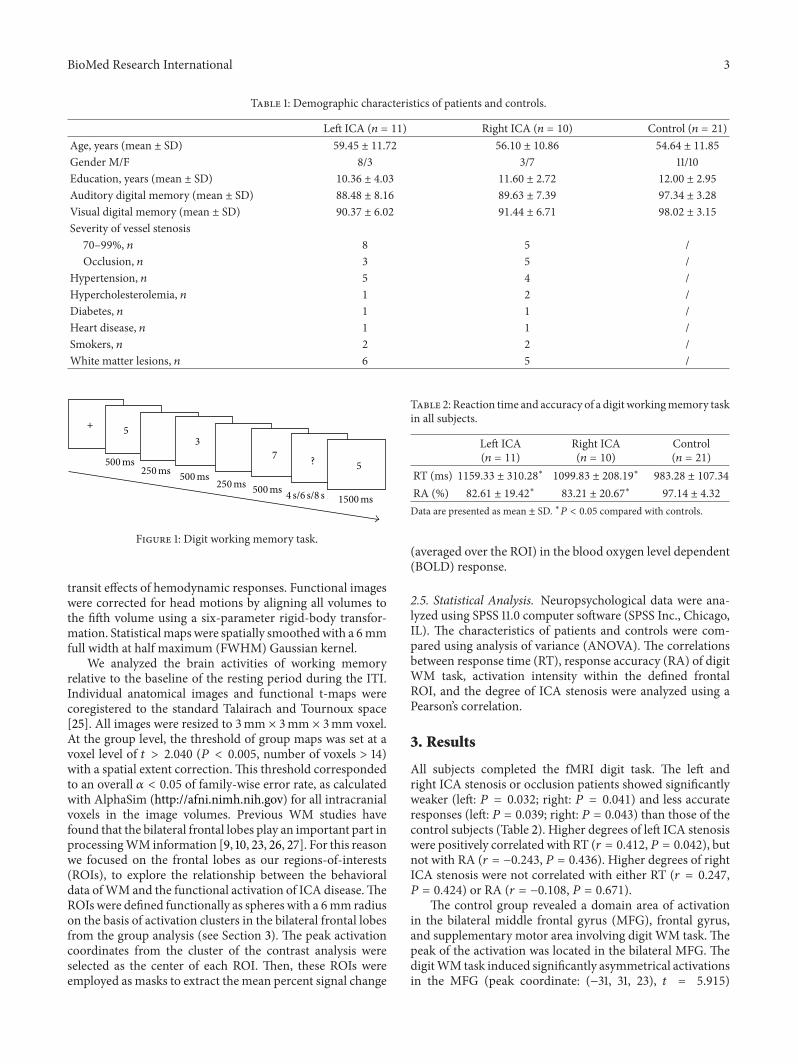
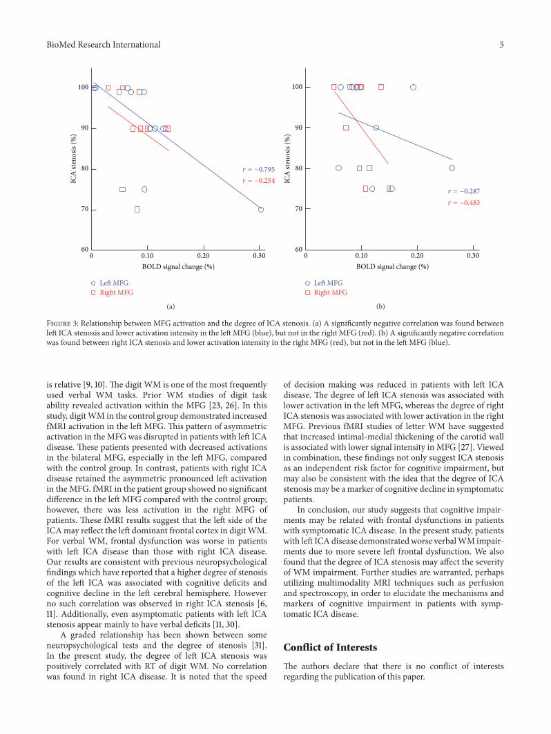
We selected the bilateral MFG for further ROI analysis.For the left ICA patients, there was a significant negativecorrelation between ICA stenosis and activation intensity inthe left MFG (𝑟 = −0.795, 𝑃 = 0.009), but not in theright MFG (𝑟 = −0.254, 𝑃 = 0.264). By contrast, therewas a significant negative correlation between the right ICAstenosis and activation intensity in the right MFG (𝑟 =−0.483, 𝑃 = 0.041), but not in the left MFG (𝑟 = −0.287,𝑃 = 0.218). Correlation graphs of MFG activation and ICAstenosis are displayed in Figure 3.
4. Discussion
The results from this study suggested that patients withleft ICA disease have more severe frontal lobe dysfunctionsthan those of age- and sex-matched controls. Comparedwith controls, the right ICA patients found slightly lessactivation in the right MFG. Such weaker MFG activity wasalso associated with a higher stenosis of ipsilateral ICA. Asignificant negative correlation was found between left ICAstenosis and activation of left MFG in the left ICA patients.Similarly, there was a significant negative correlation betweenthe right ICA stenosis and activation of right MFG in rightICA patients.
Behavioral studies have reported impaired frontal lobefunction in patients with ICA disease. Patients demonstratedWM impairments by neuropsychological assessments [4–7,11, 13]. In addition, results from fMRI and positron emissiontomography (PET) imaging suggest that the frontal cortexplays a critical role in the WM [9, 10, 23, 26, 27]. MFG isthe core region involving WM. Our results indicated thatthe activation of MFG (especially left MFG) is key regionparticipant in digit WM. However, compared with controls,MFG activation was weaker in patients with left or rightICA disease. This finding demonstrates the ability of fMRI todetect abnormal frontal lobe activation in patients with mildcognitive impairment.
y = 29
R L
Z = 26 Z = 30 Z = 34
(a)
0 5
y = 32
t value
Z = 23 Z = 27 Z = 31
(b)
Figure 2: fMRI results for a digit WM task. (a)The left ICA patientsshowed significantly less activation in the leftMFG as well as slightlyless activation in the right MFG than control subjects. (b) The rightICA patients showed slightly less activation in the right MFG onlythan control subjects.
ICA disease may cause cognitive impairment but themechanisms involved are poorly understood. Our resultssuggest that frontal lobe dysfunction may be one of thepossible mechanisms. Since fMRI is based on hemodynamiccoupling in activated brains, our results also imply that per-fusion responses may be involved. Previous studies proposethat compromised frontal lobe perfusion may be a causeof cognitive impairment in patients with ICA disease. Thusthere is the suggestion that restoring, or at least improving,frontal perfusion with carotid endarterectomy or carotidartery stenting may enhance cognitive function [28, 29]. Inthis study, we did not examine changes in cerebral bloodflow in our subjects. Future studies combining fMRI withperfusion imaging may be helpful for investigating thishypothesis.
Two forms of WM, namely, verbal and nonverbal WM,have been found to be asymmetrically represented in the leftand right frontal cortex; however, this left-right specialization
BioMed Research International 5
100
90
80
70
600 0.10 0.20 0.30
ICA
sten
osis
(%)
BOLD signal change (%)
r = −0.795
r = −0.254
Left MFGRight MFG
(a)
100
90
80
70
60
ICA
sten
osis
(%)
0 0.10 0.20 0.30
BOLD signal change (%)
Left MFGRight MFG
r = −0.287
r = −0.483
(b)
Figure 3: Relationship between MFG activation and the degree of ICA stenosis. (a) A significantly negative correlation was found betweenleft ICA stenosis and lower activation intensity in the left MFG (blue), but not in the right MFG (red). (b) A significantly negative correlationwas found between right ICA stenosis and lower activation intensity in the right MFG (red), but not in the left MFG (blue).
is relative [9, 10]. The digit WM is one of the most frequentlyused verbal WM tasks. Prior WM studies of digit taskability revealed activation within the MFG [23, 26]. In thisstudy, digitWM in the control group demonstrated increasedfMRI activation in the left MFG. This pattern of asymmetricactivation in theMFGwas disrupted in patients with left ICAdisease. These patients presented with decreased activationsin the bilateral MFG, especially in the left MFG, comparedwith the control group. In contrast, patients with right ICAdisease retained the asymmetric pronounced left activationin the MFG. fMRI in the patient group showed no significantdifference in the left MFG compared with the control group;however, there was less activation in the right MFG ofpatients. These fMRI results suggest that the left side of theICAmay reflect the left dominant frontal cortex in digitWM.For verbal WM, frontal dysfunction was worse in patientswith left ICA disease than those with right ICA disease.Our results are consistent with previous neuropsychologicalfindings which have reported that a higher degree of stenosisof the left ICA was associated with cognitive deficits andcognitive decline in the left cerebral hemisphere. Howeverno such correlation was observed in right ICA stenosis [6,11]. Additionally, even asymptomatic patients with left ICAstenosis appear mainly to have verbal deficits [11, 30].
A graded relationship has been shown between someneuropsychological tests and the degree of stenosis [31].In the present study, the degree of left ICA stenosis waspositively correlated with RT of digit WM. No correlationwas found in right ICA disease. It is noted that the speed
of decision making was reduced in patients with left ICAdisease. The degree of left ICA stenosis was associated withlower activation in the left MFG, whereas the degree of rightICA stenosis was associated with lower activation in the rightMFG. Previous fMRI studies of letter WM have suggestedthat increased intimal-medial thickening of the carotid wallis associated with lower signal intensity in MFG [27]. Viewedin combination, these findings not only suggest ICA stenosisas an independent risk factor for cognitive impairment, butmay also be consistent with the idea that the degree of ICAstenosis may be amarker of cognitive decline in symptomaticpatients.
In conclusion, our study suggests that cognitive impair-ments may be related with frontal dysfunctions in patientswith symptomatic ICA disease. In the present study, patientswith left ICAdisease demonstratedworse verbalWM impair-ments due to more severe left frontal dysfunction. We alsofound that the degree of ICA stenosis may affect the severityof WM impairment. Further studies are warranted, perhapsutilizing multimodality MRI techniques such as perfusionand spectroscopy, in order to elucidate the mechanisms andmarkers of cognitive impairment in patients with symp-tomatic ICA disease.
Conflict of Interests
The authors declare that there is no conflict of interestsregarding the publication of this paper.
6 BioMed Research International
References
[1] M.M. B. Breteler, “Vascular risk factors for Alzheimer’s disease:an epidemiologic perspective,” Neurobiology of Aging, vol. 21,no. 2, pp. 153–160, 2000.
[2] H. Henon, I. Durieu, D. Guerouaou, F. Lebert, F. Pasquier, andD. Leys, “Poststroke dementia: incidence and relationship toprestroke cognitive decline,” Neurology, vol. 57, no. 7, pp. 1216–1222, 2001.
[3] R. Rao, “The role of carotid stenosis in vascular cognitive im-pairment,” European Neurology, vol. 46, no. 2, pp. 63–69, 2001.
[4] F. C. Bakker, C. J.M. Klijn, A. Jennekens-Schinkel, and L. J. Kap-pelle, “Cognitive disorders in patients with occlusive disease ofthe carotid artery: a systematic review of the literature,” Journalof Neurology, vol. 247, no. 9, pp. 669–676, 2000.
[5] F. C. Bakker, C. J. M. Klijn, A. Jennekens-Schinkel, I. vander Tweel, C. A. F. Tulleken, and L. J. Kappelle, “Cognitiveimpairment in patients with carotid artery occlusion andipsilateral transient ischemic attacks,” Journal of Neurology, vol.250, no. 11, pp. 1340–1347, 2003.
[6] J. E. Kim, B. R. Lee, J. E. Chun et al., “Cognitive dysfunctionin 16 patients with carotid stenosis: detailed neuropsychologicalfindings,” Journal of Clinical Neurology, vol. 3, no. 1, pp. 9–17,2007.
[7] R. Rao, S. Jackson, and R. Howard, “Neuropsychologicalimpairment in stroke, carotid stenosis, and peripheral vasculardisease: a comparison with healthy community residents,”Stroke, vol. 30, no. 10, pp. 2167–2173, 1999.
[9] E. E. Smith and J. Jonides, “Neuroimaging analyses of humanworking memory,” Proceedings of the National Academy ofSciences of theUnited States of America, vol. 95, no. 20, pp. 12061–12068, 1998.
[10] L. E. Nystrom, T. S. Braver, F. W. Sabb, M. R. Delgado, D. C.Noll, and J. D. Cohen, “Working memory for letters, shapes,and locations: fMRI evidence against stimulus-based regionalorganization in human prefrontal cortex,” NeuroImage, vol. 11,no. 5, part 1, pp. 424–446, 2000.
[11] S. C. Johnston, E. S. O’Meara, T. A. Manolio et al., “Cognitiveimpairment and decline are associated with carotid arterydisease in patients without clinically evident cerebrovasculardisease,”Annals of InternalMedicine, vol. 140, no. 4, pp. 237–247,2004.
[12] L. E. Philipose, H. Alphs, V. Prabhakaran, andA. E.Hillis, “Test-ing conclusions from functional imaging of working memorywith data from acute stroke,” Behavioural Neurology, vol. 18, no.1, pp. 37–43, 2007.
[13] L. K. Sztriha, D. Nemeth, T. Sefcsik, and L. Vecsei, “Carotidstenosis and the cognitive function,” Journal of the NeurologicalSciences, vol. 283, no. 1-2, pp. 36–40, 2009.
[14] North American Symptomatic Carotid Endarterectomy TrialCollaborators, “Beneficial effect of carotid endarterectomy insymptomatic patients with high-grade carotid stenosis,” TheNew England Journal of Medicine, vol. 325, no. 7, pp. 445–453,1991.
[15] L. H. Bonati, S. T. Engelter, and P. A. Lyrer, “Carotid arterystenting,” SwissMedicalWeekly, vol. 142,Article IDw13619, 2012.
[16] S. L. Xu and Z. Y. Wu, “Development of the clinical memoryscale,” Acta Psychologica Sinica, vol. 18, pp. 100–108, 1986.
[17] S. K. Sutherland, S. E. Purdon, C. H. Lai, L. J. Wang, G. Z. Liu,and J. J. Shan, “Memory enhancement from twoweeks’ exposure
to North American ginseng extract HT1001 in young andmiddle aged healthy adults,” The Open Nutraceuticals Journal,vol. 3, pp. 20–24, 2010.
[18] R. C. Oldfield, “The assessment and analysis of handedness: theedinburgh inventory,”Neuropsychologia, vol. 9, no. 1, pp. 97–113,1971.
[19] R. Sakakibara, T. Hattori, T. Uchiyama, and T. Yamanishi, “Uri-nary function in elderly people with and without leukoaraiosis:relation to cognitive and gait function,” Journal of NeurologyNeurosurgery and Psychiatry, vol. 67, no. 5, pp. 658–660, 1999.
[20] P. Gong, A. Zheng, D. Chen et al., “Effect of BDNF Val66Metpolymorphism on digital workingmemory and spatial localiza-tion in a healthy chinese han population,” Journal of MolecularNeuroscience, vol. 38, no. 3, pp. 250–256, 2009.
[21] S. Y. Shu, Y. M. Wu, X. M. Bao et al., “A new area in the humanbrain associated with learning and memory: immunohisto-chemical and functional MRI analysis,” Molecular Psychiatry,vol. 7, no. 9, pp. 1018–1022, 2002.
[22] M. Raabe, V. Fischer, D. Bernhardt, and M. W. Greenlee,“Neural correlates of spatial working memory load in a delayedmatch-to-sample saccade task,” Neuroimage, vol. 71, pp. 84–91,2013.
[23] H. C. Bergmann, M. Rijpkema, G. Fernandez, and R. P. C.Kessels, “Distinct neural correlates of associativeworkingmem-ory and long-term memory encoding in the medial temporallobe,” NeuroImage, vol. 63, no. 2, pp. 989–997, 2012.
[24] R. W. Cox, “AFNI: software for analysis and visualization offunctional magnetic resonance neuroimages,” Computers andBiomedical Research, vol. 29, no. 3, pp. 162–173, 1996.
[25] J. Talairach and P. Tournoux, Co-Planar Stereotactic Atlas of theHuman Brain-3-Dimensional Proportional System: AnApproachto Cerebral Imaging, Thieme Medical Publishers, Stuttgart,Germany, 1988.
[26] F. E. Cooper, M. Grube, K. Von Kriegstein et al., “Distinctcritical cerebellar subregions for components of verbal workingmemory,” Neuropsychologia, vol. 50, no. 1, pp. 189–197, 2012.
[27] A. P. Haley, L. H. Sweet, J. Gunstad et al., “Verbal workingmemory and atherosclerosis in patients with cardiovasculardisease: an fMRI study,” Journal of Neuroimaging, vol. 17, no. 3,pp. 227–233, 2007.
[28] K. Kishikawa, M. Kamouchi, Y. Okada, T. Inoue, S. Ibayashi,and M. Iida, “Effects of carotid endarterectomy on cerebralblood flow and neuropsychological test performance in patientswith high-grade carotid stenosis,” Journal of the NeurologicalSciences, vol. 213, no. 1-2, pp. 19–24, 2003.
[29] W. Mlekusch, I. Mlekusch, M. Haumer et al., “Improvement ofneurocognitive function after protected carotid artery stenting,”Catheterization and Cardiovascular Interventions, vol. 71, no. 1,pp. 114–119, 2008.
[30] M. Silvestrini, I. Paolino, F. Vernieri et al., “Cerebral hemo-dynamics and cognitive performance in patients with asymp-tomatic carotid stenosis,” Neurology, vol. 72, no. 12, pp. 1062–1068, 2009.
[31] E. B. Mathiesen, K. Waterloo, O. Joakimsen, S. J. Bakke, E.A. Jacobsen, and K. H. Bønaa, “Reduced neuropsychologicaltest performance in asymptomatic carotid stenosis: the TromsøStudy,” Neurology, vol. 62, no. 5, pp. 695–701, 2004.