Objectives. The purpose of this research was to study, in macerated adult human mandibles, the height of the lingula and providemorphometric data for its location considering aspects such as shape of the lingula, gender, and race. Material and Methods.132 macerated mandibles of Brazilian adult individuals, both sexes, Amerindian and Caucasian, were used. The distances: frommandibular notch to lingula; from anterior margin of ramus of mandible to lingula; from posterior margin of ramus of mandibleto lingula; from mandibular base to lingula, and the height of lingula were obtained. To perform these measurements we used adigital caliper. The variables such as gender and race were analyzed. Results. The mean values found for the height of lingula and itslocation were determined according to the gender, race, and the lingula shape. Conclusion. This research provides additional dataon height of the lingula and morphometric data for its location considering aspects such as shape of the lingula, gender, and race,information that had not been reported in the literature to date. We emphasize that a careful study considering gender and ethnicgroup makes procedures involving the region of lingula safer.
1. Introduction
The mandibular foramen (MF) corresponds to the openingof mandibular canal through which it penetrates the infe-rior alveolar vascular-nervous bundle [1], which is situatedinferiorly and posteriorly to the greatest prominence of thelingula [2]. The lingula medially limits the MF [1]; thus,many authors consider this structure as an ideal anatomicallandmark [3] to determine the position of the MF whenperforming certain surgical procedures, such as sagittal splitramus osteotomy (SSRO) technique [2]. In this procedure, thehorizontal osteotomy should be made just above the lingulaand extended posteriorly to it in order to make a safe splitwith less potential for nerve injury [4, 5].
The correct identification of MF is important to avoidcomplications not only during the performance of surgicalprocedures involving the region of lingula, but also in theinferior alveolar nerve anesthesia [6].The authors report thatthis proceduremay fail between 29% and 35%of cases and the
anatomical variations are considered the major responsiblefor these failures [7, 8]. Inasmuch as this is an internalstructure that cannot be palpated clinically, standard teachingis to use landmarks to estimate its location [9]. Many authorshave used the lingula as a landmark for the location of theMF; however, they have not considered important variablessuch as the shape of lingula, gender, and race.
The purpose of this research was to study, in maceratedadult humanmandibles, the height of the lingula and providemorphometric data for its location considering aspects suchas shape of the lingula, gender, and race. We intend to con-tribute to guide maxillofacial surgeons to perform a surgicalprocedure involving the region of lingula safer, preventingcomplications.
2. Material and Method
One hundred thirty-two macerated mandibles of Brazilianadult individuals, of both sexes, Amerindian and Caucasian,
Hindawi Publishing CorporationBioMed Research InternationalVolume 2015, Article ID 873751, 7 pageshttp://dx.doi.org/10.1155/2015/873751
2 BioMed Research International
(a) (b)
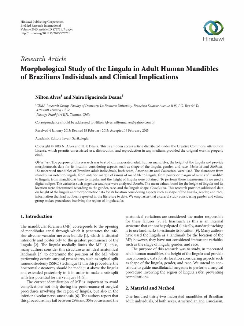
Figure 1: Medial view of the ramus of mandible. (a) Illustration of the height of the lingula (h). (b) Illustration of the distances measures:MN-L, A-L, P-L, and MB-L.
belonging to the Department of Morphology and Genet-ics, UNIFESP (Sao Paulo, Brazil), were used. Edentulousmandibles and/or those that did not contain information onsex, age, and race were excluded from this study. Only whollyor partially dentate hemimandibles that had at least onemolarin the hemiarcade that are being studied were included.
Regarding the shape, the lingula was classified into fourtypes, according to Tuli et al. [10]: triangular, truncated,nodular, and assimilated. To determine the height of thelingula, the distance from the uppermost point of the lingula(L) to the lowest point of the mandibular foramen (MF) (L-MF) was mesured (Figure 1(a)). To determine the preciselocation of lingula, the following distances were mesured:from mandibular notch (MN) to lingula (MN-L); fromanterior margin of ramus of mandible (A) to lingula (A-L); from posterior margin of ramus of mandible (P) tolingula (P-L); from mandibular base (MB) to lingula (MB-L)(Figure 1(b)).
To perform thesemeasurements, we used a digital caliper.The variables such as gender and race were analyzed.The dataobtained were tabulated and analyzed using ANOVA and tstudent, as applicable. Statistically significant 𝑃 < 0.05 wasconsidered.
Additionally we calculated the ratio of the lingula toobtain the anteroposterior position of lingula on the ramusof mandible, using the following formula: (A-L)/A-L + P-L.A smaller ratio of the lingula indicates that the lingula wouldbe located at most anterior portion of ramus of mandible. Toobtain the superoinferior position of lingula on the ramus ofmandible, we used the following formula: MN-L + MB-L/3to analyze whether it was located in the upper, medium, orlower thirds.
3. Results
The total was 253 hemimandibles, 96 Amerindian males(AM), 69 Caucasian males (CM), 62 Amerindian females(AF), and 26 Caucasian females (CF). In the statistical tests,
we have not included the assimilated shape due to the smallnumber of samples obtained.
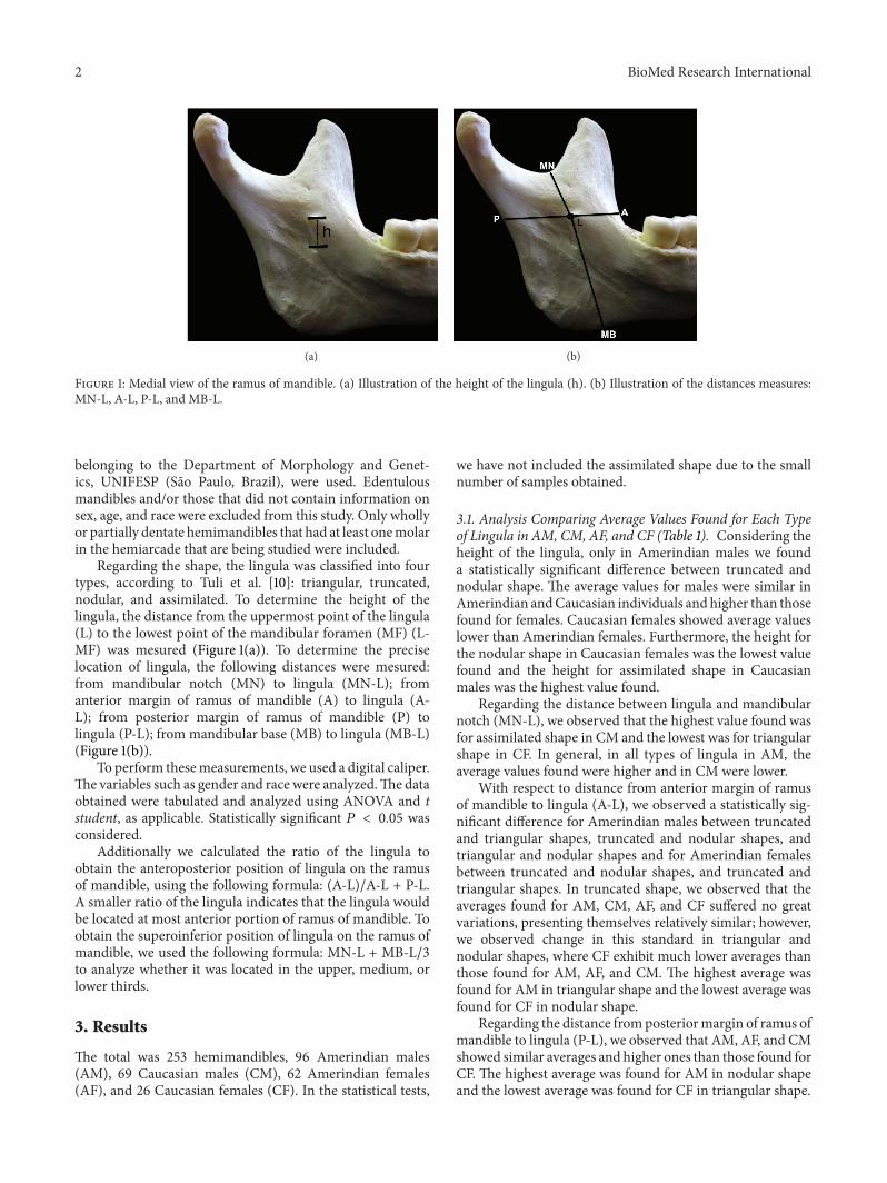
3.1. Analysis Comparing Average Values Found for Each Typeof Lingula in AM, CM, AF, and CF (Table 1). Considering theheight of the lingula, only in Amerindian males we founda statistically significant difference between truncated andnodular shape. The average values for males were similar inAmerindian andCaucasian individuals andhigher than thosefound for females. Caucasian females showed average valueslower than Amerindian females. Furthermore, the height forthe nodular shape in Caucasian females was the lowest valuefound and the height for assimilated shape in Caucasianmales was the highest value found.
Regarding the distance between lingula and mandibularnotch (MN-L), we observed that the highest value found wasfor assimilated shape in CM and the lowest was for triangularshape in CF. In general, in all types of lingula in AM, theaverage values found were higher and in CM were lower.
With respect to distance from anterior margin of ramusof mandible to lingula (A-L), we observed a statistically sig-nificant difference for Amerindian males between truncatedand triangular shapes, truncated and nodular shapes, andtriangular and nodular shapes and for Amerindian femalesbetween truncated and nodular shapes, and truncated andtriangular shapes. In truncated shape, we observed that theaverages found for AM, CM, AF, and CF suffered no greatvariations, presenting themselves relatively similar; however,we observed change in this standard in triangular andnodular shapes, where CF exhibit much lower averages thanthose found for AM, AF, and CM. The highest average wasfound for AM in triangular shape and the lowest average wasfound for CF in nodular shape.
Regarding the distance fromposteriormargin of ramus ofmandible to lingula (P-L), we observed that AM, AF, and CMshowed similar averages and higher ones than those found forCF. The highest average was found for AM in nodular shapeand the lowest average was found for CF in triangular shape.
BioMed Research International 3
Table 1: Average height of the lingula (in millimeters) and average of the following measurements: mandibular notch to lingula (MN-L);anterior margin of ramus of mandible to lingula (A-L); posterior margin of ramus of mandible to lingula (P-L); mandibular base to lingula(MB-L) (in millimeters), for Amerindian and Caucasian males and females, 𝑃 value and standard deviation (SD).
Amerindian male Caucasian male Amerindian female Caucasian female Total meanHeight
(—) could not be calculated; ∗statistically significant difference. The 𝑃 value was found on analysis of the shapes of lingula in each group studied.
Regarding the distance from mandibular base to lingula(MB-L), we have found that CM showed averages consider-ably higher than AM, AF, and CF in all types of lingula andCF showed the lowest values. The highest average was foundfor CM in truncated shape and the lowest was found for CFin nodular shape, being observed as a difference of 8.16mmbetween these two averages.
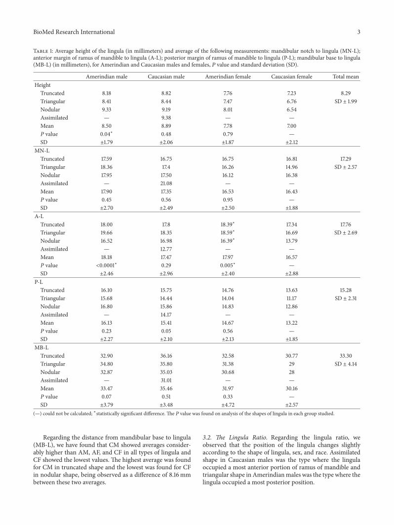
3.2. The Lingula Ratio. Regarding the lingula ratio, weobserved that the position of the lingula changes slightlyaccording to the shape of lingula, sex, and race. Assimilatedshape in Caucasian males was the type where the lingulaoccupied a most anterior portion of ramus of mandible andtriangular shape in Amerindianmales was the type where thelingula occupied a most posterior position.
4 BioMed Research International
Table 2: Lingula ratio found for each shape of lingula in Amerindian male, Caucasian male, Amerindian female, and Caucasian female.
Amerindian male Caucasian male Amerindian female Caucasian femaleTruncated 0.52 0.53 0.55 0.55Triangular 0.60 0.55 0.56 0.59Nodular 0.49 0.51 0.52 0.51Assimilated — 0.47 — —(—) could not be calculated.
Table 3: 𝑃 value (betweenmale and female, Amerindian and Caucasian individuals) for height and the followingmeasurements: mandibularnotch to lingula (MN-L); anterior margin of ramus of mandible to lingula (A-L); posterior margin of ramus of mandible to lingula (P-L);mandibular base to lingula (MB-L).
Truncated Triangular NodularM × F C × A M × F C × A M × F C × A
In general terms, the nodular shape appeared to beoccupied a most anterior portion of ramus of mandible andtriangular shape most posterior in all individuals (Table 2).
3.3. Analysis of Superoinferior Position of the Lingula. ForAM, the nodular and triangular shapes were located in thesuperior third of ramus of mandible and only the truncatedshape was located in the middle third. For CM, all types werelocated in the superior third. For AF, only nodular shape waslocated in the superior third; the truncated and triangularshapes were located in the middle third. For CF, all shapeswere located in the middle third.
We observed that in all cases the lingula was located nearthe intersection line between the superior and middle thirds,no wider than 1mm, except in nodular shape for CF, whichwas in themiddle third, 2mm away from the intersection linebetween the superior and middle thirds.
3.4. Gender Analysis. Analyzing gender differences, we ob-served that there was a statistically significant difference forthe truncated shape in the height of the lingula and inMN-L,P-L, and MB-L distances; for the triangular shape in MN-L,P-L, and MB-L distances, regarding the nodular shape, onlyA-L distance showed no difference between sexes (Table 3).
3.5. Analysis between Amerindian and Caucasian Individuals.Analyzing differences between Amerindian and Caucasianindividuals, we observed that there was a statistically sig-nificant difference only in P-L distance for truncated andtriangular shapes. Additionally, we found statistically signif-icant difference between Amerindian and Caucasian malesin P-L (𝑃 = 0.04) and MB-L (𝑃 = 0.0007). BetweenAmerindian and Caucasian females, we found statisticallysignificant difference in A-L (𝑃 = 0.01), P-L (𝑃 = 0.005),and MB-L (𝑃 = 0.04) distances (Table 3).
4. Discussion
Morphometric data on the lingula and its location werestudied in different researches, addressing different popula-tions (Table 4); however, we found that there are no studiesanalyzing the location of the lingula according to the shapeof the lingula in adult individuals; furthermore, we found nostudies comparing races and there are few that analyze genderdifferences.
Sekerci et al. [11], studying a population of Turkish chil-dren, reported that there was statistical difference betweengender in the triangular and nodular shapes. The heightof lingula shows differences between genders and varies indifferent populations [12]. In our study, we found significantdifferences betweenmales and females in height for truncatedand nodular shapes. We observed that sexual differentiationwas well set for other distances, such as MN-L, in thetriangular and nodular shapes, P-L and MB-L for all shapes.Only for A-L distance, we did not observe difference betweengenders. Sekerci and Sisman [12] and Jansisyanont et al. [13]claim that, in females, the distances are shorter than or nearlyequal to those found in males; we observed in our study thatthemean values for height, MN-L, P-L, andMB-L were lowerin females and only the A-L distance was lower or equivalentto those found in males, confirming that male mandibles aregenerally larger than female mandibles [9].
Amerindian females showed mean values higher thanCaucasian females in height and in all distances, exceptin MN-L. Amerindian and Caucasian males showed sim-ilar values in height and in all distances, except MB-Lin which Caucasian males showed higher average values.We found significant differences in P-L and MB-L betweenAmerindian and Caucasian males and in A-L, P-L, and MB-L between Amerindian and Caucasian females. Accordingto our results, there was significant difference betweenCaucasian and Amerindian females in the anteroposterior
BioMed Research International 5
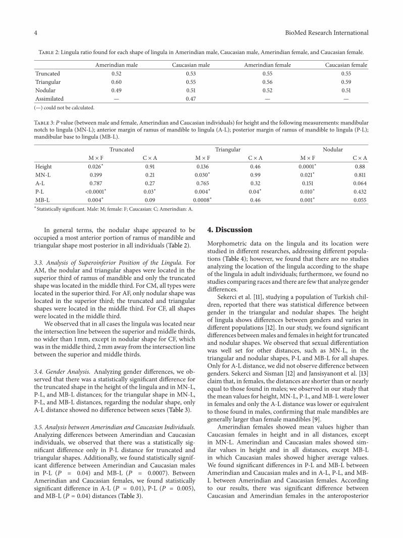
Table 4: Mean values (in millimeters) for height and the following measurements: mandibular notch to lingula (MN-L); anterior margin oframus of mandible to lingula (A-L); posterior margin of ramus of mandible to lingula (P-L); mandibular base to lingula (MB-L) reported byother authors.
Authors References Population Height MN-L A-L P-L MB-L
Monnazzi et al. [2] — 5.82 16.38 16.5 14.63 27.09—
Kim et al. [5] Koreans — 15.1 17.4 — —
Sekerci and Sisman [12] Turkish
7RF9.08RM7.24LF8.58LM7.97∗
13.95RF17.21RM14.17LF15.93LM15.3∗
15.97RF18.23RM15.6LF17.26LM16.7∗
12.43RF13.61RM12LF
14.03LM13∗
30.93RF35.53RM30.94LF36.32LM
—
Jansisyanont et al. [13] Thai
8RM8.1RF8.4LM8.5LF8.2∗
16.9RM16RF16.8LM15.9LF16.6∗
20.9RM20.2RF20.6LM20.1LF20.6∗
18.2RM18.4RF17.4LM17.3LF18∗
————
Gite and Padhye [14] Indian — 16.2 — — —Samanta and Kharb [15] North Indian 5.5 15.4 20 15 —Woo et al. [16] Koreans 10.51 19.82 18.6 16.1 —
Shah et al. [17] Indian ——
18.19L18.28R
——
——
——
Nicholson [18] East Indian 8.6R9.1L
——
——
——
——
Viravudth and Plakornkul [19] Thai 8.7R8.2L
——
——
——
——
Unreported or with different measurement: (—); left: L; right: R; male: M; female: F; mean: ∗.
location of the lingula; however, the distance from the lingulato the mandibular notch was quite similar.
Sekerci and Sisman [12] claim that the height of the lin-gula varies in different populations. In our study, in Brazilianadult mandibles, the height of the lingula was higher for CMin assimilated shape (9.38mm) and lower for CF in nodularshape (6.54mm). Lower values were found by Monnazzi etal. [2] (5.82mm) and Samanta and Kharb [15] (5.5mm),and higher values were reported in koreans (10.51mm) [16].The average values of the height of the lingula, which wefound for males and females in our study, were similar tothose reported by Sekerci and Sisman [12] in the Turkishpopulation. Jansisyanont et al. [13] and Nicholson [18] foundsimilar values to those that we found for males in our study.The mean values found for the height of the lingula, in ourstudy, were determined according to the gender, race, andthe lingula shape, having great variability. This data has greatclinical relevance, since it can be used as a parameter tocarry out certain surgical procedures involving the region oflingula.
In our study, the distance from the mandibular notchto the lingula (MN-L), distance that helps surgeons to findthe lingula [12], was similar to the studies of Monnazzi etal. [2], Sekerci and Sisman [12], Jansisyanont et al. [13],Gite and Padhye [14], Viravudth and Plakornkul [19], andKositbowornchai et al. [20]. Woo et al. [16] and Shah et al.[17] found values slightly higher than ours for Koreans andIndians, respectively. According to Sekerci and Sisman [12],the length of horizontal osteotomy in bilateral SSRO should
be between 15 and 19mm; according to Jansisyanont et al.[13], it should be between 17 and 24mm. We disagree withthese authors because in our study the distance from themandibular notch to the lingula (MN-L) showed minimumvalues lower than 15mm; furthermore, most authors reportvalues lower than 17mm[2, 5, 12–15]. Based on the results thatwere obtained through our study and on the literature data,we suggest that the length of horizontal osteotomy in bilateralSSRO should be between 13mm and 24mm. In our study, thedifferences between the average values for theMN-L distanceare associated with ethnicity and gender. The average valuesthat we found for females were lower than those that wefound for males; moreover, the distances with higher averagevalues were found for Amerindian males. We emphasize thata careful study considering gender and ethnic group makesprocedures involving the region of lingula safer.
In our study, the distance from the anterior margin of themandible to the lingula (A-L) with highest average value wasfound in triangular shape for Amerindian males (19.66mm),and the lowest average value was found in assimilated shapefor Caucasianmales (12.77mm).The average values found forfemales, in our study, were higher than those reported for theTurkish [12], while the average values found for males weresimilar. Highermean values were found for North Indian andThai, with 20 and 20.6mm, respectively [12, 15]. In our studywe observed great variability in the average values for A-L,with lower average values for assimilated and nodular shapes,allowing us to claim that the most anterior or posteriorlocation of the lingula and consequently the location of the
6 BioMed Research International
mandibular foramen are associated with each type of lingula.This distance showed no statistically significant differencebetween gender and race.
The distance from the posterior margin of the mandibleto the lingula (P-L) was lower in the Turkish population [12]than that found in our study, in both females and males.Monnazzi et al. [2] found similar average values to thosefound in our study. The same occurred with Samanta andKharb [15] who worked with North Indian population andwith Woo et al. [16] who worked with Koreans. Jansisyanontet al. [13] reported the highest value found for this distancein theThai population, with 18mm.The lowest average valuewas found in our study for Caucasian female in triangularshape, with 11.17mm. In our study, this distance showedmarked sexual difference between races.
Regarding the distance from the mandibular base to thelingula (MB-L), the values found for the Turkish population[12] were similar to those found in our study. The valuesreported by Monnazzi et al. [2] were higher than ours. In ourstudy, Caucasian males showed the highest mean values andCaucasian females the lowest. This distance showed markeddifference between sexes.
The lingula ratio (LR) provides relevant data to aid insurgical planning. We note that, in general, lingulas withtriangular shape are located slightly more posterior than lin-gulas with nodular shape, which can determine change in thelevel where the osteotomy should be performed in bilateralSSRO or the inferior alveolar nerve block, since the lingulamarks the entrance of the inferior alveolar neurovascularbundle.TheLR ranged from0.47 for assimilated shape to 0.60for triangular shape. The value reported in the literature forLR varied from 0.53 [13] to 0.56 [12, 15]. Based on our results,we agree with Kositbowornchai et al. [20] when they claimthat the lingula was positioned slightly posterior to the centerof the widht of the ramus.
As additional data for location of the lingula, we dividedthe ramus ofmandible into three thirds: superior,middle, andinferior to analyze its superoinferior position. We observedthat in all cases the lingula was located near the intersectionline between the superior and middle thirds, except for thenodular shape in Caucasian females, who was in the middlethird, 2mm away from the intersection line, between thesuperior and middle thirds. The lingula was usually locatedin the middle third in females and in the superior third inmales.
Based on our results, we emphasize that in planninga surgery involving the region of lingula, such as sagittalsplit ramus osteotomy (SSRO) technique, the surgeon musttake into account aspects such as gender, race, and shape oflingula. Another important issue that should be consideredis a possible anatomical variation in the medial region ofthe ramus of mandible, where there may be an accessorymandibular foramen located above the lingula [21, 22] or nearthe mandibular notch [23]. Such variations are not commonbut result in increased risk of complications involving theinferior alveolar nerve [21].
Alves and Candido [24] affirm that the major factorsinvolved in the failure of inferior alveolar nerve block are theaccessory innervations and the improper placement of needle
due to improper evaluation of landmarks. We consider itvery important, when performing the inferior alveolar nerveblock, to use the lingula as a landmark for the location of theMF considering aspects such as the shape of lingula, gender,and ethnic group. This is relevant not only for the successof anesthesia but also for preventing damage to vascular andneural elements.
5. Conclusions
This research provides additional data on height of the lingulaand morphometric data for its location considering aspectssuch as shape of the lingula, gender, and race, informationthat had not been reported in the literature to date.
Comparing men and women, we found that the heightof the lingula and the measured distances change accordingto the shape of the lingula (truncated, triangular, or nodular)determining variations in the lingula position.These changeswere not so marked when comparing Amerindian and Cau-casian individuals. Despite the gender, differences are moreapparent than the differences between races; both of themmust be taken into account in surgical procedures performedin the lingula region or inferior alveolar nerve block.
Themean values found for the height of the lingula, in ourstudy, were determined according to the gender, race, and thelingula shape, showing great variability. This data has greatclinical relevance, since it can be used as a parameter to carryout surgical procedures performed in the lingula region orinferior alveolar nerve block.
Considering the anteroposterior position, we concludethat the lingula was positioned slightly posterior to thecenter of the width of the ramus of mandible. Regardingthe superoinferior position, the lingula was located in thesuperior or middle third of the ramus, in general, 1mm awayfrom the intersection line of these two thirds.
Based on our study and on the literature data, wesuggest that the length of horizontal osteotomy in bilateralSSRO should be between 13mm and 24mm; however, weemphasize that a careful study considering gender and ethnicgroupmakes procedures involving the region of lingula safer.
Conflict of Interests
The authors declare that they have no conflict of interests.
Acknowledgment
This study was supported by Department of Morphology andGenetics, UNIFESP, Sao Paulo, Brazil.
References
[1] N. Alves and P. L. Candido, Anatomia para o cirurgiao-dentista,Gen-Santos, Sao Paulo, Brazil, 2nd edition, 2013.
[2] M. S. Monnazzi, L. A. Passeri, M. F. R. Gabrielli, P. D. A. Bolini,W. R. S. de Carvalho, and H. Da Costa Machado, “Anatomicstudy of the mandibular foramen, lingula and antilingula indry mandibles, and its statistical relationship between the true
BioMed Research International 7
lingula and the antilingula,” International Journal of Oral andMaxillofacial Surgery, vol. 41, no. 1, pp. 74–78, 2012.
[3] G. Salgado, H. Oscar Inzunza, M. Cantın et al., “Evaluation ofmandibular anatomy related to sagittal split ramus osteotomy,”International Journal of Morphology, vol. 30, no. 1, pp. 30–39,2012.
[4] B. N. Epker, “Modifications in the sagittal osteotomy of themandible,” Journal of Oral Surgery, vol. 35, no. 2, pp. 157–159,1977.
[5] H.-J. Kim, H.-Y. Lee, I.-H. Chung, I.-H. Cha, and C.-K. Yi,“Mandibular anatomy related to sagittal split ramus osteotomyinKoreans,”YonseiMedical Journal, vol. 38, no. 1, pp. 19–25, 1997.
[6] T. Yoshida, T. Nagamine, T. Kobayashi et al., “Impairment of theinferior alveolar nerve after sagittal split osteotomy,” Journal ofCranio-Maxillo-Facial Surgery, vol. 17, no. 6, pp. 271–278, 1989.
[7] W. D. Robertson, “Clinical evaluation of mandibular conduc-tion anesthesia,”General Dentistry, vol. 27, no. 5, pp. 49–51, 1979.
[8] T. P. Levy, “An assessment of the Gow-Gates mandibular blockfor third molar surgery,” The Journal of the American DentalAssociation, vol. 103, no. 1, pp. 37–41, 1981.
[9] A. Afsar, D. A. Haas, P. E. Rossouw, and R. E. Wood, “Radio-graphic localization of mandibular anesthesia landmarks,”OralSurgery, Oral Medicine, Oral Pathology, Oral Radiology, andEndodontics, vol. 86, no. 2, pp. 234–241, 1998.
[10] A. Tuli, R. Choudhry, S. Choudhry, S. Raheja, and S. Agarwal,“Variation in shape of the lingula in the adult humanmandible,”Journal of Anatomy, vol. 197, part 2, pp. 313–317, 2000.
[11] A. E. Sekerci, K. Cantekin, and M. Aydinbelge, “Cone beamcomputed tomographic analysis of the shape, height, andlocation of the mandibular lingula in a population of children,”BioMed Research International, vol. 2013, Article ID 825453, 8pages, 2013.
[12] A. E. Sekerci and Y. Sisman, “Cone-beam computed tomogra-phy analysis of the shape, height, and location of themandibularlingula,” Surgical and Radiologic Anatomy, vol. 36, no. 2, pp. 155–162, 2014.
[13] P. Jansisyanont, W. Apinhasmit, and S. Chompoopong, “Shape,height and location of the lingula for sagittal ramus osteotomyinThais,” Clinical Anatomy, vol. 22, no. 7, pp. 787–793, 2009.
[14] M. Gite and M. Padhye, “Location of lingula from sigmoidnotch in an indian population—a radiographic study,” ScientificJournal, vol. 1, 2007.
[15] P. P. Samanta and P. Kharb, “Morphological analysis of the lin-gula in dry adult humanmandibles of north Indian population,”Journal of Cranio-Maxillary Diseases, vol. 1, no. 1, pp. 7–11, 2012.
[16] S. S. Woo, J. Y. Cho, W. H. Park, I. H. Yoo, Y. S. Lee, andK. S. Shim, “A study of mandibular anatomy for orthognathicsurgery in Koreans,” Journal of the Korean Association of Oraland Maxillofacial Surgeons, vol. 28, no. 2, pp. 126–131, 2002.
[17] K. Shah, P. Shah, and A. Parmar, “Study of the location of themandibular foramina in Indian drymandibles,”Global ResearchAnalysis, vol. 2, no. 7, pp. 128–130, 2013.
[18] M. L. Nicholson, “A study of the position of the mandibularforamen in the adult humanmandible,”Anatomical Record, vol.212, no. 1, pp. 110–112, 1985.
[19] Y. Viravudth and V. Plakornkul, “The mandibular foramen inThais,” Siriraj Hospital Gazette, vol. 41, pp. 551–554, 1989.
[20] S. Kositbowornchai, M. Siritapetawee, T. Damrongrungruanget al., “Shape of the lingula and its localization by panoramicradiograph versus dry mandibular measurement,” Surgical andRadiologic Anatomy, vol. 29, no. 8, pp. 689–694, 2007.
[21] N. Alves and N. F. Deana, “Morphometric study of mandibularforamen inmacerated skulls to contribute to the development ofsagittal split ramus osteotomy (SSRO) technique,” Surgical andRadiologic Anatomy, vol. 36, no. 9, pp. 839–845, 2014.
[22] A. R. Freire, A. C. Rossi, F. B. Prado, P. H. F. Caria, and P. R.Botacin, “Incidence of the mandibular accessory foramina inBrazilian population,” Journal of Morphological Sciences, vol. 29,no. 3, pp. 171–173, 2012.
[23] E. Cvetko, “Bilateral anomalous high position of themandibularforamen: a case report,” Surgical and Radiologic Anatomy, vol.36, no. 6, pp. 613–616, 2014.
[24] N. Alves and P. L. Candido, Anatomia para o curso de Odon-tologia geral e especıfica, Santos Editora, Sao Paulo, Brazil, 3rdedition, 2012.