Spurdle et al. Breast Cancer Research (2014) 16:3419 DOI 10.1186/s13058-014-0474-y
RESEARCH ARTICLE Open Access
Refined histopathological predictors of BRCA1and BRCA2 mutation status: a large-scale analysisof breast cancer characteristics from the BCAC,CIMBA, and ENIGMA consortiaAmanda B Spurdle1*, Fergus J Couch2, Michael T Parsons1, Lesley McGuffog3, Daniel Barrowdale3, Manjeet K Bolla3,Qin Wang3,4, Sue Healey1, Rita Katharina Schmutzler5, Barbara Wappenschmidt5, Kerstin Rhiem5, Eric Hahnen5,Christoph Engel6, Alfons Meindl7, Nina Ditsch8, Norbert Arnold9, Hansjoerg Plendl10,11, Dieter Niederacher12,Christian Sutter13, Shan Wang-Gohrke14, Doris Steinemann15, Sabine Preisler-Adams16, Karin Kast17,Raymonda Varon-Mateeva18, Steve Ellis3, Debra Frost3, Radka Platte3, Jo Perkins3, D Gareth Evans19, Louise Izatt20,Ros Eeles21, Julian Adlard22, Rosemarie Davidson23, Trevor Cole24, Giulietta Scuvera25, Siranoush Manoukian25,Bernardo Bonanni26, Frederique Mariette27,28, Stefano Fortuzzi27,28, Alessandra Viel29, Barbara Pasini30,31,Laura Papi32, Liliana Varesco33, Rosemary Balleine34, Katherine L Nathanson35, Susan M Domchek35,Kenneth Offitt36, Anna Jakubowska37, Noralane Lindor38, Mads Thomassen39, Uffe Birk Jensen40, Johanna Rantala41,Åke Borg42, Irene L Andrulis43,44, Alexander Miron45, Thomas VO Hansen46, Trinidad Caldes47,Susan L Neuhausen48, Amanda E Toland49, Heli Nevanlinna50, Marco Montagna51, Judy Garber52,Andrew K Godwin53, Ana Osorio54,55, Rachel E Factor56, Mary B Terry57, Timothy R Rebbeck35, Beth Y Karlan58,Melissa Southey59, Muhammad Usman Rashid60,61, Nadine Tung62, Paul DP Pharoah63, Fiona M Blows64,Alison M Dunning64, Elena Provenzano64, Per Hall65, Kamila Czene65, Marjanka K Schmidt66, Annegien Broeks66,Sten Cornelissen66, Senno Verhoef66, Peter A Fasching67,68, Matthias W Beckmann68, Arif B Ekici69,Dennis J Slamon67,70, Stig E Bojesen71,72,73, Børge G Nordestgaard71,72,73, Sune F Nielsen71,72, Henrik Flyger74,Jenny Chang-Claude75, Dieter Flesch-Janys76, Anja Rudolph75, Petra Seibold75, Kristiina Aittomäki77,Taru A Muranen50, Päivi Heikkilä78, Carl Blomqvist79, Jonine Figueroa80, Stephen J Chanock80, Louise Brinton80,Jolanta Lissowska81, Janet E Olson82, Vernon S Pankratz82, Esther M John83,84, Alice S Whittemore84,Dee W West83,84, Ute Hamann60, Diana Torres60,85, Hans Ulrich Ulmer86, Thomas Rüdiger87, Peter Devilee88,Robert AEM Tollenaar89, Caroline Seynaeve90, Christi J Van Asperen91, Diana M Eccles92, William J Tapper92,Lorraine Durcan92, Louise Jones93, Julian Peto94, Isabel dos-Santos-Silva94, Olivia Fletcher95, Nichola Johnson95,Miriam Dwek96, Ruth Swann96, Anita L Bane97,98, Gord Glendon99, Anna M Mulligan100,101, Graham G Giles102,103,Roger L Milne102,103, Laura Baglietto102,103, Catriona McLean104, Jane Carpenter105, Christine Clarke106,Rodney Scott107,108, Hiltrud Brauch109,110, Thomas Brüning111, Yon-Dschun Ko112, Angela Cox113, Simon S Cross114,Malcolm WR Reed113, Jan Lubinski37, Katarzyna Jaworska-Bieniek37, Katarzyna Durda37, Jacek Gronwald37,Thilo Dörk115, Natalia Bogdanova116, Tjoung-Won Park-Simon116, Peter Hillemanns116, Christopher A Haiman117,Brian E Henderson117, Fredrick Schumacher117, Loic Le Marchand118, Barbara Burwinkel119,120,Frederik Marme119,121, Harald Surovy119,120, Rongxi Yang119,120, Hoda Anton-Culver122, Argyrios Ziogas122,Maartje J Hooning123, J Margriet Collée124, John WM Martens123, Madeleine MA Tilanus-Linthorst125,
* Correspondence: [email protected] of Genetics and Computational Biology, QIMR BerghoferMedical Research Institute, 300 Herston Road, Brisbane, QLD 4006, AustraliaFull list of author information is available at the end of the article
Spurdle et al. Breast Cancer Research (2014) 16:3419 Page 2 of 16
Hermann Brenner126, Aida Karina Dieffenbach126, Volke Arndt126, Christa Stegmaier127, Robert Winqvist128,Katri Pylkäs128, Arja Jukkola-Vuorinen129, Mervi Grip130, Annika Lindblom131, Sara Margolin132, Vijai Joseph36,Mark Robson36, Rohini Rau-Murthy36, Anna González-Neira133, José Ignacio Arias134, Pilar Zamora135,Javier Benítez54,136, Arto Mannermaa137,138, Vesa Kataja137,139, Veli-Matti Kosma137,138, Jaana M Hartikainen137,138,Paolo Peterlongo27, Daniela Zaffaroni25, Monica Barile26, Fabio Capra27,28, Paolo Radice140, Soo H Teo141,142,Douglas F Easton3,4, Antonis C Antoniou3, Georgia Chenevix-Trench1, David E Goldgar143, on behalf of ABCTBInvestigators, EMBRACE Group, GENICA Network, HEBON Group and kConFab Investigators
Abstract
Introduction: The distribution of histopathological features of invasive breast tumors in BRCA1 or BRCA2 germlinemutation carriers differs from that of individuals with no known mutation. Histopathological features thus haveutility for mutation prediction, including statistical modeling to assess pathogenicity of BRCA1 or BRCA2 variants ofuncertain clinical significance. We analyzed large pathology datasets accrued by the Consortium of Investigators ofModifiers of BRCA1/2 (CIMBA) and the Breast Cancer Association Consortium (BCAC) to reassess histopathologicalpredictors of BRCA1 and BRCA2 mutation status, and provide robust likelihood ratio (LR) estimates for statisticalmodeling.
Methods: Selection criteria for study/center inclusion were estrogen receptor (ER) status or grade data available forinvasive breast cancer diagnosed younger than 70 years. The dataset included 4,477 BRCA1 mutation carriers, 2,565BRCA2 mutation carriers, and 47,565 BCAC breast cancer cases. Country-stratified estimates of the likelihood ofmutation status by histopathological markers were derived using a Mantel-Haenszel approach.
Results: ER-positive phenotype negatively predicted BRCA1 mutation status, irrespective of grade (LRs from 0.08 to0.90). ER-negative grade 3 histopathology was more predictive of positive BRCA1 mutation status in women 50 yearsor older (LR = 4.13 (3.70 to 4.62)) versus younger than 50 years (LR = 3.16 (2.96 to 3.37)). For BRCA2, ER-positive grade 3phenotype modestly predicted positive mutation status irrespective of age (LR = 1.7-fold), whereas ER-negative grade 3features modestly predicted positive mutation status at 50 years or older (LR = 1.54 (1.27 to 1.88)). Triple-negative tumorstatus was highly predictive of BRCA1 mutation status for women younger than 50 years (LR = 3.73 (3.43 to 4.05)) and50 years or older (LR = 4.41 (3.86 to 5.04)), and modestly predictive of positive BRCA2 mutation status in women 50 yearsor older (LR = 1.79 (1.42 to 2.24)).
Conclusions: These results refine likelihood-ratio estimates for predicting BRCA1 and BRCA2 mutation status by usingcommonly measured histopathological features. Age at diagnosis is an important variable for most analyses, and grade ismore informative than ER status for BRCA2 mutation carrier prediction. The estimates will improve BRCA1 and BRCA2variant classification and inform patient mutation testing and clinical management.
IntroductionIt is well established that BRCA1-related breast tumors,as a group, differ from non-BRCA1 tumors in terms ofhistological phenotype. Tumors of BRCA1 mutation car-riers are more likely to be high-grade with medullarysubtype features, including greatly increased mitoticcount, pushing margins, lymphocytic infiltrate, trabecu-lar growth pattern, and necrosis [1-3]. Consistent withoverrepresentation of a basal phenotype, a number ofimmunohistochemical (IHC) markers have been shownto be of value in assessing BRCA1 tumor phenotype infemale patients, including estrogen receptor (ER), pro-gesterone receptor (PR), human Epidermal Growth FactorReceptor 2 (HER2), p53, cytokeratin 5/6 (CK5/6), cytoker-atin 14 (CK14), cytokeratin 17 (CK17), and epidermalgrowth factor receptor (EGFR) [4-8]. In addition, severalstudies reported that reduced expression of CK8/18 can
discriminate the basal tumors of BRCA1 mutation car-riers from basal tumors of noncarriers [9,10], whereasloss of phosphatase and tensin homolog (PTEN), to-gether with triple-negative (TN; ER-, PR-, HER2-) status,was reported to improve the sensitivity of BRCA1 muta-tion prediction in a study of Asian breast cancer patients[11]. The introduction of PTEN to BRCA1 mutation-prediction algorithms is supported by single-cell analysesof temporal somatic events in BRCA1 breast tumor tis-sue, which revealed that loss of PTEN is an early eventin the development of BRCA1 basal-like tumors, whereasTP53 mutations occur first in most luminal BRCA1tumors [12].The breast tumor phenotype of female BRCA2 fe-
male mutation carriers is less distinctive than that ofBRCA1 mutation carriers [1,13,14]. Nevertheless, re-ports based on IHC or expression array analysis have
Spurdle et al. Breast Cancer Research (2014) 16:3419 Page 3 of 16
shown that BRCA2 breast tumors are predominantlyof the luminal B subtype [13,15], and are more likelythan non-BRCA2 tumors to be ER positive and highgrade, with reduced tubule formation and continuouspushing margins [2,13].A number of these histopathological features have
been incorporated into prediction models or havebeen proposed as selection criteria for prioritizingtesting of breast cancer patients for BRCA1 andBRCA2 mutations [11,16-24]. These findings have alsoserved as the basis for including independently pre-dictive tumor histopathological features as a compo-nent of the multifactorial likelihood model for clinicalclassification of BRCA1/2 variants of uncertain signifi-cance [25]. The current iteration of the model in-cludes likelihood ratio (LR) estimates of pathogenicityfor combined ER and grade or combined ER, CK5/6,and CK14 status, for analysis of BRCA1 variants, andtubule formation for BRCA2 [26-29]. However, these LRestimates were derived from analyses of relatively smalldatasets including a maximum of 600 mutation carriersand 288 noncarriers [4,6], and have not been directlyvalidated.We conducted analyses of large pathology datasets ac-
crued by the Consortium of Investigators of Modifiers ofBRCA1/2 (CIMBA) and the Breast Cancer AssociationConsortium (BCAC) to reassess previously reportedhistopathological predictors of BRCA1 and BRCA2 mu-tation status. The results provide more-refined LR esti-mates for downstream multifactorial likelihood analysisand for prediction of BRCA1 and BRCA2 mutationstatus.
MethodsAccess to data and ethics approvalsENIGMA (Evidence-based Network for the Interpret-ation of Germline Mutant Alleles) is a research con-sortium aimed to improve methods to assess theclinical significance in breast cancer susceptibilitygenes [30]. Considerable overlap in membership existsbetween ENIGMA, CIMBA, and BCAC. As a collab-oration between the three consortia, investigators inENIGMA accessed CIMBA and BCAC datasets forapproved pathology-related analyses relevant to thepurposes of ENIGMA. The collection of clinical, path-ology, and genetic data by CIMBA and BCAC hasbeen previously approved for ongoing research studiesby the local ethics committee relevant to each of theparticipating CIMBA and BCAC studies, and allparticipants provided informed consent to the rele-vant participating CIMBA and BCAC sites for suchongoing studies.Research analyses specific to this study were carried
out using only de-identified data, with approval from the
Human Research Ethics Committee of the QIMR BerghoferMedical Research Institute, and the Institutional ReviewBoard of the University of Utah.
Sample setsCIMBAThe Consortium of Investigators of Modifiers of BRCA1/2(CIMBA; [31]) is a consortium established to conductlarge-scale research studies of carriers of germline BRCA1or BRCA2 pathogenic mutations [32]. Specifically, carriersof variants of uncertain significance are ineligible for entryinto CIMBA. The major focus is discovery and validationof genetic factors that modify risk of breast and ovariancancer in BRCA1 and BRCA2 mutation carriers, with con-sideration of risk stratified by tumor histologic features.Contributing centers provide information relevant to ana-lyses, including year of birth, age at diagnosis of breastand/or ovarian cancer, cancer behavior (invasive, in situ),basic histology, and other pathology measures for breastand ovarian tumors from study participants. Pathology in-formation is extracted mainly from pathology reports, al-though a small subset of contributing centers haveconducted centralized pathology review and/or supple-mented clinical IHC results with research testing of tumormaterial (for example, 5% of ER pathology results werecentrally reviewed) [33]. All CIMBA centers with ERand grade data available in the CIMBA database thatwere from countries with pathology data available frompopulation (presumed noncarrier) reference cases inBCAC (see later) were included in the analyses. Vari-ables included were as follows: gene mutated, mutationnomenclature (and mutation type, for example, truncat-ing, missense, and so on), date of birth, age and date ofdiagnosis of breast cancer(s), breast cancer behavior, ERstatus, PR status, HER2 status, Cytokeratin 5 or 5/6 sta-tus, and grade. No CK14 IHC results were available. Nodual-mutation carriers were found. Only invasive breastcancer cases diagnosed before age 70 years were in-cluded, to reduce the likelihood of phenocopy tumorsnot directly related to mutation status. Samples were in-cluded irrespective of ovarian cancer diagnoses. For indi-viduals with two breast cancers (20% of cases), thebreast cancer diagnosed closest in time to the entry intothe CIMBA cohort was included preferentially.
BCACThe Breast Cancer Association Consortium (BCAC [34])was established to discover and validate genetic factorsassociated with risk of breast cancer in the generalpopulation [35]. BCAC also studies risk factors associ-ated with tumor subtypes and tumor histologic features,and pathology data from participating centers are de-rived from pathology reports or center-specific researchefforts. BCAC pathology data were checked and cleaned
Table 1 Subjects in CIMBA and BCAC datasets with breasttumor ER or grade status, by country
Country CIMBA BCAC
Number BRCA1 Number BRCA2 Number BCACnoncarriers
Australia 363 293 2,014
Canada 97 57 927
Denmark 201 151 2,318
Finland 55 64 2,607
Germany 982 493 9,503
Italy 547 362 270
Netherlands 113 32 4,181
Poland 247 0 2,527
Spain 91 102 358
Sweden 158 34 5,266
United Kingdom 642 388 12,989
USA 981 589 4,605
Total 4,477 2,565 47,565
Data for primary breast tumor. ER, breast tumor estrogen receptor status; BCAC,Breast Cancer Association Consortium; CIMBA, Consortium for Investigator ofModifiers of BRCA1 and BRCA2.ER status was missing for 548 (12.2%) BRCA1 carriers, 292 (11.4%) BRCA2carriers and 4,942 (10.4%) presumed noncarriers. Histological grade wasmissing for 890 (19.9%) BRCA1 carriers, 555 (21.6%) BRCA2 carriers, and 6,020(12.7%) presumed noncarriers.
Spurdle et al. Breast Cancer Research (2014) 16:3419 Page 4 of 16
centrally [36]. BCAC centers were selected for inclusionin this analysis based on availability of ER and gradedata. Studies in BCAC in which cases were ascertainedon the basis of tumor characteristics (for example, theTN consortium) were excluded. Variables provided foranalyses were as follows: study type (to identify within-study strata, and/or to define cohorts with familialcases), age at diagnosis of breast cancer(s), breast cancerbehavior, ER status, PR status, HER2 status, CK5 or 5/6status, and grade. No CK14 IHC results were available.The study design was noted as selected (familial and/orage-selected, relevant for 13 studies) or unselected (frompopulation-based or hospital-based design), based onstudy-ascertainment criteria provided by the principalinvestigators of individual BCAC sites.BRCA1 and BRCA2 germline mutation testing results
were provided by 13 of the 36 BCAC studies (compris-ing 12% of BCAC individuals overall), nine of whichused age/family history selection criteria for case ascer-tainment (with testing for 4% to 100% of these ninestudies). The 345 known mutation carriers (189 BRCA1,156 BRCA2) identified in BCAC were excluded. Analysisincluded subjects known to be noncarriers or untestedfor BRCA1/2 mutations, with relevant pathology infor-mation for primary invasive breast cancer diagnosisyounger than age 70 years. As for CIMBA, for individ-uals with two breast cancers (only 5% of all BCAC casesconsidered), the breast cancer diagnosed closest in timeto the entry into the cohort was included preferentially.
Statistical analysisER or grade data were available for 4,477 BRCA1 muta-tion carriers, 2,565 BRCA2 mutation carriers, and 47,565BCAC breast cancer cases with no known mutation inBRCA1 or BRCA2 (presumed noncarriers). The numbersof subjects by country are shown in Table 1. Only coun-tries with ≥200 cases in BCAC and ≥100 carriers inCIMBA were included in analyses to minimize potentialbias due to country-specific patterns of pathology assess-ment. ER-negative, PR-negative, and HER2-negative tu-mors were categorized as triple-negative (TN). All othercombinations of known ER, PR, and HER2 status for asingle breast tumor were categorized as “Not TN.”CIMBA and BCAC studies contributing pathology dataare noted in Additional file 1: Table S1. Final samplesizes for analyses are reported in footnotes to Tables 2and 3, and Additional file 1, Tables S2 to S4.CK5/6 IHC data were available for only 128 BRCA1
carriers, 78 BRCA2 carriers and 6,796 BCAC cases withvalid data on ER status. Numbers of carriers reducedfurther after country-matching, and frequencies differedsignificantly between countries for carriers. Cytokeratinanalyses were thus not pursued further.
All statistical analyses were performed by using STATAversion 12 (StatCorp, College Station, TX, USA). Statisticalsignificance was defined as P <0.05.We first examined whether family history was related
to the predictor variables of interest in the BCAC sam-ple set. Family-history information, defined as first-degree relative with breast cancer, was available for30,223 individuals (7,547 reporting a family history ofbreast cancer). Logistic regression analyses were per-formed to predict ER status, grade 3, or TN status as afunction of family history (defined as first-degree relativewith breast cancer), adjusting for age at diagnosis andcountry. No significant effect was observed for familyhistory on any of these histopathologic features, so wedid not consider family history further in any analyses.To identify the most important predictors of mutation
status to be used in estimation of the likelihood ratiosfor classification of variants, we undertook a series of lo-gistic regression analyses. These analyses comparedBRCA1 and BRCA2 with the BCAC set. A sequentialseries of models with country and age (younger than50 years versus 50 years or older) as a starting point andthen adding ER, grade, and the ER/grade combination totest for interaction between ER and grade. For thosecases who had data on TN status, we examined ER, ERand grade, ER TN, grade TN, and last, models with ER,grade, and TN. Likelihood ratio tests were used to deter-mine the most parsimonious models for each gene.
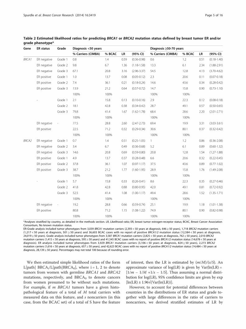
Table 2 Estimated likelihood ratios for predicting BRCA1 or BRCA2 mutation status defined by breast tumor ER and/orgrade phenotype*
Gene ER status Grade Diagnosis <50 years Diagnosis ≥50-70 years
% Carriers (CIMBA) % BCAC LR (95% CI) % Carriers (CIMBA) % BCAC LR (95% CI)
ER positive 80.8 71.2 1.15 (1.08-1.22) 74.9 80.1 0.90 (0.82-0.98)
100% 100% 100% 100%
*Analyses stratified by country, as detailed in the methods section. LR, Likelihood ratio; ER, breast tumor estrogen-receptor status; BCAC, Breast Cancer AssociationConsortium, No known mutation status.ER-Grade analysis included tumor phenotypes from 3,039 BRCA1 mutation carriers (2,393 < 50 years at diagnosis, 646 ≥ 50 years), 1,718 BRCA2 mutation carriers(1,217 < 50 years at diagnosis, 501 ≥ 50 years) and 36,603 BCAC cases with no report of positive BRCA1/2 mutation status (12,584 < 50 years at diagnosis,24,019 ≥ 50 years). Grade analysis included tumor phenotypes from 3,587 BRCA1 mutation carriers (2,825 < 50 years at diagnosis, 762 ≥ 50 years), 2,010 BRCA2mutation carriers (1,415 < 50 years at diagnosis, 595 ≥ 50 years) and 41,545 BCAC cases with no report of positive BRCA1/2 mutation status (14,678 < 50 years atdiagnosis). ER analysis included tumor phenotypes from 3,929 BRCA1 mutation carriers (3,106 < 50 years at diagnosis, 824 ≥ 50 years), 2,273 BRCA2mutation carriers (1,616 < 50 years at diagnosis, 657 ≥ 50 years), and 42,623 BCAC cases with no report of positive BRCA1/2 mutation status (14,484 < 50 years atdiagnosis, 28,139 ≥ 50 years). Percentages may not total 100 because of rounding error.
Spurdle et al. Breast Cancer Research (2014) 16:3419 Page 5 of 16
We then estimated simple likelihood ratios of the formL[path| BRCAi/L[path|BRCA0], where i = 1, 2 to denotetumors from women with germline BRCA1 and BRCA2mutations, respectively, and BRCA0 to denote cancersfrom women presumed to be without such mutations.For example, if m BRCA1 tumors have a given histo-pathological feature of a total of M total carriers withmeasured data on this feature, and s noncarriers (in thiscase, from the BCAC set) of a total of S have the feature
of interest, then the LR is estimated by (m/M)/(s/S). Anapproximate variance of log(LR) is given by Var(ln(LR) =[1/m – 1/M +1/s – 1/S]. Thus assuming a normal distri-bution for log(LR), 95% confidence limits are given by exp[ln(LR) ± 1.96√(Var(ln(LR))].However, to account for potential differences between
countries in the distributions of ER status and grade to-gether with large differences in the ratio of carriers tononcarriers, we derived stratified estimates of LR by
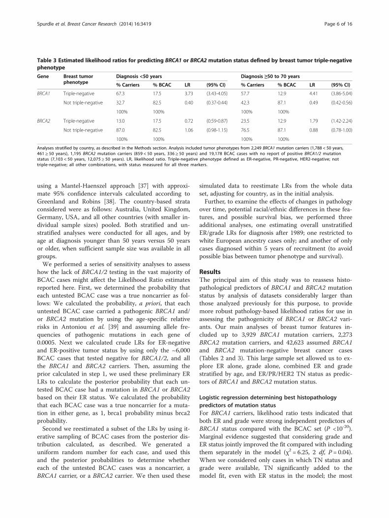
Table 3 Estimated likelihood ratios for predicting BRCA1 or BRCA2 mutation status defined by breast tumor triple-negativephenotype
Gene Breast tumorphenotype
Diagnosis <50 years Diagnosis ≥50 to 70 years
% Carriers % BCAC LR (95% CI) % Carriers % BCAC LR (95% CI)
Not triple-negative 87.0 82.5 1.06 (0.98-1.15) 76.5 87.1 0.88 (0.78-1.00)
100% 100% 100% 100%
Analyses stratified by country, as described in the Methods section. Analysis included tumor phenotypes from 2,249 BRCA1 mutation carriers (1,788 < 50 years,461 ≥ 50 years), 1,195 BRCA2 mutation carriers (859 < 50 years, 336 ≥ 50 years) and 19,178 BCAC cases with no report of positive BRCA1/2 mutationstatus (7,103 < 50 years, 12,075 ≥ 50 years). LR, likelihood ratio. Triple-negative phenotype defined as ER-negative, PR-negative, HER2-negative; nottriple-negative; all other combinations, with status measured for all three markers.
Spurdle et al. Breast Cancer Research (2014) 16:3419 Page 6 of 16
using a Mantel-Haenszel approach [37] with approxi-mate 95% confidence intervals calculated according toGreenland and Robins [38]. The country-based strataconsidered were as follows: Australia, United Kingdom,Germany, USA, and all other countries (with smaller in-dividual sample sizes) pooled. Both stratified and un-stratified analyses were conducted for all ages, and byage at diagnosis younger than 50 years versus 50 yearsor older, when sufficient sample size was available in allgroups.We performed a series of sensitivity analyses to assess
how the lack of BRCA1/2 testing in the vast majority ofBCAC cases might affect the Likelihood Ratio estimatesreported here. First, we determined the probability thateach untested BCAC case was a true noncarrier as fol-lows: We calculated the probability, a priori, that eachuntested BCAC case carried a pathogenic BRCA1 and/or BRCA2 mutation by using the age-specific relativerisks in Antoniou et al. [39] and assuming allele fre-quencies of pathogenic mutations in each gene of0.0005. Next we calculated crude LRs for ER-negativeand ER-positive tumor status by using only the ~6,000BCAC cases that tested negative for BRCA1/2, and allthe BRCA1 and BRCA2 carriers. Then, assuming theprior calculated in step 1, we used these preliminary ERLRs to calculate the posterior probability that each un-tested BCAC case had a mutation in BRCA1 or BRCA2based on their ER status. We calculated the probabilitythat each BCAC case was a true noncarrier for a muta-tion in either gene, as 1, brca1 probability minus brca2probability.Second we reestimated a subset of the LRs by using it-
erative sampling of BCAC cases from the posterior dis-tribution calculated, as described. We generated auniform random number for each case, and used thisand the posterior probabilities to determine whethereach of the untested BCAC cases was a noncarrier, aBRCA1 carrier, or a BRCA2 carrier. We then used these
simulated data to reestimate LRs from the whole dataset, adjusting for country, as in the initial analysis.Further, to examine the effects of changes in pathology
over time, potential racial/ethnic differences in these fea-tures, and possible survival bias, we performed threeadditional analyses, one estimating overall unstratifiedER/grade LRs for diagnosis after 1989; one restricted towhite European ancestry cases only; and another of onlycases diagnosed within 5 years of recruitment (to avoidpossible bias between tumor phenotype and survival).
ResultsThe principal aim of this study was to reassess histo-pathological predictors of BRCA1 and BRCA2 mutationstatus by analysis of datasets considerably larger thanthose analyzed previously for this purpose, to providemore robust pathology-based likelihood ratios for use inassessing the pathogenicity of BRCA1 or BRCA2 vari-ants. Our main analyses of breast tumor features in-cluded up to 3,929 BRCA1 mutation carriers, 2,273BRCA2 mutation carriers, and 42,623 assumed BRCA1and BRCA2 mutation-negative breast cancer cases(Tables 2 and 3). This large sample set allowed us to ex-plore ER alone, grade alone, combined ER and gradestratified by age, and ER/PR/HER2 TN status as predic-tors of BRCA1 and BRCA2 mutation status.
Logistic regression determining best histopathologypredictors of mutation statusFor BRCA1 carriers, likelihood ratio tests indicated thatboth ER and grade were strong independent predictors ofBRCA1 status compared with the BCAC set (P <10-20).Marginal evidence suggested that considering grade andER status jointly improved the fit compared with includingthem separately in the model (χ2 = 6.25, 2 df, P = 0.04).When we considered only cases in which TN status andgrade were available, TN significantly added to themodel fit, even with ER status in the model; the most
Spurdle et al. Breast Cancer Research (2014) 16:3419 Page 7 of 16
parsimonious model included ER, grade, and TN status,which was significantly better than any model with onlytwo of these included (χ2 = 83.8, 1 df, P <10-20). For BRCA2both ER and grade were highly significant predictors ofmutation status, and the interaction of ER and grade wasalso quite significant (χ2 = 28.3, 2 df, P <10-6). Theaddition of TN did not improve the model fit signifi-cantly (P = 0.14) when ER and grade were included in themodel. We thus considered ER, grade, and TN status in de-riving likelihood ratio estimates for BRCA-mutation status.
ER and grade as predictors of mutation statusThe estimated likelihood ratios for predicting BRCA1 orBRCA2 mutation status defined by breast tumor ER-grade phenotype, adjusted for country by using stratifiedanalysis, are shown in Table 2. Results based on pooleddata unstratified for country, including cell counts, areshown in Additional file 1: Table S2. In general, theMantel-Haenszel stratified LR estimates were quite simi-lar to the pooled estimates, with stratified estimatesmost often closer to 1.0 (although not always). Signifi-cant between-country heterogeneity for the estimatedlikelihood ratios was most often observed with grade ra-ther than ER or TN status. ER-positive cases were lesslikely to be carriers of a BRCA1 mutation, irrespective ofgrade. Conversely, ER-negative cases with high-grade tu-mors were more likely to be BRCA1 mutation carriers.Further, our analyses showed that ER-positive grade 3tumors were modestly predictive of positive BRCA2 mu-tation status (Table 2). The association of BRCA2 muta-tion status with ER-positive high-grade tumors was notsubstantially different for women diagnosed at youngeror older than age 50 years (LR <50 years = 1.77 (95% CI,1.60 to 1.95), LR ≥50 years = 1.76 (95% CI, 1.49 to 2.08)).However, ER-negative grade 3 tumor status was mod-estly predictive of positive BRCA2 mutation status inwomen diagnosed at 50 years or older (LR, 1.54; 95% CI =1.27 to 1.88).It is well known that ER and grade status are corre-
lated, with ER-negative tumors more likely to presentwith high grade. Consistent with this, relatively few casesappeared in any of the sample sets with ER-negativegrade 1 tumors. However, we estimated LRs for ER aloneand grade alone to allow inclusion of pathology data inmodels for predicting BRCA1 and BRCA2 mutation sta-tus, in instances in which information for only one ofthese variables is available (Table 2). For example, for awoman diagnosed with breast cancer at 50 years orolder, the LR in favor of positive BRCA1 mutation statuswould be 3.5 if her tumor were known to be ER negativebut grade status was unknown, and 2.4 if reported asgrade 3 without information on ER status.An acknowledged caveat to the inclusion of pathology
data in multifactorial likelihood modeling is the underlying
assumption that missense and in-frame deletions consid-ered to be pathogenic mutations will exhibit the sametumor histopathological characteristics as do truncatingmutations. The dataset in this study included 398 knownpathogenic BRCA1 missense mutation carriers (mainlyC61G), and 44 pathogenic BRCA2 missense mutation car-riers with information on ER status or grade. Comparingthe missense variants with the truncating set of mutations,we found no significant association of BRCA1 mutationtype with ER status (OR = 0.9; 95% CI, 0.7 to 1.2; P =0.4)or grade (OR = 1.15; 95% CI, 0.9 to 1.4; P =0.2) or BRCA2(OR = 2.7; CI, 0.9 to 7.6; =0.07 for ER; OR = 0.6 0.3 –to1.2; P =0.14 for grade), although power was quite limitedfor BRCA2.
Triple-negative (TN) phenotype in BRCA1 and BRCA2carriersSecondary country-stratified analysis of 2,249 BRCA1,1,195 BRCA2 and 19,178 assumed mutation-negativebreast cancer cases (Table 3) indicated that TN tumorstatus is highly predictive of BRCA1 mutation status forwomen diagnosed at younger than 50 years (LR = 3.73;95% CI, 3.43 to 4.05) and at age 50 years or older (LR =4.41; 95% CI 3.86 to 5.04), and results were little differ-ent for unstratified analysis (see Additional file 1: Table S3,also displaying cell counts).Results also indicated that TN phenotype is modestly
predictive of BRCA2 mutation status in cases diagnosedat age 50 years or older (LR, 1.79; 95% CI = 1.42 to 2.24).This observation is explained by the lower frequency ofthe TN phenotype in noncarriers (12.9% 50 years orolder) versus BRCA2 mutation carriers (23.5% 50 yearsor older). Additional analysis considering grade and TNstatus combined (see Additional file 1: Table S4) did notshow substantial improvement over LRs estimated forER and grade combined (Table 2) or TN status (Table 3),although numbers in some cells were limited.
Sensitivity analysesWith respect to the possible consequences of contamin-ation by missed mutation carriers in the BCAC sampleset, we first estimated which BCAC-untested cases weremore likely to be an undetected mutation carrier, andthen re-estimated a subset of the LRs by using iterativesampling of the control dataset. Based on age-specificrelative risks, we estimated that there could be at most796 BRCA1 (1.7%) and 433 BRCA2 (0.9%) undetectedcarriers in the reference dataset of 47,565 BCAC cases.Based on age and crude ER, LR estimated from truenon-carriers in BCAC, of 41,515 BCAC cases whosegenetic status was unknown, 34,869 (84%) had posteriorprobabilities of being a true BRCA1/2-negative casegreater than 0.95, with the minimum posterior probabil-ity being 0.89. Repeating this sampling process a total of
Spurdle et al. Breast Cancer Research (2014) 16:3419 Page 8 of 16
5 times, the number of BRCA1 carriers within the BCACset ranged from 688 to 784, and the number of BRCA2carriers ranged from 410 to 455 (total carriers, 1,114 to1,194). Re-estimation of a subset of LRs indicated thatthe LRs assuming all BCAC cases do not carry a patho-genic BRCA1 or BRCA2 mutation is quite close to whatwe would expect, had all individuals been tested. ForER-negative Grade 3 cases diagnosed at younger than50 years, the original LR for BRCA1 mutation status, as-suming all BCAC cases were non-carriers, was 3.16,whereas the five replicates from iterative analysis rangedfrom 3.22 to 3.25. For TN tumor phenotype, the originalLR for BRCA1 mutation status was 3.73, whereas themedian of the five replicates was 3.76.In additional sensitivity analyses, we recalculated un-
stratified LRs for ER and grade combined, restricting theanalyses to the subset of 36,522 (33,260 BCAC, 3,252CIMBA) breast cancer cases of European ancestry, ofwhich 31,374 (28,364 BCAC, 3,010 CIMBA) were diag-nosed within 5 years of interview, and 40,874 (36,414BCAC, 4,460 CIMBA) were diagnosed after 1989. Re-sults were similar to those from the overall analyses,with LR estimates consistently within the confidence in-tervals of the overall analyses.
DiscussionHistopathological predictors of mutation statusThis study assessing histopathological predictors ofBRCA1 and BRCA2 mutation status is based on the lar-gest sample set reported to date, and so provides more-precise estimates that account for age at diagnosis as apotential confounder. We also provide age-stratified LRsfor ER alone and grade alone, which, although not aspredictive as ER and grade combined, will facilitate in-clusion of minimal pathology information in multifactor-ial modeling of individually rare variants.Further, we provide, for the first time, LR estimates for
TN status that can be applied when grade information isnot recorded, with estimates associated with TN statuscomparable to those for ER-negative-grade 3 (for BRCA1)and ER-positive-grade 3 (for BRCA2). Altogether, these re-fined LRs will improve the clinical classification of BRCA1and BRCA2 variants, particularly those identified inwomen with later age at diagnosis.Our ER-grade analysis results for BRCA1 are consist-
ent with results from analysis of raw data for a smallerdataset of 600 BRCA1 carriers aged younger than 60 yearsand 258 age-matched non-carriers from the Breast Can-cer Linkage Consortium, which yielded LRs of 1.94 (95%CI = 1.05 to 3.56) and 2.95 (95% CI = 2.41 to 3.62) for ER-negative grade 2 and ER-negative grade 3 tumors,respectively [26,27]. However, the current study demon-strates that ER-negative grade 2 or 3 status is more pre-dictive of positive BRCA1 status in women diagnosed at
older than 50 years compared with younger than 50 (forexample, for ER-negative-grade 3, LR ≥50 years is 4.13(95% CI = 3.70 to 4.62) versus LR <50 years of 3.16 (95%CI = 2.96 to 3.37); Phet <0.0001. These observations re-flect the fact that although the overall proportion of ER-negative high-grade tumors is lower for older onset(54.5%) than younger onset (67.1%) BRCA1 carriers (aspreviously reported [33,40]), the proportion of ER-negative high-grade tumors differs much more markedlyfor older-onset (12.8%) than younger-onset (20.8%) caseswith no identified mutation in BRCA1 or BRCA2.In addition, not reported in previous smaller studies
[6,7,41], our results show that ER-positive grade 2 or 3status is a stronger negative predictor of BRCA1 muta-tion status in women diagnosed before age 50 yearscompared with those diagnosed at age 50 years or older.These patterns reflect changes in the frequency of ERstatus and grade as a function of age in the non-carriercases, rather than large changes in the frequency ofthese features in the carriers. Similarly, the findings forBRCA2 are consistent with those from a previous studyof 157 BRCA2 mutation carriers and 314 mutation-negative familial breast cancer cases, which indicatedthat BRCA2-associated tumors were more likely to beER-positive than were control tumors, when accountingfor grade (OR, 2.09; 95% CI, 1.21 to 3.63; P =0.008) [13].However, age-stratified analysis highlighted that ER-
negative grade 3 tumor status modestly predicted posi-tive BRCA2 mutation status in women diagnosed at age50 years or older, indicating that grade is a more import-ant factor than ER status in predicting BRCA2 tumors.We attempted to assess pathology difference by muta-tion type (missense versus truncating), an issue that hasnot previously been addressed rigorously because of thelimited availability of pathology information for provenhigh-risk missense mutations. However, even in our verylarge dataset, the number of proven pathogenic missensemutations remained small, and it is apparent that futureeven larger studies will be needed to address this question.The associations between BRCA1 mutation status and
TN phenotype are consistent with those observed forER-negative, high-grade tumors. They are also consistentwith prior evidence that BRCA1 mutation carriers areenriched for the “basal” tumor phenotype that is highlyconcordant with TN status. A recent meta-analysisassessing the prevalence of BRCA1 mutations in TN ver-sus non-TN breast cancer patients from largely high-riskbreast cancer populations [42] estimated a risk of 5.65(95% CI, 4.15 to 7.69) based on analysis of 236 BRCA1mutation carriers and 2,297 non-carriers. In addition,these authors predicted that approximately two in ninewomen with TN breast cancer and additional high-riskfeatures (early onset or family history) harbor a BRCA1mutation [42]. TN status has not been obviously linked
Spurdle et al. Breast Cancer Research (2014) 16:3419 Page 9 of 16
to BRCA2 mutation status previously; however, a recentstudy of 43 deleterious BRCA1/2 mutation carriers iden-tified from screening of 409 Chinese familial breast can-cer cases reported that TN phenotype was more likely tobe exhibited by both BRCA1 (P =0.001, 69%, n = 16) andBRCA2 (P =0.01, 46%, n = 27) carriers identified in theircohort, compared with non-carriers (23%; n = 366) [43].In contrast, a similar study of 221 Korean familial breastcancer patients [44] identified 81 deleterious mutationcarriers, and demonstrated increased TN phenotype forBRCA1 mutation carriers (P <0.00001,57%, n = 35), butnot BRCA2 mutation carriers (P =0.9, 13.9%, n = 36)compared with non-carriers (13%, n = 130). Neither ofthese studies presented their findings for cases stratifiedby diagnosis age 50 years or older.Our study has shown that TN phenotype is modestly
predictive of BRCA2 mutation status in cases diagnosedat 50 years or older, due to a lower TN frequency innon-carriers versus BRCA2 mutation carriers in this agegroup. Reassuringly, these TN frequency differences mir-ror the results seen for ER-negative grade 3 status innon-carriers and BRCA2 mutation carriers, an analysisbased on a much larger sample set.
Possible impact of study limitationsWe acknowledge several limitations of our study. Ideally,our reference group would have been drawn from thesame source as the mutation carriers, as there may bedifferences between non-BRCA familial cases and unse-lected cases. However, in the subset of 30,233 BCACcases that had data on family history, we did not see anysignificant differences between this group and theremainder of the sample in terms of the pattern of histo-logical features, nor with those who indicated no first-or second-degree relatives with breast cancer.In our analyses, we are implicitly assuming that testing
for BRCA1/2 mutations was independent of the histo-pathology features used for prediction of mutation sta-tus. Although recently some features with therapeuticimplications, such as TN status, are being used as a cri-terion for testing in some centers, we believe that thevast majority of our CIMBA carriers were tested solelyon the basis of their family history. This analysis as-sumes that mutation testing in CIMBA sample sets wasnot directed by tumor histology. Mutation status wasnot known for all BCAC samples. However, mutationtesting of BCAC samples had been performed for manystudies with selected design that might be expected tobe enriched for BRCA1 and BRCA2 mutation carriers,and these known mutation carriers were excluded fromanalysis.Further, our sensitivity analyses suggest that, at very
most, 2.5% of BCAC cases might carry an undetectedmutation, and also show that our results would not be
substantially affected by this level of contamination ofthe reference group.The various sensitivity analyses conducted for the ER-
grade dataset provided no convincing evidence for obviousdifferences for the factors being assessed. We did not seeany marked difference in LR estimates for analyses re-stricted to individuals of European ancestry, but the smallnumbers of cases from other ethnic/racial groups did notallow us to assess reliably tumor histopathological featuresfor other ethnic groups, and so may not be generalizableto patients of non-European ancestry. Although it is pos-sible that variation in pathology grading and IHC testingmethods might occur between countries or over time, ourinvestigations provided no evidence that such differenceswould meaningfully confound interpretation of the results,and thus should not limit the use of the information gen-erated for multifactorial likelihood analysis of BRCA1 orBRCA2 variants across continents.
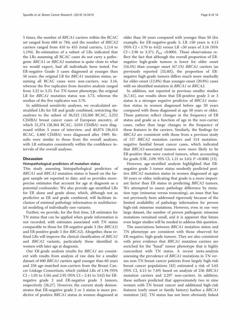
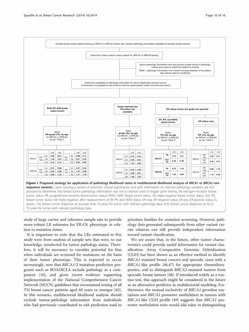
Use of revised LR estimates for future multifactoriallikelihood analysesThis study has re-estimated the likelihood of BRCA1 orBRCA2 mutation status associated with breast tumorfeatures commonly measured in the clinical setting, byanalyzing much larger datasets than previously used forthis purpose. Our findings provide measures of confi-dence in the individual LR estimates, and in particular,allow age at diagnosis to be incorporated into the path-ology component of the multifactorial likelihood model.Figure 1 provides a flowchart indicating the proposedapplication of pathology-based LRs, dependent on whatbreast tumor pathology information is available for avariant carrier. As indicated, ER-grade LRs should be ap-plied in preference to other pathology LR estimates,where both ER and grade information is available. TheER-grade LRs were derived from analysis of the largestsample sizes and thus have the greatest precision, andapplication of 12 strata provided by three grade categoriesrefines both positive and negative prediction of mutationstatus. For example, a patient with a high-grade ER-negative tumor is three- to fourfold more likely to carry aBRCA1 mutation than not, whereas a patient with a low-grade ER-positive tumor is about 10 times more likely tobe mutation-negative than mutation-positive. Given thatgrade and ER are almost universally used to assess prog-nosis and predict response to antiestrogen therapies, thesefeatures are generally readily available on standard path-ology reports.This study could not provide a comparison to existing
LR estimates of BRCA1 mutation status based on ER-CKstatus, determined from analysis of 182 BRCA1 and 109age-matched cases [6]. However, we caution that very largeconfidence limits exist around the previously estimatedLRs for ER-CK characteristics, and recommend further
Female breast cancer patient carrying a BRCA1 or BRCA2 variant with relevant pathology information available for primary breast cancers
Ignore pathology information from any primary breast cancer if pathology criteria were used to select the patient for testing
(Note – pathology information from variant-carrying relatives of the patient may still be used for modelling)
Determine criteria used to select patient for BRCA1 or BRCA2 testing
Determine availability of pathology information for each independent primary tumour. If information is available for two tumours from the same patient, select only the most recent.
ER status only ER, PR, and HER2
status known
ApplyTN LRs, by age
for BRCA1 or BRCA2 as per Table 3
Apply ER-grade LRs, by agefor BRCA1 or BRCA2
as per Table 2
Apply grade LRs, by age
for BRCA1 or BRCA2as per Table 2
Apply ER LRs, by age
for BRCA1 or BRCA2 as per Table 2
Grade reported but ER status not
availableER status known but grade not reported
<50 ≥50
BRCA1
G1 0.13 0.12
G2 0.38 0.57
G3 1.67 2.20
BRCA2
G1 0.33 0.35
G2 0.88 0.81
G3 1.08 1.52
<50 ≥50 <50 ≥50
BRCA1 ER-neg
G1 0.59 0.51ER-pos
G1 0.08 0.11
G2 1.36 2.34 G2 0.21 0.34
G3 3.16 4.13 G3 0.64 0.90
BRCA2ER-neg
G1 0.51 0.86ER-pos
G1 0.37 0.32
G2 0.49 0.89 G2 1.07 0.89
G3 0.69 1.54 G3 1.77 1.76
<50 ≥50
BRCA1
ER-neg
2.60 3.31
ER-pos
0.32 0.37
BRCA2
ER-neg
0.66 1.18
ER-pos
1.15 0.90
<50 ≥50
BRCA1
TN 3.73 4.41
Not TN 0.40 0.49
BRCA2
TN 0.72 1.79
Not TN 1.06 0.88
Both ER AND grade status known
Figure 1 Proposed strategy for application of pathology likelihood ratios in multifactorial likelihood analysis of BRCA1 or BRCA2 raresequence variants. Cases carrying a variant of uncertain clinical significance, and with information on relevant pathology variables, are firstassessed to determine that breast tumor pathology information was not a criterion used to trigger gene testing. ER, estrogen-receptor breasttumor status; PR, progesterone-receptor breast tumor status; HER2, HER2 breast tumor status; TN, triple-negative breast tumor status; Not TN,breast tumor status not triple-negative, after measurement of ER, PR, and HER2 status; ER-neg, ER-negative status; ER-pos, ER-positive status; G,grade; <50, breast cancer diagnosis at younger than 50 years for tumor with relevant pathology data; ≥50, breast cancer diagnosis at 50 to70 years for tumor with relevant pathology data.
Spurdle et al. Breast Cancer Research (2014) 16:3419 Page 10 of 16
study of large carrier and reference sample sets to providemore-robust LR estimates for ER-CK phenotype in rela-tion to mutation status.It is important to note that the LRs estimated in this
study were from analysis of sample sets that were, to ourknowledge, unselected for tumor pathology status. There-fore, it will be necessary to consider potential for biaswhen individuals are screened for mutations on the basisof their tumor phenotype. This is expected to occurincreasingly, now that BRCA1/2 mutation-prediction pro-grams such as BOADICEA include pathology as a com-ponent [16], and given recent evidence supportingimplementation of the National Comprehensive CancerNetwork (NCCN) guidelines that recommend testing of allTN breast cancer patients aged 60 years or younger [45].In this scenario, multifactorial likelihood analysis shouldexclude tumor-pathology information from individualswho had previously contributed to risk prediction used to
prioritize families for mutation screening. However, path-ology data generated subsequently from other variant car-rier relatives can still provide independent informationtoward variant classification.We are aware that, in the future, other tumor charac-
teristics could provide useful information for variant clas-sification. Array Comparative Genomic Hybridization(CGH) has been shown as an effective method to identifyBRCA1-mutated breast cancers and sporadic cases with aBRCA1-like profile [46,47] for appropriate chemothera-peutics, and to distinguish BRCA2-mutated tumors fromsporadic breast tumors [48]. If introduced widely as a rou-tine test, this approach might be considered in the futureas an alternative predictor in multifactorial modeling. Fur-thermore, the mutual exclusivity of BRCA1-germline mu-tations and BRCA1 promoter methylation in tumors withBRCA1-like CGH profile [49] suggests that BRCA1 pro-moter methylation tests would add value in distinguishing
Spurdle et al. Breast Cancer Research (2014) 16:3419 Page 11 of 16
somatic from germline loss of BRCA1 function, as is estab-lished for clinical testing triage and variant classificationrelating to MLH1 mismatch repair cancer-predispositiongene [50].Alternatively, genome-wide tumor-methylation profiles
may prove of value to distinguish between individualwith and without a germline BRCA1 mutation [51]. Fur-ther, additional substratification of currently used histo-logical features may add value in prediction of mutationstatus. Options include PTEN loss of expression inaddition to TN status as a marker of BRCA1 mutation sta-tus [11], or gene-expression arrays to identify BRCA2 mu-tation carriers among the subset of luminal B tumors [15].Recent research has also shown the value of considering
further stratification of breast cancer subtype in the pre-diction of BRCA mutation status. For example althoughER-negative status clearly predicts BRCA1 mutation sta-tus, even ER-positive BRCA1-related breast cancers aremore likely to be grade 3, CK14+, and show high mitoticrate compared with ER-positive sporadic cancers [52].In addition, possibilities exist to extend histopatho-
logical analyses to tumors other than female breast can-cer. The combination of modified Nottingham grade 3serous or undifferentiated histology, prominent intrae-pithelial lymphocytes, marked nuclear atypia with giantnuclei, and high mitotic index has recently been re-ported to be a significant predictor of BRCA1 mutationstatus in women with epithelial ovarian cancer [53]. Fur-ther, breast tumors of male BRCA2 mutation carriers aremore likely to present as high-grade, PR-negative, andrelatively high rates of HER2-positivity with a micropapil-lary component to histology have been reported [54,55].Investigation of these features in larger sample sizesshould be considered in the future.Although this article has focused on the utility of his-
topathologic features of breast cancers in the context ofthe classification of variants in the BRCA1 and BRCA2genes, these results should also be useful in a range ofother applications. The information provided in themain tables can be used to estimate sensitivities andspecificities of histopathological predictors by broad age-group (for example, triple-negative tumor status has sen-sitivity of 0.67 and specificity 0.82 for detection ofBRCA1 mutation status in women diagnosed at youngerthan age 50 years, whereas the sensitivity is 0.57 and thespecificity 0.87 for women diagnosed at age 50 or older.As such, these results, in conjunction with other predic-tors of mutation status, could be useful to guide system-atic genetic testing of germline DNA from patients todetermine the appropriateness of the use of PARP inhib-itors in therapy. The results arising from this study arealso likely to inform future development of parallelmodels, which estimate the probability of an individualcarrying a BRCA1 or BRCA2 mutation, to determine
eligibility and/or priority for genetic testing (in particular,the BOADICEA model, which has recently been updatedto include additional histopathologic characteristics fromlarge data resources [56]).
ConclusionsThe results from this large-scale analysis refine likeli-hood ratio estimates for predicting BRCA1 and BRCA2mutation status by using commonly measured histo-pathological features. We demonstrate the importanceof considering age at diagnosis for analyses, and showthat grade is more informative than ER status for BRCA2mutation-carrier prediction. The estimates will improveBRCA1 and BRCA2 variant classification by using multi-factorial likelihood analysis, and inform patient mutationtesting and clinical management.
Additional file
Additional file 1: Table S1. CIMBA and BCAC sample sets included inanalysis.* Table S2. Unstratified estimated likelihood ratios (LRs) forpredicting BRCA1 or BRCA2 mutation status defined by breast tumorER-grade phenotype. Table S3. Unstratified estimated LRs for predictingBRCA1 or BRCA2 mutation status defined by breast tumor triple-negativephenotype. Table S4. Unstratified estimated LRs for predicting BRCA1 orBRCA2 mutation status defined by breast tumor TN-grade phenotype.*
AbbreviationsBCAC: Breast Cancer Association Consortium; CGH: Array ComparativeGenomic Hybridization; CIMBA: Consortium of Investigators of Modifiers ofBRCA1/2; CK14: cytokeratin 14; CK17: cytokeratin 17; CK5/6: cytokeratin 5/6;EGFR: epidermal growth factor receptor; ENIGMA: Evidence-based Networkfor the Interpretation of Germline Mutant Alleles; ER: estrogen receptor;HER2: Human epidermal growth factor receptor 2;IHC: immunohistochemical; LR: likelihood ratio; NCCN: NationalComprehensive Cancer Network; PR: progesterone receptor;PTEN: phosphatase and tensin homolog; TN: triple negative.
Competing interestsThe authors declare that they have no competing interests.
Authors’ contributionsABS, FJC, and DEG conceived and designed the study. ABS and MTPcoordinated data collation for analysis, and DEG performed the statisticalanalysis. ABS and DEG drafted the manuscript. ACA, LMcG, DB, and SHcoordinated the cleaning of pathology data, risk factor data, and mutationclassification for CIMBA, overseen by GC-T. MKS, MKB, and QW coordinated thecollation and cleaning of pathology and risk-factor data for BCAC, overseen byDFE. FJC, RKS, BW, KR, EH, CE, A Meindl, ND, NA, HP, DN, C Sutter, SW-G, DS,SP-A, KK, RV-M, SDE, DF, RP, J Perkins, DGE, LI, RE, JA, RD, TC, GS, S Manoukian, BBonanni, F Mariette, SF, AV, BP, LP, LV, RB, KLN, SMD, KO, AJ, NL, MT, UBJ, JR, AB,ILA, A Miron, TvOH, TC, SLN, AET, HN, MM, J Garber, AKG, AO, REF, MBT, TRR,BYK, MS, MUR, NT, PDPP, FMB, AMD, EP, P Hall, KC, MKS, AB, SC, SV, PAF, MWB,ABE, DJS, SEB, BGN, SFN, HF, JC-C, DF-J, AR, PS, KA, TAM, P Heikkila, CB, JF, SJC, LBrinton, J Lissowska, JEO, VSP, EMJ, ASW, DWW, UH, DT, HUU, TR, PD, RAEMT, CSeynaeve, CJVA, DME, WJT, LD, LJ, J Peto, IdS-S, OF, NJ, MD, R Swann, ALB, GG,AMM, GGG, RLM, L Baglietto, CM, JC, CC, R Scott, H Brauch, TB, Y-DK, AC, SSC,MWRR, J Lubinski, KJ-B, KD, J Gronwald, TD, NB, T-WP-S, P Hillemanns, CAH, BEH,FS, LLM, B Burwinkel, F Marme, HS, RY, HA-C, AZ, MJH, JMC, JWMM, MMAT-L,HB, AKD, VA, C Stegmaier, RW, KP, AJ-V, MG, AL, S Margolin, VJ, MR, RR-M, AG-N,JIA, PZ, JB, AM, VK, V-MK, JMH, PP, DZ, MB, FC, PR, S-HT, and GC-T contributed tobaseline study design and recruitment of subjects included in the analysis, andprovided access to genetic, epidemiologic, and pathology data from thesecharacterized cohorts. All authors read and approved the final manuscript.
Spurdle et al. Breast Cancer Research (2014) 16:3419 Page 12 of 16
AcknowledgementsAmanda Spurdle is supported by an NHMRC Senior Research Fellowship, andaspects of this research were funded by Australian NHMRC Project grant ID1010719. This work was supported in part by NIH grants CA128978 andCA116167, an NIH specialized program of research excellence in breastcancer to the Mayo Clinic (P50 CA116201), and the Breast Cancer ResearchFoundation. CIMBA data management was supported by Cancer Research-UKgrant C12292/A11174 and C1287/A10118. ACA is a Cancer Research-UK SeniorCancer Research Fellow. BCAC data management was funded by CancerResearch UK (C1287/A10118 and C1287/A12014) and by the EuropeanCommunity’s Seventh Framework Programme under grant agreement 223175(HEALTH-F2-2009-223175).
Study-specific AcknowledgementsABCS was supported by Dutch Cancer Society grants NKI 2007-3839; 2009 4363.ABCFS thanks Maggie Angelakos, Judi Maskiell, Gillian Dite, and Helen Tsimiklis.The Australian Breast Cancer Tissue Bank is generously supported by theNational Health and Medical Research Council of Australia, The CancerInstitute NSW, and the National Breast Cancer Foundation.The work of the BBCC was partly funded by ELAN-Fond of the UniversityHospital of Erlangen.The BBCS is funded by Cancer Research UK and Breakthrough Breast Cancerand acknowledges NHS funding to the NIHR Biomedical Research Centre,and the National Cancer Research Network (NCRN).The work of the Breast Cancer Family Registry (BCFR) centers (BCFR-AU(ABCFS), BCFR-NC, BCFR-NY, BCFR-ON (OFBCR), BCFR-PA (FCCC), and BCFR-UTwas supported by grant UM1 CA164920 from the National Cancer Institute.The content of this manuscript does not necessarily reflect the views orpolicies of the National Cancer Institute or any of the collaborating centersin the Breast Cancer Family Registry, nor does mention of trade names,commercial products, or organizations imply endorsement by the USGovernment or the Breast Cancer Family Registry.BCFR-ON (OFBCR) work was additionally supported by the Canadian Institutesof Health Research “CIHR Team in Familial Risks of Breast Cancer” program.BIDMC is supported by the Breast Cancer Research Foundation.For BRICOH, data were collected under NIH R01 CA74415. SLN is holder ofthe Morris and Horowitz Families Endowed Professorship.The BSUCH study was supported by the Dietmar-Hopp Foundation, theHelmholtz Society, and the German Cancer Research Center (DKFZ).CBCS thanks Anne-Marie Gerdes and Bent Ejletsen for clinical data.The CGPS was supported by the Chief Physician Johan Boserup and LiseBoserup Fund, the Danish Medical Research Council, and Herlev Hospital.CGPS thanks staff and participants of the Copenhagen General PopulationStudy. For the excellent technical assistance, thanks to Dorthe UldallAndersen, Maria Birna Arnadottir, Anne Bank, and Dorthe Kjeldgård Hansen.The Danish Breast Cancer Group (DBCG) is acknowledged for the tumorinformation.The work of CNIO was partially supported by Spanish Association againstCancer (AECC08), RTICC 06/0020/1060, FISPI08/1120, Mutua MadrileñaFoundation (FMMA) and SAF2010-20493.MBCSG and CONSIT TEAM is supported by grants from the ItalianAssociation for Cancer Research (AIRC) and by funds from the Italian citizenswho allocated the 5/1,000 share of their tax payment according to Italianlaws in support of the Fondazione IRCCS Istituto Nazionale Tumori to SMand of the IRCCS AOU San Martino-IST to LV and from FiorGen Foundationfor Pharmacogenomics to LP. MBCSG and CONSIT TEAM thank BernardPeissel of the Fondazione IRCCS Istituto Nazionale Tumori (INT), Milan, Italy;Laura Ottini and Giuseppe Giannini of the Sapienza University, Rome, Italy;Antonella Savarese and Aline Martayan of the Istituto Nazionale TumoriRegina Elena, Rome, Italy ; Maria Grazia Tibiletti and Daniela Furlan of theOspedale di Circolo-Università dell’Insubria, Varese, Italy; Stefania Tommasi ofthe Istituto Nazionale Tumori “Giovanni Paolo II, Bari”, Italy, and Loris Bernardand the personnel of the Cogentech Cancer Genetic Test Laboratory, Milan,Italy.The University of Westminster curates the DietCompLyf database created byand funded by Against Breast Cancer Registered Charity No. 1121258.DKFZ are grateful to all the patients for their participation in this study.The HEBON ([email protected]) study is supported by the Dutch Cancer Societygrants NKI1998-1854, NKI2004-3088, NKI2007-3756, the NWO grant 91109024,the Pink Ribbon grant 110005 and the BBMRI grant CP46/NWO. The
Hereditary Breast and Ovarian Cancer Research Group Netherlands (HEBON),Coordinating center: Netherlands Cancer Institute, Amsterdam, The Netherlands.EMBRACE ([email protected]) is supported by Cancer Research UKGrants C1287/A10118 and C1287/A11990. D. Gareth Evans and Fiona Lallooare supported by an NIHR grant to the Biomedical Research Centre,Manchester. The Investigators at The Institute of Cancer Research and TheRoyal Marsden NHS Foundation Trust are supported by an NIHR grant to theBiomedical Research Centre at The Institute of Cancer Research and TheRoyal Marsden NHS Foundation Trust. Ros Eeles and Elizabeth Bancroft aresupported by Cancer Research UK Grant C5047/A8385, Department ofLaboratory Medicine and Pathology, Mayo Clinic, 200 First Street SW,Rochester, MN 55905, USA.ESTHER was supported in part by the Baden-Württemberg State Ministry ofScience, Research and Arts; and by the German Federal Ministry of Educationand Research. Additional cases were recruited in the context of the VERDIstudy, which was supported by a grant from the German Cancer Aid(Deutsche Krebshilfe). We thank all the individuals who took part in thisstudy and all the researchers, clinicians, technicians, and administrative staffwho have enabled this work to be carried out.The FCCC thanks Ms. JoEllen Weaver, and Dr. Betsy Bove for their technicalsupport. The authors acknowledge support from The University of KansasCancer Center (P30 CA168524) and the Kansas Bioscience Authority EminentScholar Program. AKG was funded by 5U01CA113916, R01CA140323, and bythe Chancellors Distinguished Chair in Biomedical Sciences Professorship.GC-HBOC was kindly supported by the German Cancer Aid to R. K.Schmutzler (grant 109076). We are very thankful to all family members whoparticipated in this study.The GENICA ([email protected]) was funded by the FederalMinistry of Education and Research (BMBF) Germany grants 01KW9975/5,01KW9976/8, 01KW9977/0 and 01KW0114, the Robert Bosch Foundation,Stuttgart, Deutsches Krebsforschungszentrum (DKFZ), Heidelberg, Institute forPrevention and Occupational Medicine of the German Social AccidentInsurance, Institute of the Ruhr University Bochum (IPA), as well as theDepartment of Internal Medicine, Evangelische Kliniken Bonn gGmbH,Johanniter Krankenhaus Bonn, Germany. The GENICA network (Dr. MargareteFischer-Bosch-Institute of Clinical Pharmacology, Stuttgart, and University ofTübingen, Germany; (HB, Wing-Yee Lo, Christina Justenhoven), Departmentof Internal Medicine, Evangelische Kliniken Bonn gGmbH, JohanniterKrankenhaus, Bonn, Germany (YDK, Christian Baisch), Institute of Pathology,University of Bonn, Germany (Hans-Peter Fischer), Molecular Genetics ofBreast Cancer, Deutsches Krebsforschungszentrum (DKFZ) Heidelberg,Germany [Ute Hamann], Institute for Prevention and Occupational Medicineof the German Social Accident Insurance, Institute of the Ruhr UniversityBochum (IPA), Germany [TB, Beate Pesch, Sylvia Rabstein, Anne Lotz], Instituteof Occupational Medicine and Maritime Medicine, University Medical CenterHamburg-Eppendorf, Germany [Volker Harth]). Molecular Genetics of BreastCancer, Deutsches Krebsforschungszentrum (DKFZ), Im Neuenheimer Feld580, 69120 Heidelberg, Germany. University of Tübingen, Geschwister-Scholl-Platz, 72074 Tübingen, Germany. Dr. Margarete Fischer-Bosch Institute ofClinical Pharmacology, Auerbachstraße 112, 70376 Stuttgart, Germany.Institute for Prevention and Occupational Medicine of the German SocialAccident Insurance (IPA), Bürkle-de-la-Camp-Platz 1, 44789 Bochum, Germany.Institute of Occupational Medicine and Maritime Medicine, University MedicalCenter Hamburg-Eppendorf, Martinistrasse 52, 20246 Hamburg, Germany.Institute of Pathology, Medical Faculty of the University of Bonn, Sigmund-Freud-Str. 25, Haus 372, 53127 Bonn, Germany.Department of InternalMedicine, Evangelische Kliniken Bonn gGmbH, and Johanniter Krankenhaus,Johanniterstrasse 3, 53113 Bonn, Germany.The GESBC was supported by the Deutsche Krebshilfe e. V. [70492], andgenotyping in part by the state of Baden-Württemberg through the MedicalFaculty of the University of Ulm [P.685].The HABCS study was supported by an intramural grant from HannoverMedical School.HCSC was supported by grant RD12/0036/006. Instituto de Salud Carlos III(FEDER). Spanish Ministry of Science.The HEBCS was financially supported by the Helsinki University CentralHospital Research Fund, Academy of Finland (266528), the Finnish CancerSociety and the Sigrid Juselius Foundation. HEBCS thanks Kirsimari Aaltonen,Karl von Smitten, and Irja Erkkilä.IOVHBOCS was supported by Ministero dell’Istruzione, dell’Università e dellaRicerca and Ministero della Salute.
Spurdle et al. Breast Cancer Research (2014) 16:3419 Page 13 of 16
Financial support for KARBAC was provided through the regional agreementon medical training and clinical research (ALF) between Stockholm CityCouncil and Karolinska Institutet, and from the Stockholm CancerFoundation and the Swedish Cancer Society.The KBCP was financially supported by the special Government Funding(EVO) of Kuopio University Hospital grants, Cancer Fund of North Savo, theFinnish Cancer Organizations, the Academy of Finland, and by the strategicfunding of the University of Eastern Finland.KConFab ([email protected]) is supported by grants from theNational Breast Cancer Foundation, the National Health and MedicalResearch Council (NHMRC), and by the Queensland Cancer Fund, the CancerCouncils of New South Wales, Victoria, Tasmania, and South Australia, andthe Cancer Foundation of Western Australia. GCT is an NHMRC SeniorPrincipal Research Fellow. RB was a Cancer Institute NSW Clinical ResearchFellow. We thank Heather Thorne, Eveline Niedermayr, all the kConFabresearch nurses and staff, the heads and staff of the Family Cancer Clinics.Peter MacCallum Cancer Centre, 7 St Andrews Pl, East Melbourne VIC 3002,Australia.The MARIE study was supported by the Deutsche Krebshilfe e.V. [70-2892-BRI], the Hamburg Cancer Society, the German Cancer Research Center, andthe Federal Ministry of Education and Research (BMBF) Germany [01KH0402].MARIE thanks Alina Vrieling, Katharina Buck, Muhabbet Celik, Ursula Eilber,and Sabine Behrens.MAYO is supported by NIH grant CA128978, an NCI Specialized Program ofResearch Excellence (SPORE) in Breast Cancer (CA116201), a U.S. Departmentof Defense Ovarian Cancer Idea award (W81XWH-10-1-0341) and a grantfrom the Breast Cancer Research Foundation.MBCSG is supported by grants from the Italian Association for Cancer Research(AIRC) and by funds from the Italian citizens who allocated the 5/1,000 share oftheir tax payment in support of the Fondazione IRCCS Istituto Nazionale Tumori,according to Italian laws (INT-Institutional strategic projects “5 × 1000”).The MCBCS was supported by the NIH grant CA128978 and a SpecializedProgram of Research Excellence (SPORE) in Breast Cancer [CA116201], theBreast Cancer Research Foundation and a generous gift from the David F.and Margaret T. Grohne Family Foundation and the Ting Tsung and WeiFong Chao Foundation.MCCS cohort recruitment was funded by VicHealth and Cancer CouncilVictoria. The MCCS was further supported by Australian NHMRC grants209057, 251553 and 504711 and by infrastructure provided by CancerCouncil Victoria.The MEC was support by NIH grants CA63464, CA54281, CA098758, andCA132839.MSKCC is supported by grants from the Breast Cancer Research Foundationand Robert and Kate Niehaus Clinical Cancer Genetics Initiative.OBCS thank Meeri Otsukka, Kari Mononen, Jukka Moilanen, Saila Kauppila.OBCS was supported by research grants from the Finnish CancerFoundation, the Academy of Finland Centre of Excellence grant 251314, theSigrid Juselius Foundation, the University of Oulu, and the Oulu UniversityHospital Research Fund.OFBCR was supported by grant UM1 CA164920 from the National CancerInstitute. The content of this manuscript does not necessarily reflect theviews or policies of the National Cancer Institute or any of the collaboratingcenters in the Breast Cancer Family Registry (BCFR), nor does mention oftrade names, commercial products, or organizations imply endorsement bythe US Government or the BCFR.The ORIGO study was supported by the Dutch Cancer Society (RUL 1997-1505) and the Biobanking and Biomolecular Resources Research Infrastructure(BBMRI-NL CP16).OSU CCG thanks Leigha Senter, Kevin Sweet, Caroline Craven, and MichelleO’Conor, who were instrumental in accrual of study participants,ascertainment of medical records, and database management.The CNIO-BCS was supported by the Genome Spain Foundation, the RedTemática de Investigación Cooperativa en Cáncer and grants from theAsociación Española Contra el Cáncer and the Fondo de InvestigaciónSanitario (PI11/00923 and PI081120). The Human Genotyping-CEGEN Unit(CNIO) is supported by the Instituto de Salud Carlos III. Thanks to GuillermoPita, Charo Alonso, Daniel Herrero, Nuria Álvarez, Pilar Zamora, PrimitivaMenendez, and the Human Genotyping-CEGEN Unit (CNIO).The PBCS was supported by the Intramural Research Programs of theDivision of Cancer Epidemiology and Genetics and Center for CancerResearch of the National Cancer Institute.
The pKARMA study was supported by Märit and Hans Rausings InitiativeAgainst Breast Cancer.POSH was supported by Cancer Research UK (grant refs A7572, A11699,C22524) and Breast Cancer Campaign 2005NOV53.RBCS thanks Petra Bos, Jannet Blom, Ellen Crepin, Anja Nieuwlaat, AnnetteHeemskerk, and the Erasmus MC Family Cancer Clinic. The RBCS was fundedby the Dutch Cancer Society (DDHK 2004-3124, DDHK 2009-4318).The SASBAC study was supported by funding from the Agency for Science,Technology and Research of Singapore (A*STAR), the US National Institutesof Health (NIH), and the Susan G. Komen Breast Cancer Foundation.SBCS thanks Sue Higham, Helen Cramp, Ian Brock, Dan Connley, andSabapathy Balasubramanian. The SBCS was supported by Yorkshire CancerResearch S295, S299, S305PA.SEARCH was supported by grants CRUK A490/A11021, C490/A16561.SEARCH thanks Marie Mack and Mitul Shah.SKKDKFZS are grateful to all the patients for their participation. We thank thephysicians, other hospital staff and research assistants who contributed tothe patient recruitment, data collection, and sample preparation.SWE-BRCA collaborators are supported by the Swedish Cancer Society.The IHCC and SZBCS studies were supported by Grant PBZ_KBN_122/P05/2004.The UCIBCS component of this research was supported by the NIH(CA58860, CA92044) and the Lon V Smith Foundation (LVS39420).UPENN is funded by National Institutes of Health (NIH) (R01-CA102776 andR01-CA083855; Breast Cancer Research Foundation; Susan G. Komen Foundationfor the cure, Basser Research Center for BRCA.The Women’s Cancer Program (WCP) at the Samuel Oschin ComprehensiveCancer Institute is funded by the American Cancer Society Early DetectionProfessorship (SIOP-06-258-01-COUN).
Author details1Department of Genetics and Computational Biology, QIMR BerghoferMedical Research Institute, 300 Herston Road, Brisbane, QLD 4006, Australia.2Department of Laboratory Medicine and Pathology, Mayo Clinic, 200 FirstStreet SW, Rochester, MN 55905, USA. 3Centre for Cancer GeneticEpidemiology, Department of Public Health and Primary Care, University ofCambridge, Cambridge, UK. 4Centre for Cancer Genetic Epidemiology,Department of Oncology, University of Cambridge, Cambridge, UK. 5Centerfor Hereditary Breast and Ovarian Cancer, Center for Integrated Oncology(CIO) and Center for Molecular Medicine Cologne (CMMC), Medical Faculty,University of Cologne and University Hospital Cologne, Cologne, Germany.6Institute for Medical Informatics, Statistics and Epidemiology, University ofLeipzig, Härtelstrasse 16-18, 04107 Leipzig, Germany. 7Division ofGynaecology and Obstetrics, Technische Universität München, IsmaningerStraße 22, 81675 Munich, Germany. 8Department of Gynaecology andObstetrics, Ludwig-Maximilians-Universität, Maistrasse 11, 80337 Munich,Germany. 9Department of Gynaecology and Obstetrics, University Hospital ofSchleswig-Holstein, Campus Kiel, Christian-Albrechts University Kiel, Kiel,Germany. 10Institute of Human Genetics, University Hospital ofSchleswig-Holstein, Campus Kiel, Christian-Albrechts University Kiel, Kiel,Germany. 11University Medical Center Schleswig-Holstein, Kiel, Germany.12Department of Gynaecology and Obstetrics, University Hospital Düsseldorf,Heinrich-Heine University Düsseldorf, Düsseldorf, Germany. 13Institute ofHuman Genetics, University Hospital Heidelberg, Im Neuenheimer Feld 366,69120 Heidelberg, Germany. 14Department of Gynaecology and Obstetrics,University Hospital Ulm, Albert-Einstein-Allee 23, 89081 Ulm, Germany.15Institute of Cell and Molecular Pathology, Hannover Medical School,Hannover, Germany. 16Institute of Human Genetics, University of Münster,Münster, Germany. 17Department of Gynecology and Obstetrics, UniversityHospital Carl Gustav Carus, Technische Universität Dresden, Dresden,Germany. 18Institute of Human Genetics, Campus Virchov Klinikum, ChariteBerlin, Germany. 19Genetic Medicine, Manchester Academic Health SciencesCentre, Central Manchester University Hospitals NHS Foundation Trust,Manchester, UK. 20Clinical Genetics, Guy’s and St. Thomas’ NHS FoundationTrust, London, UK. 21Oncogenetics Team, The Institute of Cancer Researchand Royal Marsden NHS Foundation Trust, Sutton, UK. 22Yorkshire RegionalGenetics Service, Chapel Allerton Hospital, Leeds, UK. 23Department ofClinical Genetics, Southern General Hospital, 1345 Glovan Rd, Glasgow G514TF, UK. 24West Midlands Regional Genetics Service, Birmingham Women’sHospital Healthcare NHS Trust, Edgbaston, Birmingham, UK. 25Unit of MedicalGenetics, Department of Preventive and Predictive Medicine, FondazioneIRCCS Istituto Nazionale dei Tumori (INT), Via Giacomo Venezian, 1, 20133
Spurdle et al. Breast Cancer Research (2014) 16:3419 Page 14 of 16
Milan, Italy. 26Division of Cancer Prevention and Genetics, Istituto Europeo diOncologia (IEO), Via Giuseppe Ripamonti, 435, 20141 Milan, Italy. 27IFOM,Fondazione Istituto FIRC di Oncologia Molecolare, Via Adamello, 16, 20139Milan, Italy. 28Cogentech Cancer Genetic Test Laboratory, Via Adamello, 16,20139 Milan, Italy. 29Division of Experimental Oncology 1, CRO AvianoNational Cancer Institute, Via Franco Gallini 2, 33081 Aviano, PN, Italy.30Department of Medical Sciences, University of Turin, Via Santena 19, 10126,Turin, Italy. 31AOU Città della Salute e della Scienza, corso Bramante 8810126, Turin, Italy. 32Unit of Medical Genetics, Department of Biomedical,Experimental and Clinical Sciences, University of Florence, Viale Pieraccini 650139 Florence, Italy. 33Unit of Hereditary Cancer, IRCCS AOU San Martino -IST Istituto Nazionale per la Ricerca sul Cancro, largo Rosanna Benzi 10 16132Genoa, Italy. 34Western Sydney and Nepean Blue Mountains Local HealthDistricts, Westmead Millennium Institute for Medical Research, University ofSydney, 176 Hawkesbury Rd, Westmead, NSW 2145, Australia. 35AbramsonCancer Center, University of Pennsylvania, 3400 Civic Center Boulevard,Philadelphia, PA 19104, USA. 36Clinical Genetics Service, Department ofMedicine, Memorial Sloan-Kettering Cancer Center, 417 East 68th Street, NewYork, NY, 10021, USA. 37Department of Genetics and Pathology, PomeranianMedical University, Połabska 4, 70-115 Szczecin, Poland. 38Department ofHealth Sciences Research, Mayo Clinic, 13400 E. Scottsdale Blvd., Scottsdale,AZ, USA. 39Department of Clinical Genetics, Odense University Hospital,Sonder Boulevard 29, Odense, C, Denmark. 40Department of Clinical Genetics,Aarhus University Hospital, Brendstrupgaardsvej 21C, Aarhus, N, Denmark.41Department of Clinical Genetics, Karolinska University Hospital L5:03,Stockholm S-171 76, Sweden. 42Department of Oncology, Clinical Sciences,Lund University and Skåne University Hospital, Lund, Sweden. 43Departmentof Molecular Genetics, University of Toronto, 1 King’s College Circle, Toronto,ON M5S 1A8, Canada. 44Lunenfeld-Tanenbaum Research Institute of MountSinai Hospital, 600 University Avenue, Toronto, ON M5G 1X5, Canada.45Department of Genetics and Genome Services, Case Western ReserveUniversity Medical School, 2109 Adelbert Rd, Cleveland, OH 44106-4955, USA.46Center for Genomic Medicine, Rigshospitalet, Copenhagen UniversityHospital, Blegdamsvej 9, DK-2100 Copenhagen, Denmark. 47MolecularOncology Laboratory, Hospital Clinico San Carlos, IdISSC, Martin Lagos s/n,Madrid, Spain. 48Beckman Research Institute of City of Hope, 1500 EastDuarte Rd, Duarte, CA 91010, USA. 49Divison of Human Cancer Genetics,Departments of Internal Medicine and Molecular Virology, Immunology andMedical Genetics, Comprehensive Cancer Center, The Ohio State Universit,998 Biomedical Research Tower, Columbus, OH, USA. 50Department ofObstetrics and Gynecology, University of Helsinki and Helsinki UniversityCentral Hospital, Haartmaninkatu 8, Helsinki FI-00029 HUS, Finland.51Immunology and Molecular Oncology Unit, Istituto Oncologico Veneto IOV -IRCCS, Via Gattamelata 64, Padua, Italy. 52Dana-Farber Cancer Institute, 450Brookline Avenue, Boston, MA, USA. 53Department of Pathology and LaboratoryMedicine, University of Kansas Medical Center, 3901 Rainbow Boulevard,4019Wahl Hall East, MS 3040 Kansas, KS, USA. 54Human Genetics Group, HumanCancer Genetics Program, Spanish National Cancer Research Centre (CNIO), C/Melchor Fernández, Almagro 3, 28029 Madrid, Spain. 55Biomedical Network onRare Diseases (CIBERER), Madrid, Spain. 56Department of Pathology, University ofUtah Health Sciences Center, 50 N Medical Dr, Salt Lake City, UT 84132, USA.57Department of Epidemiology, Columbia University, New York, NY, USA.58Women’s Cancer Program at the Samuel Oschin Comprehensive CancerInstitute, Cedars-Sinai Medical Center, 8700 Beverly Boulevard, Suite 290 W, LosAngeles, CA, USA. 59Genetic Epidemiology Laboratory, Department ofPathology, University of Melbourne, Parkville, Victoria, Australia. 60MolecularGenetics of Breast Cancer, Deutsches Krebsforschungszentrum (DKFZ), ImNeuenheimer Feld 580 69120 Heidelberg, Germany. 61Department of BasicSciences, Shaukat Khanum Memorial Cancer Hospital and Research Centre(SKMCH & RC) 7A, Block R3, Johar, Pakistan. 62331 Brookline Avenue, Boston, MA02215, USA. 63Department of Public Health and Primary Care, University ofCambridge, Cambridge, UK. 64Department of Oncology, University ofCambridge, Cambridge, UK. 65Department of Medical Epidemiology andBiostatistics, Karolinska Institutet, Nobels väg 12A, Stockholm SE-17177, Sweden.66Netherlands Cancer Institute, Antoni van Leeuwenhoek Hospital, Plesmanlaan121, 1066 CX Amsterdam, Netherlands. 67David Geffen School of Medicine,Department of Medicine Division of Hematology and Oncology, University ofCalifornia at Los Angeles, 10833 Le Conte Avenue, Los Angeles, CA 90095, USA.68Department of Gynecology and Obstetrics, University Breast Center Franconia,University Hospital Erlangen, Friedrich-Alexander UniversityErlangen-Nuremberg, Comprehensive Cancer Center Erlangen-EMN,
Universitätsstrasse 21-23, 91054 Erlangen, Germany. 69Institute of HumanGenetics, University Hospital Erlangen, Friedrich Alexander UniversityErlangen-Nuremberg, Schlossplatz 4, 91054 Erlangen, Germany. 70JonssonComprehensive Cancer Center, University of California-Los Angeles, 10833 LeConte Ave, Los Angeles, CA 90024, USA. 71Copenhagen General PopulationStudy, Herlev Hospital, Copenhagen University Hospital, Herlev Ringvej 74, 2730Herlev, Denmark. 72Department of Clinical Biochemistry, Herlev Hospital,Copenhagen University Hospital, Herlev Ringvej 74, 2730 Herlev, Denmark.73Faculty of Health and Medical Sciences, University of Copenhagen,Blegdamsvej 3B, 2200 Copenhagen N, Denmark. 74Department of BreastSurgery, Herlev Hospital, Copenhagen University Hospital, Herlev Ringvej 74,2730 Herlev, Denmark. 75Division of Cancer Epidemiology, German CancerResearch Center (DKFZ), Im Neuenheimer Feld 280, 69120 Heidelberg, Germany.76Department of Cancer Epidemiology/Clinical Cancer Registry and Institute forMedical Biometrics and Epidemiology, University Clinic Hamburg-Eppendorf,Martinistraße 52, 20246 Hamburg, Germany. 77Department of Clinical Genetics,University of Helsinki and Helsinki University Central Hospital, Haartmaninkatu 8,Helsinki FI-00029, HUS, Finland. 78Department of Pathology, Helsinki UniversityCentral Hospital, Haartmaninkatu 8, Helsinki FI-00029, HUS, Finland.79Department of Oncology, University of Helsinki and Helsinki University CentralHospital, Haartmaninkatu 8, Helsinki FI-00029, HUS, Finland. 80Division of CancerEpidemiology and Genetics, National Cancer Institute, 9609 Medical CenterDrive, Rockville, MD 20850, USA. 81Department of Cancer Epidemiology andPrevention, M. Sklodowska-Curie Memorial Cancer Center & Institute ofOncology, Warsaw, Poland. 82Department of Health Sciences Research, MayoClinic, 200 First Street SW, Rochester, MN 55905, USA. 83Cancer PreventionInstitute of California, 2201 Walnut Avenue #300, Fremont, CA 94538, USA.84Department of Health Research and Policy, Stanford University School ofMedicine, 291 Campus Drive, Stanford, CA 94305, USA. 85Institute of HumanGenetics, Pontificia University Javeriana, Carrera 7, Bogotá, DC 11001000,Colombia. 86Frauenklinik der Stadtklinik Baden-Baden, Balger Straße 50, 76532Baden-Baden, Germany. 87Institute of Pathology, Städtisches Klinikum Karlsruhe,Moltkestraße 90, 76133 Karlsruhe, Germany. 88Department of Human Genetics &Department of Pathology, Leiden University Medical Center, Einthovenweg 20,2333, ZC Leiden, Netherlands. 89Department of Surgical Oncology, LeidenUniversity Medical Center, Einthovenweg 20, 2333, ZC Leiden, Netherlands.90Family Cancer Clinic, Department of Medical Oncology, Erasmus MC-Danielden Hoed Cancer Centrer, Groene Hilledijk 301, 3075 EA Rotterdam,Netherlands. 91Department of Clinical Genetics, Leiden University MedicalCenter, Einthovenweg 20, 2333 ZC Leiden, Netherlands. 92Faculty of Medicine,University of Southampton, University Road, Southampton SO17 1BJ, England.93Queen Mary University London, Mile End Road, London E1 4NS, England.94Department of Non-Communicable Disease Epidemiology, London School ofHygiene and Tropical Medicine, Keppel Street, London WC1E 7HT, UK.95Breakthrough Breast Cancer Research Centre, The Institute of Cancer Research,237 Fulham Road, London SW3 6JB, UK. 96Department of Molecular andApplied Biosciences, Faculty of Science and Technology, University ofWestminster, 115 New Cavendish Street, London W1W 6UW, UK. 97Departmentof Pathology & Molecular Medicine, Juravinski Hospital, Concession St, Hamilton,Ontario L8V 1C3, Canada. 98Cancer Centre, McMaster University, Hamilton, ONL8V 1C4, Canada. 99Ontario Cancer Genetics Network, Lunenfeld-TanenbaumResearch Institute of Mount Sinai Hospital, 600 University Avenue, Toronto, ONM5G 1X5, Canada. 100Department of Pathology, University Health Network,Toronto, ON M5G 2C4, Canada. 101Department of Laboratory Medicine andPathobiology, University of Toronto, 1 King’s College Circle, Toronto, ON M5S1A8, Canada. 102Cancer Epidemiology Centre, Cancer Council Victoria, 615 StKilda Rd, Melbourne, Victoria 3004, Australia. 103Centre for Epidemiology andBiostatistics, Melbourne School of Population and Global Health, The Universityof Melbourne, The University of Melbourne, Level 1, 723 Swanston Street,Melbourne, Victoria 3010, Australia. 104Anatomical Pathology, The AlfredHospital, 55 Commercial Rd, Melbourne, Victoria 3004, Australia. 105AustralianBreast Cancer Tissue Bank, Westmead Millennium Institute, University of Sydney,Darcy Rd, Sydney NSW 2145, Australia. 106Westmead Institute for CancerResearch, University of Sydney, Darcy Road, Sydney NSW 2145, Australia.107Discipline of Medical Genetics, School of Biomedical Sciences and Pharmacy,Faculty of Health, University of Newcastle, University Drive, Callaghan NSW 2305,Australia. 108Division of Genetics, Hunter Area Pathology Service, John HunterHospital, Lookout Road, New Lambton Heights, Newcastle NSW 2305, Australia.109University of Tübingen, Geschwister-Scholl-Platz, 72074 Tübingen, Germany.110Dr. Margarete Fischer-Bosch Institute of Clinical Pharmacology,Auerbachstraße 112, 70376 Stuttgart, Germany. 111Institute for Prevention and
Spurdle et al. Breast Cancer Research (2014) 16:3419 Page 15 of 16
Occupational Medicine of the German Social Accident Insurance (IPA),Bürkle-de-la-Camp-Platz 1, 44789 Bochum, Germany. 112Evangelische KlinikenBonn gGmbH, Johanniter Krankenhaus, Johanniterstraße 3, 53113 Bonn,Germany. 113Department of Oncology, CRUK/YCR Sheffield Cancer ResearchCentre, University of Sheffield, Beech Hill Road, Sheffield S10 2RX, UK.114Academic Unit of Pathology, Department of Neuroscience, University ofSheffield, 385a Glossop Road, Sheffield S10 2HQ, UK. 115Clinics of RadiationOncology, Hannover Medical School, Carl-Neuberg-Straße 1, 30625 Hannover,Germany. 116Department of Obstetrics and Gynaecology, Hannover MedicalSchool, Carl-Neuberg-Straße 1 30625 Hannover, Germany. 117Department ofPreventive Medicine, Keck School of Medicine, University of Southern California,Health Science Campus, 1975 San Pablo St., Los Angeles, CA 90033, USA.118Epidemiology Program, Cancer Research Center, University of Hawaii, 701Ilalo Street, Honolulu, HI 96813, USA. 119Department of Obstetrics andGynecology, University of Heidelberg, Im Neuenheimer Feld 672, 69120Heidelberg, Germany. 120Molecular Epidemiology Group, German CancerResearch Center (DKFZ), Im Neuenheimer Feld 280, 69120 Heidelberg, Germany.121National Center for Tumor Diseases, University of Heidelberg, ImNeuenheimer Feld 280, 69120 Heidelberg, Germany. 122Department ofEpidemiology, University of California Irvine, 224 Irvine Hall, Irvine, CA 92697,USA. 123Department of Medical Oncology, Family Cancer Clinic, ErasmusUniversity Medical Center, P.O. Box 52013008 AE Rotterdam, Netherlands.124Department of Clinical Genetics, Family Cancer Clinic, Erasmus UniversityMedical Center, P.O. Box 20403000 CA Rotterdam, Netherlands. 125Departmentof Surgical Oncology, Erasmus University Medical Center, DoctorMolewaterplein 50-60, Rotterdam, Netherlands. 126Division of ClinicalEpidemiology and Aging Research, German Cancer Research Center (DKFZ), ImNeuenheimer Feld 280, 69120 Heidelberg, Germany. 127Saarland CancerRegistry, Präsident-Baltz-Straße 5, 66119 Saarbrücken, Germany. 128Laboratory ofCancer Genetics and Tumor Biology, Department of Clinical Chemistry andBiocenter Oulu, University of Oulu, NordLab Oulu/Oulu University Hospital,Aapistie 5A, FI-90220 Oulu, Finland. 129Department of Oncology, Oulu UniversityHospital, University of Oulu, Kajaanintie 50, FI-90220 Oulu, Finland.130Department of Surgery, Oulu University Hospital, University of Oulu,Kajaanintie 50, FI-90220 Oulu, Finland. 131Department of Molecular Medicineand Surgery, Karolinska Institutet, Nobels väg 12A, Stockholm SE-17177, Sweden.132Department of Oncology and Pathology, Karolinska Institutet, Nobels väg12A, Stockholm SE-17177, Sweden. 133Human Genotyping-CEGEN Unit, HumanCancer Genetics Program, Spanish National Cancer Research Centre (CNIO), C/Melchor Fernández, Almagro 3 28029, Madrid, Spain. 134Servicio de CirugíaGeneral y Especialidades, Hospital Monte Naranco, Av Doctores FernándezVega, 107 33012 Oviedo, Asturias, Spain. 135Servicio de Oncología Médica,Hospital Universitario La Paz, Paseo de la Castellana, 261, 28046 Madrid, Spain.136Centro de Investigación en Red de Enfermedades Raras (CIBERER), Valencia,Spain. 137School of Medicine, Institute of Clinical Medicine, Pathology andForensic Medicine, University of Eastern Finland, Yliopistonranta 1C, FI-70211Kuopio, Finland. 138Imaging Center, Department of Clinical Pathology, KuopioUniversity Hospital, Puijonlaaksontie 2, 70210 Kuopio, Finland. 139Cancer Center,Kuopio University Hospital, Puijonlaaksontie 2, 70210 Kuopio, Finland. 140Unit ofMolecular Bases of Genetic Risk and Genetic Testing, Department of Preventiveand Predictive Medicine, Fondazione IRCCS Istituto Nazionale dei Tumori (INT),Via Giacomo Venezian 1, 20133 Milan, Italy. 141Cancer Research InitiativesFoundation, Sime Darby Medical Centre, 1 Jalan SS 12/1a, Ss 12, Subang Jaya,Selangor, Malaysia. 142Faculty of Medicine, University Malaya Cancer ResearchInstitute, University Malaya, 50603 Kuala Lumpur, Malaysia. 143Department ofDermatology and Huntsman Cancer Institute, University of Utah School ofMedicine, Salt Lake City, UT 84112, USA.
Received: 2 April 2014 Accepted: 5 November 2014
References1. Armes JE, Egan AJ, Southey MC, Dite GS, McCredie MR, Giles GG, et al. The
histologic phenotypes of breast carcinoma occurring before age 40 years inwomen with and without BRCA1 or BRCA2 germline mutations: apopulation-based study. Cancer. 1998;83:2335–45.
2. Lakhani SR, Jacquemier J, Sloane JP, Gusterson BA, Anderson TJ, van deVijver MJ, et al. Multifactorial analysis of differences between sporadic breastcancers and cancers involving BRCA1 and BRCA2 mutations. J Natl CancerInst. 1998;90:1138–45.
3. Southey MC, Ramus SJ, Dowty JG, Smith LD, Tesoriero AA, Wong EE, et al.Morphological predictors of BRCA1 germline mutations in young womenwith breast cancer. Br J Cancer. 2011;104:903–9.
4. Lakhani SR, Van De Vijver MJ, Jacquemier J, Anderson TJ, Osin PP, McGuffogL, et al. The pathology of familial breast cancer: predictive value ofimmunohistochemical markers estrogen receptor, progesterone receptor,HER-2, and p53 in patients with mutations in BRCA1 and BRCA2. J ClinOncol. 2002;20:2310–8.
5. Foulkes WD, Stefansson IM, Chappuis PO, Begin LR, Goffin JR, Wong N, et al.Germline BRCA1 mutations and a basal epithelial phenotype in breastcancer. J Natl Cancer Inst. 2003;95:1482–5.
6. Lakhani SR, Reis-Filho JS, Fulford L, Penault-Llorca F, van der Vijver M, ParryS, et al. Prediction of BRCA1 status in patients with breast cancer usingestrogen receptor and basal phenotype. Clin Cancer Res. 2005;11:5175–80.
7. Foulkes WD, Metcalfe K, Sun P, Hanna WM, Lynch HT, Ghadirian P, et al.Estrogen receptor status in BRCA1- and BRCA2-related breast cancer: theinfluence of age, grade, and histological type. Clin Cancer Res. 2004;10:2029–34.
8. Foulkes WD, Brunet JS, Stefansson IM, Straume O, Chappuis PO, Begin LR,et al. The prognostic implication of the basal-like (cyclin E high/p27 low/p53+/glomeruloid-microvascular-proliferation+) phenotype of BRCA1-related breast cancer. Cancer Res. 2004;64:830–5.
9. Laakso M, Loman N, Borg A, Isola J. Cytokeratin 5/14-positive breast cancer:true basal phenotype confined to BRCA1 tumors. Mod Pathol.2005;18:1321–8.
10. Mulligan AM, Pinnaduwage D, Bane AL, Bull SB, O'Malley FP, Andrulis IL.CK8/18 expression, the basal phenotype, and family history in identifyingBRCA1-associated breast cancer in the Ontario site of the breast cancerfamily registry. Cancer. 2011;117:1350–9.
11. Phuah SY, Looi LM, Hassan N, Rhodes A, Dean S, Taib NA, et al. Triple-negative breast cancer and PTEN (phosphatase and tensin homologue) lossare predictors of BRCA1 germline mutations in women with early-onset andfamilial breast cancer, but not in women with isolated late-onset breastcancer. Breast Cancer Res. 2012;14:R142.
12. Martins FC, De S, Almendro V, Gonen M, Park SY, Blum JL, et al. Evolutionarypathways in BRCA1-associated breast tumors. Cancer Discov. 2012;2:503–11.
13. Bane AL, Beck JC, Bleiweiss I, Buys SS, Catalano E, Daly MB, et al. BRCA2mutation-associated breast cancers exhibit a distinguishing phenotypebased on morphology and molecular profiles from tissue microarrays. Am JSurg Pathol. 2007;31:121–8.
14. Lakhani SR, Gusterson BA, Jacquemier J, Sloane JP, Anderson TJ, van deVijver MJ, et al. The pathology of familial breast cancer: histological featuresof cancers in families not attributable to mutations in BRCA1 or BRCA2. ClinCancer Res. 2000;6:782–9.
15. Larsen MJ, Kruse TA, Tan Q, Laenkholm AV, Bak M, Lykkesfeldt AE, et al.Classifications within molecular subtypes enables identification of BRCA1/BRCA2 mutation carriers by RNA tumor profiling. PLoS One. 2013;8:e64268.
16. Mavaddat N, Rebbeck TR, Lakhani SR, Easton DF, Antoniou AC.Incorporating tumour pathology information into breast cancer riskprediction algorithms. Breast Cancer Res. 2010;12:R28.
17. Antoniou AC, Cunningham AP, Peto J, Evans DG, Lalloo F, Narod SA, et al.The BOADICEA model of genetic susceptibility to breast and ovariancancers: updates and extensions. Br J Cancer. 2008;98:1457–66.
18. Evans DG, Howell A, Ward D, Lalloo F, Jones JL, Eccles DM. Prevalence ofBRCA1 and BRCA2 mutations in triple negative breast cancer. J Med Genet.2011;48:520–2.
19. Evans DG, Lalloo F, Cramer A, Jones EA, Knox F, Amir E, et al. Addition ofpathology and biomarker information significantly improves theperformance of the Manchester scoring system for BRCA1 and BRCA2testing. J Med Genet. 2009;46:811–7.
20. Rummel S, Varner E, Shriver CD, Ellsworth RE. Evaluation of BRCA1mutations in an unselected patient population with triple-negative breastcancer. Breast Cancer Res Treat. 2013;137:119–25.
21. Gonzalez-Angulo AM, Timms KM, Liu S, Chen H, Litton JK, Potter J, et al.Incidence and outcome of BRCA mutations in unselected patients withtriple receptor-negative breast cancer. Clin Cancer Res. 2011;17:1082–9.
22. Andres R, Pajares I, Balmana J, Llort G, Ramon YCT, Chirivella I, et al.Association of BRCA1 germline mutations in young onset triple-negativebreast cancer (TNBC). Clin Transl Oncol. 2013;16:280–4.
23. Vargas AC, Da Silva L, Lakhani SR. The contribution of breast cancerpathology to statistical models to predict mutation risk in BRCA carriers.Fam Cancer. 2010;9:545–53.
Spurdle et al. Breast Cancer Research (2014) 16:3419 Page 16 of 16
24. de la Cruz J, Andre F, Harrell RK, Bassett Jr RL, Arun B, Mathieu MC, et al.Tissue-based predictors of germ-line BRCA1 mutations: implications fortriaging of genetic testing. Hum Pathol. 2012;43:1932–9.
25. Goldgar DE, Easton DF, Deffenbaugh AM, Monteiro AN, Tavtigian SV, CouchFJ. Integrated evaluation of DNA sequence variants of unknown clinicalsignificance: application to BRCA1 and BRCA2. Am J Hum Genet.2004;75:535–44.
26. Spurdle AB, Lakhani SR, Healey S, Parry S, Da Silva LM, Brinkworth R, et al.Clinical classification of BRCA1 and BRCA2 DNA sequence variants: the valueof cytokeratin profiles and evolutionary analysis–a report from the kConFabInvestigators. J Clin Oncol. 2008;26:1657–63.
27. Chenevix-Trench G, Healey S, Lakhani S, Waring P, Cummings M, Brinkworth R,et al. Genetic and histopathologic evaluation of BRCA1 and BRCA2 DNAsequence variants of unknown clinical significance. Cancer Res. 2006;66:2019–27.
29. Sweet K, Senter L, Pilarski R, Wei L, Toland AE. Characterization of BRCA1ring finger variants of uncertain significance. Breast Canc Res Treat.2010;119:737–43.
30. Spurdle AB, Healey S, Devereau A, Hogervorst FB, Monteiro AN, NathansonKL, et al. ENIGMA–evidence-based network for the interpretation ofgermline mutant alleles: an international initiative to evaluate risk andclinical significance associated with sequence variation in BRCA1 and BRCA2genes. Hum Mutat. 2012;33:2–7.
31. The Consortium of Investigators of Modifiers of BRCA1 and BRCA2.[http://ccge.medschl.cam.ac.uk/consortia/cimba/]
32. Chenevix-Trench G, Milne RL, Antoniou AC, Couch FJ, Easton DF, GoldgarDE. An international initiative to identify genetic modifiers of cancer risk inBRCA1 and BRCA2 mutation carriers: the Consortium of Investigators ofModifiers of BRCA1 and BRCA2 (CIMBA). Breast Cancer Res. 2007; 9:104.
33. Mavaddat N, Barrowdale D, Andrulis IL, Domchek SM, Eccles D, NevanlinnaH, et al. Pathology of breast and ovarian cancers among BRCA1 and BRCA2mutation carriers: results from the Consortium of Investigators of Modifiersof BRCA1/2 (CIMBA). Cancer Epidemiol Biomarkers Prev. 2012;21:134–47.
34. Breast Cancer AC. [http://ccge.medschl.cam.ac.uk/consortia/bcac/]35. Breast Cancer Association Consortium. Commonly studied single-nucleotide
polymorphisms and breast cancer: results from the Breast Cancer AssociationConsortium. J Natl Cancer Inst. 2006;98:1382–96.
36. Broeks A, Schmidt MK, Sherman ME, Couch FJ, Hopper JL, Dite GS, et al.Low penetrance breast cancer susceptibility loci are associated with specificbreast tumor subtypes: findings from the Breast Cancer AssociationConsortium. Hum Mol Genet. 2011;20:3289–303.
37. Rothman KJ, Boice JD. Epidemiologic analysis with a programmablecalculator. Brookline, MA: Epidemiology Resources Inc.; 1982.
38. Greenland S, Robins JM. Estimation of a common effect parameter fromsparse follow-up data. Biometrics. 1985;41:55–68.
39. Antoniou A, Pharoah PD, Narod S, Risch HA, Eyfjord JE, Hopper JL, et al.Average risks of breast and ovarian cancer associated with BRCA1 or BRCA2mutations detected in case Series unselected for family history: a combinedanalysis of 22 studies. Am J Hum Genet. 2003;72:1117–30.
40. Tung N, Wang Y, Collins LC, Kaplan J, Li H, Gelman R, et al. Estrogenreceptor positive breast cancers in BRCA1 mutation carriers: clinical riskfactors and pathologic features. Breast Cancer Res. 2010;12:R12.
41. Vaziri SA, Krumroy LM, Elson P, Budd GT, Darlington G, Myles J, et al. Breasttumor immunophenotype of BRCA1-mutation carriers is influenced by ageat diagnosis. Clin Cancer Res. 2001;7:1937–45.
42. Tun NM, Villani G, Ong K, Yoe L, Bo ZM. Risk of having BRCA1 mutation inhigh-risk women with triple-negative breast cancer: a meta-analysis. ClinGenet. 2013;85:43–8.
43. Zhang J, Pei R, Pang Z, Ouyang T, Li J, Wang T, et al. Prevalence andcharacterization of BRCA1 and BRCA2 germline mutations in Chinesewomen with familial breast cancer. Breast Canc Res Treat. 2012;132:421–8.
44. Seong MW, Kim KH, Chung IY, Kang E, Lee JW, Park SK, et al. A multi-institutional study on the association between BRCA1/BRCA2 mutationalstatus and triple-negative breast cancer in familial breast cancer patients.Breast Canc Res Treat. 2014;146:63–9.
45. Sharma P, Klemp JR, Kimler BF, Mahnken JD, Geier LJ, Khan QJ, et al.Germline BRCA mutation evaluation in a prospective triple-negative breastcancer registry: implications for hereditary breast and/or ovarian cancersyndrome testing. Breast Cancer Res Treat. 2014;145:707–14.
46. Lips EH, Laddach N, Savola SP, Vollebergh MA, Oonk AM, Imholz AL, et al.Quantitative copy number analysis by Multiplex Ligation-dependent ProbeAmplification (MLPA) of BRCA1-associated breast cancer regions identifiesBRCAness. Breast Cancer Res. 2011;13:R107.
47. Schouten PC, van Dyk E, Braaf LM, Mulder L, Lips EH, de Ronde JJ, et al.Platform comparisons for identification of breast cancers with a BRCA-likecopy number profile. Breast Cancer Res Treat. 2013;139:317–27.
48. Joosse SA, Brandwijk KI, Devilee P, Wesseling J, Hogervorst FB, Verhoef S,et al. Prediction of BRCA2-association in hereditary breast carcinomas usingarray-CGH. Breast Cancer Res Treat. 2012;132:379–89.
49. Lips EH, Mulder L, Oonk A, van der Kolk LE, Hogervorst FB, Imholz AL, et al.Triple-negative breast cancer: BRCAness and concordance of clinical featureswith BRCA1-mutation carriers. Br J Cancer. 2013;108:2172–7.
50. Parsons MT, Buchanan DD, Thompson B, Young JP, Spurdle AB. Correlationof tumour BRAF mutations and MLH1 methylation with germline mismatchrepair (MMR) gene mutation status: a literature review assessing utility oftumour features for MMR variant classification. J Med Genet. 2012;49:151–7.
51. Flanagan JM, Cocciardi S, Waddell N, Johnstone CN, Marsh A, Henderson S,et al. kConFab I, Khanna K, Lakhani S, Boshoff C, Chenevix-Trench G: DNAmethylome of familial breast cancer identifies distinct profiles defined bymutation status. Am J Hum Genet. 2010;86:420–33.
52. Kaplan JS, Schnitt SJ, Collins LC, Wang Y, Garber JE, Montgomery K, et al.Pathologic features and immunophenotype of estrogen receptor-positivebreast cancers in BRCA1 mutation carriers. Am J Surg Pathol. 2012;36:1483–8.
53. Fujiwara M, McGuire VA, Felberg A, Sieh W, Whittemore AS, Longacre TA.Prediction of BRCA1 germline mutation status in women with ovariancancer using morphology-based criteria: identification of a BRCA1 ovariancancer phenotype. Am J Surg Pathol. 2012;36:1170–7.
54. Deb S, Jene N, Fox SB. Genotypic and phenotypic analysis of familial malebreast cancer shows under representation of the HER2 and basal subtypesin BRCA-associated carcinomas. BMC Cancer. 2012;12:510.
55. Ottini L, Silvestri V, Rizzolo P, Falchetti M, Zanna I, Saieva C, et al. Clinicaland pathologic characteristics of BRCA-positive and BRCA-negative malebreast cancer patients: results from a collaborative multicenter study in Italy.Breast Cancer Res Treat. 2012;134:411–8.
56. Lee AJ, Cunningham AP, Kuchenbaecker KB, Mavaddat N, Easton DF,Antoniou AC. Consortium of Investigators of Modifiers of B, Breast CancerAssociation C. BOADICEA breast cancer risk prediction model: updates tocancer incidences, tumour pathology and web interface. Br J Cancer.2014;110:535–45.
Submit your next manuscript to BioMed Centraland take full advantage of:
• Convenient online submission
• Thorough peer review
• No space constraints or color figure charges
• Immediate publication on acceptance
• Inclusion in PubMed, CAS, Scopus and Google Scholar
• Research which is freely available for redistribution
Submit your manuscript at www.biomedcentral.com/submit