Hindawi Publishing CorporationEvidence-Based Complementary and Alternative MedicineVolume 2013, Article ID 513542, 11 pageshttp://dx.doi.org/10.1155/2013/513542
Research ArticleReishi Protein LZ-8 Induces FOXP3+ Treg Expansion viaa CD45-Dependent Signaling Pathway and Alleviates AcuteIntestinal Inflammation in Mice
Hsien-Yeh Hsu,1,2,3 Yen-Chou Kuan,4,5 Tung-Yi Lin,1,3 Shu-Ming Tsao,1 Jason Hsu,6
Li-Juan Ma,1 and Fuu Sheu4,5
1 Department of Biotechnology and Laboratory Science in Medicine, National Yang-Ming University, No. 155, Section 2,Li-Nong Street, Taipei 11221, Taiwan
2The Genomics Research Center, Academia Sinica, Taipei 11574, Taiwan3 Program in Molecular Medicine, National Yang-Ming University and Academia Sinica, Taipei 11574, Taiwan4Center for Biotechnology, National Taiwan University, No. 1, Section 4, Roosevelt Road, Taipei 10673, Taiwan5Department of Horticulture, National Taiwan University, No. 1, Section 4, Roosevelt Road, Taipei 10673, Taiwan6 Fordham University, New York, NY 10458, USA
LZ-8, an immunomodulatory protein isolated from Ganoderma lucidum (also known as Ling-Zhi or Reishi), has been shown topromote cell proliferation and IL-2 production in T cells. In this study, we show that LZ-8 induces the expansion of both murineand human CD4+ T cells into FOXP3+ regulatory T (Treg) cells. LZ-8 treatment was found to stimulate a 4-fold and a 10-foldexpansion in the Treg populations of murine and human primary CD4+ T cells, respectively. In addition, the expression of CTLA-4and IL-10 was induced in LZ-8-treated CD4+ T cells. Using neutralizing antibodies and gene-deficient T-cell lines, we also foundthat LZ-8 promotes Treg expansion through a CD45-mediated signaling pathway and that the CD18-dependent induction of IL-2was involved in Treg formation and IL-10 production.The suppressive activity of LZ-8 was confirmed using amurinemodel of DSS-induced colitis; the disease was alleviated by the adoptive transfer of LZ-8-treated CD4+ T cells. In conclusion, a new regulatoryfunction for LZ-8 was identified, and the molecular mechanisms underlying this function were elucidated.
1. Introduction
Ganoderma lucidum (also known as Ling-Zhi or Reishi)is a medicinal mushroom that is widely appreciated as atraditional Chinese medicine throughout the world. It hasbeen well documented that Reishi possesses a broad range ofpharmacological properties including antitumor [1], immunomodulatory [2], and anti-inflammatory activities [3].
LZ-8 is an immunomodulatory protein that can beisolated from Reishi. LZ-8 was first discovered by Kino etal. [4], and its nucleotide sequence and structure have beencharacterized in several studies [5, 6]. LZ-8 activates murineeffector T cells [2], activates antigen-presenting cells [7],stimulates cell proliferation and IL-2 production in human
T cells [8], and promotes the maturation of human dendriticcells [9].
Regulatory T (Treg) cells are characterized by the expres-sion of FOXP3, although other Treg subpopulations havebeen described, including IL-10-producing Tr1 cells, TGF-𝛽-producing Th3 cells, and CD4+CD25+ T cells [10]. Tregs areindispensable for the control of pathogenic autoreactivity andthe maintenance of immuno homeostasis. These cells exerttheir regulatory functions through variousmechanisms, suchas the secretion of IL-10 and TGF-𝛽, the provision ofinhibitory signals via cell-cell interactions, and altering theavailability of factors required for immuno activation, suchas IL-2 [11]. IL-2 is an immuno-potentiating cytokine thatenhances T-cell survival, T-cell proliferation, and effector
2 Evidence-Based Complementary and Alternative Medicine
cell differentiation and has been termed the “T-cell growthfactor.” Mice deficient in IL-2 signaling develop severeautoimmuno disease due to impaired Treg development[12–14], which confirms the importance of IL-2 for Tregdifferentiation and function.
Ulcerative colitis (UC) is a type of inflammatory boweldisease (IBD) that is characterized by leukocyte infiltrationand the increased presence of inflammatory cytokines inthe intestine, and DSS-induced colitis has been used as amodel to study UC in mice. Oral ingestion of DSS causes adisruption in the intestinal barrier, which leads to intestinalinflammation that resembles the symptoms ofUC. Tregs havebeen shown to play an essential role in the regulation ofintestinal homeostasis, which has been confirmed by the factthat a genetic defect in Treg development can lead to a severeautoimmune response in the intestines [15].
We have previously demonstrated the mechanism bywhich LZ-8 stimulates IL-2 production in human T cells [8],and we have also suggested that LZ-8 has the potential toactivate T-cell differentiation. In this study, we show that LZ-8 promotes Treg expansion, and we elucidate the underlyingmolecular mechanisms of this process. Furthermore, wealso demonstrate the potential of LZ-8-treated T cells toattenuate intestinal inflammation using a murine model ofDSS-induced colitis.
2. Materials and Methods
2.1. Reagents and Antibodies. Anti-human CD3 and CD28antibodies, APC-conjugated anti-human CD25 antibody,PE-conjugated anti-human CTLA-4 antibody, and PE-conjugated anti-human FOXP3 staining sets were purchasedfrom eBioscience (San Diego, CA, USA). Anti-human IL-2and CD18 antibodies were purchased from Biolegend (SanDiego, CA, USA). PHAwas purchased from Sigma (St. Louis,MO, USA). The CFSE-staining kits were purchased fromMolecular Probes (Eugene, OR, USA). Recombinant humanIL-2 was purchased from ProSpec-Tany TechnoGene Ltd.(Israel). Phosphor-PLC𝛾I (T783) antibody was purchasedfrom Stressgen Inc. (Ann Arbor, MI, USA). Phosphor-ZAP-70 (T319, T352) antibody was purchased from Cell SignalingTechnologies (Danvers, MA, USA). Protein tyrosine phos-phatase CD45 inhibitor was purchased from Calbiochem-Novabiochem Corp. (San Diego, CA, USA).
2.2. Preparation of Recombinant LZ-8 (rLZ-8). The LZ-8protein produced in Saccharomyces cerevisiae was preparedas described in our previous report [2]. Briefly, the LZ-8 gene(NCBI M58032.1) was cloned into the pYEX-S1 plasmid andtransformed into S. cerevisiaeDBY747.The LZ-8 was purifiedby fast protein liquid chromatography using a HiTrap Q col-umn (Amersham Biosciences, Uppsala, Sweden). The purityof LZ-8 was >98% as analyzed by SDS-PAGE. The endotoxinlevel of purified LZ-8 was <0.012 EU/𝜇g as determined by theLimulus amoebocyte lysate assay.
2.3.Mice andCell Cultures. MaleBALB/cmice between 6 and8 weeks of age were obtained from the National LaboratoryAnimal Center of Taiwan. All animal experiments and
maintenance were performed according to the regulationsset by the institutional animal care and use committee ofNational Yang-Ming University (approval number 951207).Mouse CD4+ T cells were purified from splenocytes bynegative selection using magnetic beads (Miltenyi Biotech,Auburn, CA, USA). The cells were stimulated for 3 days withLZ-8 (1 𝜇g/mL) or anti-CD3 plus anti-CD28 mAbs (1 𝜇g/mLeach), and these cells, together with nonstimulated controlcells incubated for 3 days, were used in the following studies.For the adoptive transfer experiments, 1.5 × 10⁶ cells wereinjected intraperitoneally into each mouse. Primary humanCD4+ T cells were purified from peripheral blood mononu-clear cells (PBMC) by negative selection using magneticbeads. Jurkat, J.RT3-T3.5 (TCR-deficient) and J45.01 (CD45-deficient), human T-cell lines were obtained from the ATCC(Rockville, MD, USA), and the cells were cultured in RPMI1640 medium supplemented with 10% fetal bovine serumand 2mM l-glutamine (Life Technologies, Inc., MD, USA).Cells, including control cells, were stimulated with eitherLZ-8 (1 𝜇g/mL) or anti-CD3 plus anti-CD28 mAbs (1 𝜇g/mLeach), and these cells, in addition to unstimulated cells, werethen incubated for growth. In some experiments, the cellswere pretreated with the indicated reagents for 1 h prior tostimulation. All cells were cultured in flat-bottom 24-wellculture plates at a density of 1 × 10⁶ cells/mL at 37∘C in ahumidified atmosphere supplemented with 5% CO
2.
2.4. Induction of Colitis and theAssessment of ColonicDamage.Male BALB/c mice between 6 and 8 weeks of age wereweighed and randomly distributed into five groups (n = 5).Colitis was induced by adding 4% (w/v) DSS to the drinkingwater for 7 days. One group that was administered normaldrinking water served as the control group. Intraperitonealinjections of CD4+ T cells or PBS as a vehicle were carried out1 day before DSS induction. The body weights of each mousewere recorded, and the mice were sacrificed on day 7. Thecolons were removed and rinsed with ice-cold PBS. Segments(0.5 cm in length) of the distal colonwere removed, fixedwith10% formalin, and stainedwith hematoxylin and eosin for thehistopathological analysis.
2.5. Analysis of Treg Expansion and Functioning. Treg expan-sion was analyzed by flow cytometry, and the data arepresented as the percent of positive fluorescent cells presentamong the total cells. In brief, the cells were harvested andresuspended in ice-cold PBS, and the expression of CD25,CTLA4, and FOXP3 was detected using fluorescence-labeledmAb. Cell-free supernatants were collected to determine IL-2 and IL-10 production using ELISA kits (R&D,Minneapolis,MN, USA), according to the manufacturer’s protocols.
2.6. Mix Leukocytes Reaction. For the suppressor cells,human CD4+ T cells were stimulated with LZ-8 (1 𝜇g/mL),anti-CD3 plus anti-CD28 mAbs (1 𝜇g/mL each), or withoutstimulation for 3 days. For the responder cells, human CD4+cells were labeled with 1𝜇M CFSE following the manufac-turer’s protocol and then cultured with PHA (2 𝜇g/mL) for 4days in the presence or absence of suppressers.The suppressorto responder ratio was 1 : 3 (2.5 × 10⁵ : 7.5 × 10⁵). The celldivision was monitored by flow cytometry.
Evidence-Based Complementary and Alternative Medicine 3
2.7. Analysis of Protein Phosphorylation by Western Blotting.These experiments were carried out as described previously[8].
2.8. Statistical Analysis. Significant differences between theexperimental and control groups were examined by ananalysis of variance, and the statistical significance was setat P < 0.05. The data are representative of at least threeindependent experiments and are expressed as the mean ±SD.
3. Results
3.1. LZ-8 Stimulates Treg Expansion in Murine and HumanCD4+ T Cells. Although it has been shown that LZ-8stimulates an immuno response by activating T cells andmonocytes, it remains unknown whether LZ-8 may havean immuno suppressive function. To address this question,primary murine and human CD4+ T cells were culturedwith LZ-8 (1𝜇g/mL) for 72 h, and Treg cell expansion wasexamined using flow cytometry. LZ-8 treatment expandedthe population of CD25+FOXP3+ Treg by approximately 4-fold and 10-fold when using murine and human CD4+ Tcells, respectively, (Figure 1(a)). The expression of CTLA-4,an immuno suppressive surface molecule, on LZ-8-treatedCD4+ T cells was also upregulated following treatment(Figure 1(b)). Interestingly, a time-course study of secretedcytokines in the culture supernatants revealed that LZ-8stimulated IL-2 production at 24 h, which was followed byenhanced IL-10 production at 120 h (Figure 1(c)). The cellstreated with CD3/28 antibodies also showed a significantincrease in Treg expansion, as well as increased expression ofCTLA-4, IL-2, and IL-10 (Figure 1).
The TGF-𝛽 presented in the culture supernatant was alsoexamined. However, neither LZ-8 nor CD3/CD28 treatmentstimulated TGF-𝛽 production (Figure 1(d)). Notably, we alsodiscovered that as comparable to CD3/CD28 stimulation, LZ-8 induced IFN-𝛾 secretion by CD4+ T cells (Figure 1(d)). Totest whether the effect of LZ-8-induced CD4+ T cells wasimmuno-enhancing or immuno-suppressive, we conducteda mix leukocytes reaction. LZ-8-induced CD4+ T cells wereable to inhibit PHA-induced T-cell proliferation by 40%;however, the cell proliferation rates were similar in the cellsthat were cocultured with the control and the CD3/CD28-induced T cells (Figure 1(e)). Collectively we confirmed thatLZ-8 was capable of inducing the expansion of IL-2 and IL-10-expressing Tregs and that the LZ-8-induced CD4+ T cellshad regulatory activity.
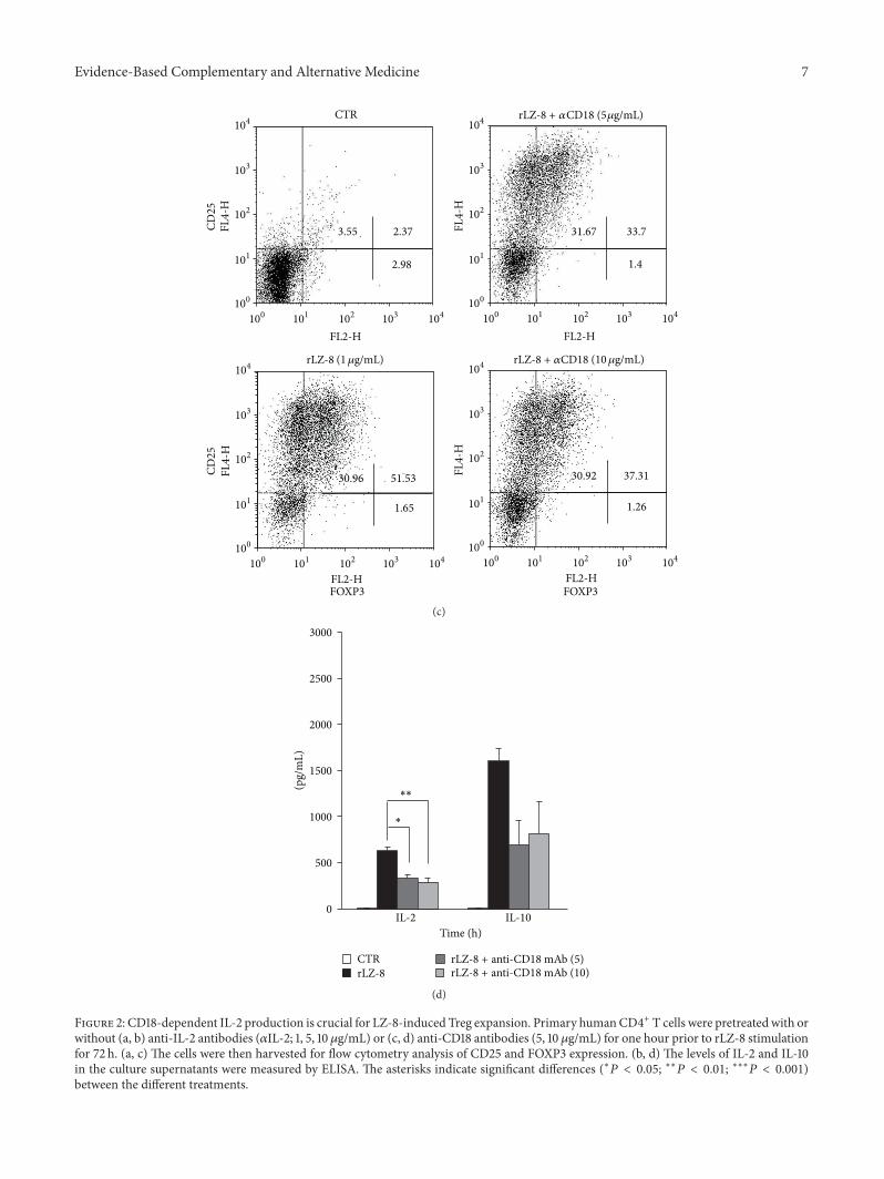
3.2. CD18-Dependent IL-2 Production Is Crucial for LZ-8-Induced Treg Expansion. IL-2 has the capacity to promoteT-cell differentiation [12–14], and our time-course cytokinestudy also indicated that there was IL-2 consumption duringTreg expansion induced by LZ-8 (Figure 1(c)). To determinewhether IL-2 is involved in LZ-8-induced Treg expansion, weneutralized IL-2 by applying anti-IL-2 antibodies (𝛼IL-2; 1, 5,10 𝜇g/mL) 1 h prior to LZ-8 stimulation. As expected, 𝛼IL-2 completely blocked IL-2 secretion (Figure 2(b)). Notably,𝛼IL-2 also significantly blocked LZ-8-induced Treg expan-sion (Figure 2(a)) and IL-10 production (Figure 2(b)) in a
dose-dependent manner. It has been reported that LZ-8enhances expression of ICAM-1 [16], a ligand of the integrinCD18, and we discovered that blocking CD18 with neutral-izing antibodies 1 h prior to LZ-8 stimulation significantlyreduced IL-2 production (Figure 2(d)). This blockade of theCD18 pathway also decreased LZ-8-induced Treg develop-ment and IL-10 production (Figures 2(c) and 2(d)). Takentogether, these findings suggested that CD18 signaling isinvolved in LZ-8-induced IL-2 production, highlighting theindispensable role of IL-2 in LZ-8-induced Treg expansion.
3.3. LZ-8 Promotes Treg Expansion through a CD45-MediatedSignal Pathway. Despite the fact that many studies haveshown the capacity of LZ-8 to stimulate CD4+ T cells,the interaction between LZ-8 and T cells remains unclear.Therefore, we investigated the potential receptor for LZ-8 onhumanCD4+ T cells using various T-cell lines, namely, Jurkat(WT), J.RT3-T3.5 (TCR−/−), and J45.01 (CD45−/−) cells. Thestimulation of Jurkat cells with LZ-8 and anti-CD3/28 anti-bodies (𝛼CD3/28; 1 𝜇g/mL each) expanded the population ofTregs (Figure 3(a)). As expected, 𝛼CD3/28 stimulation failedto induceTreg expansion inTCR−/− J.RT3-T3.5 cells, whereasLZ-8 continued to induce Treg expansion in TCR−/− cells(Figure 3(a)). These results suggest that the TCR is not thereceptor of LZ-8. Notably, it has been shown that LZ-8 failsto induce a significant increase in Treg expansion in CD45−/−J45.01 cells (Figure 3(a)), which indicates that CD45 is likelythe primary receptor that mediates the interactions betweenhuman CD4+ T cells and LZ-8. To elucidate the pathwaydownstream of CD45, J.RT3-T3.5 cells were stimulated withLZ-8, harvested, and then lysed for immunoblot analysis. LZ-8 stimulation increased the amount of phosphorylated Zap-70 and phospholipase C-𝛾 (Figure 3(b)), indicating that LZ-8 activates human T cells via CD45-dependent activationof T-cell proximal signaling. This finding was confirmedusing a CD45 inhibitor (CD45i; 1, 3𝜇M), where the appli-cation of CD45i significantly reduced LZ-8-induced Tregexpansion (Figure 3(c)). Taken as a whole, these findingsdemonstrate that LZ-8-induced Treg expansion occurs viaCD45-mediated T-cell proximal signaling.
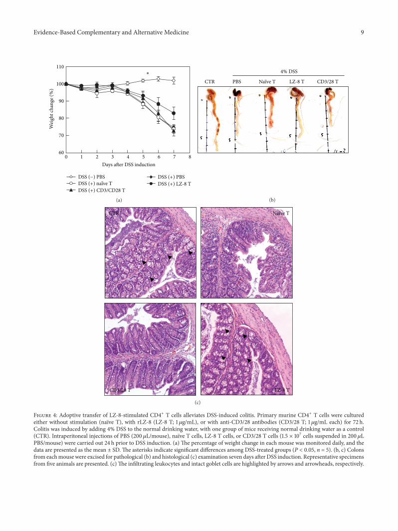
3.4. Adoptive Transfer of LZ-8-Stimulated CD4+ T CellsAlleviates DSS-Induced Colitis. To evaluate whether LZ-8-induced Tregs possess immuno suppressive capabilities, weemployed a murine model of DSS-induced acute colitis asdescribed in Section 2.4. The weight loss of mice receivingLZ-8-stimulated CD4+ T cells (TLZ-8) was less severe thanthat observed in otherDSS-treatedmice andwas significantlylower than that of mice receiving naıve CD4+ T cells (Tnaıve;Figure 4(a)). Furthermore, colon samples from mice receiv-ing TLZ-8 showed the greatest similarity to those of controlmice (Figure 4(b)). Reductions in colon length among micereceiving PBS, Tnaıve cells, or 𝛼CD3/28-stimulated CD4+ T(T𝛼CD3/28) cells were also observed (Figure 4(b)). Moreover,
during the histological analysis, a normal colon structureand normal crypt morphology were observed in the con-trol mice (Figure 4(c), upper left, UL), whereas leukocyteinfiltration and goblet cell depletion (as indicated by the
4 Evidence-Based Complementary and Alternative Medicine
Evidence-Based Complementary and Alternative Medicine 5
0
250
500
750
1000
1250
1500
1750
2000
12 24 72 120Time (h)
0
250
500
750
1000
1250
1500
1750
2000
12 24 72 120Time (h)
TGF-𝛽
(pg/
mL)
IFN
-𝛾(p
g/m
L)
CTRCD3/28rLZ-8
CTRCD3/28rLZ-8
(d)
CFSE
M1
M1
M1
M1
Even
tsEv
ents
Even
tsEv
ents
FL1-H
Control CD4+ T cells
PHA-activated CD4+ T cells+
Control CD4+ T cells
PHA-activated CD4+ T cells+
CD3/CD28-induced CD4+ T cells
PHA-activated CD4+ T cells+
LZ8-induced CD4+ T cells
17.3%
47.9%
31.3%
52%
100 101 102 103 104
FL1-H10
0
0
0
0
256
256
256
256
0 101 102 103 104
FL1-H100 101 102 103 104
FL1-H100 101 102 103 104
(e)
Figure 1: LZ-8 stimulates Treg expansion in murine and human CD4+ T cells. Primary murine and human CD4+ T cells were culturedfor 72 h, either in the absence of stimulation as the control (CTR), in the presence of stimulation with recombinant LZ-8 antibody (rLZ-8;1 𝜇g/mL), or in the presence of stimulation with anti-CD3/28 antibodies (CD3/CD28; 1 𝜇g/mL each, (a, b). The cells were then harvestedfor flow cytometry analysis of CD25, FOXP3, and CTLA-4 expression. (c and d) The levels of IL-2, IL-10, IFN-𝛾, and TGF-𝛽 in the culturesupernatants were measured by ELISA. (e)The cell proliferation of CD4+ T cells treated with or without PHA (2𝜇g/mL) and then coculturedwith indicated CD4+ T cells was monitored by cell-trace CFSE fluorescence via flow cytometry.
6 Evidence-Based Complementary and Alternative MedicineCD
25
CD25
FOXP3
FOXP3 FOXP3 FOXP3
FOXP3
CTR
2.16 3.5
3.35
14.87 33.79
6.51
16.44 54.63
4.02
15.19 37.37
5.87
16.27 32.25
6
FL2-H
FL4-
H
FL2-H
FL4-
H
FL4-
H
FL4-
H
FL4-
H
FL2-H FL2-H
FL2-H
100
100 101 102 103 104 100 101 102 103 104
101
102
103
104
100
101
102
103
104
100 101 102 103 104100
101
102
103
104
100 101 102 103 104100
101
102
103
104
100 101 102 103 104100
101
102
103
104rLZ8 + 𝛼IL-2 (5 𝜇g/mL)
rLZ-8 + 𝛼IL-2 rLZ-8 + 𝛼IL-2 (10𝜇g/mL)
rLZ-8 (1𝜇g/mL)
(1𝜇g/mL)
(a)
0
500
1000
1500
2000
2500
3000
IL-2 IL-10Time (h)
(pg/
mL)
∗
CTRrLZ-8rLZ-8 + anti-IL-2 mAb (1)
rLZ-8 + anti-IL-2 mAb (5)rLZ-8 + anti-IL-2 mAb (10)
∗∗∗
(b)
Figure 2: Continued.
Evidence-Based Complementary and Alternative Medicine 7
FOXP3 FOXP3
CD25
CD25
CTR
3.55 2.37
2.98
31.67 33.7
1.4
30.92 37.31
1.26
30.96 51.53
1.65
FL4-
H
FL2-H
104
103
102
101
100
100 101 102 103 104
FL4-
H
FL2-H
104
103
102
101
100
100 101 102 103 104
FL4-
H
FL2-H
103
104
102
101
100
100 101 102 103 104
FL4-
H
FL2-H
104
103
102
101
100
100 101 102 103 104
rLZ-8 + 𝛼CD18 (5 𝜇g/mL)
rLZ-8 rLZ-8 + 𝛼CD18 (1𝜇g/mL) (10𝜇g/mL)
(c)
0
500
1000
1500
2000
2500
3000
(pg/
mL)
IL-2 IL-10Time (h)
∗
∗∗
CTRrLZ-8
rLZ-8 + anti-CD18 mAb (5)rLZ-8 + anti-CD18 mAb (10)
(d)
Figure 2: CD18-dependent IL-2 production is crucial for LZ-8-induced Treg expansion. Primary humanCD4+ T cells were pretreated with orwithout (a, b) anti-IL-2 antibodies (𝛼IL-2; 1, 5, 10𝜇g/mL) or (c, d) anti-CD18 antibodies (5, 10 𝜇g/mL) for one hour prior to rLZ-8 stimulationfor 72 h. (a, c) The cells were then harvested for flow cytometry analysis of CD25 and FOXP3 expression. (b, d) The levels of IL-2 and IL-10in the culture supernatants were measured by ELISA. The asterisks indicate significant differences (∗𝑃 < 0.05; ∗∗𝑃 < 0.01; ∗∗∗𝑃 < 0.001)between the different treatments.
8 Evidence-Based Complementary and Alternative Medicine
Figure 3: LZ-8 promotes Treg expansion via a CD45-mediated signal pathway. (a) Human T-cell Jurkat (WT), J.RT3-T3.5 (TCR−/−), andJ45.01 (CD45−/−) cells were cultured without stimulation as the control (CTR), with stimulation by rLZ-8 (1, 2 𝜇g/mL), or with stimulation byanti-CD3/28 antibodies (CD3/CD28; 1, 2 𝜇g/mL) for 72 h. The cells were then harvested for flow cytometry analysis of CD25 and FOXP3expression. (b) J.RT3-T3.5 cells were stimulated with or without rLZ-8 (2𝜇g/mL) or with or without anti-CD3/28 antibodies (CD3/28;2𝜇g/mL) for the indicated time periods. The total cell lysates were collected for protein phosphorylation analysis. (c) Primary human CD4+T cells were pretreated with or without CD45 phosphatase inhibitor (CD45i; 1, 3𝜇M) for one hour prior to rLZ-8 stimulation for 72 h. Thecells were then harvested for flow cytometry analysis of CD25 and FOXP3 expression.
Evidence-Based Complementary and Alternative Medicine 9
0 1 2 3 4 5 6 7 8Days after DSS induction
110
100
90
80
70
60
∗
DSS (−) PBS
DSS (+) CD3/CD28 T
DSS (+) PBSDSS (+) LZ-8 TDSS (+) naïve T
Wei
ght c
hang
e (%
)
(a)
CTR PBS Naïve T LZ-8 T CD3/28 T
4% DSS
(b)
CTR Naïve T
LZ-8 TCD3/28 T
(c)
Figure 4: Adoptive transfer of LZ-8-stimulated CD4+ T cells alleviates DSS-induced colitis. Primary murine CD4+ T cells were culturedeither without stimulation (naıve T), with rLZ-8 (LZ-8 T; 1𝜇g/mL), or with anti-CD3/28 antibodies (CD3/28 T; 1 𝜇g/mL each) for 72 h.Colitis was induced by adding 4% DSS to the normal drinking water, with one group of mice receiving normal drinking water as a control(CTR). Intraperitoneal injections of PBS (200 𝜇L/mouse), naıve T cells, LZ-8 T cells, or CD3/28 T cells (1.5 × 10⁷ cells suspended in 200𝜇LPBS/mouse) were carried out 24 h prior to DSS induction. (a) The percentage of weight change in each mouse was monitored daily, and thedata are presented as the mean ± SD. The asterisks indicate significant differences among DSS-treated groups (P < 0.05, n = 5). (b, c) Colonsfrom eachmouse were excised for pathological (b) and histological (c) examination seven days after DSS induction. Representative specimensfrom five animals are presented. (c)The infiltrating leukocytes and intact goblet cells are highlighted by arrows and arrowheads, respectively.
10 Evidence-Based Complementary and Alternative Medicine
arrows and arrowheads, respectively) were observed in theDSS-treated mice. In addition, the adoptive transfer of TLZ-8(Figure 4(c), LR), but not of Tnaıve (Figure 4(c), UR) orT𝛼CD3/28 (Figure 4(c), LL), significantly reduced leukocyte
infiltration and goblet cell depletion.
4. Discussion
The immunomodulatory activities of LZ-8 with regard toimmuno cells have been extensively studied [2, 7–9]; however,few reports have explored the immuno suppressive activityor regulatory functions of LZ-8. In our previous research,we reported that LZ-8 directly induced the activation andIL-2 production of murine and human CD4+ T cells inthe absence of antigen-presenting cells [2, 8]. In this study,using both murine and human CD4+ T cells, we found thatLZ-8 expanded CD25+FOXP3+ Tregs (Figure 1(a)). We alsonoticed that 𝛼CD3/28 stimulation expanded the proportionof CD25+FOXP3+ Treg cells (Figure 1(a)). These findings arein agreement with those of Chen et al., who demonstratedthat the application of a superagonistic anti-CD28 antibody(supCD28mAb, D665) expanded FOXP3+ regulatory T cells[17]. However, the anti-CD28 antibody used in this studywas a different clone (CD28.6, eBioscience Cat. No. 16-0288). In addition, we showed that LZ-8-induced CD4+ Tcells expressed a higher level of CTLA-4 (Figure 1(b)), a keymolecule involved in controlling regulatory T-cell function.Furthermore, this effect corresponded to the expansion ofTregs and the colitis suppressive effect of LZ-8-stimulatedCD4+ T cells (Figure 4).
Notably, we discovered that LZ-8-induced CD4+ T cellswere also able to secrete IFN-𝛾, a proinflammatory cytokine(Figure 1(d)). This showed that although LZ-8 expanded theCD25+FOXP3+ Treg population, other effector cells that liewithin the population might also be expanded. It has beenreported that a certain subpopulation of Helios− cells wereobserved to secret effector cytokines such as the IFN-𝛾 [18].Therefore, in order to specify the subtype of LZ-8-inducedCD4+ T cells, further examinations on surface markers suchas the Helios and the CD127 should be carried out in thefuture. Nevertheless, using a mix leukocytes reaction, wedemonstrated the immuno regulatory activity of the LZ-8-induced CD4+ T cell (Figure 1(e)).
It has been previously demonstrated that FOXP3+ thy-mocytes are dramatically reduced in IL-2/IL-2R deficientmice and that neutralization of IL-2 using 𝛼IL-2 mono-clonal antibody results in autoimmuno gastritis in mice[12–14]. These findings highlight the critical role of IL-2signaling in the development and functioning of regulatoryT cells. In the present study, we demonstrated that LZ-8-induced IL-2 production was required for LZ-8-inducedTreg expansion and functioning. Furthermore, it has beenreported that CD18 signaling is crucial for the developmentaland suppressive functions of both innate and peripherallyinduced Tregs [19]. We found that blocking CD18 signalingresulted in a significant reduction in IL-2 production andreduced Treg expansion among LZ-8-induced CD4+ T cells(Figure 2). Taken together, we propose that CD18-mediatedIL-2 production is critical to LZ-8-induced Treg expansion.
Although it has been previously reported that LZ-8 inducesT-cell activation [2, 7–9], the molecular receptor responsiblefor LZ-8-mediated T-cell activation remains unknown. Inthis study, we demonstrated using gene knockout and inhibi-tion studies that CD45 appears to act as a major downstreammolecule for LZ-8 in human T cells (Figure 3). Finally, inthis context, we also demonstrated that LZ-8 activates Tcells through a CD45-dependent T-cell proximal signalingpathway.
FOXP3+ cells, CD4+CD25+FOXP3+ cells, and IL-10-secreting CD4+CD25+ T cells all play key roles in theregulation and suppression of DSS-induced colitis [20, 21].In addition, adoptive transfer of Tregs was shown to preventthe development of colitis and has previously been shown toattenuate established IBD [22, 23] Thus, therapies targetingTregs should provide promising insights for the treatment ofIBD and are under intense investigation [24, 25]. We haveshown here that LZ-8-induced CD4+ T cells alleviates colitis(Figure 4), and it has previously been shown that LZ-8 pos-sesses immunosuppressive activity and can prevent insulitisin nonobese diabetic mice and prolong allograft survival [26,27]. Thus, our results suggest that these suppressive effectsmay be a result of Treg expansion. Nonetheless, althoughwe have demonstrated the potential of LZ-8-induced CD4+T cell on ameliorating mice colitis, further experiments arerequired to define the effect and action mechanism of LZ-8as an immuno therapeutic agent.
In conclusion, we showed that LZ-8 induces Treg expan-sion and that LZ-8-induced Treg expansion is dependent onCD18-dependent IL-2 production; furthermore, this induc-tion seemed to occur via CD45-mediated T-cell proximalsignaling. In addition, we demonstrated that LZ-8-inducedTregs were capable of alleviating acute colitis. Thus, thesestudies provide new insights into the immuno modulatorymechanism(s) of LZ-8 and suggest that LZ-8 has the potentialto help control intestinal inflammation.
Conflict of Interests
The authors declare no financial interest or commercial con-flict of interests.
Acknowledgments
This work was supported by Grants from The National Sci-ence Council (NSC) of Taiwan (NSC 99-2320-B-010-010-MY3, NSC 101-2627-M-010-003, NSC 100-2627-M-010-003-,and NSC 100-2627-M-010-004), and The Ministry of Edu-cation (The Top University Project: 100AC-BI1 and 102AC-P664) to H.-Y. Hsu, as well as Grants from NSC of Taiwan,(NSC 99C-3245-2 and NSC 100C-3642-1) to F. Sheu. Theauthors thank Ms. S.-T. Weng for her technical help andassistance with this paper.
References
[1] M.M.Martınez-Montemayor, R. R. Acevedo, E. Otero-Franqui,L. A. Cubano, and S. F. Dharmawardhane, “Ganoderma lucidum(Ling-Zhi) inhibits cancer cell growth and expression of key
Evidence-Based Complementary and Alternative Medicine 11
molecules in inflammatory breast cancer,” Nutrition and Can-cer, vol. 63, no. 7, pp. 1085–1094, 2011.
[2] C. H. Yeh, H. C. Chen, J. J. Yang, W. I. Chuang, and F. Sheu,“Polysaccharides PS-G and protein LZ-8 from Reishi (Gano-derma lucidum) exhibit diverse functions in regulating murinemacrophages and T lymphocytes,” Journal of Agricultural andFood Chemistry, vol. 58, no. 15, pp. 8535–8544, 2010.
[3] R. Hanaoka, Y. Ueno, S. Tanaka et al., “The water-solubleextract from cultured medium of Ganoderma lucidum (Ling-Zhi) mycelia (Designated as MAK) ameliorates murine colitisinduced by trinitrobenzene sulphonic acid,” Scandinavian Jour-nal of Immunology, vol. 74, no. 5, pp. 454–462, 2011.
[4] K. Kino, A. Yamashita, K. Yamaoka et al., “Isolation andcharacterization of a new immunomodulatory protein, LingZhi-8 (LZ-8), from Ganoderma lucidum,” Journal of BiologicalChemistry, vol. 264, no. 1, pp. 472–478, 1989.
[5] S. Tanaka, K. Ko, K. Kino et al., “Complete amino acidsequence of an immunomodulatory protein, ling zhi-8 (LZ-8). An immunomodulator from a fungus, Ganoderma lucidum,having similarity to immunoglobulin variable regions,” Journalof Biological Chemistry, vol. 264, no. 28, pp. 16372–16377, 1989.
[6] L. Huang, F. Sun, C. Liang et al., “Crystal structure of LZ-8 fromthemedicinal fungusGanoderma lucidum,” Proteins, vol. 75, no.2, pp. 524–527, 2009.
[7] C. C. Lin, Y. L. Yu, C. C. Shih et al., “A novel adjuvant Ling Zhi-8 enhances the efficacy of DNA cancer vaccine by activatingdendritic cells,” Cancer Immunology, Immunotherapy, vol. 60,no. 7, pp. 1019–1027, 2011.
[8] H. Y. Hsu, K. F. Hua, W. C. Wu et al., “Reishi immuno-modulation protein induces interleukin-2 expression via pro-tein kinase-dependent signaling pathways within human Tcells,” Journal of Cellular Physiology, vol. 215, no. 1, pp. 15–26,2008.
[9] Y. L. Lin, Y. C. Liang, Y. S. Tseng et al., “An immunomodulatoryprotein, Ling Zhi-8, induced activation and maturation ofhuman monocyte-derived dendritic cells by the NF-𝜅B andMAPK pathways,” Journal of Leukocyte Biology, vol. 86, no. 4,pp. 877–889, 2009.
[10] S. Sakaguchi, “Naturally arising Foxp3-expressing CD25+ CD4+regulatory T cells in immunological tolerance to self and non-self,” Nature Immunology, vol. 6, no. 4, pp. 345–352, 2005.
[11] K. J. Wood and S. Sakaguchi, “Regulatory T cells in transplan-tation tolerance,” Nature Reviews Immunology, vol. 3, no. 3, pp.199–210, 2003.
[12] M. F. Bachmann and A. Oxenius, “Interleukin 2: from immun-ostimulation to immunoregulation and back again,” EMBOReports, vol. 8, no. 12, pp. 1142–1148, 2007.
[13] P. Hoffmann, R. Eder, T. J. Boeld et al., “Only the CD45RA+subpopulation of CD4+CD25 high T cells gives rise to homo-geneous regulatory T-cell lines upon in vitro expansion,” Blood,vol. 108, no. 13, pp. 4260–4267, 2006.
[14] S. Letourneau, C. Krieg, G. Pantaleo, and O. Boyman, “IL-2-and CD25-dependent immunoregulatory mechanisms in thehomeostasis of T-cell subsets,” Journal of Allergy and ClinicalImmunology, vol. 123, no. 4, pp. 758–762, 2009.
[15] A. Izcue, J. L. Coombes, and F. Powrie, “Regulatory lymphocytesand intestinal inflammation,” Annual Review of Immunology,vol. 27, pp. 313–338, 2009.
[16] M. Haak-Frendscho, K. Kino, T. Sone, and P. Jardieu, “LingZhi-8: a novel T cell mitogen induces cytokine production andupregulation of ICAM-1 expression,” Cellular Immunology, vol.150, no. 1, pp. 101–113, 1993.
[17] J. Chen, L. Xie, S. Toyama et al., “The effects of Foxp3-expressingregulatory T cells expanded with CD28 superagonist antibodyin DSS-induced mice colitis,” International Immunopharmacol-ogy, vol. 11, no. 5, pp. 610–617, 2011.
[18] Y. C. Kim, R. Bhairavabhotla, J. Yoon et al., “Oligodeoxynu-cleotides stabilize Helios-expressing Foxp3+ human T regula-tory cells during in vitro expansion,” Blood, vol. 119, no. 12, pp.2810–2818, 2012.
[19] M. Marski, S. Kandula, J. R. Turner, and C. Abraham, “CD18 isrequired for optimal development and function of CD4+CD25+T regulatory cells,” Journal of Immunology, vol. 175, no. 12, pp.7889–7897, 2005.
[20] C.Mottet, H.H.Uhlig, and F. Powrie, “Cutting edge: cure of col-itis by CD4+CD25+ regulatory T cells,” Journal of Immunology,vol. 170, no. 8, pp. 3939–3943, 2003.
[21] H. H. Uhlig, J. Coombes, C. Mottet et al., “Characterizationof Foxp3+CD4+CD25+ and IL-10-secreting CD4+CD25+ T cellsduring cure of colitis,” Journal of Immunology, vol. 177, no. 9, pp.5852–5860, 2006.
[22] M. C. Fantini, C. Becker, I. Tubbe et al., “Transforming growthfactor beta induced FoxP3+ regulatory T cells suppress Th1mediated experimental colitis,” Gut, vol. 55, no. 5, pp. 671–680,2006.
[23] C. Toms and F. Powrie, “Control of intestinal inflammation byregulatory T cells,”Microbes and Infection, vol. 3, no. 11, pp. 929–935, 2001.
[24] E. K. Boden and S. B. Snapper, “Regulatory T cells in inflamma-tory bowel disease,” Current Opinion in Gastroenterology, vol.24, no. 6, pp. 733–741, 2008.
[25] B. Sitohy, S. Hammarstrom, A. Danielsson, and M. L. Ham-marstrom, “Basal lymphoid aggregates in ulcerative colitiscolon: a site for regulatory T cell action,” Clinical and Experi-mental Immunology, vol. 151, no. 2, pp. 326–333, 2008.
[26] K. Kino, K. Mizumoto, T. Sone et al., “An immunomodulatingprotein, Ling Zhi-8 (LZ-8) prevents insulitis in non-obesediabetic mice,” Diabetologia, vol. 33, no. 12, pp. 713–718, 1990.
[27] L. G. van der Hem, J. A. van der Vliet, C. F. M. Bocken, K. Kino,A. J. Hoitsma, and W. J. M. Tax, “Ling Zhi-8: studies of a newimmunomodulating agent,” Transplantation, vol. 60, no. 5, pp.438–443, 1995.


![CD45 Immunohistochemistry in Mouse Kidney [Abstract]](https://static.documents.pub/doc/80x56/61fda0a5835b935d1a626f51/cd45-immunohistochemistry-in-mouse-kidney-abstract.jpg)


