Research ArticleSafety Evaluation of Oral Toxicity of Carica papaya Linn.Leaves: A Subchronic Toxicity Study in Sprague Dawley Rats
Zakiah Ismail,1 Siti Zaleha Halim,1 Noor Rain Abdullah,1,2 Adlin Afzan,1
Badrul Amini Abdul Rashid,1 and Ibrahim Jantan3
1 Herbal Medicine Research Center, Institute for Medical Research, Jalan Pahang, 50588 Kuala Lumpur, Malaysia2 Infectious Disease Research Center, Institute for Medical Research, Jalan Pahang, 50588 Kuala Lumpur, Malaysia3 Drug and Herbal Research Center, Faculty of Pharmacy, Universiti Kebangsaan Malaysia, Jalan Raja Muda Abdul Aziz,50300 Kuala Lumpur, Malaysia
Correspondence should be addressed to Siti Zaleha Halim; [email protected]
Received 28 February 2014; Accepted 28 July 2014; Published 29 October 2014
The subchronic toxicity effect of the leaf extract of Carica papaya Linn. in Sprague Dawley (SD) rats was investigated in this study.The extract was prepared by dissolving the freeze dried extract of the leaves in distilled water and was administered orally to SD rats(consisted of 10 rats/sex/group) at 0 (control), 0.01, 0.14, and 2 g/kg body weight (BW) for 13 weeks. General observation, mortality,and food and water intake were monitored throughout the experimental period. Hematological and biochemical parameters,relative organ weights, and histopathological changes were evaluated. The study showed that leaf extract when administered for 13weeks did not cause any mortality and abnormalities of behavior or changes in body weight as well as food and water intake. Therewere no significant differences observed in hematology parameters between treatment and control groups; however significantdifferences were seen in biochemistry values, for example, LDH, creatinine, total protein, and albumin. However, these changeswere not associated with histopathological changes. In conclusion, the results suggested that daily oral administration of rats withC. papaya leaf extract for 13 weeks at a dose up to fourteen times the levels employed in traditional medicine practice did not causeany significant toxic effect.
1. Introduction
Carica papaya Linn. (family: Caricaceae) is primarily cul-tivated for its fruit and the young leaves are consumed as avegetable by the Malay community in Malaysia. Differentparts of the plant have been used in traditional medicine totreat various diseases. The juice of the green leaves is con-sumed as beverage to treat malarial fever [1]. The leaves arealso used to treat digestive disorders and other disturbancesof the gastrointestinal tract. The fruit is used to treat highfever, cough, and anorexia and the seed has been used totreat digestive disorders, to improve protein digestion, and toexpel intestinal worms [2]. The root is used to treat urinarydisorders while the bark is used to treat toothache [3].
Many studies have been conducted to evaluate the bio-logical activities of various parts of C. papaya. The fruit
and seed of C. papaya have showed bacteriostatic activityagainst several enteropathogens in human [4]. It was reportedthat experimentally induced diabetic rats showed signifi-cant wound healing after treatment with the aqueous extractof the fruit of C. papaya [5]. The aqueous extract of theleaves of the plant showed significant protective effect againstalcohol induced oxidative damage to the gastric mucosa inrats [6], and it was effective in healing induced wound inrats [7]. The aqueous extract of the leaves also exhibited theability to inhibit tumor cell lines [8] and this activity waspotentiated with phenolic compounds [9, 10]. Xanthine oxi-dase was found to be present in the leaf extract and thus ithas potential to work as an anti-gout agent [11]. The crudeethyl acetate extract of C. papaya leaves showed high anti-plasmodial activity against Plasmodium falciparum and P.falciparum-resistant strains [12]. The ethanol extract of
Hindawi Publishing CorporationEvidence-Based Complementary and Alternative MedicineVolume 2014, Article ID 741470, 10 pageshttp://dx.doi.org/10.1155/2014/741470
2 Evidence-Based Complementary and Alternative Medicine
C. papaya leaves showed potential as a source of drugs againsturinary tract microbes [13].
Phytochemical studies on the leaves of C. papaya showedthe presence of various compounds including piperidinealkaloids such as carpaine, pseudocarpaine, and dehydro-carpaines I and II [14–17]. The alkaloids have been shownto be teratogenic to livestock as they were found to inhibitthe fetal movement [18] and exhibited antiamoebic activity[19]. Ekong et al. [20] also reported that the aqueous extractof C. papaya leaves given to pregnant rats showed somedefects in fetus.The leaves can be considered to possess somenutritional value as theywere found to containmultinutrientsand iron [21]. Recently we identified carpaine, malic acid,quinic acid, six malic acid derivatives, and four flavonolglycosides in the leaf extract of the plant [22].
Toxicity evaluation of C. papaya leaves becomes moreimportant as they are not only consumed widely as foodbut also prepared and used as a traditional medicine. Acutetoxicity study which involved a single dose administrationof C. papaya leaf juice and is followed by fourteen days’observation in rats up to 2 g/kg BW showed dehydration asdemonstrated by an increase in red cell mass [23]. We havealso carried out a repeated dose 28-day oral toxicity study ofthe leaf extract in rats (subacute toxicity study) and the resultsindicated that the plant extract did not cause mortality, therewere no treatment-related changes, and all organs did notreveal morphological alterations [22]. We also have carriedout a clinical trial to investigate the platelet elevating propertyin dengue patients after having received C. papaya leaf juicesfor 3 consecutive days [24].
The present study, a subchronic toxicity evaluation of C.papaya leaf extract, was carried out to determine long termconsumption effect after repeated doses of C. papaya leafextract in a thirteen-week oral toxicity study.
2. Materials and Methods
2.1. Plant Material. The fresh leaves of Carica papaya L.“Sekaki” were purchased by the Malaysian AgriculturalResearch and Development Institute (MARDI). A represen-tative sample of this plant was authenticated at the ForestResearch Institute Malaysia (FRIM), Kepong, with voucherspecimen number 007/10. Leaves were collected and washedunder running tap water and then cut into small pieces andjuice was extracted using a juicer (Panasonic, Shah Alam,Malaysia). The resulting juice, without addition of water, wasthen poured into a glass container and left frozen in a freezer.The juice was lyophilized resulting in a dark green powder(2.6% w/w yield). Phytochemical analysis had been carriedout for chemical fingerprinting [22]. The doses used in thetoxicity studywere calculated based on the bodyweight (BW)of the rats. The test samples were prepared by dissolving thepowder in distilled water to obtain concentrations of 0.01,0.14, and 2 g/kg BW.
2.2. Animals. Male and female Sprague Dawley (SD) ratsaged between six and seven weeks and weighed between90 and 100 g were used in this study. The SD rats were
obtained from the Laboratory Animal Resource Unit, Medi-cal Resource Research Center, Institute for Medical Research(IMR), Kuala Lumpur. The use of laboratory animals andthe study design were approved by the Institutional AnimalCare andUsedCommittee (IACUC) (ACUCnumberACUC/KKM02 (1/2009)).TheGuidelines of Handling of LaboratoryAnimals by the Ministry of Health Malaysia were followedthroughout the experiments [25].
The animals were housed individually in a stainless-steelwire-mesh cage with size 6H× 11D× 16W cm andmaintainedat room temperature (27 ± 2∘C) with humidity of 65.85 ±6.76% and with 12 h alternate artificial and natural light anddark cycle. Room temperature and relative humidity wererecorded daily using a temperature datalogger (TempRHDatalogger BG-DL-01/01B). Each animal was identified bya cage card. They were fed with a pellet diet with ZeiglerRodent NIH-31 irradiated auto wafer feeds (Zeigler Bros,Pennsylvania, USA) and given an unlimited supply of reverseosmosis water. The animals were acclimatized to laboratoryconditions for seven days prior to the experiments.
2.3. Experimental Design. The subchronic toxicity study wascarried out according to the OECD Guidelines for the“Repeated Dose 90-day Oral Toxicity Study in Rodents,” no.408 [26] with some modifications. The modifications werein the temperature and humidity of the room. Forty femaleand 40 male rats were randomly assigned into four groupswhichweremade up of one control group and three treatmentgroups (𝑛 = 10 rats/sex/group).The treatment group receivedlyophilized C. papaya leaf juice diluted in water to thenecessary dosage while the control group received water only.
2.4. Selection of Doses and Oral Administration of the Extracts.Dose selection was based on the acute toxicity and subacutetoxicity studies carried out previously, where the highest doseof 2 g/kg BW did not exhibit any acute effects on the rats(NOAEL) [23]. Therefore, a dose of 2 g/kg BW was selectedfor the highest dose in this study. The medium (0.14 g/kgBW) and low dose (0.01 g/kg BW) levels were selected basedon the local traditional preparation of C. papaya juice fortreatment of fever in humans. The C. papaya leaf juiceat different concentrations was administered orally usingintubation needle on a daily basis for 13 weeks. The rats wereweighed weekly and the amount of C. papaya leaf juice tobe given was recalculated based on the new body weight toensure a constant dose volume per kg BWat all times. Controlrats were administered the same volume of drinking water asthe amount given to the test groups.
2.5. Parameters Measured during the Study
2.5.1. General Observation and Mortality. General observa-tions were carried out twice daily for mortality, moribundand ill health, or reaction to treatment. These observationsinclude changes in skin, fur, eyes, mucus membranes, behav-ior pattern, tremors, salivation, diarrhea, sleep, and coma.Theobservation was carried out according to the Guidance Doc-ument on the Recognition, Assessment and Use of Clinical
Evidence-Based Complementary and Alternative Medicine 3
Signs as Humane Endpoints for Experimental Animals Usedin Safety Evaluation [27].
2.5.2. Body Weight and Food and Water Consumption. Indi-vidual rat was weighed before the commencement of theexperiment and then weighed once on day 7 of every week.Final body weights were recorded prior to the schedulednecropsy. Each cage was supplied with calculated amounts offood and water.The amounts of left over food and water weremeasured weekly and the differences were regarded as food(g/rat/week) and water consumption (mL/rat/week).
2.5.3. Hematological and Biochemical Analysis. The rats werefasted overnight by removing all food from the cages butwere allowed access to water ad libitum before blood wascollected. Rats were anaesthetized with light ether and bloodsamples were collected via direct heart puncture and putinto two types of tubes, one with anticoagulant (EDTA) andthe other without any additives. After withdrawing bloodfrom the rats, they were sacrificed with an overdose of ether.The anticoagulated blood samples (EDTA) were analyzedimmediately for hematology parameters using HematologyAnalyzer (Medonic CA 620 VET, Stockholm, Sweden).Theseparameters include total and differential leukocyte count(WBC), erythrocyte counts (RBC), hemoglobin concentra-tion (HGB), hematocrit (HCT), mean cell volume (MCV),mean corpuscular hemoglobin (MCH), mean corpuscularhemoglobin concentration (MCHC), and platelet counts(PLT). The blood without any additives was used in thebiochemistries and the parameters which include serumtotal protein, albumin, alkaline phosphatase (ALP), aspartateaminotransferase (AST), alanine aminotransferase (ALT)urea, creatinine, uric acid, creatinine kinase (CK), lactatedehydrogenase (LDH), 𝛼-hydroxybutyrate dehydrogenase(HBDH), cholesterol, triglycerides, and glucose were deter-mined. The tests were done using the biochemistry analyser(Vitalab Selecta, E-series, Netherlands).
2.5.4. Gross Findings and Organ Weights. Complete post-mortem examinations were carried out on all animals. Therats were dissected, and their internal organs were carefullyexamined for any pathological changes. Subsequently theorgans were removed and weighed. The organs were thelungs, heart, liver, stomach, spleen, gastrointestinal, kidneys,testes, and adrenals.The relative organ weight (ROW) of eachorgan was calculated using the following equation:
ROW = [Absolute organ weight (g)
Body weight of rat on sacrifice day (g)]
× 100.
(1)
The organs were preserved in 10% buffered formalin forsubsequent histopathological examination.
2.5.5. Histopathology. For histopathological examination, arepresentative tissue or the whole organ (depending on thesize and weight) was taken and processed further to make
Figure 1: Mean body weight (g) of male and female rats admin-istered daily after subchronic treatment orally for 13 weeks withC. papaya leaf extract in SD rats. Values are expressed as mean ±standard deviation (𝑛 = 10/group).
slides using the standard procedure for histology slides andwas stained with Hematoxylin and Eosin. The slides wereexamined using a light microscope (Olympus BX 51, Tokyo,Japan).
2.6. Statistical Analysis. The data were analyzed using SPSSprogram, version 14. The one-way analysis of variance(ANOVA) was used to test for significant differences betweenthe experimental groups andwas followed byTukey’sHSD formultiple comparisons [28, 29]. Nonparametric test methodswere usedwhen the distribution of certain variable(s) differedfrom normal. The nonparametric methods employed werethe Kruskal-Wallis test for pairwise comparison. Results with𝑃 < 0.05 were considered statistically significant. The resultswere expressed as mean value (𝑥) and standard deviation(SD) for each variable measured.
3. Results
3.1. Survival and General Observation. There was no treat-ment related death reported in any of the experiment groupsas well as in the control group. Neither physical nor behav-ioral changes were observed in any of the groups throughoutthe study period of 13 weeks.
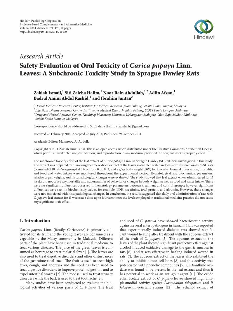
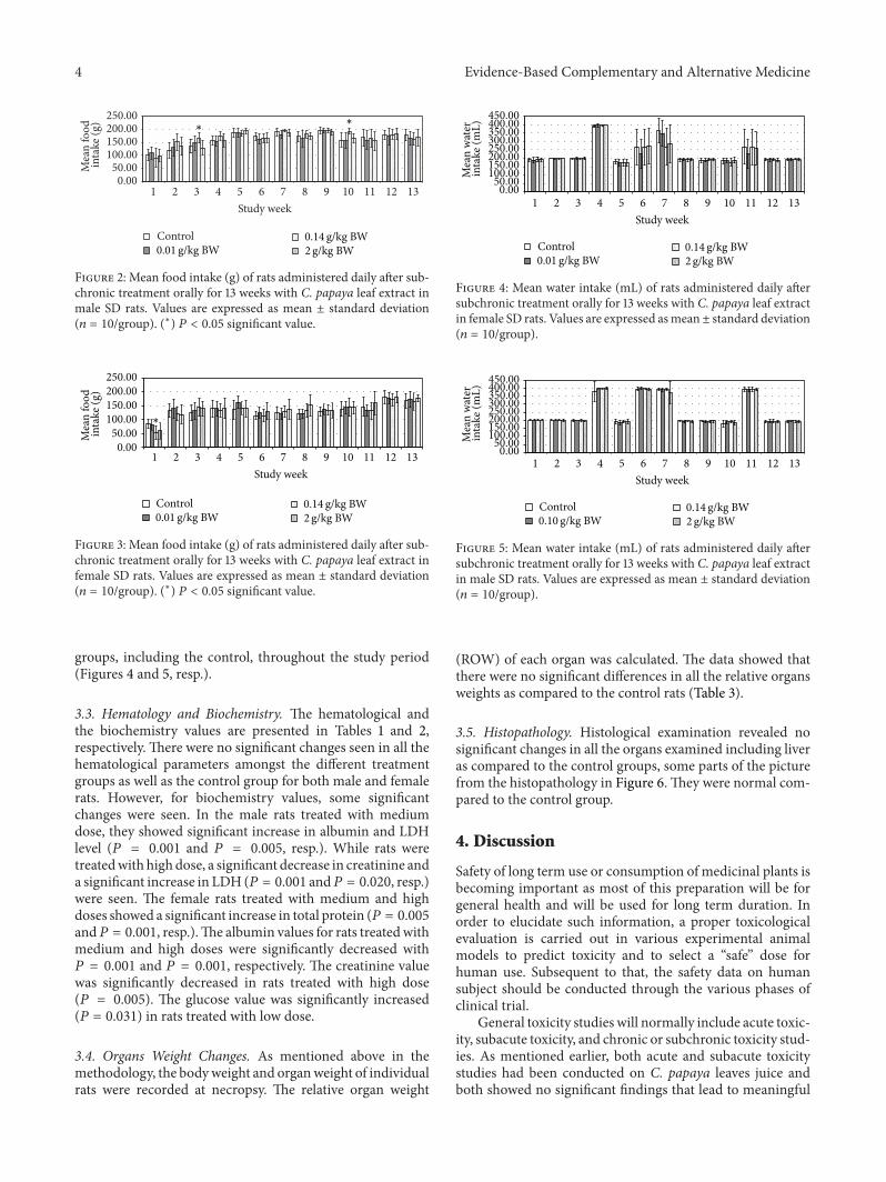
3.2. Body Weight Changes and Food and Water Consumption.The initial (day 0) body weights of the female rats were436.00±2.33 g, whereas for themale rats were 545.00±1.14 g.Their body weights were gradually increased as noted inweekly measurements and presented in Figure 1. No signif-icant difference in body weight changes was noted betweenthe control group and any of the treated groups at any timeof the 13-week period.Themale test group receiving mediumdose in week 3 and week 10 showed significant increase infood consumption (𝑃 = 0.008, 𝑃 = 0.012, resp.) (Figure 2).While the female test group which received medium doseshowed significant decrease (𝑃 = 0.010) in food consumptionduring week 1 (Figure 3). However, there is no significantdifference in the amount of water consumed in all various
4 Evidence-Based Complementary and Alternative Medicine
0.0050.00
100.00150.00200.00250.00
1 2 3 4 5 6 7 8 9 10 11 12 13
Mea
n fo
od in
take
(g)
Study week
Control
∗∗
0.01 g/kg BW0.14 g/kg BW2g/kg BW
Figure 2: Mean food intake (g) of rats administered daily after sub-chronic treatment orally for 13 weeks with C. papaya leaf extract inmale SD rats. Values are expressed as mean ± standard deviation(𝑛 = 10/group). (∗) 𝑃 < 0.05 significant value.
0.0050.00
100.00150.00200.00250.00
1 2 3 4 5 6 7 8 9 10 11 12 13
Mea
n fo
od
inta
ke (g
)
Study week
∗
Control0.01 g/kg BW
0.14 g/kg BW2g/kg BW
Figure 3: Mean food intake (g) of rats administered daily after sub-chronic treatment orally for 13 weeks with C. papaya leaf extract infemale SD rats. Values are expressed as mean ± standard deviation(𝑛 = 10/group). (∗) 𝑃 < 0.05 significant value.
groups, including the control, throughout the study period(Figures 4 and 5, resp.).
3.3. Hematology and Biochemistry. The hematological andthe biochemistry values are presented in Tables 1 and 2,respectively. There were no significant changes seen in all thehematological parameters amongst the different treatmentgroups as well as the control group for both male and femalerats. However, for biochemistry values, some significantchanges were seen. In the male rats treated with mediumdose, they showed significant increase in albumin and LDHlevel (𝑃 = 0.001 and 𝑃 = 0.005, resp.). While rats weretreatedwith high dose, a significant decrease in creatinine anda significant increase in LDH (𝑃 = 0.001 and𝑃 = 0.020, resp.)were seen. The female rats treated with medium and highdoses showed a significant increase in total protein (𝑃 = 0.005and𝑃 = 0.001, resp.).The albumin values for rats treatedwithmedium and high doses were significantly decreased with𝑃 = 0.001 and 𝑃 = 0.001, respectively. The creatinine valuewas significantly decreased in rats treated with high dose(𝑃 = 0.005). The glucose value was significantly increased(𝑃 = 0.031) in rats treated with low dose.
3.4. Organs Weight Changes. As mentioned above in themethodology, the bodyweight and organweight of individualrats were recorded at necropsy. The relative organ weight
0.0050.00
100.00150.00200.00250.00300.00350.00400.00450.00
1 2 3 4 5 6 7 8 9 10 11 12 13
Mea
n w
ater
inta
ke (m
L)
Study week
Control0.01 g/kg BW
0.14 g/kg BW2g/kg BW
Figure 4: Mean water intake (mL) of rats administered daily aftersubchronic treatment orally for 13 weeks with C. papaya leaf extractin female SD rats. Values are expressed asmean± standard deviation(𝑛 = 10/group).
Figure 5: Mean water intake (mL) of rats administered daily aftersubchronic treatment orally for 13 weeks with C. papaya leaf extractin male SD rats. Values are expressed as mean ± standard deviation(𝑛 = 10/group).
(ROW) of each organ was calculated. The data showed thatthere were no significant differences in all the relative organsweights as compared to the control rats (Table 3).
3.5. Histopathology. Histological examination revealed nosignificant changes in all the organs examined including liveras compared to the control groups, some parts of the picturefrom the histopathology in Figure 6.They were normal com-pared to the control group.
4. Discussion
Safety of long term use or consumption of medicinal plants isbecoming important as most of this preparation will be forgeneral health and will be used for long term duration. Inorder to elucidate such information, a proper toxicologicalevaluation is carried out in various experimental animalmodels to predict toxicity and to select a “safe” dose forhuman use. Subsequent to that, the safety data on humansubject should be conducted through the various phases ofclinical trial.
General toxicity studies will normally include acute toxic-ity, subacute toxicity, and chronic or subchronic toxicity stud-ies. As mentioned earlier, both acute and subacute toxicitystudies had been conducted on C. papaya leaves juice andboth showed no significant findings that lead to meaningful
Evidence-Based Complementary and Alternative Medicine 5
Table 1: Hematological values of control group and rats treated with C. papaya leaf extract measured during the subchronic toxicity study.
interpretation of toxic effect. The acute toxicity study ofC. papaya leaf juice in rats showed dehydration as demon-strated by an increase in red cell mass [23]. We have alsocarried out a repeated dose 28-day oral toxicity study ofa similar leaf extract in rats and the results indicated thatthe plant extract did not cause mortality, there were notreatment-related changes, and all organs did not revealmorphological alterations [22]. However the biochemistryvalues (total protein, AST, ALT, and ALP) revealed somechanges although they are non-dose dependent. Thus, asubchronic toxicity evaluation of C. papaya leaf extract isnecessary to confirm the finding especially when the dosingis given for a longer period in this thirteen-week oral toxicitystudy.The same aqueous juice extract of C. papaya leaves wasalso used in this study and such formulationwas the form thattraditionally had been consumed [30].
From the above presented result, it was showed that dailyadministration of theC. papaya leaves juice for 13weeks at thechosen doses did not show any changes in the general behav-iors of treated rats, all gained weight normally correspondingto the food and water intake. It was noted that during weeks4, 6, 7, and 11 the water intake was increased in both femaleandmale rats in all groups. However there were no significantdifferences between groups as compared to the control group.These increases of water intake did not show any change ordirectly proportional to body weight and food intake; hence,it was more of an accidental finding and was not related tothe administration of C. papaya leaf juice. As evidenced bythe absence of toxic symptoms, there were no changes in
food intake and body weight in those weeks. As seen in theprevious studies, there were no hematological changes seenin all the parameters measured including the platelets andhematocrits.
Thebiochemistry values showed some significant changesin the liver enzymes as in the previous 2 studies; howeverthere is no dose related pattern was observed. At the currentsubchronic study, all the liver enzymes (AST, ALT, and ALP)except LDH were not significantly different from the controlgroup in both male and female rats. The LDH was seento be increasing significantly but only in male group withmedium (with increased albumin) and high doses (withdecreased level of creatinine), while in female group, suchfinding was not noted. Previous researchers describe possibleliver dysfunction when such studies were conducted for theherbal product/plant. Everds [31] reported that an increase inalbumin level maybe associated to abnormal liver function ordehydration state. As discussed byRamaiah [32], abnormalityin AST, ALT, and ALP levels is more specific for liver cellinjury. Garba and Ubom [33] and Kotoh et al. [34] reportedthat LDH also can be used as indicator in liver cell injuresas the enzymes are released from the injured hepatocytesalthough the specificity to indicate liver disease is lowercompared with AST, ALT and ALP. This is due to the factthat serum LDH in livermore rapidly declines and alsomightindicate the presence of red cell hemolysis rather than livercell injuries. The other authors, Preus et al. [35] and Oloyedeand Sunmonu [36], mention the LDH as a useful indicatorfor cardiac damage. In the current study, the finding is most
6 Evidence-Based Complementary and Alternative Medicine
Table 2: Biochemistry values of control group and rats treated with C. papaya leaf extract measured during the subchronic toxicity study.
Male rats Control 0.01 g/kg BW 0.14 g/kg BW 2 g/kg BWLiver profile
Glucose (mmol/L) 8.88 ± 5.92 15.17 ± 5.62∗ 9.24 ± 3.04 6.85 ± 1.73Values are expressed as mean ± standard deviation (𝑛 = 10/group). ALP: alkaline phosphatase, AST: aspartate aminotransferase, ALT: alanine aminotrans-ferase, CK: creatinine kinase, LDH: lactate dehydrogenase, and HBDH: 𝛼-hydroxybutyrate dehydrogenase. ∗𝑃 value less than 0.05 (𝑃 < 0.05), significantvalue.
likely to indicate liver damaged rather than red cell hemolysisor cardiac damaged although the other specific liver enzymesAST, ALT, and ALP were not significantly different, whereasin the female group, the medium and high group showedsignificant increase in total protein, 𝑃 = 0.005 and 𝑃 =0.001, respectively, but their albumin valueswere significantlydecreased with 𝑃 = 0.001 and 𝑃 = 0.001, respectively. This
could be due to the fact that other protein fractions otherthan albumin might be increased and thus need to be furtherinvestigated.
As the rats are aging and their size increases, their physicalactivities reduced and hence this could be the explanationfor the significant reduction of creatinine level in both maleand female rats treated with the highest dose. This is in
Evidence-Based Complementary and Alternative Medicine 7
Table 3: Organ weight values of control group and rats treated with C. papaya leaf extract measured during the subchronic toxicity study.The relative organ weight per 100 g body weight recorded at the end of the study.
Organ Control 0.01 g/kg BW 0.14 g/kg BW 2 g/kg BWMale rats
Values are expressed as mean ± standard deviation (𝑛 = 10/group).
reference to reports by Omer [37], Attia and Nasr [38]which described that creatinine values were affected by thevariation of body weight or physical activity. Creatinine andurea are waste products of protein excreted from kidney andare indicators for kidney damage. Thus this reduction ofcreatinine indicates that there was no potential kidney effectand this was supported by the urea value which remainednormal as compared to the control group.
Tarkang et al. [39] also conducted study on aqueous aswell as ethanol extracts of the leaves of C. papaya for 28and 90 days. The finding showed no abnormalities in liverenzymes and renal biochemistries in rat after administrationof C. papaya leaf extract for 28 and 90 days. Only ethanolextract showed some changes in the liver and renal toxicityat the dose 1 g/kg BW. This different finding could be dueto the fact that different extract was used in the study. Itwas air-dried and extracted either with water or ethanol,while juice extracted from the fresh leaves was used in thisstudy.
5. Conclusions
In conclusion, the administration of rats with low, medium,and high doses of fresh juice of C. papaya leaf extract for 13weeks did not cause any changes in body weight, food intake,and water level. There were also no significant differencesobserved in hematology parameters between treatment andcontrol groups. There were significant differences in bio-chemistry values, such as the LDH, creatinine, total protein,and albumin. However, these changes were not associatedwith histopathological changes and were not dose depen-dent. Such finding on possible liver dysfunction needs tobe confirmed with proper hepatotoxicity study protocol toelucidate extension of the toxic effect if any. The oral doseof C. papaya leaf extract was more than 2 g/kg BW andno observed adverse effect level (NOAEL) of the extractfor both female and male rats was 2 g/kg BW per day for13 weeks extracts on Sprague Dawley rats for the presentstudy.
8 Evidence-Based Complementary and Alternative Medicine
(i) (ii)
PV
CV
CV
CV
S
H
(a)
(i) (ii)
H
PV
S
(b)
Figure 6: Histological structure of liver from control (a) medium 0.14 g/kg BW (b) group of male SD rat showing the Central vein (CV),hepatocyte (H), the sinusoid (S) and the portal vein (PV). The structure showed there were normal as compared to the control. (i) 4x, (ii)40x, (H&E Staining).
Conflict of Interests
The authors declare that there is no conflict of interestsregarding the publication of this paper.
Acknowledgments
Theauthors thank theDirectorGeneral ofHealth,Ministry ofHealth, Malaysia, and also Director of the Institute for Med-ical Research (IMR), Kuala Lumpur, for the permission topublish this paper. This study was supported by the NationalInstitute of Health, Ministry of Health, Malaysia. Thanks arealso due to the staff at the Herbal Medicine Research Centerfor their contribution to this study. The authors are grateful
to Dr. Naseem Malik from the Laboratory Animal ResourceUnit, IMR, for rendering technical assistance.Thanks are alsodue to Dr. Murizal Zainol and Dr. Hussin Muhammad forreviewing the paper.
References
[1] H. C. Ong, B. N. Ruzalila, and P.Milow, “Traditional knowledgeof medicinal plants among the malay villagers in KampungTanjung Sabtu, Terengganu, Malaysia,” Indian Journal of Tradi-tional Knowledge, vol. 10, no. 3, pp. 460–465, 2011.
[2] HMRC, Compendium Medicinal Plants Used in Malaysia, vol.1, Herbal Medicine Research Centre, Institute for MedicalResearch, Kuala Lumpur, Malaysia, 2002.
Evidence-Based Complementary and Alternative Medicine 9
[3] K. L. Krishna, M. Paridhavi, and J. A. Patel, “Review on nutri-tional, medicinal and pharmacological properties of Papaya(Carica papaya Linn.),” Natural Product Radiance, vol. 7, no. 4,pp. 364–373, 2008.
[4] J. A. Osato, L. A. Santiago, G. M. Remo, M. S. Cuadra, andA. Mori, “Antimicrobial and antioxidant activities of unripepapaya,” Life Sciences, vol. 53, no. 17, pp. 1383–1389, 1993.
[5] B. S. Nayak, L. P. Pereira, and D. Maharaj, “Wound healingactivity of Carica papaya L. In experimentally induced diabeticrats,” Indian Journal of Experimental Biology, vol. 45, no. 8, pp.739–743, 2007.
[6] M. Indran, A. A. Mahmood, and U. R. Kuppusamy, “Protectiveeffect of Carica papaya L leaf extract against alcohol inducedacute gastric damage and blood oxidative stress in rats,” WestIndian Medical Journal, vol. 57, no. 4, pp. 323–326, 2008.
[7] A. A. Mahmood, K. Siddiq, and I. Salmah, “Wound healingacivity of Carica papaya L. aqueous leaf extract rats,” Journal ofMolecular and Advance Sciences, vol. 1, pp. 398–401, 2005.
[8] N. Otsuki, N. H. Dang, E. Kumagai, A. Kondo, S. Iwata, andC.Morimoto, “Aqueous extract of Carica papaya leaves exhibitsanti-tumor activity and immunomodulatory effects,” Journal ofEthnopharmacology, vol. 127, no. 3, pp. 760–767, 2010.
[9] A. Murukami, H. Ohigashi, and K. Koshimizu, “Possible anti-tumour promoting properties of traditionalThai food items andsome of their active constituents,”Asia Pasific Journal of ClinicalNutrition, vol. 3, pp. 185–191, 1994.
[10] A. Canini, D. Alesiani, G. D’Arcangelo, and P. Tagliatesta, “Gaschromatography-mass spectrometry analysis of phenolic com-pounds fromCarica papayaL. leaf,” Journal of FoodCompositionand Analysis, vol. 20, no. 7, pp. 584–590, 2007.
[11] S. M. N. Azmi, P. Jamal, and A. Amid, “Purification of Xanthineoxidase inhibitor fromcarica papaya leaves using reversed phaseflash column chromatography (RPFCC)-high performance thinlayer chromatography (HPTLC),” Australian Journal of Basicand Applied Sciences, vol. 6, no. 1, pp. 117–122, 2012.
[12] P. Melariri, W. Campbell, P. Etusim, and P. Smith, “Antiplas-modial properties and bioassay-guided fractionation of ethylacetate extracts fromCarica papaya leaves,” Journal of Parasitol-ogy Research, vol. 2011, Article ID 104954, 7 pages, 2011.
[13] M. Yusha’u, F. C. Onourah, and Y. Murtala, “In-vitro sensitivitypattern of someurinary tract isolated toCarica papaya extracts,”Bayero Journal of Pure and Applied Sciences, vol. 2, no. 2, pp. 75–78, 2009.
[14] E. M. Burdick, “Carpaine: an alkaloid of Carica papaya—itschemistry and pharmacology,” in Proceedings of the 8th AnnualMeeting of the Society for Economic Botany, University ofMiami,1967.
[15] A. U. Ogan, “The basic constituents of the leaves of Caricapapaya,” Phytochemistry, vol. 10, no. 10, pp. 2544–2547, 1971.
[16] C. S. Tang, “New macrocyclic, Δ1-piperideine alkaloids frompapaya leaves: dehydrocarpaine I and II,” Phytochemistry, vol.18, no. 4, pp. 651–652, 1979.
[17] V. U. Khuzhaev and S. F. Aripova, “Pseudocarpaine fromCaricaPapaya,” Chemistry of Natural Compounds, vol. 36, no. 4, p. 418,2000.
[18] B. T. Green, S. T. Lee, K. E. Panter et al., “Actions of piperidinealkaloid teratogens at fetal nicotinic acetylcholine receptors,”Neurotoxicology and Teratology, vol. 32, no. 3, pp. 383–390, 2010.
[19] L. Tona, K. Kambu, N. Ngimbi, K. Cimanga, and A. J. Vlietinck,“Antiamoebic and phytochemical screening of some Congolesemedicinal plants,” Journal of Ethnopharmacology, vol. 61, no. 1,pp. 57–65, 1998.
[20] M. B. Ekong, M. U. Akpan, T. B. Ekanem, and M. I. Akpaso,“Morphometric malformations in fetal rats following treatmentwith aqueous leaf extract of Carica papaya,” Asian Journal ofMedical Sciences, vol. 2, no. 1, pp. 18–22, 2011.
[21] P. B. Ayoola and A. Adeyeye, “Phytochemical and nutrient eval-uation ofCarica papaya (Pawpaw) leaves,” International Journalof Research and Reviews in Applied Sciences, vol. 5, no. 3, p. 325,2010.
[22] A. Afzan, N. R. Abdullah, S. Z. Halim et al., “Repeated dose28-days oral toxicity study of Carica papaya L. leaf extract inSprague Dawley rats,” Molecules, vol. 17, no. 4, pp. 4326–4342,2012.
[23] S. Z. Halim, N. R. Abdullah, A. Afzan, B. A. Abdul Rashid, I.Jantan, and Z. Ismail, “Study of acute toxicity of Carica papayaleaf extract in Sprague Dawley rats,” Journal of Medicinal PlantsResearch, vol. 5, no. 10, pp. 1867–1872, 2011.
[24] S. Subenthiran, T. C. Choon, K. C. Cheong et al., “Caricapapaya leaves juice significantly accelerates the rate of increasein platelet count among patients with dengue fever and denguehaemorrhagic fever,” Evidence-Based Complementary andAlter-native Medicine, vol. 2013, Article ID 616737, 7 pages, 2013.
[25] Ministry of Health (MOH), Principle and Guide to Ethical Useof Laboratory Animals, Institute for Medical Research, 2000.
[26] Organization for EconomicCooperationDevelopment (OECD),Guideline for the Testing of Chemicals: Repeated Dose 90-DayOral Toxicity Study in Rodents (OECD No. 408), 1998.
[27] Organization for EconomicCooperationDevelopment (OECD),Guidance Document on the Recognition, Assessment and Use ofClinical Signs as Human Endpoints for Experimental AnimalsUsed in Safety Evaluation, Environmental Health and SafetyMonograph Series on Testing and Assessment (OECD #19),OECD, 2000.
[28] N. Crichton, “Information point: tukey multiple comparisontest,” Journal of Clinical Nursing, vol. 8, pp. 299–304, 1999.
[29] P. R. Hinton, C. Brownlow, I. McMurray, and B. Cozens, SPSSExplained, Routledge, New York, NY, USA, 2004.
[30] J. T. Mukinda and P. F. K. Eagles, “Acute and sub-chronic oraltoxicity profiles of the aqueous extract of Polygala fruticosa infemale mice and rats,” Journal of Ethnopharmacology, vol. 128,no. 1, pp. 236–240, 2010.
[31] N. Everds, “Hematology of the mouse,” in The LaboratoryMouse, H. J. Hedrich, G. Bullock, and P. Petrusz, Eds., pp. 271–285, Elsevier Academic Press, London, UK, 2004.
[32] S. K. Ramaiah, “A toxicologist guide to the diagnostic interpre-tation of hepatic biochemical parameters,” Food and ChemicalToxicology, vol. 45, no. 9, pp. 1551–1557, 2007.
[33] I. H. Garba and G. A. Ubom, “Total serum lactate dehydroge-nase activity in acutePlasmodium falciparummalaria infection,”Singapore Medical Journal, vol. 46, no. 11, pp. 632–634, 2005.
[34] K. Kotoh,M. Enjoji,M. Kato,M.Kohjima,M.Nakamuta, andR.Takayanagi, “A new parameter using serum lactate dehydroge-nase and alanine aminotransferase level is useful for predictingthe prognosis of patients at an early stage of acute liver injury:a retrospective study,” Comparative Hepatology, vol. 7, article 6,2008.
[35] M. Preus, B. Karsten, and A. S. Bhargava, “Serum isoenzymepattern of creatine kinase and lactate dehydrogenase in variousanimal species,” Journal of Clinical Chemistry and Clinical Bio-chemistry, vol. 27, no. 10, pp. 787–790, 1989.
[36] O. B. Oloyede and T. O. Sunmonu, “Decrease in activities ofselected rat liver enzymes following consumption of chemical
10 Evidence-Based Complementary and Alternative Medicine
effluent,” Journal of Applied Science and EnvironmentalManage-ment, vol. 12, no. 2, pp. 95–100, 2008.
[37] S. A. Omer, “Normal values of some serochemical parametersin male and female German Shepherd dogs in Sudan,” AssiutVeterinary Medical Journal, vol. 55, no. 120, pp. 110–115, 2009.
[38] A. M. Attia and H. M. Nasr, “Dimethoate -induced changes inbiochemical parameters of experimental rat serum and its neu-tralization by black seed (Nigella sativa L.) oil,” Slovak Journal ofAnimal Science, vol. 42, no. 2, pp. 87–94, 2009.
[39] P. A. Tarkang, G. A. Agbor, T. D. Armelle, T. L. R. Yamthe, K.David, and Y. S. Mengue Ngadena, “Acute and chronic toxicitystudies of the aqueous and ethanol leaf extracts ofCarica papayaLinn in Wistar rats,” Journal of Natural Products and PlantResources, vol. 2, no. 5, pp. 617–627, 2012.