Sciani et al. Journal of Venomous Animals and Toxins including Tropical Diseases 2013, 19:33http://www.jvat.org/content/19/1/33
RESEARCH Open Access
Cathepsin B/X is secreted by Echinometra lucuntersea urchin spines, a structure rich in granular cellsand toxinsJuliana Mozer Sciani1,2, Marta Maria Antoniazzi3, Adriana da Costa Neves3 and Daniel Carvalho Pimenta1,2*
Abstract
Background: Echinometra lucunter is a common American sea urchin responsible for the majority of the marineaccidents in Brazil. Although not lethal, these accidents are reported to be extremely painful. Recently, our groupdescribed the presence of toxins in its spines that contribute to the pathological reactions. Additionally, we haveobserved that the E. lucunter spines can regenerate when broken. In the present work we evaluated the enzymaticactivities of sea urchin spine extracts in order to identify an enzyme that could contribute not only to the toxicity,but also participate in the spine growth and regeneration.
Results: The spine aqueous extract was tested for peptidase activity, with synthetic substrates, in the presence andabsence of inhibitors and activators. For proper enzyme classification, the FRET-substrate cleavage pattern,pH-dependency activity and Western-blot analyses were performed. The spine extract was able to cleave Z-R-MCAand Abz-GIVRAK(Dnp)-OH following pre-incubation with DTT, and was inhibited by E-64. Furthermore, thedouble-peaked pH curve (5 and 7) and the cleavage site proportion (4:6, R↓A:A↓K) indicate the presence of bothmono and dicarboxypeptidase activities. Moreover, in Western-blot analysis, the spine extract was positive foranti-cathepsin B antibody.
Conclusions: E. lucunter spines extracts presented a cysteine peptidase activity that was identified as cathepsinB/X that would participate in the remodeling and growth processes of the spine, as well as in the inflammatoryresponse to the accident.
BackgroundCathepsin cysteine peptidases belong to the C1 cysteinepeptidase family of the CA clan, which is also known asthe papain family, and comprise a large number of en-zymes from both prokaryotes and eukaryotes [1]. Twomajor cathepsin groups have been described based ontheir tissue distribution: the first group is composed ofubiquitously expressed members, including cathepsin B,C, F, H, L, O and X, while the second group consists ofcathepsin J, K, S and W which have shown restricted ex-pression in certain tissues. Moreover, cathepsins can be
* Correspondence: [email protected] of Biochemistry and Biophysics, Butantan Institute, São Paulo,São Paulo State, Brazil2Center of Marine Biology, University of São Paulo (USP), São Sebastião, SãoPaulo State, BrazilFull list of author information is available at the end of the article
divided into three major subgroups based on theirsequence homology and specific amino acid motifs;these include the cathepsin B-like, cathepsin L-like andcathepsin F-like genes. Cathepsins are synthesized asinactive precursors and become activated after pro-teolytic removal of an N-terminal propeptide [2-4].These enzymes constitute major components of the
lysosomal proteolytic system responsible for protein deg-radation and turnover, playing an important role in main-taining homeostasis in organisms. Movement of lysosomalcathepsins towards the cell membrane or their secretionoutside the cell may lead to degradation of the extra-cellular matrix. This process is usually pathological andcontributes to the development of many serious humandiseases such as cancer, arthritis, osteoporosis, Alzheimer'sdisease, multiple sclerosis, inflammation etc. [3,4].
td. This is an open access article distributed under the terms of the Creativeommons.org/licenses/by/2.0), which permits unrestricted use, distribution, andiginal work is properly cited.
Sciani et al. Journal of Venomous Animals and Toxins including Tropical Diseases 2013, 19:33 Page 2 of 8http://www.jvat.org/content/19/1/33
Cathepsin X (EC 3.4.18.1, nomenclature according tothe NC-IUBMB) – previously known as carboxypeptidaseLB, cathepsin IV, cathepsin B2, cathepsin P, cathepsin Y,cathepsin Z, cathepsin Z1, CTSZ g.p., cysteine-type car-boxypeptidase or lysosomal carboxypeptidase B – is acarboxypeptidase that preferentially degrades substratesas carboxymonopeptidases. It cleaves substrates contai-ning Arg at the antepenultimate position as carboxydi-peptidases, demonstrating a very unusual switchingbetween monopeptidyl and dipeptidyl peptidases, henceits initial name cathepsin B2. Moreover, it exhibits littleor no endopeptidase activity, another reason for suchassessment [5,6].Cathepsin X and cathepsin B (EC 3.4.22.1) share seve-
ral features. The superimposition of cathepsin X (DOI:10.2210/pdb1deu/pdb) and cathepsin B (DOI: 10.2210/pdb1csb/pdb) structures indicates that His23 of cathepsinX occupies a region in space which partially overlapsHis110 of cathepsin B, a residue considered to be respon-sible for the exopeptidase activity of the latter enzyme.Cathepsin B is known to hydrolyze substrates through adipeptidyl carboxypeptidase pathway, and also displays alower but significant endopeptidase activity [5,7].Cathepsins are involved in the digestion of yolk pro-
teins in oocytes, fertilized eggs and the yolk sac. More-over, cathepsins are also normally found in non-ovariantissues of fish where they can be involved in cellular deg-radation of proteins in other process including death,spawning or starvation; as well as having bacteriolyticand defensive roles [8].Echinometra lucunter is a sea urchin commonly found
in the Brazilian shoreline whose spines are composed bycalcium and/or magnesium carbonate. Accidents withhumans are frequent, usually involving several stings onfeet or hands. There are cells within the calcified matrixthat are able to secrete toxins that may take part intothe accident, complementing the mechanical traumacaused by the spines [9-12]. These cells may also be in-volved in the regeneration process, which is a knownmechanism triggered whenever a spine breaks [13].In this work we identified the presence of a cysteine
peptidase activity in the aqueous extract of E. lucunterspines. By analyzing the enzyme kinetic parameters,antibody (Ab) recognition pattern and histological ob-servations, we classified this enzyme as a cathepsin B/Xthat could be involved in the regeneration process ofthe broken spines, as well as in the defense of the seaurchin.
MethodsReagentsAll the employed reagents were purchased from SigmaCo. (St. Louis, USA), unless otherwise stated.
Sea urchin spine extractSpecimens of E. lucunter were collected in São Paulo,Brazil (23°49’53”S; 45°31’18”O), under license number13852-1 from the Brazilian Institute of Environment andRenewable Natural Resources (IBAMA). Animals werecollected without distinction of sex, age or size. Spineswere removed by cutting the connective tissue with ascissor, then they were washed very quickly with distil-lated water to remove sand and algae, and immediatelyimmerged in ammonium acetate (100 mM) for 24 hours,at 4°C. After that, the solution was centrifuged at 9500 gfor seven minutes, and the supernatant was used in theexperiments.The protein content was assessed by reactivity to
Bradford reagent (compared to albumin curve as stan-dard), to kinetic assays and western blotting experiments.Analysis and fractioning by HPLC were performed in
a gel-filtration column (TSKgel® Super SW2000, 46 ×300 mm, TOSOH Bioscience, Japan) and the contentswere eluted with a solution of 1 M NaHPO4 and 1 MNaCl, pH 6.7, under a constant flow of 0.3 mL.min-1.Fractions were collected at one-minute intervals andwere all assayed by enzymatic effect using FRET sub-strates (described in “Kinetic assays” section).
Kinetic assaysZ-R-MCA (carbobenzoxy-L-arginine-7-amino-4-methyl-coumarin) was purchased from Sigma and the FRET sub-strates were a kind gift of Aminotech P&D Ltda (Brazil).Hydrolysis of Z-R-MCA substrate was monitored by the
fluorescence emission (λex330 nm and λem430 nm) at30°C in 100 mM ammonium acetate (CH3COONH4),pH 7.4, in a SpectraMax® Gemini XPS spectrofluorimeter(Molecular Devices, USA). The enzyme solution wasadded to the 96-well microplate containing the substratesolution and the increase in fluorescence over time wascontinuously recorded for up to 40 minutes. Alternatively,the enzyme solution was preincubated with 2 mM DTTfor ten minutes, at room temperature. PMSF (1 mM),aprotinin (0.3 μM), EDTA (1 mM) and E-64 (9 μM) wereindividually added to the DTT-activated enzyme solution,prior to the substrate addiction for catalytic mechanismassessment.The FRET substrates Abz-GIVRAK(Dnp)-OH (Abz:
o-aminobenzoic acid; Dnp: 2,4-dinitrophenyl) and Abz-GIVRAKQ-EDDnp [EDDnp; N-(2,4-dinitrophenyl)-ethy-lenediamine] were employed for exo- and endopeptidaseactivity, respectively. The assay was performed in 100 mMCH3COONH4, containing 200 mM NaCl, pH 4.5 andthe fluorescence was measured at λex 320 nm λem = 420 nmin a SpectraMax® M2 spectrofluorimeter (MolecularDevices, USA), in a 1 cm path-length cuvette. The in-crease in fluorescence with time was continuously re-corded for up to ten minutes. The aqueous extract was
Sciani et al. Journal of Venomous Animals and Toxins including Tropical Diseases 2013, 19:33 Page 3 of 8http://www.jvat.org/content/19/1/33
also assayed over FITC-conjugated casein, as describedby Twining [14].The pH-dependent activity of the enzyme over the
hydrolysis of Z-R-MCA was determined by assaying theenzyme activity (Vmax) in CH3COONH4 buffer solutionsranging from pH 2.0 to 8.0 (1 unit interval).Active site titration was performed with E-64, according
to Salvesen and Nagase [15], using Abz-GIVRAK(Dnp)-OH substrate. Cleavage site identification was achieved byincubation of spine extract and Abz-GIVRAK(Dnp)-OHsubstrate and then manually separating the hydrolysisproducts by RP-HPLC, followed by mass spectrometryanalyses.Non-linear regression data fitting was performed and
the kinetic parameters were calculated according toWilkinson [16], using the Grafit® software (UK).
Western blottingSDS-PAGE (10%) was used to separate proteins ofthe spine extract (10 μg, determined by Bradford assay),according to the method described by Laemmli [17], andsubsequently transferred onto nitrocellulose membranes.Briefly, membranes were blocked for one hour with sha-king at 4°C in 0.3% serum albumin in Tris-buffered salinewith Tween-20 (TBS-T). Membranes were incubated withprimary antibody, a mouse monoclonal anti-cathepsinB or anti-cathepsin K, 1:250 (Sigma Co., USA) for tenminutes, in SNAP i.d.® Protein Detection System (Millipore,USA). Membranes were washed three times for tenminutes each with TBS-T. Horseradish-peroxidase-con-jugated secondary antibody (1:333) was added for tenminutes, followed by a wash in TBS-T. Protein signals weredetected using enhanced chemiluminescence Westernblotting detection reagents (GE Healthcare, UK).
Histology and immunohistochemistryFor such analyses, spines were removed in loco and im-mediately fixed by immersion in Karnovsky solution[18]. After 48 hours, spines were decalcified in a solution
Figure 1 Photography of the Echinometra lucunter spine tip: (A) intac
of 4% EDTA, pH 7.2, under constant agitation for 4 to 6hours, dehydrated in ethanol series (70 to 100%) and em-bedded in glycol methacrylate (Leica Microsystems,Germany). Transversal 2-μm sections were obtained witha Microm HM340 E® (Thermo Fisher Scientific, USA)microtome. Sections were stained with toluidine blue-basic fuchsin.Immunohistochemical reactions were performed using
monoclonal anti-cathepsin B produced in mouse (SigmaAldrich, MO). Three-micrometer sections of spine weredeparaffinized, rehydrated, and incubated in 6% aqueoushydrogen peroxide in methanol (1:1) for 30 minutes toquench endogenous peroxidase activity. The slides werenot submitted to antigen retrieval treatment. The sectionswere incubated with anti-B cathepsin at 8 μg/mL for twohours at room temperature. ADVANCE® HRP system(Dako, USA) was used to detect cathepsin antibodies. Thespecimens were lightly counterstained with Mayer’shematoxylin, dehydrated, and mounted onto glass cover-slips and xylene-based mounting medium. No immuneserum was used as negative control.Photomicrographs were obtained in an Olympus BX51®
microscope coupled to an Olympus QColor 5® camera(Tokyo, Japan), using Image-Pro® Express software (MediaCybernetics, USA) for image capture.
ResultsAs described for other animals, the presence of peptidasesis important to the growth and regeneration of eggs, shellsand exosqueleton [19]. The regeneration process wasobserved in the tip of Echinometra lucunter spines, asshowed in Figure 1. Then, a screening for proteolytic ac-tivities was performed.When the spine extract (12 μg) was assayed over Z-R-
MCA substrate, no proteolytic activity was observed.However, when the spine extract was preincubated withDTT, an activity was observed, as shown in Figure 2 – Aand Table 1. E-64 was the only one that could inhibit thisactivity, indicating the presence of cysteine peptidases.
t spine; (B) regeneration process.
A B
C D
E F
Figure 2 Enzymatic characterization of the spine extract. Kinetic data of velocity over concentration of substrate, after the incubation ofspine aqueous extract (12 μg) with (A) Z-R-MCA and (B) Abz-GIVRAK(Dnp)-OH. (C) Enzyme titration using E-64 inhibitor, performed overAbz-GIVRAK(Dnp)-OH substrate. (D) Determination of pH for optimum activity of spine aqueous extract, over Abz-GIVRAK(Dnp)-OH substrate.(E) HPLC profile, in λ = 365 nm, of the products of complete hydrolysis of the Abz-GIVRAK(Dnp)-OH substrate by spine aqueous extract. (F) Massspectrometry analysis of the products of Abz-GIVRAK(Dnp)-OH hydrolysis by spine aqueous extract.
Table 1 Kinetic parameter for the hydrolyses of FRET andfluorogenic peptides by E. lucunter aqueous extractcathepsin B/X
Abz-GIVRAKQ-EDDnp ND2 ND ND1As determined by HPLC product peak area.2Not determined due to poor hydrolysis.
Sciani et al. Journal of Venomous Animals and Toxins including Tropical Diseases 2013, 19:33 Page 4 of 8http://www.jvat.org/content/19/1/33
In order to better evaluate the nature of the cysteine pep-tidase present in the spine extract, the specific FRET sub-strate Abz-GIVRAK(Dnp)-OH was tested (Figure 2 – D,Table 1), as well as its C-terminal blocked analog Abz-GIVRAKQ-EDDnp. When Abz-GIVRAK(Dnp)-OH wasused, an important activity was observed (Figure 2 – B,Table 1), while Abz-GIVRAK-EDDnp was virtually resis-tant to hydrolysis (Table 1), indicating an exopeptidaseactivity. This spine extract failed to cleave the C-terminalFRET substrate, and neither was able to cleave theFITC-conjugated casein (data not shown), behaving as acarboxypeptidase.The inhibition of proteolytic activity over Abz-GIVRAK
(Dnp)-OH by E-64 is shown in Figure 2 – C. The aqueousspine extract employed throughout this work (correspon-ding to circa 600 spines; ten animals and 0.12 mg.mL-1
protein) contained 30 nM cysteine peptidase, as titratedwith E-64. The pH-dependence of this hydrolysis was
also analyzed, as presented in Figure 2 – D, which showstwo maximums pH: 4.4 and 6.8.C18-RP-HPLC was performed to isolate the cleavage
products over Abz-GIVRAK(Dnp)-OH, as shown inFigure 2 – E, that were identified by MS (Figure 2 – F).
Sciani et al. Journal of Venomous Animals and Toxins including Tropical Diseases 2013, 19:33 Page 5 of 8http://www.jvat.org/content/19/1/33
Two cleavage sites were identified: after arginine andafter alanine.In order to confirm the nature of cathepsin, Western
blotting (WB) experiments were performed with anti-cathepsin B antibody and anti-cathepsin K. The rathercomplex protein composition of the spine extract hasbeen already assessed by SDS-PAGE (Figure 3 – A),so the recognition pattern of WB analysis by anti-cathepsin B was considered specific (Figure 3 – B) [12].There was no recognition using anti-cathepsin K andwhen higher exposition of the membrane was employed,unspecific background staining was observed (data notshown).A gel-filtration separation was performed and all the
fractions were assayed for cysteine peptidase activityover Z-R-MCA. One can observe that only the highermolecular mass fractions presented proteolytic activity(Figure 3 – C), in accordance to the WB analysis.Toluidine blue-fuchsin stained histological sections of
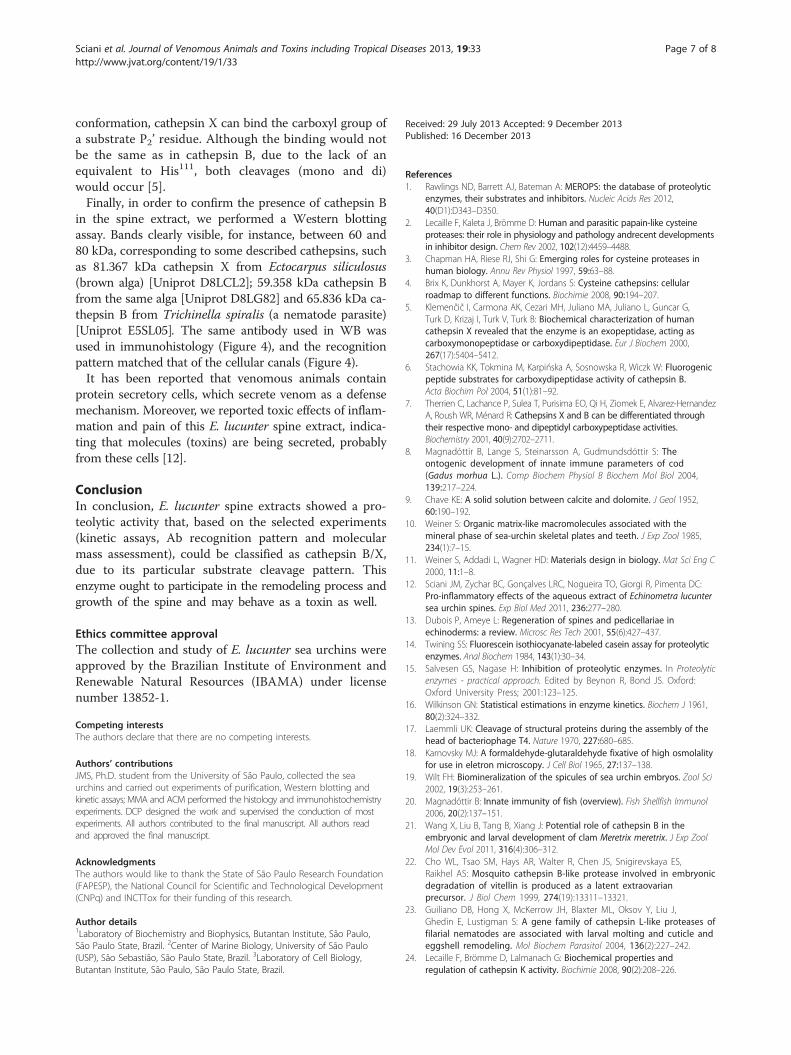
the spine (Figure 4 – A) show that the spaces within thecalcified matrix are walled by a continuous filamentousstructure forming a framework for longitudinal inter-connected compartments (or canals) containing severaldifferent types of cells, some of them full of cytoplasmicgranules. These cells seem to be proportionally moreconcentrated at the spine tip (data not shown).Immunohistochemical test for anti-cathepsin B anti-
body was performed in transversal spine sections. It ispossible to observe the positive (brownish staining)along the decalcified matrix (Figure 4 – B), in the samelocation where cells were observed in the section,stained by toluidine-fuchsin (Figure 4 – A).
DiscussionThe regeneration of the calcified matrix is a knownprocess that has been described for some groups (reviewby Dubois and Ameye [13]). However, this event stillremains unclear at certain points. Spine regeneration is apositive constitutive process taking place in E. lucunter asdepicted in Figure 1. So, we chose to biochemically inves-tigate this process. Spine tip regeneration must be veryimportant for this animal, since its defense relies mainlyon the mechanical trauma inflicted by the spines, which isassociated with the inflammatory reaction caused by themolecules present in the spine [12].A known molecule, frequently associated to matrix
remodeling processes, is the proteolytic enzyme ca-thepsin, particularly the cysteine peptidase cathepsins.The involvement of these proteins in digestion of yolkproteins in oocytes, fertilized eggs and the yolk sachave already been described [20]. Wang et al. [21] veri-fied the participation of a cathepsin B in the embryonicand larval development of Meretrix meretrix. The activ-ity of the enzyme altered significantly the shell length of
the animals, which was shorter when the enzyme wasinhibited.In cods, such enzymes are primarily involved in the
proteolytic digestion of the egg vitellogenin but mayhave an additional defensive role, as has been demon-strated, for example, in the mucus of adult fish [8]. Inthe yellow-fever mosquito (Aedes aegypti), for example,vitellogenic cathepsin B-like protease participates inembryonic degradation of vitellin [22]. In nematodes,this family of enzymes has a role in molting and incuticle and eggshell remodeling, involving proteasesfor the degradation of cuticular proteins. Additionally,proteases may also be involved in the processing of pro-proteins that are subsequently incorporated into thenew cuticle [23]. Moreover, its mammal relative, cathep-sin K, has been associated to osteoclast in bone remo-deling [24].In this work, we report a cysteine peptidase activity that
was detected in E. lucunter aqueous spine extract. Thiscysteine peptidase cathepsin activity, seemly unique (e.g.,due to one single enzyme) was termed as cathepsin B/X,for it presents both carboxi mono- and dipeptidyl pepti-dase activities, and virtually no endopeptidase activity[25]. Moreover, complementary assays (cleavage pattern ofspecific substrates, DTT activation, active site E-64 in-hibition, pH dependence of the activity, molecular massevaluation and Ab recognition pattern) corroborate suchstatement. We used the commercial specific FRET sub-strate Abz-GIVRAK(Dnp)-OH (Aminotech P&D Ltda.,Brazil) and obtained kinetic parameters (KM, kcat) verysimilar to those described for cathepsin B, X and cruzain[26]. Its C-terminal blocked analog, Abz-GIVRAKQ-EDDnp, was virtually resistant to the spine extract, as wellas the FITC-casein, excluding a cathepsin L activity on theaqueous extract.Cotrin et al. [26] verified the cleavage site of cathepsins
B and X with the Abz-GIVRAK(Dnp)-OH: cathepsin Bcleaved 100% after arginine residue and cathepsin Xcleaved 100% after alanine, the two cleavage sites observedwith the spine extract. The KM value observed for cathep-sin B by Cotrin et al. [26] was 5.9 μM, similar for the spineextract, which was calculated 8.57 μM. Other cleavagesobserved by the authors were by cathepsin L, after valine(55%) and after arginine (45%). We did not observedcleavage after valine, so we can discard the presence ofthis type of cathepsin. Moreover, we did not verify anendopeptidase activity, what confirm the absence ofcathepsin L.Although an approximately 6:4 cleavage ratio (carbo-
xydi:carboxymonopeptidyl peptidase activity) could be ob-served for Abz-GIVRAK(Dnp)-OH after incubation withthe aqueous spine extract, this fact could be explained bythe switching catalytic mechanism of cathepsin X. It isbased on structural data: the crystal structure of cathepsin
0 5 10 15 20 250
200
400
600
800
0
1
2
3
4
5
min
A22
0
V, A
UF
/s
kDa M SE
kDa
10575
50
5 µg 10 µg 20µg
97
55
21
30
160
A B C
Figure 3 Proteic characterization of the spine extract. (A) SDS-PAGE (10%) of aqueous extract of spine. M =molecular mass standard,SE = spine extract. Arrows indicate the molecular masses. (B) Western blotting of 5, 10 and 20 μg aqueous extract of spine, incubated withanti-cathepsin B antibody. Left lane, molecular mass standard. (C) Chromatogram obtained by gel filtration of spine aqueous extract. Dashed linesindicate the enzymatic activity.
Sciani et al. Journal of Venomous Animals and Toxins including Tropical Diseases 2013, 19:33 Page 6 of 8http://www.jvat.org/content/19/1/33
X suggests that the positively charged imidazolium ring ofHis23 can switch between two conformations. In the con-formation observed in the crystal structure, this ring canbind to the carboxyl group of asubstrate P1’ residue, whichcan form an additional hydrogen bond with the hydrogen
Figure 4 Immunohistochemical test for anti-cathepsin B antibody wawith toluidin-fuchsin. (B) Spine section incubated with anti-cathepsin B. (Cpossible to observe the positive (brownish staining) along the decalcified msections stained by toluidine-fuchsin (A).
of NE1 atom of Trp202. On the other hand, a modelingstudy has suggested that the ring of His23 can be broughtinto the position equivalent to the position of His110 incathepsin B structure by simple rotations about the χ1 andχ2 angles. With the His23 ring in the cathepsin B-like
s performed in transversal spine sections. (A) Spine section stainedand D) zoomed images, corresponding to A and B, respectively. It isatrix (B), in the same location where cells were observed in the
Sciani et al. Journal of Venomous Animals and Toxins including Tropical Diseases 2013, 19:33 Page 7 of 8http://www.jvat.org/content/19/1/33
conformation, cathepsin X can bind the carboxyl group ofa substrate P2’ residue. Although the binding would notbe the same as in cathepsin B, due to the lack of anequivalent to His111, both cleavages (mono and di)would occur [5].Finally, in order to confirm the presence of cathepsin B
in the spine extract, we performed a Western blottingassay. Bands clearly visible, for instance, between 60 and80 kDa, corresponding to some described cathepsins, suchas 81.367 kDa cathepsin X from Ectocarpus siliculosus(brown alga) [Uniprot D8LCL2]; 59.358 kDa cathepsin Bfrom the same alga [Uniprot D8LG82] and 65.836 kDa ca-thepsin B from Trichinella spiralis (a nematode parasite)[Uniprot E5SL05]. The same antibody used in WB wasused in immunohistology (Figure 4), and the recognitionpattern matched that of the cellular canals (Figure 4).It has been reported that venomous animals contain
protein secretory cells, which secrete venom as a defensemechanism. Moreover, we reported toxic effects of inflam-mation and pain of this E. lucunter spine extract, indica-ting that molecules (toxins) are being secreted, probablyfrom these cells [12].
ConclusionIn conclusion, E. lucunter spine extracts showed a pro-teolytic activity that, based on the selected experiments(kinetic assays, Ab recognition pattern and molecularmass assessment), could be classified as cathepsin B/X,due to its particular substrate cleavage pattern. Thisenzyme ought to participate in the remodeling process andgrowth of the spine and may behave as a toxin as well.
Ethics committee approvalThe collection and study of E. lucunter sea urchins wereapproved by the Brazilian Institute of Environment andRenewable Natural Resources (IBAMA) under licensenumber 13852-1.
Competing interestsThe authors declare that there are no competing interests.
Authors’ contributionsJMS, Ph.D. student from the University of São Paulo, collected the seaurchins and carried out experiments of purification, Western blotting andkinetic assays; MMA and ACM performed the histology and immunohistochemistryexperiments. DCP designed the work and supervised the conduction of mostexperiments. All authors contributed to the final manuscript. All authors readand approved the final manuscript.
AcknowledgmentsThe authors would like to thank the State of São Paulo Research Foundation(FAPESP), the National Council for Scientific and Technological Development(CNPq) and INCTTox for their funding of this research.
Author details1Laboratory of Biochemistry and Biophysics, Butantan Institute, São Paulo,São Paulo State, Brazil. 2Center of Marine Biology, University of São Paulo(USP), São Sebastião, São Paulo State, Brazil. 3Laboratory of Cell Biology,Butantan Institute, São Paulo, São Paulo State, Brazil.
Received: 29 July 2013 Accepted: 9 December 2013Published: 16 December 2013
References1. Rawlings ND, Barrett AJ, Bateman A: MEROPS: the database of proteolytic
enzymes, their substrates and inhibitors. Nucleic Acids Res 2012,40(D1):D343–D350.
2. Lecaille F, Kaleta J, Brömme D: Human and parasitic papain-like cysteineproteases: their role in physiology and pathology andrecent developmentsin inhibitor design. Chem Rev 2002, 102(12):4459–4488.
3. Chapman HA, Riese RJ, Shi G: Emerging roles for cysteine proteases inhuman biology. Annu Rev Physiol 1997, 59:63–88.
4. Brix K, Dunkhorst A, Mayer K, Jordans S: Cysteine cathepsins: cellularroadmap to different functions. Biochimie 2008, 90:194–207.
5. Klemenčič I, Carmona AK, Cezari MH, Juliano MA, Juliano L, Guncar G,Turk D, Krizaj I, Turk V, Turk B: Biochemical characterization of humancathepsin X revealed that the enzyme is an exopeptidase, acting ascarboxymonopeptidase or carboxydipeptidase. Eur J Biochem 2000,267(17):5404–5412.
6. Stachowia KK, Tokmina M, Karpińska A, Sosnowska R, Wiczk W: Fluorogenicpeptide substrates for carboxydipeptidase activity of cathepsin B.Acta Biochim Pol 2004, 51(1):81–92.
7. Therrien C, Lachance P, Sulea T, Purisima EO, Qi H, Ziomek E, Alvarez-HernandezA, Roush WR, Ménard R: Cathepsins X and B can be differentiated throughtheir respective mono- and dipeptidyl carboxypeptidase activities.Biochemistry 2001, 40(9):2702–2711.
8. Magnadóttir B, Lange S, Steinarsson A, Gudmundsdóttir S: Theontogenic development of innate immune parameters of cod(Gadus morhua L.). Comp Biochem Physiol B Biochem Mol Biol 2004,139:217–224.
9. Chave KE: A solid solution between calcite and dolomite. J Geol 1952,60:190–192.
10. Weiner S: Organic matrix-like macromolecules associated with themineral phase of sea-urchin skeletal plates and teeth. J Exp Zool 1985,234(1):7–15.
11. Weiner S, Addadi L, Wagner HD: Materials design in biology. Mat Sci Eng C2000, 11:1–8.
12. Sciani JM, Zychar BC, Gonçalves LRC, Nogueira TO, Giorgi R, Pimenta DC:Pro-inflammatory effects of the aqueous extract of Echinometra lucuntersea urchin spines. Exp Biol Med 2011, 236:277–280.
13. Dubois P, Ameye L: Regeneration of spines and pedicellariae inechinoderms: a review. Microsc Res Tech 2001, 55(6):427–437.
15. Salvesen GS, Nagase H: Inhibition of proteolytic enzymes. In Proteolyticenzymes - practical approach. Edited by Beynon R, Bond JS. Oxford:Oxford University Press; 2001:123–125.
17. Laemmli UK: Cleavage of structural proteins during the assembly of thehead of bacteriophage T4. Nature 1970, 227:680–685.
18. Karnovsky MJ: A formaldehyde-glutaraldehyde fixative of high osmolalityfor use in eletron microscopy. J Cell Biol 1965, 27:137–138.
19. Wilt FH: Biomineralization of the spicules of sea urchin embryos. Zool Sci2002, 19(3):253–261.
20. Magnadóttir B: Innate immunity of fish (overview). Fish Shellfish Immunol2006, 20(2):137–151.
21. Wang X, Liu B, Tang B, Xiang J: Potential role of cathepsin B in theembryonic and larval development of clam Meretrix meretrix. J Exp ZoolMol Dev Evol 2011, 316(4):306–312.
22. Cho WL, Tsao SM, Hays AR, Walter R, Chen JS, Snigirevskaya ES,Raikhel AS: Mosquito cathepsin B-like protease involved in embryonicdegradation of vitellin is produced as a latent extraovarianprecursor. J Biol Chem 1999, 274(19):13311–13321.
23. Guiliano DB, Hong X, McKerrow JH, Blaxter ML, Oksov Y, Liu J,Ghedin E, Lustigman S: A gene family of cathepsin L-like proteases offilarial nematodes are associated with larval molting and cuticle andeggshell remodeling. Mol Biochem Parasitol 2004, 136(2):227–242.
24. Lecaille F, Brömme D, Lalmanach G: Biochemical properties andregulation of cathepsin K activity. Biochimie 2008, 90(2):208–226.
Sciani et al. Journal of Venomous Animals and Toxins including Tropical Diseases 2013, 19:33 Page 8 of 8http://www.jvat.org/content/19/1/33
25. Sciani JM, Antoniazzi MM, Jared C, Pimenta DC: Cathepsin B/X is secretedby Echinometra lucunter sea urchin spines, a structure rich in granularcells and toxins. Toxicon 2012, 60(2):151–152.
26. Cotrin SS, Puzer L, de Souza Judice WA, Juliano L, Carmona AK, Juliano MA:Positional-scanning combinatorial libraries of fluorescence resonanceenergy transfer peptides to define substrate specificity ofcarboxydipeptidases: assays with human cathepsin B. Anal Biochem 2004,335(2):244–252.
doi:10.1186/1678-9199-19-33Cite this article as: Sciani et al.: Cathepsin B/X is secreted byEchinometra lucunter sea urchin spines, a structure rich in granular cellsand toxins. Journal of Venomous Animals and Toxins including TropicalDiseases 2013 19:33.
Submit your next manuscript to BioMed Centraland take full advantage of:
• Convenient online submission
• Thorough peer review
• No space constraints or color figure charges
• Immediate publication on acceptance
• Inclusion in PubMed, CAS, Scopus and Google Scholar
• Research which is freely available for redistribution
Submit your manuscript at www.biomedcentral.com/submit