Detection of ascaridoid nematode parasitesin the important marine food-fish Congermyriaster (Brevoort) (Anguilliformes:Congridae) from the Zhoushan Fishery,ChinaHui-Xia Chen1, Lu-Ping Zhang1, David I. Gibson2, Liang Lü1, Zhen Xu3, Hai-Tao Li3, Hui-Dong Ju1 and Liang Li1*
Abstract
Background: The whitespotted conger Conger myriaster (Brevoort) (Anguilliformes: Congridae) is an extremelymarketable food fish, commonly consumed as sashimi or sushi in some Asian countries (i.e. Japan, Korea andChina). Conger myriaster is also suspected as being an extremely important source of human anisakidosis. However,there is currently very little information on the levels of infection with ascaridoid nematode parasites in thiseconomically important marine fish. The aims of the present study are to determine the species composition,prevalence and mean intensity of ascaridoid parasites of C. myriaster caught in the Zhoushan Fishery.
Results: A total of 1142 third-stage ascaridoid larvae were isolated from 204 C. myriaster. The overall prevalence ofinfection was 100% (mean intensity 5.6). Nine species of such larvae were accurately identified using integrativetaxonomic techniques involving both morphological and genetic data; these included Anisakis pegreffii, A. typicaand A. simplex (sensu stricto) × A. pegreffii, Hysterothylacium fabri, H. aduncum, H. sinense, H. amoyense, H.zhoushanense and Raphidascaris lophii. Although high levels of infection and species richness were revealed in C.myriaster, most of the ascaridoid parasites (1135 individuals) were collected from the body cavity and visceralorgans of the fish and only seven individuals of A. pegreffii were found in the musculature.
Conclusions: This study represents the first report C. myriaster from the Zhoushan Fishery being heavily infectedwith third-stage ascaridoid larvae. Among the ascaridoid larvae parasitic in this fish, an important etiological agentof human anisakidosis, A. pegreffii (L3), represents the predominant species. The genus Hysterothylacium has thehighest species richness, with H. fabri (L3) being the most prevalent species. This high level of infection of A.pegreffii (L3) in C. myriaster suggests a high risk of anisakidosis or associated allergies for people consuming raw orpoorly cooked fish originating from this marine area. These findings provide important basic information on theoccurrence and infection parameters of ascaridoid nematodes in this economically important marine fish. They alsohave significant implications for the prevention and control of human anisakidosis when conger eels from theZhoushan Fishery are consumed.
Keywords: Nematode, Anisakidosis, Ascaridoidea, Conger myriaster, Zhoushan Fishery, East China Sea
* Correspondence: [email protected] Laboratory of Animal Physiology, Biochemistry and Molecular Biology ofHebei Province, College of Life Sciences, Hebei Normal University, 050024Shijiazhuang, Hebei Province, People’s Republic of ChinaFull list of author information is available at the end of the article
BackgroundAnisakidosis (anisakiasis) is a zoonotic disease well-recognized by the seafood industry [1–5]. Humans be-come infected by the accidental ingestion of raw orundercooked fish flesh contaminated by ascaridoid lar-vae, especially anisakids [5–8]. Most cases of human ani-sakidosis are caused by Anisakis simplex (sensu stricto),A. pegreffii and Pseudoterranova decipiens, but species ofHysterothylacium and Contracaecum have also been im-plicated [7, 9–12]. During the past three decades, morethan 20,000 cases of human anisakidosis have been re-ported globally, and over 90% of cases are from Japan[6]. Although only one case of human anisakidosis hasbeen reported in mainland China [13], due to the spreadof exotic foods (sushi, sashimi, etc.) and the growingconsumption of raw or undercooked seafood in main-land China, more attention needs to be paid to thisdisease.The whitespotted conger Conger myriaster (Brevoort)
(Anguilliformes: Congridae) has been considered one of
the most common and marketable food-fishes in someAsian countries (i.e. Japan, Korea and China), where it isfavoured for consumption raw as sashimi or sushi [14–17]. This fish is mainly distributed between the EastChina Sea and the waters of Korea and Japan [18]. TheZhoushan Fishery off the coast of China is thought to bethe most important fishing ground worldwide for C.myriaster [19, 20]. However, there is currently very littleinformation on the levels of infection with ascaridoidparasites in this economically important marine fish.Therefore, the aims of the present study are to deter-mine the species composition, prevalence and mean in-tensity of ascaridoid parasites in C. myriaster caught inthe Zhoushan Fishery.
MethodsParasite collectionA total of 204 Conger myriaster (Brevoort) (Anguilli-formes: Congridae), with a total length (TL) rangingfrom 25.0–65.0 cm, was dissected, and the body cavity
Fig. 1 Conger myriaster (Brevoort) (Anguilliformes: Congridae) caught in the Zhoushan Fishery, China, heavily infected with ascaridoid nematodeparasites. a Fish host. b, d, e Large numbers of ascaridoid nematodes present in the visceral organs. c Ascaridoid nematode present inthe muscles
Chen et al. Parasites & Vectors (2018) 11:274 Page 2 of 12
and visceral organs (i.e. digestive tract, mesentery, liverand gonads) were examined for nematode parasites (Fig.1). The musculature of 54 of these fish (26.5%) (TL 25.0–50.0 cm) was sliced into thin slivers (1.0–2.0 mmthick), and then visually inspected for parasites underwhite light. All of the fish were caught by commercialtrawlers in the Zhoushan Fishery (29°30'–31°00'N, 121°30'–125°00'E) in the South China Sea off China. Thenematodes isolated were washed in physiological saline,then fixed and stored in 80% ethanol until studied.
Morphological identificationThe morphology of the nematode larvae was observedusing light and scanning electron microscopy. For scan-ning electron microscopy, specimens were prepared ac-cording to the methods used in previous studies [21, 22].The following morphological characters were used forthe identification of different morphotypes of larvalnematodes as in previous studies [22–27], including theposition of the excretory pore, the absence and presenceof an intestinal caecum and a ventricular appendix andtheir relative lengths, and the morphology of the ventric-ulus and tail tip.
Molecular identificationFor larval morphotypes with large numbers of individuals,the polymerase chain reaction followed by restriction frag-ment length polymorphism (PCR-RFLP) analysis and tar-geted sequencing of the internal transcribed spacer (ITS1-5.8S-ITS2) region of the ribosomal DNA (rDNA) wereused for genetic identification. The Column GenomicDNA Isolation Kit (Shanghai Sangon, China) wasemployed to extract the genomic DNA of each worm ac-cording to the manufacturer’s instructions. The ITS1-5.8S-ITS2 region was amplified by PCR using the primersNC5 (5'-GTA GGT GAA CCT GCG GAA GGA TCA T-3') and NC2 (5'-TTA GTT TCT TTT CCT CCG CT-3')[28] under the cycling conditions described previously[25]. PCR products were checked on GoldView-stained 1.5% agarose gel and purified by the Column PCR ProductPurification Kit (Shanghai Sangon, China). PCR-RFLPanalysis was performed independently using two restric-tion enzymes HinfI and HhaI (Thermo Scientific, Wal-tham, MA, USA) according to a previous study [29]. ThePCR products were digested according to the manufactur-er's recommendations. The digested samples were sub-jected to electrophoresis on 2% agarose gels and thenphotographed. Representative samples per distinct RFLPprofile set were selected for the sequencing of the ITS1-5.8S-ITS2 region. Sequencing was carried out using a Dye-DeoxyTerminator Cycle Sequencing Kit (v.2, Applied Bio-systems, California, USA) and an automated sequencer(ABI-PRISM 377). Sequencing for each sample was car-ried out for both strands. Sequences were aligned using
ClustalW2 and adjusted manually. The ITS1-5.8S-ITS2sequences determined were compared (using the algo-rithm BLASTn) with those available in the National Cen-ter for Biotechnology Information (NCBI) database(http://www.ncbi.nlm.nih.gov). For larval morphotypeswith small numbers of individuals, the directly targeted se-quencing of the ITS1-5.8S-ITS2 region was used for fur-ther genetic identification of species with the sameprimers and methods mentioned above.
Phylogenetic analysisPhylogenetic analyses of the ITS1-5.8S-ITS2 sequencedata obtained herein were undertaken for bothNeighbour-Joining (NJ) and Maximum likelihood (ML)methods using MEGA 6 [30]. We used a built-in func-tion in MEGA 6 [30] to select a best-fitting substitutionmodel for the sequences using the Bayesian informationcriterion [31]. The Kimura two-parameter model of nu-cleotide substitution was identified as optimal. Ascarislumbricoides was chosen as the outgroup. Reliabilitiesfor both NJ and ML trees were tested using 1000 boot-strap replications [32] and nodes with bootstrap valuesexceeding 70 were considered well supported [33].
ResultsMorphological identificationA total of 1142 third-stage ascaridoid larvae were iso-lated from C. myriaster. Based on morphological charac-ters (Figs. 2, 3; Table 1), we found that these larvaerepresented six different morphotypes belonging to threedifferent genera: Anisakis, Hysterothylacium and Raphi-dascaris. Among these larvae, 630 individuals were iden-tified morphologically as Anisakis type I of Berland(1961) [23]. Four different morphotypes of Hysterothyla-cium spp. larvae were distinguished, including the Hys-terothylacium larval type of Smith (1983) [24] with 28individuals, Hysterothylacium larval type of Guo et al.(2014) [27] with 23 individuals, Hysterothylacium larvaltype of Li et al. (2012) [25] with 11 individuals and Hys-terothylacium larval type IV of Shamsi et al. (2013) [26]with 447 individuals. Only three third-stage larvae ofRaphidascaris, identified morphologically as Raphidas-caris larval type of Zhao et al. (2016) [22], were found inthe present study (see Table 2 for details).
Molecular identificationAll the Anisakis samples were further identified by PCR-RFLP analysis (Table 2). Digestion of the PCR productsusing HhaI produced two different RFLP profiles, i.e.628 samples with two bands (c.550 and 430 bp) and onlytwo samples with four bands (c.320, 240, 180 and 160bp). Digestion with HinfI yielded three different RFLPprofiles, 621 samples with three bands (c.370, 330 and250 bp), only two samples with two bands (c.620 and
Chen et al. Parasites & Vectors (2018) 11:274 Page 3 of 12
350 bp) and seven samples with four bands (c.620, 370,300 and 250 bp). According to the molecular taxonomickey based on the PCR-RFLP patterns of Anisakis speciesobtained by the digestion of ITS amplicons with endo-nucleases HhaI or HinfI [29], we assigned these Anisakisthird-stage larvae to A. pegreffii, A. typica and A. simplex(sensu stricto) × A. pegreffii (a recombinant genotype).The ITS region was sequenced for 43 randomly selectedindividuals of A. pegreffii, two individuals of A. typicaand seven individuals of A. simplex (sensu stricto) × A.pegreffii identified by the PCR-RFLP analysis (Table 2).There was no nucleotide variation detected among the43 ITS sequences of A. pegreffii (MF539758-MF539767),two ITS sequences of A. typica (MF539771, MF539775)and seven ITS sequences of A. simplex (sensu stricto) ×A. pegreffii (MF539768-MF539770, MF539772-MF539774, MF539776). Pairwise comparison betweenour genetic data and the ITS sequences of Anisakis spp.registered in GenBank proved 100% identical to A.pegreffii (AY821738, AY821740, AY821745, JQ934867,JQ934871, JQ900763, JQ934869, KP301519, KF032066,KJ011486, EU624343), A. typica (EU346093, KC928262,KF356670-KF356671, JX523715, JN968930) and A. sim-plex (sensu stricto) × A. pegreffii (AB894874).
For the further identification of Hysterothylacium spp.third-stage larvae, the ITS region was sequenced for 45randomly selected individuals of Hysterothylacium larvaltype IV of Shamsi et al. (2013) [26] and all of the individ-uals of the other three larval morphotypes (Table 2). Fourdifferent genotypes (MF539793, MF539794, MF539796,MF539790) were detected among the 45 ITS sequences ofHysterothylacium larval type IV of Shamsi et al. (2013)[26] (MF539787-MF539796) obtained herein, which dis-played 0–0.2% nucleotide variability. Pairwise compari-sons between the present data and the ITS sequences ofHysterothylacium spp. registered in GenBank showed 0–0.3% nucleotide differences with H. fabri (KC852206,JQ520158, JX974558, KF736939-KF736944). Thus, weconsidered that the present nematode larvae, referred toas Hysterothylacium larval type IV of Shamsi et al. (2013)[26], belong to H. fabri. No nucleotide variability was de-tected among the 23 ITS sequences of Hysterothylaciumlarval type HL of Guo et al. (2014) [27] (MF539797-MF539806). Pairwise comparisons between the presentdata and the ITS sequences of Hysterothylacium spp. reg-istered in GenBank showed them to be 100% identicalwith H. sinense (KX817293-KX817295, KX110078,KX084795). Thus, we confirmed that the nematode larvae
Fig. 2 Anterior and posterior extremities of ascaridoid larval morphotypes isolated from Conger myriaster (Brevoort) (Anguilliformes: Congridae)caught in the Zhoushan Fishery, China (excretory pore arrowed). a, b, Anisakis type I of Berland (1961) [23]. c, d, Hysterothylacium larval type ofSmith (1983) [24]. e, f Hysterothylacium larval type IV of Shamsi et al. (2013) [26]. g, h Hysterothylacium larval type of Guo et al. (2014) [27]. i, jHysterothylacium larval type of Li et al. (2012) [25]. k, l Raphidascaris larval type of Zhao et al. (2016) [22]
Chen et al. Parasites & Vectors (2018) 11:274 Page 4 of 12
referred to as Hysterothylacium larval type HL of Guo etal. (2014) [27] belong to H. sinense. Only one genotypewas detected among the 28 ITS sequences of specimensreferred to as Hysterothylacium larval type of Smith(1983) [24] (MF539777-MF539786). Pairwise comparisonsbetween the present data and the ITS sequences of Hyster-othylacium spp. registered in GenBank showed 0–0.5%nucleotide differences with H. aduncum (KF736937,HM437225, KP318743-KP318782, KP276150, KT852542).Thus, we considered that the specimens referred to as Hys-terothylacium larval type of Smith (1983) [24] represent H.aduncum. There were seven different genotypes detectedamong the 11 ITS sequences of Hysterothylacium larvaltype of Li et al. (2012) [25] (MF539807-MF539813,MF539814-MF539817) obtained herein, which displayed0–1.7% nucleotide variability (Table 3). Pairwise compari-sons between the present data and the ITS sequences of
Hysterothylacium spp. registered in GenBank showed thatseven individuals (MF539807-MF539813) exhibited 0–0.3% nucleotide differences with H. amoyense (KP252130-KP252133, EU828749), and four individuals (MF539814-MF539817) exhibited 0–1.4% nucleotide differences withH. zhoushanense (KP326556, KP326549-KP326551,JX028277-JX028282). We considered, therefore, that the11 individuals of Hysterothylacium larval type of Li et al.(2012) [25] represent two different species, H. zhousha-nense and H. amoyense, with 0.8–1.7% nucleotide variationin the ITS region between these species. Only three third-stage larvae of Raphidascaris, identified morphologically asRaphidascaris larval type of Zhao et al. (2016) [22], werefound. The ITS region was directly sequenced for all ofthese Raphidascaris larvae (Table 2) and the three ITS se-quences (MF539818-MF539820) were identical. Compari-son of our genetic data with the ITS sequences of
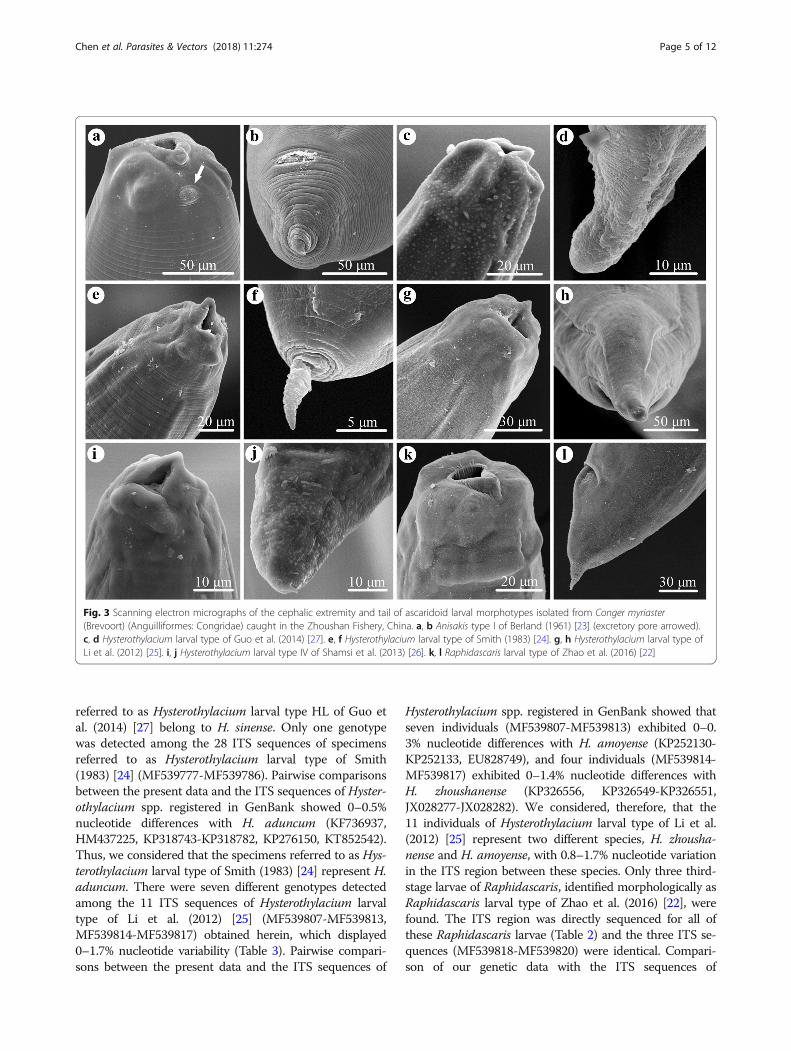
Fig. 3 Scanning electron micrographs of the cephalic extremity and tail of ascaridoid larval morphotypes isolated from Conger myriaster(Brevoort) (Anguilliformes: Congridae) caught in the Zhoushan Fishery, China. a, b Anisakis type I of Berland (1961) [23] (excretory pore arrowed).c, d Hysterothylacium larval type of Guo et al. (2014) [27]. e, f Hysterothylacium larval type of Smith (1983) [24]. g, h Hysterothylacium larval type ofLi et al. (2012) [25]. i, j Hysterothylacium larval type IV of Shamsi et al. (2013) [26]. k, l Raphidascaris larval type of Zhao et al. (2016) [22]
Chen et al. Parasites & Vectors (2018) 11:274 Page 5 of 12
Raphidascaris spp. registered in GenBank showed them tobe 100% identical with R. lophii (KP262039, KP326520-KP326531, KP326533-KP326538, KP419720). Conse-quently, we considered these Raphidascaris larvae to beconspecific with R. lophii. All of the ITS sequences of thelarval nematode parasites obtained herein are deposited inthe GenBank database under the accession numbers(MF539758-MF539820).
Phylogenetic analysisOur results revealed that the ascaridoid nematodes se-lected for phylogenetic analysis were divided into twodistinct clades (families), the Anisakidae (which includesspecies of Anisakis, Pseudoterranova and Contracaecum)and the Raphidascarididae (which includes species ofHysterothylacium and Raphidascaris) with strong sup-port (Fig. 4). In Anisakis, A. simplex (JX535521), A.
Table 1 Morphometric data for ascaridoid larval morphotypes isolated from Conger myriaster (Brevoort) (Anguilliformes: Congridae)caught in the Zhoushan Fishery, China (measurements in mm)
Abbreviations: BL body length, OL oesophagus length, VL ventriculus length, VW ventriculus width, ICL intestinal caecum length, VAL ventricular appendix length,EC distance from excretory pore to cephalic end, TL tail length, ATB Anisakis type I of Berland (1961) [23], HTS Hysterothylacium larval type of Smith (1983) [24],HTG Hysterothylacium larval type of Guo et al. (2014) [27], HTL Hysterothylacium larval type of Li et al. (2012) [25], HTIV Hysterothylacium larval type IV of Shamsi etal. (2013) [26], RTZ Raphidascaris larval type of Zhao et al. (2016) [22]
Table 2 Infection data of ascaridoid nematode larvae isolated from Conger myriaster (Brevoort) (Anguilliformes: Congridae) caught inthe Zhoushan Fishery, China, and samples selected for molecular analysis
aRandomly selectedAbbreviations: ATB Anisakis type I of Berland (1961) [23], HTS Hysterothylacium larval type of Smith (1983) [24], HTG Hysterothylacium larval type of Guo et al. (2014)[27], HTL Hysterothylacium larval type of Li et al. (2012) [25], HTIV Hysterothylacium larval type IV of Shamsi et al. (2013) [26], RTZ Raphidascaris larval type of Zhaoet al. (2016) [22]
Chen et al. Parasites & Vectors (2018) 11:274 Page 6 of 12
Table 3 Sequence polymorphisms (highlighted in bold) revealed at alignment positions of the ITS region among the differentindividuals of Hysterothylacium larval type of Li et al. (2012) [25] obtained in the present study
Fig. 4 Maximum likelihood (ML) and Neighbour-Joining (NJ) trees showing the phylogenetic relationships of ascaridoid nematode species detected inthe present study (shown in colour). Ascaris lumbricoides was chosen as the outgroup
Chen et al. Parasites & Vectors (2018) 11:274 Page 7 of 12
pegreffii (MF539758, KX110076), A. simplex (sensustricto) × A. pegreffii (MF539768, AB894874), A. ziphi-darum (JQ912691), A. nascettii (JQ912692) and A.typica (MF539771, JQ912690) grouped together, repre-senting Anisakis type I of Berland (1961) [23], and A.brevispiculata (JQ912694), A. paggiae (JQ912695) andA. physeteris (JQ912693) formed a group, representingAnisakis type II. The results showed that A. pegreffii, A.simplex and A. simplex (sensu stricto) × A. pegreffii ex-hibit a very close relationship and that A. typica was thebasal branch in the Anisakis type I group. In Hysterothy-lacium, the four different genotypes of H. fabri(MF539793, MF539794, MF539796, MF539790) ob-tained herein and the previously reported ITS sequenceof H. fabri (JQ520158) clustered together and as a sister-group to H. reliquens (KX786293). Hysterothylaciumsinense (MF539798, KX817295) formed a sister relation-ship with H. liparis (KF601896). Hysterothylacium adun-cum (MF539777, KX110074) and H. auctum (AF115571)also exhibited a close relationship (Fig. 4). The seven dif-ferent genotypes of Hysterothylacium larval type of Li etal. (2012) [25] obtained herein were divided into twoclades: three genotypes (MF539808, MF539809,MF539811) clustered with the previously reported ITSdata for H. amoyense (KP252133) and four genotypes(MF539814-MF539817) grouped together with a previ-ously reported ITS sequence of H. zhoushanense(KP326551) (Fig. 4). Raphidascaris lophii (MF539818)was sister to R. trichiuri (FJ009682) with high branchsupport scores (Fig. 4).
Infection levelsThe overall prevalence of infection for ascaridoid larvaein C. myriaster was 100% (mean intensity 5.6 worms/fish). Anisakis pegreffii (L3) was the predominant spe-cies. The prevalence and mean intensity of A. typica andA. simplex (sensu stricto) × A. pegreffii was distinctlylower than that of A. pegreffii. Hysterothylacium fabri(L3) represented the most prevalent species among theHysterothylacium larvae collected from C. myriaster.The infection parameters of the other four species ofHysterothylacium and R. lophii are presented in Table 2.Moreover, in the 54 fish whose muscles were sliced intothin slivers, only seven Anisakis larvae, identified genet-ically as A. pegreffii, were found in the musculature, witha prevalence of 2.5% and a mean intensity of 1.0.
DiscussionConger myriaster represents one of the most valuablefishery resources in some Asian countries (i.e. Japan,Korea and China) [14–17], but it has also been consid-ered an extremely important agent of anisakidosis inKorea [15, 34]. However, our present knowledge of thespecies composition, prevalence and mean intensity of
ascaridoid parasites in C. myriaster remains very limited.To date, only one preliminary investigation of the occur-rence of ascaridoid larvae in this fish has been under-taken; it was carried out in 1992 and based onspecimens purchased from a fish market in Seoul [34].The study showed that the overall prevalence of infec-tion was 57.7% (of the only 26 fish investigated) [34] anddistinctly lower than that (100%) found in C. myriastercollected from the Zhoushan Fishery in Chinese waters.However, the mean intensity (90.1) in the previous studyis much higher than the value in our investigation (meanintensity only 5.6). The different infection parameters ofascaridoid larvae in C. myriaster caught in Korean andChinese waters may be a result of the different geo-graphical locations or the sampling time (July to Augustin 1990 vs October in 2010 and 2016 and April in 2012and 2013). In addition, the species composition of ascar-idoid larvae detected from C. myriaster caught in Ko-rean and Chinese waters is similar. These larval types, i.e. Anisakis type I of Berland (1961) [23], Hysterothyla-cium larval type of Smith (1983) [24], Hysterothylaciumlarval type IV of Shamsi et al. (2013) [26] and Hyster-othylacium larval type of Guo et al. (2014) [27], werefound in both our study and the previous one [34], butRaphidascaris larval type of Zhao et al. (2016) [22] isonly reported in our study. Unfortunately, in the previ-ous study these larvae were not accurately determinedto the species level because only morphological methodswere used.The accurate identification of ascaridoid larvae to the
species level is essential for an evaluation of the molecu-lar epidemiology of the disease [21, 22, 35]. Recently, thecombination of PCR-RFLP analysis and targeted sequen-cing of the ITS region has been widely used for large-scale studies on the identification of ascaridoid larvae tothe species level [11, 29, 35–40]. Consequently, in thepresent study, in order to accurately identify large num-bers of ascaridoid larvae isolated from C. myriaster inthe Zhoushan Fishery, both morphological methods andmolecular approaches, including PCR-RFLP analysisand/or targeted sequencing of the ITS region, wereemployed, and the six ascaridoid larval types classifiedherein were identified genetically as nine species.According to some previous studies [12, 41–45], most
cases of human anisakidosis in Europe and Korea areknown to be caused by A. pegreffii (L3), which is widelydistributed in the South Atlantic, the Mediterranean Sea,Australian and Chinese waters (including the YellowSea, the East China Sea and the Taiwan Strait) [27, 29,35, 36, 40, 46–50]. During the present survey, a highlevel of A. pegreffii infection with an overall prevalenceof 99.0%, was also revealed in C. myriaster, but most ofthe A. pegreffii were found in the body cavity and vis-ceral organs of the fish, which are not eaten by humans,
Chen et al. Parasites & Vectors (2018) 11:274 Page 8 of 12
and only seven third-stage larvae of A. pegreffii were de-tected from the musculature of seven C. myriaster(prevalence 13.0% and mean intensity 1.0). The relativelylow prevalence and intensity of Anisakis specimens de-tected in the musculature of C. myriaster suggest a rela-tively low probability of human infection whenconsuming C. myriaster as sashimi or sushi. However,the post-mortem migration of Anisakis larvae from thefish body cavity and/or visceral organs to the muscula-ture can occur after the death of the fish [7, 51, 52],which could increase the risk of anisakidosis when con-suming raw or undercooked fish. Consequently, we sug-gest the removal of the viscera from C. myriaster assoon after capture as possible, which would be a usefuland practical preventive measure against human anisaki-dosis [15].Our genetic data and phylogenetic analysis indicated
that the third-stage larvae of Anisakis type I of Berland(1961) [23] obtained herein represent three species: A.pegreffii, A. typica and A. simplex (sensu stricto) × A.pegreffii (Fig. 4). However, based only on morphologicalcharacters (i.e. the morphology of the cephalic region, thelength of the oesophagus, and the length and morphologyof the ventriculus and tail), it is almost impossible to dis-tinguish the third-stage larvae of the three species [37, 46].The infection levels of A. typica (L3) and A. simplex (sensustricto) × A. pegreffii (L3) are distinctly lower than that ofA. pegreffii (see Table 2 for details). This situation can bereadily understood if one considers the different distribu-tional patterns of these Anisakis species in Chinese waters.Anisakis typica is mainly distributed in the tropical andsubtropical waters [48] and is considered to be the pre-dominant species in the South China Sea [37], but it hasalso been reported from marine fish species in other Chin-ese waters (i.e. Auxis tapeinosoma and Chelidonichthyskumu in the Yellow Sea, Trichiurus lepturus in the EastChina Sea and Scomber australasicus in the Taiwan Strait)[29, 40, 46]. The recombinant genotype A. simplex (sensustricto) × A. pegreffii is found in both temperate and sub-tropical waters and has frequently been reported fromvarious marine fishes in the Yellow Sea, East China Seaand Taiwan Strait, but all reports indicate low levels ofprevalence and intensity [40, 46, 53]. The present study isthe first record of A. simplex (sensu stricto) × A. pegreffiifrom C. myriaster in the East China Sea. The significanceof A. typica and A. simplex (sensu stricto) × A. pegreffii ascausative agents of human anisakidosis is distinctly lowerthan that of A. pegreffii. To date, only one case of humananisakidosis caused by the recombinant genotype A. sim-plex (sensu stricto) × A. pegreffii has been reported; thiswas from Japan [54]. Anisakis typica has not been con-firmed as a pathogen causing human anisakidosis.Species of Hysterothylacium are common nematode
parasites of marine fishes worldwide [27, 35]. Marine
fishes can act as both the paratenic/intermediate and/orthe definitive hosts of Hysterothylacium spp. [55, 56].However, most of species of this genus are commonlyconsidered as non-pathogenic to humans. So far, onlyone case of human anisakidosis apparently caused by H.aduncum has been reported [9]. Among the ascaridoidlarvae parasitic in C. myriaster, Hysterothylacium has thehighest species richness (five species). Based on the rela-tive length of the intestinal caecum and ventricular ap-pendix, and the morphology of the tail,Hysterothylacium larvae can be readily assigned to fourdifferent morphotypes (Figs. 2, 3, Table 1). However, it isimpractical and problematic to identify these differentlarval morphotypes to the species level using morpho-logical characters alone [25], thus molecular data wereused for the exact identification of species. Hysterothyla-cium fabri (L3) was the most prevalent species amongthe Hysterothylacium spp. larvae obtained herein. Theprevalence of H. fabri (L3) in C. myriaster was similar tothat reported for Liparis tanakae (Gilbert & Burke)(Scorpaeniformes: Liparidae) collected from the Yellowand East China Seas (prevalence 30.0%) [27]. Thepresent study represents the first record of H. fabri (L3)in C. myriaster. Hysterothylacium aduncum is the mostcommon marine ascaridoid parasite, being reportedfrom more than 220 fish species belonging to 70 familiesin 22 orders throughout the world [57]. Recently, someauthors reported H. aduncum (L3) in L. tanakae (preva-lence of 100%, mean intensity 26.7) and Pseudorhombuscinnamoneus (Temminck & Schlegel) (Pleuronecti-formes: Paralichthyidae) (prevalence 81.2%, mean inten-sity 2.7) in Chinese waters [27, 35]. The prevalence andmean intensity of H. aduncum (L3) in these two studieswere considerably higher than the values in the presentstudy (prevalence 11.8%, mean intensity 1.2). Hysterothy-lacium sinense possibly represents a species endemic toChinese waters; its type-host is C. myriaster. The third-stage larvae of H. sinense have been reported from P.cinnamoneus in the Yellow Sea off China with a preva-lence of 100% and mean intensity of 17.4 [35]. In con-trast, we found H. sinense (L3) from C. myriaster in theEast China Sea with a prevalence of only 10.3% and amean intensity of 1.1. Hysterothylacium amoyense (L3)has been reported from several marine fishes, includingScomber japonicus and Trichiurus lepturus in the EastChina Sea, Halieutaea stellata in the South China Sea[21, 39] and Platycephalus indicus (L.) (Scorpaeniformes:Platycephalidae) in the Persian Gulf [58]. In the presentstudy, the prevalence of H. amoyense (L3) (3.4%) in C.myriaster is distinctly lower than reported in the previ-ous investigation of Halieutaea stellata (24.0%) [21]. Inthe case of the third-stage larvae of H. zhoushanense, wefound this species in C. myriaster for the first time, witha prevalence of 2.0% and mean intensity of 1.0. Previously,
Chen et al. Parasites & Vectors (2018) 11:274 Page 9 of 12
it has been reported from Pseudorhombus oligodon (Blee-ker) (Pleuronectiformes: Paralichthyidae) in the EastChina Sea, with a prevalence of 5.3% and mean intensityof 7.0 [25].Our phylogenetic analyses showed that A. pegreffii, A.
simplex (sensu stricto) × A. pegreffii and A. simplex(sensu stricto) have much closer relationships than withA. typica. Consequently, the present results agree withseveral previous phylogenetic studies [22, 35, 37, 40, 47,48]. Some authors have considered that H. zhoushanenseis closely related to H. amoyense, and it is indeed almostimpossible to distinguish the third-stage larvae of thesetwo species based on morphological characters [25].However, adults of H. zhoushanense, which have well-developed lateral alae, can be readily differentiated fromthose of H. amoyense, which lack alae. Our phylogeneticanalysis corroborated H. zhoushanense and H. amoyenseas separate species and confirmed their sister relation-ship (Fig. 4).Conger myriaster is bathydemersal and a voracious
predator. The high levels of nematode infections andspecies richness in this fish host are likely related to itsfeeding habit, which is the main factor affecting parasitecommunity structure [59]. To date, more than 50 speciesof marine fishes, 40 species of crustaceans, 15 species ofmolluscs and four species of annelids have been reportedin its diet [60]. Most of these prey fishes and inverte-brates can act as paratenic or intermediate hosts of theabove-mentioned ascaridoid nematode species. Westrongly recommended two traditional methods for re-ducing the risk of human anisakidosis when C. myriasterfrom the Zhoushan Fishery is consumed, i.e. adequatecooking (60 °C for 1–2 min) or freezing (-10 °C for 24 h)of the fish, both of which should kill anisakids [61].Lastly, the visual inspection of pieces of fish for parasiteswhen preparing sashimi or sushi may be also a goodprophylactic measure against human anisakidosis, be-cause Anisakis larvae can be detected and eliminated ifthe pieces of fish are sliced thinly.
ConclusionsConger myriaster caught in the Zhoushan Fishery washeavily infected with ascaridoid third-stage larvae.Among the ascaridoid larvae parasitic in this fish, theimportant etiological agent of human anisakidosis, A.pegreffii (L3), represented the predominant species. Thishigh infection level of A. pegreffii (L3) in C. myriastersuggests a high risk of anisakidosis or associated allergiesfor people consuming raw or poorly cooked raw fish ori-ginating from this marine area. The genus Hysterothyla-cium had the highest species richness, and H. fabri (L3)was the most prevalent species among the Hysterothyla-cium larvae. The findings of the present study provideimportant basic data on the occurrence and infection
parameters of ascaridoid nematodes in this economicallyimportant marine fish. They also have significant impli-cations for the prevention and control of human anisaki-dosis, which is possible when the flesh of fish caught inthe Zhoushan Fishery off the coast of China is con-sumed raw or inadequately cooked.
AcknowledgementsWe are grateful to Associate Professor Ji-Biao Li (Instrumental Analysis Center,Hebei Normal University) for the technical support with the SEM.
FundingThis study was supported by the National Natural Science Foundation ofChina (NSFC-31572231, NSFC-31750002), Natural Science Foundation ofHebei Province (NSFH-C2016205088) and the Youth Top Talent Support Pro-gram of Hebei Province to LL.
Availability of data and materialsITS sequences of the ascaridoid nematode parasites obtained in this studywere deposited in the GenBank database under the accession numbersMF539758-MF539820. Voucher specimens are deposited in College of LifeSciences, Hebei Normal University, Hebei Province, China, under the acces-sion numbers HBNU-F2017020L-F2017029L.
Authors’ contributionsHXC, LPZ and LL conceived the study, identified and sequenced samples,and analysed the data. HXC, ZX, HTL, HDJ and LL collected the parasitesfrom fish. LLü contributed to the phylogeny. LL and DIG wrote themanuscript. All authors read and approved the final manuscript.
Ethics approvalThis study was conducted according to the Hebei Normal UniversityExperiments in Animals Policy and was approved by the Animal EthicsCommittee of Hebei Normal University as complying with the AnimalProtection Law of the People’s Republic of China.
Competing interestsThe authors declare that they have no competing interests.
Publisher’s NoteSpringer Nature remains neutral with regard to jurisdictional claims inpublished maps and institutional affiliations.
Author details1Key Laboratory of Animal Physiology, Biochemistry and Molecular Biology ofHebei Province, College of Life Sciences, Hebei Normal University, 050024Shijiazhuang, Hebei Province, People’s Republic of China. 2Department of LifeSciences, Natural History Museum, Cromwell Road, London SW7 5BD, UK.3Medical College of Hebei University of Engineering, 056002 Handan, HebeiProvince, People’s Republic of China.
Received: 28 December 2017 Accepted: 16 April 2018
References1. Ahmed FE. Review: Assessing and managing risk due to consumption of
seafood contaminated with micro-organisms, parasites, and natural toxins inthe US. Int J Food Sci Tech. 1992;27:243–60.
2. Butt AA, Aldridge KE, Sanders CV. Infections related to the ingestion ofseafood. Part I: Parasitic infections and food safety. Lancet Infect Dis. 2004;4:294–300.
3. Chai JY, Murrell KD, Lymbery AJ. Fish-borne parasitic zoonoses: status andissues. Int J Parasitol. 2005;35:1233–54.
4. Anantanawat S, Kiermeier A, McLeod C, Sumner J. A semi-quantitative riskassessment of harmful parasites in Australian finfish. South Australia: SouthAustralian Research & Development Institute; 2012. p. 36.
5. Audicana MT, Ansotegui IJ, de Corres LF, Kennedy MW. Anisakis simplex:dangerous - dead and alive? Trends Parasitol. 2002;18:20–5.
Chen et al. Parasites & Vectors (2018) 11:274 Page 10 of 12
6. Hochberg NS, Hamer DH. Anisakidosis: perils of the deep. Clin Infect Dis.2010;51:806–12.
7. Suzuki J, Murata R, Hosaka M, Araki J. Risk factors for human Anisakisinfection and association between the geographic origins of Scomberjaponicus and anisakid nematodes. Int J Food Microbiol. 2010;31:88–93.
8. Shamsi S, Suthar J. A revised method of examining fish for infection withzoonotic nematode larvae. Int J Food Microbiol. 2016;227:13–6.
9. Yagi K, Nagasawa K, Ishikura H, Nakagawa A, Sato N, Kikuchi K, et al. Femaleworm Hysterothylacium aduncum excreted from human: a case report. Jpn JParasitol. 1996;45:12–23.
10. Yu JR, Seo M, Kim YW, Oh MH, Sohn WM. A human case of gastric infectionby Pseudoterranova decipiens larva. Korean J Parasitol. 2001;39:193–6.
11. Umehara A, Kawakami Y, Ooi HK, Uchida A, Ohmae H, Sugiyama H.Molecular identification of Anisakis type I larvae isolated from hairtail fish offthe coasts of Taiwan and Japan. Int J Food Microbiol. 2010;143:161–5.
12. Mattiucci S, Fazii P, De Rosa A, Paoletti M, Megna AS, Glielmo A, et al.Anisakiasis and gastroallergic reactions associated with Anisakis pegreffiiinfection, Italy. Emerg Infect Dis. 2013;19:496–9.
13. Qin Y, Zhao Y, Ren Y, Zheng L, Dai X, Li Y, et al. Anisakiasis in China: the firstclinical case report. Foodborne Pathog Dis. 2013;10:472–4.
14. Kim J-S, Oh K-S, Lee J-S. Comparison of food component between congereel (Conger myriaster) and sea eel (Muraenesox cinereus) as a sliced raw fishmeat. J Korean Fish Soc. 2001;34:678–84.
15. Choi P, Hur JW, Lim JH, Lee JY, Kim DW, Park MI, et al. A case of gastricsubmucosal tumor suspected to be caused by Anisakis. Korean JGastrointest Endosc. 2003;27:26–30.
16. Uchida K, Nishihara M, Shinanba T, Yoshoda T, Miyamoto Y, Kakihara T.Behavioral observations of white spotted conger eel Conger myriaster inbaited traps in Tokyo Bay. Proceedings of the Design Symposium onConservation of Ecosystem (The 12th SEASTAR 2000 Workshop). Kyoto:Kyoto University; 2013. p. 109–14.
17. Park J-Y, Cho H, Kang J-H, Kim E-M, An C-M, Kim J-H, et al. Development ofDNA microarray for species identification of eels (Anguilliformes andMyxiniformes) in Korean fisheries markets. Bio Chip J. 2014;8:310–6.
18. Froese R, Pauly D, editors. FishBase. World Wide Web electronic publication.http://www.fishbase.org, version. 2017;02. Accessed 12 Nov 2017.
19. Tang Y-M, Wu C-W. On biologic habits and resources distribution ofcommon Japanese conger Conger myriaster (Brevoort). J Zhejiang Coll Fish.1988;7:19–26.
20. Yang B-Z, Tang Y-L, Liang Z-L. Selectivity of escape-hole size in tube trapsfor white-spotted conger Conger myriaster. J Ocean Limnol. 2011;29:1041–7.
21. Li L, Zhao W-T, Guo Y-N, Zhang L-P. Nematode parasites infection in thestarry batfish Halieutaea stellata (Vahl) (Lophiiformes: Ogcocephalidae) fromthe East and South China Sea. J Fish Dis. 2016;39:515–29.
22. Zhao W-T, Lü L, Chen H-X, Yang Y, Zhang L-P, Li L. Ascaridoid parasitesinfecting in the frequently consumed marine fishes in the coastal area ofChina: a preliminary investigation. Parasitol Int. 2016;65:87–98.
23. Berland B. Nematodes from some Norwegian marine fishes. Sarsia. 1961;2:1–50.24. Smith JW. Larval Anisakis simplex (Rudolphi, 1809, det. Krabbe, 1878) and
larval Hysterothylacium sp. (Nematoda: Ascaridoidea) in euphausiids(Crustacea: Malacostraca) in the North-East Atlantic and northern North Sea.J Helminthol. 1983;57:167–77.
25. Li L, Liu Y-Y, Zhang L-P. Morphological and genetic characterization ofHysterothylacium zhoushanensis sp. nov. (Ascaridida: Anisakidae) from theflatfish Pseudorhombus oligodon (Bleeker) (Pleuronectiformes:Paralichthyidae) in the East China Sea. Parasitol Res. 2012;111:2393–401.
26. Shamsi S, Gasser RB, Beveridge I. Description and genetic characterisation ofHysterothylacium (Nematoda: Raphidascarididae) larvae parasitic in Australianmarine fishes. Parasitol Int. 2013;62:320–8.
27. Guo Y-N, Xu Z, Zhang L-P, Hu Y-H, Li L. Occurrence of Hysterothylacium andAnisakis nematodes (Ascaridida: Ascaridoidea) in the Tanaka's snailfish Liparistanakae (Gilbert & Burke) (Scorpaeniformes: Liparidae). Parasitol Res. 2014;113:1289–300.
28. Zhu X, Gasser RB, Podolska M, Chilton NB. Characterisation of anisakidnematodes with zoonotic potential by nuclear ribosomal DNA sequences.Int J Parasitol. 1998;28:1911–21.
29. Chen H-Y, Shih H-H. Occurrence and prevalence of fish-borne Anisakislarvae in the spotted mackerel Scomber australasicus from Taiwanesewaters. Acta Trop. 2015;145:61–7.
30. Tamura K, Stecher G, Peterson D, Filipski A, Kumar S. MEGA6: MolecularEvolutionary Genetics Analysis version 6.0. Mol Biol Evol. 2013;30:2725–9.
31. Posada D, Crandall KA. Selecting the best-fit model of nucleotidesubstitution. Syst Biol. 2001;50:580–601.
32. Felsenstein J. Confidence limits on phylogenies: an approach using thebootstrap. Evolution. 1985;39:783–91.
33. Hillis D, Bull MJJ. An empirical test of bootstrapping as a method forassessing confidence in phylogenetic analysis. Syst Biol. 1993;42:182–92.
34. Chai J-Y, Cho S-R, Kook J, Lee S-H. Infection status of the sea eel(Astroconger myriaster) purchased from the Noryangjin fish market withanisakid larvae. Korean J Parasit. 1992;30:157–62.
35. Li L, Zhao J-Y, Chen H-X, Ju H-D, An M, Xu Z, Zhang L-P. Survey for thepresence of ascaridoid larvae in the cinnamon flounder Pseudorhombuscinnamoneus (Temminck & Schlegel) (Pleuronectiformes: Paralichthyidae). IntJ Food Microbiol. 2017;241:108–16.
36. Setyobudi E, Jeon CH, Choi K, Lee SI, Lee CI, Kim J-H. Molecularidentification of anisakid nematodes third-stage larvae isolated fromcommon squid (Todarodes pacificus) in Korea. Ocean Sci J. 2013;48:197–205.
37. Zhang L-P, Du X-J, An R-Y, Li L, Gasser RB. Identification and geneticcharacterization of Anisakis larvae from marine fishes in the South ChinaSea, using an electrophoretic-guided approach. Electrophoresis. 2013;34:888–94.
38. Pekmezci G-Z, Onuk E-E, Bolukbas C-S, Yardimci B, Gurler A-T, Acici M, et al.Molecular identification of Anisakis species (Nematoda: Anisakidae) frommarine fishes collected in Turkish waters. Vet Parasitol. 2014;201:82–94.
39. Cavallero S, Magnaboscob C, Civettini M, Boffo L, Mingarelli G, Buratti P, etal. Survey of Anisakis sp. and Hysterothylacium sp. in sardines and anchoviesfrom the North Adriatic Sea. Int J Food Microbiol. 2015;200:18–21.
40. Kong Q-M, Fan L-F, Zhang J-H, Akao N, Dong K-W, Lou D, et al. Molecularidentification of Anisakis and Hysterothylacium larvae in marine fishes fromthe East China Sea and the Pacific coast of central Japan. Int J FoodMicrobiol. 2015;199:1–7.
41. D'Amelio S, Mathiopoulos KD, Brandonisio O, Lucarelli G, Doronzo F, PaggiL. Diagnosis of a case of gastric anisakidosis by PCR-based restrictionfragment length polymorphism analysis. Parassitologia. 1999;41:591–3.
42. Paggi L, Mattiucci S, D’Amelio S. Allozyme and PCR-RFLP markers in anisakidnematodes, aethiological agents of human anisakidosis. Parassitologia. 2001;43:21–7.
43. Fumarola L, Monno R, Ierardi E, Rizzo G, Giannelli G, Lalle M, et al. Anisakispegreffi etiological agent of gastric infections in two Italian women.Foodborne Pathog Dis. 2009;6(9):1157.
44. Mattiucci S, Paoletti M, Borrini F, Palumbo M, Palmieri RM, Gomes V, et al.First molecular identification of the zoonotic parasite Anisakis pegreffii(Nematoda: Anisakidae) in a paraffin-embedded granuloma taken from acase of human intestinal anisakiasis in Italy. BMC Infect Dis. 2011;11:82.
45. Lim H, Bong-Kwang J, Cho J, Yooyen T, Shin EH, Chai JY. Moleculardiagnosis of cause of anisakiasis in humans, South Korean. Emerg Infect Dis.2015;21:342–4.
46. Du C-X, Zhang L-P, Shi M-Q, Ming Z, Hu M, Gasser RB. Elucidating theidentity of Anisakis larvae from a broad range of marine fishes from theYellow Sea, China, using a combined electrophoretic-sequencing approach.Electrophoresis. 2010;31:654–8.
47. Mattiucci S, Nascetti G. Molecular systematics, phylogeny and ecology ofanisakid nematodes of the genus Anisakis Dujardin, 1845: an update.Parasite. 2006;13:99–113.
48. Mattiucci S, Nascetti G. Advances and trends in the molecular systematics ofanisakid nematodes, with implications for their evolutionary ecology andhost-parasite co-evolutionary processes. Adv Parasitol. 2008;66:47–148.
49. Zhang L-P, Hu M, Shamsi S, Beveridge I, Li H-M, Xu Z, et al. The specificidentification of anisakid larvae from fishes from the Yellow Sea, China,using mutation scanning-coupled sequence analysis of nuclear ribosomalDNA. Mol Cell Probe. 2007;21:386–90.
50. Li J, Guo J-N, Zhou J-B, Shi W, Li W-W, Fang F, et al. Preliminaryinvestigation of Anisakis sp. third stage larvae infection of Pneumatophorusjaponicus from the Yellow Sea area, China. Chinese J Food Hyg. 2013;25:56–61.
51. Šimat V, Miletić J, Bogdanović T, Poljak V, Mladineo I. Role of biogenicamines in the post-mortem migration of Anisakis pegreffii (Nematoda:Anisakidae) larvae into fish fillets. Int J Food Microbiol. 2015;214:179–86.
52. Cipriani P, Acerra V, Bellisario B, Sbaraglia GL, Cheleschi R, Nascetti G, et al.Larval migration of the zoonotic parasite Anisakis pegreffii (Nematoda:Anisakidae) in European anchovy, Engraulis encrasicolus: Implications toseafood safety. Food Control. 2016;59:148–57.
Chen et al. Parasites & Vectors (2018) 11:274 Page 11 of 12
53. Shih H-H, Ku C-C, Wang C-S. Anisakis simplex (Nematoda: Anisakidae) third-stage larval infections of marine cage cultured cobia, Rachycentroncanadum L., in Taiwan. Vet Parasitol. 2010;171:277–85.
54. Arai T, Akao N, Seki T, Kumagai T, Ishikawa H, Ohta N, et al. Moleculargenotyping of Anisakis larvae in middle eastern Japan and endoscopicevidence for preferential penetration of normal over atrophic mucosa. PLoSOne. 2014;9:e89188.
55. KØie M. Aspects of the life-cycle and morphology of Hysterothylaciumaduncum (Rudolphi, 1802) (Nematoda, Ascaridoidea, Anisakidae). Can J Zool.1993;71:1289–96.
56. Klimpel S, Rückert S. Life cycle strategy of Hysterothylacium aduncum tobecome the most abundant anisakid fish nematode in the North Sea.Parasitol Res. 2005;97:141–9.
57. Li L, Gibson DI, Zhang L-P. An annotated catalogue of the ascaridoidnematode parasites of Chinese vertebrates. Syst Parasitol. 2016;93:1–35.
58. Najjari M, Sadjjadi SM, Derakhshanfar A, Ebrahimipour M. Hysterothylaciumamoyense in Platycephalus indicus: a Persian Gulf fish and its experimentalinfection of mouse model. Comp Clin Pathol. 2016;25:1143–9.
59. Sasal P, Niquil N, Bartoli P. Community structure of digenean parasites ofsparid and labrid fishes of the Mediterranean Sea: a new approach. JParasitol. 1999;119:635–48.
60. Choi JH, Choi SH, Kim JB, Park J-H, Oh CW. Feeding ecology of the white-spotted conger eel (Conger myriaster) in the Southern Sea of Korea. JKorean Fish Soc. 2008;41:282–8.
61. Huss HH, Embarek PKB. Assessment and management of seafood safety andquality. FAO Fisheries Technical Paper. 2004;444:60–70.
Chen et al. Parasites & Vectors (2018) 11:274 Page 12 of 12