Kawamura et al. Stem Cell Research & Therapy (2016) 7:77 DOI 10.1186/s13287-016-0334-z
RESEARCH Open Access
EDTA soluble chemical components andthe conditioned medium from mobilizeddental pulp stem cells contain an inductivemicroenvironment, promoting cellproliferation, migration, and odontoblasticdifferentiation
Rei Kawamura1,2,3, Yuki Hayashi1,4, Hiroshi Murakami2,3 and Misako Nakashima1*
Abstract
Background: The critical challenge in tissue engineering is to establish an optimal combination of stem cells,signaling morphogenetic molecules, and extracellular matrix scaffold/microenvironment. The extracellular matrixcomponents of teeth may be reconstituted as an inductive microenvironment in an ectopic tooth transplantationbioassay. Thus, the isolation and identification of the chemical components of the inductive microenvironment inpulp/dentin regeneration will accelerate progress towards the goal of tissue engineering of the tooth.
Methods: The teeth demineralized in 0.6 M hydrochloric acid were sequentially extracted by 4.0 M guanidinehydrochloride (GdnHCl), pH 7.4, and 0.5 M ethylenediaminetetraacetic acid (EDTA), pH 7.4. The extracted teeth weretransplanted into an ectopic site in severe combined immunodeficiency (SCID) mice with mobilized dental pulpstem cells (MDPSCs). The unextracted tooth served as a positive control. Furthermore, the soluble components forthe inductive microenvironment, the GdnHCl extracts, or the EDTA extracts together with or without MDPSCconditioned medium (CM) were reconstituted systematically with autoclaved teeth in which the chemicalcomponents were completely inactivated and only the physical microenvironment was preserved. Theirpulp/dentin regenerative potential and angiogenic potential were compared 28 days after ectopic toothtransplantation by histomorphometry and real-time RT-PCR analysis.
Results: Expression of an odontoblastic marker, enamelysin, and a pulp marker, thyrotropin-releasing hormonedegrading enzyme (TRH-DE), was lower, and expression of a periodontal cell marker, anti-asporin/periodontalligament-associated protein 1 (PLAP-1), was higher in the transplant of the EDTA-extracted teeth compared withthe GdnHCl-extracted teeth. The autoclaved teeth reconstituted with the GdnHCl extracts or the EDTA extracts haveweak regenerative potential and minimal angiogenic potential, and the CM significantly increased this potential.Combinatorial effects of the EDTA extracts and the CM on pulp/dentin regeneration were demonstrated in vivo,consistent with their in-vitro effects on enhanced proliferation, migration, and odontoblastic differentiation.(Continued on next page)
* Correspondence: [email protected] of Stem Cell Biology and Regenerative Medicine, NationalCenter for Geriatrics and Gerontology, Research Institute, 7-430 Morioka, Obu,Aichi 474-8511, JapanFull list of author information is available at the end of the article
Kawamura et al. Stem Cell Research & Therapy (2016) 7:77 Page 2 of 14
(Continued from previous page)
Conclusions: The EDTA-extracted teeth demonstrated significantly lower pulp/dentin regenerative potentialcompared with the GdnHCl-extracted teeth. The EDTA soluble chemical components when reconstituted with thephysical structure of autoclaved teeth serve as an inductive microenvironment for pulp/dentin regeneration,promoting cell proliferation, migration, and odontoblastic differentiation.
BackgroundThe human body has biological mechanisms for regener-ation of tissue damage by recapitulation as part of embry-onic development and morphogenesis. The criticalchallenge in tissue engineering and regeneration is to es-tablish an optimal combination of the triad of stem cells,signaling molecules, and extracellular matrix scaffold/microenvironment for pulp/dentin regeneration. Mesen-chymal stem cells (MSCs) exhibit trophic properties bybioactive factors, which directly trigger intracellular mech-anisms of damaged cells or indirectly enhance release ofthe functionally active signals by adjacent cells [1, 2]. Thecytokines, inflammatory mediators, extracellular matrixcomponents, and antimicrobial proteins released by MSCsgenerate an appropriate microenvironment for tissue re-pair [3]. Our work demonstrated that quantitatively simi-lar tissues were regenerated after transplantation of pulp,bone marrow, and adipose tissue-derived stem cells in therat brain ischemic model, the mouse hind-limb ischemicmodel, and the dog pulpitis model [4, 5]. Furthermore,conditioned medium (CM) from pulp induced a highervolume of regenerated pulp tissue compared with CMfrom bone marrow and adipose tissue-derived stem cellsin the ectopic tooth transplantation model, due to potenttrophic factors which enhance migration and angiogen-esis. The regenerated tissues, however, were quantitativelysimilar among the three distinct transplants of CM [6].These reports indicate that the regenerated tissue is inde-pendent of stem cell origin. A multitude of cell responsesare governed by complex chemical and physical cues fromthe surrounding microenvironment encompassing differ-ent length scales, from nano to micro [7, 8], although themechanisms of these are poorly understood. The fate ofprogenitor/stem cells is influenced by both soluble and in-soluble factors. Many intrinsic soluble factors, however,form matrix-associated insoluble complexes and are regu-lated in their release and activation from the surroundingmicroenvironment [9].Engineered microenvironments from the construction
of synthetic extracellular matrix-mimetic systems forprogrammed stem cell response are becoming increas-ingly a productive approach to determine structural andfunctional complexity of the physiological environmentin vivo around the cells. Tooth loss greatly affects quality
of life. Developing tissue engineering for natural toothincluding dentin/pulp complex and periodontal tissue,however, potentially overcomes limitations of conven-tional replacement with dentures or synthetic artificialdental implants. Our previous studies [4, 6] and otherstudies [10, 11] suggest the dentin matrix itself to be aninductive microenvironment for dentin/pulp regener-ation. The chemical factors contained in dentin matrixhave not been identified. Therefore, in this investigation,the pulp/dentin regenerative potential was compared inteeth extracted sequentially with hydrochloric acid(HCl), guanidine hydrochloride (GdnHCl), and ethylene-diaminetetraacetic acid (EDTA) by an ectopic toothtransplantation assay harnessing mobilized dental pulpstem cells (MDPSCs). In addition, the optimal extractfrom the teeth was examined for pulp/dentin regener-ation after reconstitution with the autoclaved teeth.
MethodsAll animal experiments were conducted using thestrict guidelines of the Animal Protocol Committees(authorization number: AGUD156) and DNA SafetyPrograms at both the National Center for Geriatricsand Gerontology, Research Institute and Aichi-GakuinUniversity.
Cell isolation and preparation of CMThe primary pulp cells were isolated from the pulp tis-sue of porcine premolar teeth with slight modification asdescribed previously [12]. The isolated cells were platedat colony-forming density (≤1 × 10⁴ cells/ml) on 35 mmdishes (BD Biosciences, San Jose, CA, USA) in Dulbecco’smodified Eagle’s medium (DMEM) (Sigma Aldrich, St.Louis, MO, USA) supplemented with 10 % fetal bovineserum (FBS) (Life Technologies, Carlsbad, CA, USA).These colony-derived DPSCs at the second passage of cul-ture at 1.5 × 10⁴ cells/100 μl in DMEM were added to theupper chambers of Costar Transwell® (Corning, Lowell,MA, USA) which was chemically pretreated to preventcell attachment. DMEM supplemented with 10 % FBS andgranulocyte-colony stimulating factor (G-CSF) (NEU-TROGIN®, 100 ng/ml; Chugai Pharmaceutical Co., Ltd,Tokyo, Japan) was added to the 24-well tissue cultureplate (BD Biosciences) in the lower chamber. After 48 h of
Kawamura et al. Stem Cell Research & Therapy (2016) 7:77 Page 3 of 14
incubation, the medium was changed to DMEM supple-mented with 10 % FBS without G-CSF. The MDPSCs bythe G-CSF gradient were harvested by 0.05 % Trypsin–EDTA (Life Technologies) prior to 60 % confluency andreplated at a 1:4 dilution. The culture medium wasswitched to DMEM without serum at 50 % confluency atthe fourth to fifth passage of culture. The CM fromMDPSCs was collected 24 h later and concentrated ~25-foldusing an ultrafiltration unit (Amicon Ultra-15 CentrifugalFilter Unit) with a 3-kDa molecular weight cutoff(Ultracel-3 membrane; Millipore, Billerica, MA, USA)and stored with proteinase inhibitors (Halt™ proteinase in-hibitor cocktail EDTA-free; Thermo Scientific, Rockford,IL, USA) at –80 °C until use. The protein concentration ofthe CM was determined by BradfordUltra™ (Expedeon,Cambridge, UK).
Sequential extraction of induction of microenvironmentactivityThe tooth roots of porcine second incisors at 1 year ofage were dissected to 6 mm in length and 2 mm inwidth, and were treated by the following process at 4 °C.The periodontal ligament was removed from the surfaceof the root by an excavator (nonextracted tooth). Theteeth were demineralized with 0.6 M HCl for 7 days(HCl-extracted tooth), and further extracted with 4 MGdnHCl in 50 mM Tris–HCl, pH 7.4 for 7 days(GdnHCl-extracted tooth) [13]. They were then extractedwith 0.5 M EDTA in 50 mM Tris–HCl, pH 7.4 for thenext 7 days (EDTA-extracted tooth) [14]. The GdnHCl ex-tracts and the EDTA extracts were concentrated respect-ively to 4 μg/ml by centrifugation with an Amicon Ultra-15 Centrifugal Filter Unit with an Ultracel-3 membrane(Millipore) and stored at –80 °C until use. After washingfour times with phosphate-buffered saline (PBS, pH 7.4),the four distinct types of the tooth (eight teeth, respect-ively) were kept in PBS at 4 °C until use.
Pulp regeneration after ectopic transplantation of thevariously extracted teethOne end of each tooth was sealed with zinc phosphatecement (Elite Cement; GC, Tokyo, Japan). The regenera-tive potential of each tooth was examined in an in-vivo
cells) at the fourth to fifth passage from four differentindividuals were injected into the teeth with collagen TE(Nitta Gelatin, Osaka, Japan). Each of the four distinctteeth was transplanted subcutaneously both as left por-tion for histological examination and right portion forreal-time RT-PCR analysis in 5-week-old severe com-bined immunodeficiency (SCID) mice (CB17; CLEA,Tokyo, Japan) (n = 16 mice, each two teeth per mouse).Each of the four distinct teeth with surrounding tissuewas harvested on day 28 for histology (n = 4 mice, re-spectively) and for real-time RT-PCR analysis (n = 4mice, respectively).The teeth were fixed in 4 % paraformaldehyde (PFA)
(Nakarai Tesque, Kyoto, Japan) at 4 °C overnight, demi-neralized with Kalkitox™ (Wako Pure Chemical Industries,Ltd, Osaka, Japan), and embedded in paraffin wax (Sigma)for histological analysis. The paraffin sections (5 μm inthickness) were stained with hematoxylin and eosin (H &E). Relative amounts of regenerative tissue were analyzedby capturing video images of the histological preparationsunder binocular microscopy (M 205 FA; Leica, Wetzlar,Germany) in four sections at 150-μm intervals for fourteeth, each transplanted with the nontreated tooth andthree type-treated teeth. Newly regenerated tissue and theroot canal were traced by on-screen image outlines usingLeica Application Suite software. The ratio of the regener-ated areas to the root canal areas were calculated (n = 4,each four distinct teeth). Cell density was analyzed aftercounterstaining with Hoechst 33342 (1:1000) on a BZ-9000 BIOREVO fluorescence microscope (KEYENCE,Osaka, Japan). The numbers of Hoechst-positive cells inthe regenerated area on day 28 were calculated in threesections of each tooth (n = 4 teeth).Immunohistological analyses with mouse anti-rat rat
endothelial cell antigen 1 (RECA1, 1:500; Sanbio BV,Uden, the Netherlands) with biotinylated horse anti-mouse Texas Red secondary antibody (1:200; VectorLab., Burlingame, CA, USA) were performed to deter-mine the level of neovascularization. The ratio of thearea of RECA1-positive newly formed capillaries to theregenerated area on day 28 was calculated in three sec-tions of each tooth (n = 4).
3' Product size Accession number
AGA 163 bp NM_013903
TAG
AATGA 199 bp NM_010080
TTT
ACGG 257 bp NM_007393
TACG
Fig. 1 (See legend on next page.)
Kawamura et al. Stem Cell Research & Therapy (2016) 7:77 Page 4 of 14
(See figure on previous page.)Fig. 1 Pulp regeneration after ectopic tooth root transplantation. Pulp regeneration after ectopic tooth root transplantation in SCID mice. Twenty-eightdays after transplantation of MDPSCs with (a, e, j, o) nonextracted tooth, (b, f, k, p) HCl-extracted tooth, (c, g, l, q) GdnHCl-extracted tooth, and (d, h, m, r)EDTA-extracted tooth. a–h H & E staining. Pulp-like cells (arrows). i Ratio of regenerated area to root canal area. j–m Hoechst 33342 staining. n Ratio ofHoechst 33342-positive cells to regenerated area. o–r Immunostaining with rat endothelial cell antigen 1 (RECA1). s Ratio of vascularization area to thetotal regenerated area. Data are expressed as means ± SD of four determinations. **P< 0.01, ***P< 0.001
Kawamura et al. Stem Cell Research & Therapy (2016) 7:77 Page 5 of 14
In-situ hybridization was performed in the regeneratedtissues on day 28 using a marker for pulp, thyrotropin-releasing hormone degrading enzyme (TRH-DE), toidentify pulp tissue regeneration as described previously[15]. The sections were examined by confocal laser mi-croscopy (TCS SP5 conventional inverted microscope;Leica), and three-dimensional structures were recon-structed by Leica Application Suite Advanced Fluores-cence (LAS AF) software (Leica). Normal pulp tissuefrom the incisors of the SCID mice was used as a posi-tive control (n = 4 teeth). Real-time RT-PCR analyseswere further performed using TRH-DE in the regener-ated tissues from each four distinct teeth 28 days aftertransplantation (n = 4).Odontoblastic differentiation was assessed by in-situ
hybridization using a marker for odontoblasts, enamelysin,as described previously [6]. DIG signals were detectedusing a TSA system. The numbers of enamelysin-positivecells along the dentinal wall in the regenerated tissue onday 28 were counted in three sections of each tooth (n = 4)by LAS AF software using confocal laser microscopy.
Expression of markers for periodontal ligament in theregenerated tissuesTo examine extracellular matrix formation, three paraf-fin sections of each tooth (n = 4) on day 28 were immu-nostained using rabbit anti-periostin (ab92460, 1:500;abcam, Cambridge, UK) and goat anti-rabbit Alexa 488-conjugated secondary antibody (ab150077, 1:200; abcam)followed by counterstaining with Hoechst 33342.The positive area relative to the regenerated areawas analyzed using a BZ9000 BIOREVO fluorescencemicroscope. The ratio of periostin-positive cells toHoechst 33342-positive cells in the regenerated tissueon day 28 was calculated in three sections of each tooth(n = 4).Furthermore, immunohistological analysis was per-
formed using a rabbit anti-asporin/periodontal ligament-associated protein 1 (PLAP-1) (ab58741, 1:500; abcam)antibody with a goat anti-rabbit Alexa 488-conjugatedsecondary antibody (ab150077, 1:200; abcam) followedby counterstaining with Hoechst 33342. The positivearea relative to the regenerated area was analyzed usinga BZ9000 BIOREVO fluorescence microscope. The ratioof PLAP-1-positive cells to Hoechst 33342-positive cellsin the regenerated tissue on day 28 was calculated inthree sections of each tooth (n = 4).
Pulp regeneration after ectopic transplantation of thereconstituted teeth with various extracts and CM fromMDPSCsNext, the optimal chemical components of inductivemicroenvironment for pulp regeneration were assessed.Nonextracted teeth were autoclaved at 121 °C, 2 atm indistilled water to inactivate the chemical components ofmicroenvironment completely and preserve only thephysical microenvironment. Some samples of the auto-clave teeth were prepared for SEM examination and ana-lyzed using a JXA-8530 F (JEOL, Aichi, Japan). Theteeth were then soaked with the GdnHCl extracts orwith the EDTA extracts with or without the CM fromMDPSCs and were lyophilized at –60 °C, 10 mmHg byFreeze Dryer 8 (Laboconco, Kansas City, KS, USA) for 4h. Each of four distinct reconstituted teeth were trans-planted and harvested for histology (n = 4) and for real-time RT-PCR analysis on day 28 (n = 4) as alreadydescribed.
Proliferation, migration, and cell adhesion assaysThe combinatorial effects of the MDPSC CM with theEDTA extracts on enhanced proliferation, migration,and cell attachment were examined to induce higher po-tential of the CM with the EDTA extracts for pulp anddentin regeneration. For the proliferation assay, afterculture without serum overnight, mouse embryonicmuscle myoblast cells (C2C12 cells; DS Pharma BiomedicalOsaka, Japan) were cultured in DMEM supplementedwith 0.2 % BSA, supplemented with the GdnHCl extractsor the EDTA extracts with or without the CM at a finalconcentration of 5 μg/ml protein. Cell counting kit-8®(Dojindo Molecular Technologies, Kumamoto, Japan)were added to the 96-well plate, and cell numbers weremeasured using a spectrophotometer at 450 nm absorb-ance at 2, 12, 24, 36, and 48 h of culture. Wells withoutcells served as negative controls.For migration, a horizontal chemotaxis assay was
performed using TAXIScan-FL® (Effector Cell Institute,Tokyo, Japan) with C2C12 cells to examine the effect ofthe GdnHCl extracts or the EDTA extracts with or with-out the CM on cell migration as described previously[16]. The cell fraction was placed into the single hole withwhich the device was held together with a stainless steelholder, and the following factors were placed into thecontra-hole: 1 μl of 5 μg/ml GdnHCl extracts or theEDTA extracts with or without the CM.
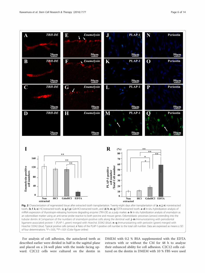
Fig. 2 Characterization of regenerated tissue after extracted tooth transplantation. Twenty-eight days after transplantation of (a, e, j, n) nonextractedtooth, (b, f, k, o) HCl-extracted tooth, (c, g, l, p) GdnHCl-extracted tooth, and (d, h, m, q) EDTA-extracted tooth. a–d In-situ hybridization analysis ofmRNA expression of thyrotropin-releasing hormone degrading enzyme (TRH-DE) as a pulp marker. e–h In-situ hybridization analysis of enamelysin asan odontoblast marker using an anti-sense probe reactive to both porcine and mouse genes. Odontoblastic processes (arrows) extending into thetubular dentin. i Comparison of the numbers of enamelysin-positive cells along the dentinal wall. j–m Immunostaining with periodontalligament-associated protein 1 (PLAP-1, green) merged with Hoechst 33342 (blue). n–q Immunostaining with periostin (green) merged withHoechst 33342 (blue). Typical positive cells (arrows). r Ratio of the PLAP-1-positive cell number to the total cell number. Data are expressed as means ± SDof four determinations. *P< 0.05, **P< 0.01 (Color figure online)
Kawamura et al. Stem Cell Research & Therapy (2016) 7:77 Page 6 of 14
For analysis of cell adhesion, the autoclaved teeth asdescribed earlier were divided in half in the sagittal planeand placed on a 24-well plate with the inside facing up-ward. C2C12 cells were cultured on the dentin in
DMEM with 0.2 % BSA supplemented with the EDTAextracts with or without the CM for 48 h to analyzetheir enhanced ability for cell adhesion. C2C12 cells cul-tured on the dentin in DMEM with 10 % FBS were used
Table 2 Relative mRNA expression of TRH-DE in regeneratedtissues of the transplants of nonextracted and extracted teethcompared with normal pulp
Kawamura et al. Stem Cell Research & Therapy (2016) 7:77 Page 7 of 14
as a positive control. The attached cells were countedafter Giemsa staining in the three areas of each tooth(n = 4) using binocular microscopic digital images ofthe three 1.0 mm × 1.0 mm rectangles scanned in aframe.
Odontoblastic and endothelial differentiation assaysThe combinatorial effects of the MDPSC CM with theEDTA extracts on enhanced odontoblastic and endothe-lial differentiation were examined to induce higher po-tential of the CM with the EDTA extracts for pulp anddentin regeneration. To analyze the enhanced odonto-blastic differentiation, C2C12 cells were cultured inDMEM with 10 % FBS, 50 mg/ml L-ascorbic acid 2-phosphate (Wako Pure Chemical Industries, Ltd), and 1mM inorganic phosphate (Pi; Sigma-Aldrich) supple-mented with the EDTA extracts alone or together withthe CM for 28 days. Bone morphogenetic protein(BMP2; kindly provided by Astellas Pharma Co., Ltd,Tokyo, Japan) at a final concentration of 100 ng/ml wasused as a positive control. RT-PCR analyses were furtherperformed using primers for odontoblast differentiationmarkers, dentin sialophosphoprotein (DSPP) andenamelysin (Table 1), in the cells from each of threedishes (n = 3).To analyze the enhanced endothelial differentiation,
human umbilical vein endothelial cells (HUVEC) werecultured in DMEM containing 2 % FBS, 1 μg/ml heparin(Lonza, Muenchensteinerstrasse, Switzerland), 1 μg/mlascorbic acid (Lonza), and 0.4 μg/ml hydrocortisone(Lonza) supplemented with the EDTA extracts alone ortogether with the CM for 14 days. Vascular endothelialgrowth factor (VEGF) (Lonza), basic fibroblast growthfactor (b-FGF) (Lonza), and insulin-like growth factor(IGF) (Lonza) at a final concentration of 1 μg/ml, re-spectively, was used as a positive control. Immunocyto-chemical analyses were performed for anti-vascularendothelial (VE)-cadherin (primary antibody, 1:50; Acris,Herford, Germany), and the positive cells were observedon a BZ-9000 BIOREVO fluorescence microscope aftercounterstaining with Hoechst 33342.
Statistical analysesData are reported as means ± SD. P values were calcu-lated using the Student’s t test and Tukey’s multiplecomparison test in SPSS 21.0 (IBM, Armonk, NY, USA).
ResultsPulp/dentin regeneration after tooth transplantationThe regenerative potential of the three distinct types ofextracted teeth was compared with control nonextractedtooth in an ectopic tooth transplantation assay of SCIDmice. Pulp-like tissue with well-organized vasculaturewas regenerated in the teeth 28 days after MDPSC trans-plantation as a positive control (Fig. 1a, e). Similar pulp-likeloose connective tissue was observed in the transplantsof the teeth extracted with HCl, GdnHCl, and EDTA(Fig. 1b–d, f–h) and in the transplant of nonextractedteeth (Fig. 1a, e). The regenerated tissue in the EDTA-extracted tooth transplant (Fig. 1m) had fewer Hoechst33342-stained cells compared with those in the nonex-tracted, HCl-extracted, and GdnHCl-extracted toothtransplants (Fig. 1j–l). The histomorphometric analysisconfirmed that the regenerated pulp area and cell dens-ity of the GdnHCl-extracted tooth transplants and theEDTA-extracted tooth transplants were significantlylower than those of the nonextracted tooth transplantson day 28 (Fig. 1n). The histomorphometric analysisconfirmed that the regenerated pulp area in the toothtransplants of the three types of treatment was signifi-cantly lower than that of the nontreatment on day 28(Fig. 1i). There were no significant differences in the re-generated area between the HCl-extracted tooth transplantand the GdnHCl-extracted tooth transplant. Transplant-ation of the EDTA-extracted teeth yielded significantly lessregenerated tissue compared with those of the other threeteeth on day 28 (Fig. 1i). These results suggest that chem-ical components extracted by EDTA may mainly generatean inductive microenvironment for pulp regeneration. Im-munostaining with a RECA1 antibody revealed neovascu-larization in the regenerated tissues by nonextracted toothtransplantation and the other three types of tooth trans-plantation (Fig. 1o–r). Histomorphometric analysis dem-onstrated that neovascularization in the nonextractedtooth transplant was significantly higher than that in theHCl-extracted, GdnHCl-extracted, and EDTA-extractedtooth transplants on day 28. There was no significant dif-ference in neovascularization between the HCl-extractedand GdnHCl-extracted tooth transplants, and a significantdifference between the EDTA-extracted tooth transplantand others (Fig. 1s). These results suggest that chemicalcomponents extracted by EDTA may mainly generate aninductive microenvironment for pulp regeneration andneovascularization.TRH-DE, a specific marker for pulp tissue, was simi-
larly expressed in the regenerated tissues of the nonex-tracted, the HCl-extracted, and the GdnHCl-extractedtooth transplants as analyzed by in-situ hybridization(Fig. 2a–c). However, there were no TRH-DE-positivecells in EDTA-extracted tooth transplant (Fig. 2d). Real-time RT-PCR analyses demonstrated similar expression
Fig. 3 (See legend on next page.)
Kawamura et al. Stem Cell Research & Therapy (2016) 7:77 Page 8 of 14
(See figure on previous page.)Fig. 3 Pulp regeneration after transplantation of the autoclaved teeth reconstituted with soluble extracts. Twenty-eight days after ectopic transplantationof MDPSCs (a, g, n, u) in the native autoclaved teeth only, and in the autoclaved teeth reconstituted (b, h, o, v) with the GdnHCl extracts, (c, i, p, w) withthe EDTA extracts, (d, j, q, x) with the MDPSC CM, (e, k, r, y) with the GdnHCl extracts and the CM, and (f, l, s, z) with the EDTA extracts and the CM. a–lH & E staining. Pulp-like cells (arrows).m Ratio of regenerated area to root canal area. n–s Hoechst 33342 staining. t Ratio of Hoechst 33342-positive cellsto regenerated area. u–z Immunostaining with RECA1. zz Ratio of vascularization area to the total regenerated area. Data are expressed as means ± SDof four determinations. *P< 0.05, **P< 0.01, #P < 0.001 to nonextracted tooth, reconstituted tooth with the MDPSC CM alone, and MDPSC CM with theGdnHCl extracts or the EDTA extracts. CM conditioned medium, RECA1 rat endothelial cell antigen 1
Kawamura et al. Stem Cell Research & Therapy (2016) 7:77 Page 9 of 14
levels of TRH-DE mRNA in the regenerated tissue of thenonextracted, HCl-extracted, and GdnHCl-extractedtooth transplants to that in normal pulp tissue, whichwas significantly higher than that of the EDTA-extractedtooth transplant (Table 2).Enamelysin-positive cells were attached to the dentinal
wall, extending their processes for some distance into thedentin in the HCl-extracted and GdnHCl-extracted toothtransplants similar to the nonextracted tooth transplant(Fig. 2e–g), and they were few in the EDTA extractedtooth transplant (Fig. 2h). The quantitative analysis dem-onstrated that the number of enamelysin-positive cells wassignificantly higher in the regenerated tissues in the nonex-tracted, HCl-extracted, and GdnHCl-extracted tooth trans-plants compared with that of the EDTA-treated toothtransplant (Fig. 2i). Furthermore, immunohistochemicalanalyses demonstrated minimal expression of PLAP-1 andperiostin, the markers for periodontal ligament in the re-generated tissues by the nonextracted, HCl-extracted, andGdnHCl-extracted tooth transplantation (Fig. 2j–q). Thequantitative analysis indicated that the number of PLAP-1-positive cells was significantly lower in the regenerated tis-sues of the nonextracted, HCl-extracted, and GdnHCl-extracted tooth transplants compared with that in theEDTA-extracted tooth transplant (Fig. 2r). These resultssuggest that the EDTA extracts may contain potentialchemical components of an inductive microenvironmentfor dentin regeneration.
Pulp/dentin regeneration after reconstitution of solubleextracts with the autoclaved teethTo examine the chemical components of inductive micro-environment, the regenerative potential of the reconsti-tuted autoclaved teeth with the EDTA extracts with orwithout the MDPSC CM was examined in the ectopictooth transplantation model. The autoclaved teeth simi-larly preserved the tubules as nonautoclaved teeth analyzedby SEM, indicating intact physical microenvironment(Additional file 1: Figure S1). The reconstituted autoclavedteeth with the GdnHCl extracts with or without the CMwere used as a control for the EDTA extracts. Pulp-like tis-sue was not regenerated in the autoclaved teeth reconsti-tuted with the GdnHCl extracts or the EDTA extracts andin the autoclaved tooth without any reconstitution 28 days
after transplantation (Fig. 3a–c, g–i). On the other hand,pulp-like tissue with well-organized vasculature was ob-served in the autoclaved tooth reconstituted with the CM(Fig. 3d, j). Furthermore, the autoclaved tooth reconsti-tuted with both the EDTA extracts and the CM (Fig. 3f, l)demonstrated a higher amount of regenerated tissue com-pared with that with the EDTA extracts alone (Fig. 3c, i) orthe CM alone (Fig. 3d, j). The histomorphometric analysisconfirmed that reconstitution of the EDTA extracts to-gether with the CM significantly enhanced the regeneratedarea compared with either alone in the autoclaved tooth(Fig. 3m). The combinatorial effect of the EDTA extractson the pulp regeneration with the CM was detected, unlikethe GdnHCl extracts (Fig. 3m). No cell was stained withHoechst 33342 nor RECA1 in the autoclaved tooth andthe reconstituted tooth of the EDTA extracts as well asthat of the GdnHCl extracts (Fig. 3n–p, u–w). The recon-stitution with the EDTA extracts together with the CMincreased the number of Hoechst-positive cells in the re-generated tissue (Fig. 3s) compared with the reconstitutionwith the CM alone (Fig. 3q). The histomorphometric ana-lysis confirmed that the regenerated pulp area and celldensity of the transplants of the tooth reconstituted withthe CM alone or the tooth reconstituted with the GdnHClextracts and the CM were significantly lower than those ofthe transplant of nontreated tooth on day 28. On the otherhand, those of the tooth transplant reconstituted both withthe EDTA extracts and the CM were significantly highercompared with the tooth transplant reconstituted with theCM or the tooth transplant reconstituted with the GdnHClextracts and the CM (Fig. 3m, t). Immunostaining with anRECA1 antibody revealed neovascularization in the regen-erated tissue of the reconstituted tooth transplants of theCM with or without the EDTA extracts (Fig. 3x, z). Histo-morphometric analysis demonstrated that there were nosignificant differences in the neovascularization betweenteeth reconstituted with the CM and with the CM with theEDTA extracts (Fig. 3zz). There were no TRH-DE-positivecells in the autoclaved tooth reconstituted with or withoutthe EDTA extracts as analyzed by in-situ hybridization(Fig. 4a, b). However, TRH-DE was similarly expressed inthe transplants of autoclaved teeth reconstituted with theCM alone and with the CM and the EDTA extractstogether (Fig. 4c, d). The real-time RT-PCR analyses
Fig. 4 Characterization of regenerated tissues after transplantation of reconstituted teeth. Twenty-eight days after ectopic transplantation ofMDPSCs (a, e, j, n) in the native autoclaved tooth, in the autoclaved teeth reconstituted (b, f, k, o) with the EDTA extracts, (c, g, l, p) with the CM,and (d, h, m, q) with the EDTA extracts and the CM. a–d In-situ hybridization analysis of expression of thyrotropin-releasing hormone degradingenzyme (TRH-DE) as a pulp marker. e–h In-situ hybridization analysis of enamelysin. Odontoblastic processes (arrows) extending into the tubulardentin. i Comparison of the numbers of enamelysin-positive cells along the dentinal wall. j–m Immunostaining with periodontal ligament-associatedprotein 1 (PLAP-1, green) merged with Hoechst 33342 (blue). A typical PLAP-1-positive cell (arrow). n–q Immunostaining with periostin (green)merged with Hoechst 33342 (blue). r Ratio of the PLAP-1-positive cell number to the total cell number. Data are expressed as means ± SD offour determinations. *P < 0.05. #P < 0.001 to nonextracted tooth, reconstituted tooth with the MDPSC CM alone, and the MDPSC CM with theEDTA extracts. CM conditioned medium (Color figure online)
Kawamura et al. Stem Cell Research & Therapy (2016) 7:77 Page 10 of 14
demonstrated that TRH-DE was similarly expressed in theregenerated tissue in the transplants of the teeth reconsti-tuted with the CM alone or with the CM and the EDTA
extracts as in normal pulp tissue (Table 3), suggesting thatthe regenerated tissue in these reconstituted teeth may bepulp tissue.
Table 3 Relative mRNA expression of TRH-DE in regeneratedtissues of the transplants of reconstituted autoclaved teeth withthe EDTA extracts, the CM alone, and the EDTA extracts withthe CM compared with normal pulp
EDTA extracts CM CM + EDTA extracts
TRH-DE 0 0.8 ± 0.3 1.0 ± 0.3
CM conditioned medium, EDTA ethylenediaminetetraacetic acid, TRH-DEthyrotropin-releasing hormone degrading enzyme
Kawamura et al. Stem Cell Research & Therapy (2016) 7:77 Page 11 of 14
There were no enamelysin-positive cells in the auto-claved tooth and the reconstituted tooth of the EDTAextracts (Fig. 4e, f ). On the other hand, there wereenamelysin-positive cells in the autoclaved tooth withthe CM and with the CM and the EDTA extracts. Statis-tical analysis showed that the number of enamelysin-positive cells was significantly higher in the autoclavedtooth reconstituted with the CM and the EDTA extractscompared with that in the CM alone reconstituted(Fig. 4i). On the other hand, there were no significantdifferences in the number of enamelysin-positive cellsbetween nontreated tooth and reconstituted autoclavedtooth. Furthermore, immunohistochemical analyses ofPLAP-1 and periostin, markers for periodontal ligament,demonstrated a positive response in the regenerative tis-sue in the reconstituted teeth with the CM or with theCM and the EDTA extracts (Fig. 4l, m). Quantitativeanalysis indicated that the number of PLAP-1-positivecells was significantly higher in the regenerated tissue ofthe CM reconstituted tooth transplant compared withthat in the nonextracted tooth and autoclaved toothreconstituted with the CM and the EDTA extracts(Fig. 4r). These results demonstrated that the EDTA ex-tracts may enhance the inductive effect of the CM onodontoblastic differentiation.
Influence of the EDTA extracts and the MDPSCs CMin vitroDuring the process of pulp/dentin regeneration, it is wellknown that the cells surrounding the tooth migrate intothe tooth canal, adhere/engraft to scaffold/microenviron-ment, and proliferate and differentiate into pulp cells/odontoblasts. Thus, to analyze the in-vivo effects of theEDTA extracts on pulp/dentin regeneration, the effectsof the EDTA extracts on proliferation, migration, cell at-tachment, and odontoblastic differentiation were exam-ined in vitro combined with or without the MDPSC CM.The proliferation analysis demonstrated that the MDPSCCM had a significantly higher effect on enhanced prolif-eration than nontreatment (Fig. 5a). However, there wereno significant differences between nontreatment andEDTA extracts. Furthermore, the CM with the EDTAextracts showed a significantly higher proliferation effectcompared with the CM alone. The EDTA extracts or theCM significantly increased the migration activity of
C2C12 cells. The EDTA extracts with the CM presentedsignificantly higher migration activity than the EDTA ex-tracts alone or the CM alone (Fig. 5b). The CM also hadsignificantly higher cell attachment ability than nontreat-ment and the EDTA extracts. Furthermore, the EDTAextracts showed no significant increase in cell attach-ment ability of the CM (Fig. 5c), suggesting no effect ofthe EDTA extracts on cell adhesion. The RT-PCR ana-lysis demonstrated that the EDTA extracts together withthe CM induced odontoblastic differentiation, althoughthose alone had no effect (Fig. 5d). The angiogenicpotential was demonstrated by immunostaining withVE-cadherin on day 14. The CM induced differentiationof HUVEC into an endothelial cell lineage positive forVE-cadherin. However, the EDTA extracts showed noincrease in cell endothelial differentiation (Fig. 5e).
DiscussionThe long-term goal of tissue engineering in dentistry isthe design and optimization of new functional teeth withdentin and pulp. The realization of this goal will over-come the limitations of artificial dental prosthesis. Thecritical challenge in tissue engineering is to optimizestem cells, morphogens to direct stem cells in the in-ductive microenvironment of extracellular matrix (ECM)scaffold.The development of novel approaches to control the
lineage of stem cell fate is a prerequisite for tissue engin-eering and regenerative medicine. The three-dimensionalmicroenvironment has a profound influence on the prolif-eration, differentiation, and maintenance of MSCs [17].The inductive microenvironment consists of physical cuesof ECM stiffness and the topology of ECM and instructivebiochemical signals in the form of soluble or ECM-boundmorphogens, growth factors, and cytokines [9, 18–21].The three-dimensional biomimetic silicon scaffold withdentinal tubule-like pones is a prerequisite for odonto-blastic differentiation of MSCs [22].In this present investigation, the biomimetic tooth
ECM scaffold for dentin and dental pulp was investi-gated systematically. The methodological approach con-sisted of sequential extraction of porcine incisor teeth by0.6 M HCl to demineralize, and extraction in 4 MGdnHCl, pH 7.4 and 0.5 M EDTA, pH 7.4. The ex-tracted tooth matrix was combined with MDPSC CM,and was evaluated in SCID mice for pulp/dentin regen-eration. In addition, the extracted ECM components ob-tained by the GdnHCl extracts and the EDTA extracts(with or without MDPSC CM) were reconstituted withnative (nonextracted) teeth that were autoclaved to in-activate the endogenous bioactive components. Thereconstituted teeth were transplanted into an ectopic sitein recipient SCID mice to unambiguously evaluate theGdnHCl extracts and the EDTA extracts. The 0.5 M
Fig. 5 Effects of the EDTA extracts and the CM on proliferation, migration, cell attachment, odontoblastic differentiation, and endothelial celldifferentiation in vitro. a Proliferation effect of the EDTA extracts and the CM analyzed by CCK-8® in C2C12 cells. b Migration effect of the EDTAextracts and the CM analyzed by TAXIScan-FL® in C2C12 cells. c Effect of the EDTA extracts and the CM on cell attachment ability analyzed byGiemza stain in C2C12 cells. d Effect of the EDTA extracts and the CM on odontoblast differentiation analyzed by RT-PCR in C2C12 cells. e Angiogeniceffect of the EDTA extracts and the CM by immunocytochemical analysis with vascular endothelial (VE)-cadherin in HUVEC. Data are expressed asmeans ± SD of four determinations. *P < 0.05, **P < 0.01. CM conditioned medium, DSPP dentin sialophosphoprotein
Kawamura et al. Stem Cell Research & Therapy (2016) 7:77 Page 12 of 14
Kawamura et al. Stem Cell Research & Therapy (2016) 7:77 Page 13 of 14
EDTA extraction reduced the inductive potential forpulp regeneration, odontoblastic differentiation, andangiogenesis/vasculogenesis in the tooth transplants.Whereas the periodontal marker PLAP-1 was expressed,the pulp marker TRH-DE was not detectable. A recentreport has demonstrated that the EDTA treatment ofthe root surface could help to expose collagen fibers onroot cementum and enhance the surface roughness, pro-moting the adherence cytokines and the release of inher-ent proteins or growth factors within the tooth [23].This report demonstrated regeneration for periodontalligament and root surface. However, the effects of EDTAtreatment for pulp regeneration are not well understood.These findings suggest that 0.5 M EDTA may extractselect biochemical components involved in inductivemicroenvironment for pulp regeneration. It is wellknown that the EDTA extracts are enriched in noncol-lagenous proteins [24] including, but not limited to,osteonectin, osteocalcin, osteopontin, bone sialopro-tein, and DSPP [25, 26]. DSPP is cleaved by a proteaseto dentin sialoprotein (DSP), dentin glycoprotein(DGP), and dentin phosphoprotein (DPP). ECM com-prises enzymes and matrix-bound growth factors aswell as intrinsic ECM components. The ECM-boundgrowth factors may regulate growth factor activity andcellular proliferation and differentiation [27, 28]. TheEDTA extracts may therefore contain a constellation ofnoncollagenous proteins and the EDTA-bound growthfactors.Based on the finding of bioactive factors in EDTA ex-
tracts we next examined the GdnHCl extracts and theEDTA extracts reconstituted with biologically inacti-vated teeth obtained by autoclaving that preservesphysical cues of the pores of the dentinal tubules. How-ever, both the GdnHCl extracts and the EDTA extractswere devoid of pulp/dentin regenerative potential. It isnoteworthy that the CM from the MDPSCs reconsti-tuted with autoclaved teeth demonstrated copiousamounts of regenerated pulp tissue. In an unexpecteddiscovery, the addition of the EDTA extracts to theMDPSC CM significantly increased pulp tissue com-pared with the MDPSC CM alone. The attachment ofMDPSCs to the dentinal wall of the autoclaved teeth isa prerequisite for pulp regeneration. The MDPSC CMcontains soluble factors including BDNF, EGF, FGF2,GDNF, GM-CSF, HGF, MMP3, NPY, NGF, PDGF,TGFβ, and VEGF [29]. The earlier work demonstratedthat the presence of multiple growth factors in theMDPSC CM was higher compared with that in theMBMSC CM and the MADSC CM. The observedhigher regenerative potential of MDPSC CM may bedue to higher secretion of trophic factors for angiogenesis,migration, and anti-apoptosis compared with BMSCs andADSCs [6, 30].
The chemical cues in the EDTA extract may induceodontoblast differentiation. The candidate molecules mayinclude DSP, DGP, and DPP [27]. DSP has an inductivepotential for mineralization in vitro [31] and has a role ininitiation of dentin mineralization and maturation ofodontoblasts [32, 33]. The MDPSC CM also inducedodontoblast differentiation in vitro [34]. It should be notedthat the DPSC CM contains BMP4, BMP7, and Wnt10a[22, 35]. As demonstrated in this investigation, the auto-claved tooth transplantation demonstrated no pulp re-generation and no odontoblast differentiation. However,combination of the EDTA extracts with the MDPSCCM with autoclaved teeth demonstrated significant pulpregeneration and odontoblast differentiation. Thecombinatorial effects on proliferation, migration, andodontoblast differentiation were also demonstratedin vitro. Thus, when transplanted with autoclaved teeth,there is collaboration between the physical cues in themicroenvironment of the autoclaved teeth and the po-tent biochemical cues present in the EDTA extracts andthe MDPSC CM. The detailed future investigations ofthe collaborative interactions between the physical cuesof the ECM and chemical cues in the EDTA extracts ofthe tooth ECM will inform and enhance the platform oftissue engineering in dentistry and oral regenerativemedicine.
ConclusionsThe EDTA-extracted teeth demonstrated significantlylower pulp/dentin regenerative potential compared withthe GdnHCl-extracted teeth. The EDTA soluble chemicalcomponents and the MDPSC CM, when reconstitutedwith the physical structure of autoclaved teeth, served asan inductive microenvironment, promoting cell prolifera-tion, migration, and odontoblastic differentiation.
Additional file 1: Figure S1. Showing the effect of autoclave treatmenton the physical microenvironment of the dentin surface. SEM imagesdemonstrating A, B nontreated dentin surface and C, D autoclaveddentin surface. (PDF 227 kb)
Author’s contributionsRK participated in the study design, performed tooth transplantation, dataanalysis, and interpretation, and drafted the manuscript. YH participated inthe tooth transplantation, contributed acquisition and analyses of in-situ and
Kawamura et al. Stem Cell Research & Therapy (2016) 7:77 Page 14 of 14
immunohistochemical data, and revised critically for important intellectualcontent. HM participated in data interpretation and writing the manuscript.MN contributed to conception and design, revised critically for importantintellectual content, and gave final approval of the version to be published.All authors read and approved the final manuscript.
Competing interestsThe authors declare that they have no competing interests.
Author details1Department of Stem Cell Biology and Regenerative Medicine, NationalCenter for Geriatrics and Gerontology, Research Institute, 7-430 Morioka, Obu,Aichi 474-8511, Japan. 2Department of Gerontology, School of Dentistry,Aichi-Gakuin University, Nagoya, Aichi 464-8651, Japan. 3Department of OralImplantology, School of Dentistry, Aichi-Gakuin University, Nagoya, Aichi464-8651, Japan. 4Department of Pediatric Dentistry, School of Dentistry,Aichi-Gakuin University, Nagoya, Aichi 464-8651, Japan.
Received: 19 November 2015 Revised: 22 February 2016Accepted: 29 April 2016
References1. Caplan AI, Dennis JE. Mesenchymal stem cells as trophic mediators. J Cell
Biochem. 2006;98:1076–84.2. Maumus M, Guérit D, Toupet K, Jorgensen C, Noël D. Mesenchymal stem
cell-based therapies in regenerative medicine: applications in rheumatology.Stem Cell Res Ther. 2011;2(2):14.
3. Yu B, Zhang X, Li X. Exosomes derived from mesenchymal stem cells. Int JMol Sci. 2014;15(3):4142–57.
4. Ishizaka R, Iohara K, Murakami M, Fukuta O, Nakashima M. Regeneration ofdental pulp following pulpectomy by fractionated stem/progenitor cells frombone marrow and adipose tissue. Biomaterials. 2012;33:2109–18.
5. Ishizaka R, Hayashi Y, Iohara K, Sugiyama M, Murakami M, Yamamoto T,Fukuta O, Nakashima M. Stimulation of angiogenesis, neurogenesis andregeneration by side population cells from dental pulp. Biomaterials.2013;34:1888–98.
6. Hayashi Y, Murakami M, Kawamura R, Ishizaka R, Fukuta O, Nakashima M.CXCL14 and MCP1 are potent trophic factors associated with cell migrationand angiogenesis leading to higher regenerative potential of dental pulpside population cells. Stem Cell Res Ther. 2015;6(6):111.
7. Guvendiren M, Burdick JA. The control of stem cell morphology anddifferentiation by hydrogel surface wrinkles. Biomaterials. 2010;31:6511–8.
8. Li H, Fu X. Mechanisms of action of mesenchymal stem cells in cutaneouswound repair and regeneration. Cell Tissue Res. 2012;348:371–7.
10. Lei M, Li K, Li B, Gao L-N, Chen F-M, Jin Y. Mesenchymal stem cellcharacteristics of dental pulp and periodontal ligament stem cells afterin vivo transplantation. Biomaterials. 2014;35(24):6332–43.
11. Shi S, Bartold PM, Miura M, Seo BM, Robey PG, Gronthos S. The efficacy ofmesenchymal stem cells to regenerate and repair dental structures. OrthodCraniofac Res. 2005;8(3):191–9.
12. Murakami M, Horibe H, Iohara K, Hayashi Y, Osako Y, Takei Y, Nakata K,Motoyama N, Kurita K, Nakashima M. The use of granulocyte-colonystimulating factor induced mobilization for isolation of dental pulp stem cellswith high regenerative potential. Biomaterials. 2013;34:9036–47.
13. Takata T, Errico JAD, Atkins KB, Berry JE, Strayhorn C, Taichman RS, SomermanMJ. Protein extracts of dentin affect proliferation and differentiation ofosteoprogenitor cells in vitro. J Periodontol. 1998;69:1247–55.
14. Sodek KL, Tupy JH, Sodek J, Grynpas MD. Relationships between boneprotein and mineral in developing porcine long bone and calvaria. Bone.2000;26:189–98.
15. Yamamoto T, Osako Y, Ito M, Murakami M, Hayashi Y, Horibe H, Iohara K,Takeuchi N, Okui N, Hirata H, Nakayama H, Kurita K, Nakashima M. Trophiceffects of dental pulp stem cells on schwann cells in peripheral nerveregeneration. Cell Transplant. 2015;25(1):183–193.
16. Iohara K, Imabayashi K, Ishizaka R, Watanabe A, Nabekura J, Ito M,Matsushita K, Nakamura H, Nakashima M. Complete pulp regeneration afterpulpectomy by transplantation of CD105+ stem cells with stromal cell-derivedfactor-1. Tissue Eng Part A. 2011;17(15–16):1911–20.
17. Scadden DT. The stem-cell niche as an entity of action. Nature. 2006;441:29.
18. Reddi AH. Role of morphogenetic proteins in skeletal tissue engineeringand regeneration. Nat Biotechnol. 1998;16(3):247–52.
19. Nakashima M, Reddi AH. The application of bone morphogenetic proteinsto dental tissue engineering. Nat Biotechnol. 2003;21(9):1025–32.
20. Das RK, Zouani OF. A review of the effects of the cell environmentphysicochemical nanoarchitecture on stem cell commitment. Biomaterials.2014;35:5278–93.
21. Griffin MF, Kalaskar DM, Butler PE, Seifalian AM. The use of adipose stemcells in cranial facial surgery. Stem Cell Rev Rep. 2014;10:671–685.
22. Miyashita S, Bellah EM, Murakami M, Iohara K, Yamamoto T, Horibe H. Kurita K,Takano-Yamamoto T, Nakashima M. Mechanical forces induce odontoblasticdifferentiation of mesenchymal stem cells on three-dimensional biomimeticscaffolds. J Tissue Eng Regen Med. 2014, Online First.
23. Zhu W, Zhang Q, Zhang Y, Cen L, Wang J. PDL regeneration via cellhoming in delayed replantation of avulsed teeth. J Transl Med. 2015;13:357.
24. Linde A, Bhown M, Butler WT. Noncollagenous protein of dentin. J BiolChem. 1980;255(12):5931–42.
25. Prasad M, Butler WT, Qin C. Dentin sialophosphoprotein inbiomineralization. Connect Tissue Res. 2010;51:404–17.
26. Yamamoto R, Oida S, Yamakoshi Y. Dentin sialophosphoprotein-derivedproteins in the dental pulp. J Dent Res. 2015;94(8):1120–7.
27. Smith AJ, Scheven BA, Takahashi Y, Ferracane JL, Shelton RM, Cooper PR.Dentine as a bioactive extracellular matrix. Arch Oral Biol. 2012;57(2):109–21.
28. Goldberg M, Six N, Chaussain C, Besten PD, Veis A, Poliard A. Dentinextracellular matrix molecules implanted into exposed pulps generatereparative dentin: a novel strategy in regenerative dentistry. J Dent Res.2009;88(5):396–9.
29. Nakashima M, Hayashi Y. Reference module in biomedical research; 2014.Chapter Dental stem cells: 1-11.
30. Murakami M, Hayashi Y, Iohara K, Osako Y, Hirose Y, Nakashima M. Trophiceffects and regenerative potential of mobilized mesenchymal stem cellsfrom bone marrow and adipose tissue as alternative cell sources for pulp/dentin regeneration. Cell Transplant. 2015;24(9):1753–65.
31. Ozer A, Yuan G, Yang G, Wang F, Li W, Yang Y, Guo F, Gao Q, Shoff L, ChenZ, Gay IC, Donly KJ, Macdougall M, Chen S. Domain of dentine sialoproteinmediates proliferation and differentiation of human periodontal ligamentstem cells. PLoS One. 2013;8(12).
32. Suzuki S, Sreenath T, Haruyama N, Honeycutt C, Terse A, Cho A, Kohler T,Müller R, Goldberg M, Kulkarni AB. Dentin sialoprotein and dentinphosphoprotein have distinct roles in dentin mineralization. Matrix Biol.2009;28:221–9.
33. Papagerakis P, Berdal A, Mesbah M, Peuchmaur M, Malaval L, Nydegger J,Simmer J, Macdougall M. Investigation of osteocalcin, osteonectin, anddentin sialophosphoprotein in developing human teeth. Bone.2002;30(2):377–85.
34. Takeuchi N, Hayashi Y, Murakami M, Alvarez FJ, Horibe H, Iohara K, Nakata K,Nakamura H, Nakashima M. Similar in vitro effects and pulp regeneration inectopic tooth transplantation by basic fibroblast growth factor andgranulocyte-colony stimulating factor. Oral Dis. 2015;21:113–22.
35. Kim NR, Lee DH, Ahn S-J, Lee I-S, Yang H-C. The differentiation-inducingeffect of conditioned media obtained from dental pulp cells. Oral Surg OralMed Oral Pathol Oral Radiol Endod. 2009;107:54–9.
• We accept pre-submission inquiries
• Our selector tool helps you to find the most relevant journal
• We provide round the clock customer support
• Convenient online submission
• Thorough peer review
• Inclusion in PubMed and all major indexing services
• Maximum visibility for your research
Submit your manuscript atwww.biomedcentral.com/submit
Submit your next manuscript to BioMed Central and we will help you at every step: