Gasser et al. Particle and Fibre Toxicology 2012, 9:17http://www.particleandfibretoxicology.com/content/9/1/17
RESEARCH Open Access
Pulmonary surfactant coating of multi-walledcarbon nanotubes (MWCNTs) influences theiroxidative and pro-inflammatory potential in vitroMichael Gasser1,2, Peter Wick2, Martin JD Clift1, Fabian Blank3, Liliane Diener2, Bing Yan4,5, Peter Gehr6,Harald F Krug2 and Barbara Rothen-Rutishauser1,3*
Abstract
Background: Increasing concern has been expressed regarding the potential adverse health effects that may beassociated with human exposure to inhaled multi-walled carbon nanotubes (MWCNTs). Thus it is imperative that anunderstanding as to the underlying mechanisms and the identification of the key factors involved in adverse effectsare gained. In the alveoli, MWCNTs first interact with the pulmonary surfactant. At this interface, proteins and lipidsof the pulmonary surfactant bind to MWCNTs, affecting their surface characteristics. Aim of the present study wasto investigate if the pre-coating of MWCNTs with pulmonary surfactant has an influence on potential adverseeffects, upon both (i) human monocyte derived macrophages (MDM) monocultures, and (ii) a sophisticated in vitromodel of the human epithelial airway barrier. Both in vitro systems were exposed to MWCNTs either pre-coatedwith a porcine pulmonary surfactant (Curosurf) or not. The effect of MWCNTs surface charge was also investigatedin terms of amino (−NH2) and carboxyl (−COOH) surface modifications.
Results: Pre-coating of MWCNTs with Curosurf affects their oxidative potential by increasing the reactive oxygenspecies levels and decreasing intracellular glutathione depletion in MDM as well as decreases the release of Tumournecrosis factor alpha (TNF-α). In addition, an induction of apoptosis was observed after exposure to Curosurfpre-coated MWCNTs. In triple cell-co cultures the release of Interleukin-8 (IL-8) was increased after exposure toCurosurf pre-coated MWCNTs. Effects of the MWCNTs functionalizations were minor in both MDM and triple cellco-cultures.
Conclusions: The present study clearly indicates that the pre-coating of MWCNTs with pulmonary surfactant morethan the functionalization of the tubes is a key factor in determining their ability to cause oxidative stress,cytokine/chemokine release and apoptosis. Thus the coating of nano-objects with pulmonary surfactant should beconsidered for future lung in vitro risk assessment studies.
* Correspondence: [email protected] Merkle Institute, University of Fribourg, Marly, Switzerland3Respiratory Medicine, Department of Clinical Research, Inselspital UniversityHospital, University of Bern, Bern, SwitzerlandFull list of author information is available at the end of the article
Gasser et al. Particle and Fibre Toxicology 2012, 9:17 Page 2 of 13http://www.particleandfibretoxicology.com/content/9/1/17
BackgroundThe ever-developing industry of nanotechnology the lasttwo decades has culminated in a plethora of new nano-objects (defined as material with one, two or three exter-nal dimensions in the nanoscale) which are being usedwithin a variety of consumer and industrial applications.Among the most prominent nano-objects are carbonnanotubes (CNTs); hollow nanofibres formed from carbon[1]. Based on their structure CNTs are classified as either,single-wall carbon nanotubes (SWCNTs), which comprisea single layer of carbon atoms, or multi-wall carbon nano-tubes (MWCNTs), comprising of multiple concentrictubes [2]. Structural and mechanical characteristics suchas an extreme strength, stiffness and robustness [3] makeCNTs interesting for the use in an infinite number ofapplications such as sporting goods, automobile productsor household items. Additionally, CNTs hold great prom-ise for application within medicine, particularly as a toolin therapeutics and diagnostics [4]. With their increasingnumber of applications, CNT emissions into the environ-ment and human exposure may increase. Mainly CNTproduction, processing and disposal may be hazardous forhumans [5]. Moreover during the use of CNT containingproducts, CNTs may be released into the environment asfor instance from abrasion or degradation of CNT con-taining products. Possible portals by which CNTs mayenter the human body include the skin, the gastro-intestinal tract and injection (nanomedicine). However, itis well accepted from previous research using nano-sizedparticles [6] and CNTs [7,8] that inhalation is the primaryexposure route to the human body if CNTs are releasedinto the environmental air.Concerns about the safety of CNTs have been raised for a
number of different reasons [3,9,10] (i) due to their smallaerodynamic diameter CNTs are hypothesised to reach thelower respiratory tract, (ii) CNTs possess, like other nano-objects, a high surface to mass ratio, thus a large surfacecan interact with the biological surroundings, and (iii) someCNTs which are fibre shaped may (if structured dimensionsare similar) behave like asbestos, or other pathogenic fibreswhich are toxic due to their needle-like shape. Moreover(iv), numerous in vivo studies (e.g.) [11-13] have shown dif-ferent types of MWCNTs to remain in the lung for up toseveral months after deposition indicating the potential forprolonged biopersistence.Recently the potential adverse effects of CNTs have
been studied on various biological systems, using differ-ent exposure methods both in vivo and in vitro [14,15].Despite the unrealistically high doses which have beenused within some of the previous studies (e.g.) [16], it isknown that subpleural fibrosis [17], granuloma forma-tion [10] and mesothelioma [16] similar to the effects ofcrocidolite asbestos fibres, can appear after in vivo expo-sures to mainly straight, stiff and extremely long CNTs.
Observed adverse effects have further been explainedby the oxidative stress paradigm [18]. In numerous stud-ies (e.g.) [19-21] an increased oxidative stress responsein vivo and in vitro has been reported causing a subse-quent (pro-) inflammatory reaction after exposures toboth straight and tangled CNTs.Although numerous studies address the adverse poten-
tial of CNTs, their comparability is often limited andresults are contradictory. Explanations for these discrep-ancies include differences in administered dose, thephysico-chemical characteristics (e.g. agglomeration/ag-gregation state, metal impurities, stiffness, length) of theCNTs studied, the exposure method of CNTs, or differ-ences in the biological system employed [15,22,23]. Thusconditions/characteristics have to be manipulated sys-tematically in order to identify key factors for their po-tential (adverse) biological effects. A promising way tomodify the properties of CNTs is the functionalizationof the surface [24,25]. Functionalization of CNTs can beused to promote the binding of specific biomolecules(such as siRNA) [26] but also to improve their biocom-patibility. The adverse effect potential of CNTs can besignificantly driven by the particular (surface) modifica-tion employed [22] whereas studies have shown both,increases and decreases in toxicity after exposures toCNTs with different surface functionalizations [27,28].Functionalization can further affect the CNTs dispersitywhich can have subsequent consequences on their celluptake and agglomeration in tissue [29].It is not only artificial surface modifications that play a
role in regards to the potential adverse effects of CNTs.The surface characteristics of MWCNTs may also bemodified by the adsorption of biomolecules following in-halation, and the subsequent interaction with the lung.Specifically, an initial coating of the MWCNTs will takeplace when they interact with pulmonary surfactantwhich is mainly produced by epithelial type II cells andwhich is located at the air-liquid interface. Surfactantconsists 85-90% of phospholipids [30], the specific sur-factant proteins (SP) -A, -B, -C, and -D (~10%) and itsmain function is the reduction of the alveolar surfacetension and keeping the gas exchange surface at optimalsize during the movements of breathing [31]. Thus, dur-ing deposition, surfactant or surfactant components willbind to the surface of MWCNTs [32,33]. Previously, thisinitial coating has not been sufficiently considered in re-spect to in vitro lung toxicity studies. The modulation ofthe adverse potential from surfactant binding is mainlydescribed for microparticles [34], however to the best ofour knowledge, not for CNTs and other nano-objects.After inhaled CNTs are coated with pulmonary surfac-tant, they may be displaced into the aqueous hypophase[35-37] and come in contact with cells of the immunesystem such as macrophages and dendritic cells, which
Gasser et al. Particle and Fibre Toxicology 2012, 9:17 Page 3 of 13http://www.particleandfibretoxicology.com/content/9/1/17
may engulf the MWCNTs and clear them from this areaof the lung [38]. Also epithelial type I cells can interactwith CNTs, as these cells mainly cover the alveolar sur-face [31].
Objectives of the study and methodological approachThe primary aim of this study therefore, was to investi-gate how a pre-coating of MWCNTs with pulmonarysurfactant may affect their potential adverse effects oncells of the air blood tissue barrier in vitro. In order tosimulate the pulmonary surfactant coating, MWCNTswere pre-coated with Curosurf, a well characterized nat-ural porcine surfactant preparation [39-41].Monocyte derived macrophage (MDM) monocultures
as well as a sophisticated 3D in vitro triple cell co-culture model of the airway epithelial barrier [42] wereexposed to the MWCNTs either pre-coated with surfac-tant or not. MWCNTs were initially compared to theirpotential to affect cell viability. Subsequently, in order tostudy an early oxidative reaction, reactive oxygen species(ROS) was quantified in MDM. For the characterizationof a prolonged oxidative stress response the intracellularantioxidant glutathione was quantified. The release ofthe cytokine TNF-α, as well as the chemokine IL-8 wasquantified in order to study their possible (pro-) inflam-matory effects.The secondary aim was to investigate the influence of
the surface charge on the MWCNTs potential adverseeffects. Therefore, non-functionalized (“pristine” or “P-MWCNTs”), carboxyl (“MWCNT-COOH”) and amino(“MWCNT-NH2”) functionalized MWCNTs were used inthe different exposures. In order to study a concentrationdependence of potential adverse effects, 2–3 different con-centrations (depending on the endpoint, see methods sec-tion for details) were applied. Eventually the differentconditions (Curosurf pre-coating, functionalization, con-centration) were statistically compared.
ResultsMWCNT characterizationDifferent MWCNT dispersions were fully characterizedprior to the exposures. A detailed overview is given inAdditional file 1: Table S1.
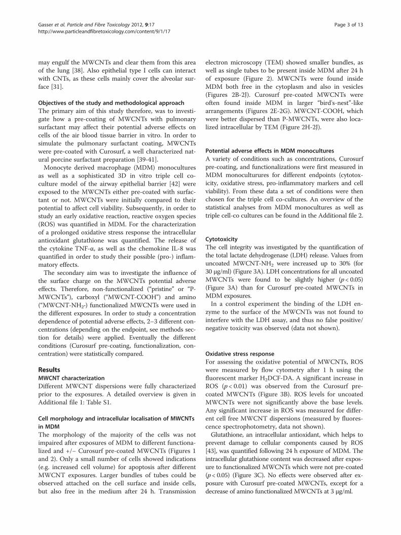
Cell morphology and intracellular localisation of MWCNTsin MDMThe morphology of the majority of the cells was notimpaired after exposures of MDM to different functiona-lized and +/− Curosurf pre-coated MWCNTs (Figures 1and 2). Only a small number of cells showed indications(e.g. increased cell volume) for apoptosis after differentMWCNT exposures. Larger bundles of tubes could beobserved attached on the cell surface and inside cells,but also free in the medium after 24 h. Transmission
electron microscopy (TEM) showed smaller bundles, aswell as single tubes to be present inside MDM after 24 hof exposure (Figure 2). MWCNTs were found insideMDM both free in the cytoplasm and also in vesicles(Figures 2B-2J). Curosurf pre-coated MWCNTs wereoften found inside MDM in larger “bird's-nest”-likearrangements (Figures 2E-2G). MWCNT-COOH, whichwere better dispersed than P-MWCNTs, were also loca-lized intracellular by TEM (Figure 2H-2J).
Potential adverse effects in MDM monoculturesA variety of conditions such as concentrations, Curosurfpre-coating, and functionalizations were first measured inMDM monoculturures for different endpoints (cytotox-icity, oxidative stress, pro-inflammatory markers and cellviability). From these data a set of conditions were thenchosen for the triple cell co-cultures. An overview of thestatistical analyses from MDM monocultures as well astriple cell-co cultures can be found in the Additional file 2.
CytotoxicityThe cell integrity was investigated by the quantification ofthe total lactate dehydrogenase (LDH) release. Values fromuncoated MWCNT-NH2 were increased up to 30% (for30 μg/ml) (Figure 3A). LDH concentrations for all uncoatedMWCNTs were found to be slightly higher (p< 0.05)(Figure 3A) than for Curosurf pre-coated MWCNTs inMDM exposures.In a control experiment the binding of the LDH en-
zyme to the surface of the MWCNTs was not found tointerfere with the LDH assay, and thus no false positive/negative toxicity was observed (data not shown).
Oxidative stress responseFor assessing the oxidative potential of MWCNTs, ROSwere measured by flow cytometry after 1 h using thefluorescent marker H2DCF-DA. A significant increase inROS (p < 0.01) was observed from the Curosurf pre-coated MWCNTs (Figure 3B). ROS levels for uncoatedMWCNTs were not significantly above the base levels.Any significant increase in ROS was measured for differ-ent cell free MWCNT dispersions (measured by fluores-cence spectrophotometry, data not shown).Glutathione, an intracellular antioxidant, which helps to
prevent damage to cellular components caused by ROS[43], was quantified following 24 h exposure of MDM. Theintracellular glutathione content was decreased after expos-ure to functionalized MWCNTs which were not pre-coated(p< 0.05) (Figure 3C). No effects were observed after ex-posure with Curosurf pre-coated MWCNTs, except for adecrease of amino functionalized MWCNTs at 3 μg/ml.
Figure 1 Light micrographs of MDM. Top images show MDM in medium and in medium containing 3% Curosurf respectively (MWCNT freecontrols). For all other conditions MDM were exposed to different functionalized and Curosurf pre-coated MWCNTs (30 μg/ml) for 24 h. A 20xmagnification was used.
Gasser et al. Particle and Fibre Toxicology 2012, 9:17 Page 4 of 13http://www.particleandfibretoxicology.com/content/9/1/17
TNF-α releaseConcentrations of the pro-inflammatory cytokine TNF-αwere assessed in the cell supernatant via an enzyme linkedimmunosorbant assay (ELISA) following 24 h exposure. In-dependent of the MWCNTs functionalization the Curosurf
pre-coating caused a decrease in the TNF-α release for the30 μg/ml exposures. This resulted in a significant (p< 0.05)decrease of 30 μg/ml exposures compared to lower con-centrations (Figure 3D) and a significant interaction of theindependent variables “Concentration” and “Pre-coating”
Figure 2 Transmission electron micrographs of MDM. MDM were exposed for 24 h to MWCNTs (30 μg/ml) under different conditions(Curosurf pre-coating, functionalization) a.: Untreated control. b.-d.: MDM exposed to uncoated P-MWCNTs. P-MWCNT agglomerates are visible atlow magnification (b); high magnification micrographs (c, d) reveal individual P-MWCNTs (black arrows). e.: MDM exposed to Curosurf pre-coatedP-MWCNTs at low magnification; f, g: higher magnifications with black arrows indicating individual MWCNTs in “bird's-nest”-like arrangements. h.-j. MDM exposed to MWCNT-COOH (without Curosurf pre-coating); arrows indicate single MWCNT-COOH. Scalebar is 5 μm for a., 2 μm for b., e.and h., 500 nm for c., f. and i., and 200 nm for d., g. and j.
Gasser et al. Particle and Fibre Toxicology 2012, 9:17 Page 5 of 13http://www.particleandfibretoxicology.com/content/9/1/17
(Additional file 2). To ensure that the decrease in TNF-α re-lease was caused by Curosurf pre-coated tubes and not fromfree surfactant, the cells were exposed to a control contain-ing cell culture medium and 3% Curosurf (see Methods sec-tion). TNF-α values for the free Curosurf were above thevalues of exposures containing Curosurf pre-coatedMWCNTs (Figure 3D). This indicates a stronger effect (on
the decrease in TNF-α release) from exposures to Curosurfpre-coated MWCNTs rather than from free Curosurf.TNF-α release in MDM was further dependent upon
the functionalization (p < 0.05) (Figure 3D), where highervalues were measured after MWCNT-NH2 exposures.In additional cell free control experiments the binding of
TNF-α cytokines to free MWCNTs and to Curosurf was
Figure 3 Endpoints measured in MDM. Several endpoints in MDM were analysed after exposure to different functionalized and pre-coatedMWCNTs. a. Cytotoxicity (lactate dehydrogenase release), b. Reactive oxygen species, c. Intracellular glutathione, d. Tumor necrosis factor-α.(For an optimized representation of group comparisons the same data are visualized in two diagrams. Values are shown relative to the mediumcontrol which corresponds to a mean concentration of 87.1 pg/ml), e. Necrosis, f. Apoptosis (For an optimized representation of groupcomparisons the same data are visualized in three diagrams.). Endpoints were assessed after 24 h exposures apart from ROS (B), which wasassessed after 1 h. Experiments have been performed in 3 to 5 repetitions. Data shows mean values ± standard deviation (SD). Different groupswere compared by ANOVA and Bonferroni post hoc tests (* = p< 0.05, ** = p< 0.01).
Gasser et al. Particle and Fibre Toxicology 2012, 9:17 Page 6 of 13http://www.particleandfibretoxicology.com/content/9/1/17
investigated by ELISA. No decrease of TNF-α levels wasdetected after the addition of either Curosurf or MWCNTsto a defined TNF-α concentration (data not shown).
Cell viabilityApoptotic and necrotic cells were assessed by using anAnnexin V staining kit via flow cytometry. Necrosis wasobserved to be significantly dependent on the Curosurfpre-coating (p < 0.05) (Figure 3E). However, the fractionof total necrotic cells was never above 5% for the differ-ent conditions studied.Apoptosis was observed to be significantly dependent
on the Curosurf pre-coating (p< 0.05). Values for Curosurfpre-coated MWCNTs were increased up to 20% (of the
total cell number). Significantly (p< 0.05) more apoptoticcells were found after exposures to MWCNT-COOHcompared to P-MWCNT exposures.
Potential adverse effects of MWCNTs on triple cell co-culturesDue to its higher complexity the triple cell co-culturemodel could only be used with a restricted set of experi-mental conditions. In MDM effects from the Curosurfpre-coating were especially pronounced for MWCNT-COOH. Thus in triple cell co-cultures only these tubeswere then used for Curosurf pre-coating.In contrast to MDM monocultures, triple cell co-cultures
were grown on transwell membrane inserts. Therefore
Gasser et al. Particle and Fibre Toxicology 2012, 9:17 Page 7 of 13http://www.particleandfibretoxicology.com/content/9/1/17
biochemical endpoint analyses were performed upon super-natants taken from below (“lower well”) and above (“upperwell”) the microporous membrane. Data from the lowerwell thus mainly represent the reaction of monocyte-derived dendritic cells (MDDC), whereas results from theupper well mainly represent the reaction of epithelial cellsand MDM (see Method section for details).
CytotoxicityCytotoxicity values were not significantly above thebaseline levels for all MWCNTs tested (Figure 4A).
Intracellular glutathione contentNo statistically significant effect was found for a specificcondition (Figure 4B). Only a tendency for a decrease in
Figure 4 Endpoints measured in triple cell co-cultures. Triple cell co-cuto different functionalized and pre-coated MWCNTs. Endpoints were assessrelease), b. Intracellular glutathione, c. Tumour necrosis factor-α (for an optvisualized in two diagrams. Values are shown relative to the medium contrupper well and 201 pg/ml for the lower well), d. Interleukin-8 (values are shconcentration of 1.39 ng/ml for the upper well and 1.37 ng/ml for the lowrepresent mean values ± SD. Different groups were compared by ANOVA a
intracellular glutathione after 24 h exposure to uncoatedMWCNTs was observed. All values of uncoated MWCNTexposures were (up to 30%) below the medium control,whereas the highest concentration of Curosurf pre-coatedMWCNT-COOH was within the range of the mediumcontrol.
TNF-α and IL-8 releaseTNF-α release was significantly (p < 0.05) lower for30 μg/ml exposures compared to 3 μg/ml exposures(Figure 4C) in the upper well (with the lowest concentra-tions for Curosurf pre-coated MWCNT-COOH). No con-centration dependent effect was measured in the lowerwell. However, in the lower well a significant (p< 0.05) ef-fect of the functionalization on the TNF-α release was
ltures consisting of MDM, MDDCs and 16HBE14o cells were exposeded after 24 h exposures. a. Cytotoxicity (lactate dehydrogenaseimized representation of group comparisons the same data areol which corresponds to a mean concentration of 249 pg/ml for theown relative to the medium control which corresponds to a meaner well). Experiments have been performed in 5 repetitions. Barsnd Bonferroni post hoc tests (* = p < 0.05).
Gasser et al. Particle and Fibre Toxicology 2012, 9:17 Page 8 of 13http://www.particleandfibretoxicology.com/content/9/1/17
found. Values for P-MWCNTs were significantly (p< 0.05)higher than for MWCNT-COOH (Figure 4C).IL-8 concentrations in the triple cell co-cultures were
significantly (p< 0.05) increased for exposures to pre-coated MWCNTs (Figure 4D) in both the upper and thelower well. Except of the Curosurf pre-coating, no other in-fluencing factors were observed to affect the IL-8 release.The ability of MWCNTs to adsorbIL-8 to the surface, eliciting a false negative toxicity,
was investigated in a previous study [44]. No significantIL-8 protein adsorption was observed.
DiscussionAs CNT production is increasing, there is an increasedneed to investigate potential adverse effects due to the pos-sible human exposure. It is imperative that an understand-ing as to the underlying mechanisms and the identificationof the key factors involved in any potential adverse effectsare gained. In the present study, two important factorsconcerning the biological interaction of MWCNTs wereinvestigated; (i) the coating of the MWCNTs surface withbiomolecules from the pulmonary surfactant Curosurf, and(ii) the functionalization of the MWCNTs surface withpositive and negative side groups. Both are modificationsof the MWCNTs surface identity which are supposed toaffect interaction of MWCNTs with their biological sur-roundings. Effects of both modifications are crucial inorder to gain knowledge regarding the mechanisms asso-ciated with MWCNTs exposure to in the biological modelbut also to identify conditions (MWCNTs pre-coating,functionalization) with the lowest adverse effect levels.In order to understand mechanisms that are underlying
a cellular response following exposure, a mono and a co-culture in vitro approach were chosen. MDM monocul-tures were first exposed to MWCNTs under submersionexposure. These cells were used as a model for alveolarmacrophages which are professional phagocytotic cellsrepresenting the first cellular “defense line” of the pulmon-ary immune system [45,46]. Primary macrophages derivedfrom human blood monocytes were used, as they areknown to retain their phenotypic differentiation more ef-fectively when compared to macrophage cell lines [47].Specific conditions of interest (concentrations, functionali-zation) were identified and then applied in an advanced 3Dmodel of the human epithelial airway barrier [38,42,47].
MWCNT uptake and cell morphologyAfter 24 h exposures of MDM, different MWCNTs werefound intracellular both agglomerated and as single tubes.As different (functionalized and pre-coated) tubes werelocated in the cytoplasm but also in vesicles, different trans-location mechanisms may be pertinent. For MWCNTsboth have been described active processes, such as endo-cytosis or phagocytosis but also piercing of the cell
membrane by single tubes or bundles [48,49]. However, ithas to be emphasized that these processes strongly dependon the MWCNTs characteristics (such as christallinity, stiff-ness, length and agglomeration state [48]) and membranepiercing is mainly described for long and stiff CNTs,whereas short and entangled CNTs are preferentiallyenclosed by the cells [48]. Another mechanism which mayexplain MWCNTs in the cytoplasm is the endosomal es-cape. Mu et al. [49] showed (with the same MWCNTs asused in the present study), that tubes which were taken upby human embryonic kidney epithelial cells (HEK293) viaendocytosis, were able to penetrate the endosomal mem-brane and escape into cytoplasm. None of the lattermechanisms can be excluded. However, a quantitative ana-lysis of subcellular localization of CNTs is necessary to def-initely elucidate this issue.Visual observations clearly revealed that Curosurf
coated MWCNTs were more often found in (intracellular)“bird's-nest”-like arrangements. This might be due to lipo-philic surfactant components which foster the adhesionamong MWCNTs as it was hypothesized in our previouscell free study [32] and other studies such as Kendall et al.[50] using carbon black. However, as dispersions withCurosurf pre-coated P-MWCNTs were clearly more stablecompared to uncoated P-MWCNTs [32], the surface-active phospholipids may also foster deagglomeration andtherefore counterbalance the former process.In addition, binding of Curosurf compounds may directly
affect uptake. For instance the surfactant protein A (SP-A)and a bovine surfactant preparation (Survanta) were foundto increase the uptake of TiO2 particles into primary rat al-veolar macrophages [51]. Konduru et al. [52] showed thatSWCNT bound phosphatidylserine represents an “eat me”signal on their surface which facilitates the recognition andinternalization by macrophages. As Curosurf containsphosphatidylserine, the pre-coating possibly also enhanceuptake in the here presented MWCNT exposures. How-ever, not only the binding of biomolecules, which wasshown to increase cellular uptake (e.g. Konduru et al. [52]or Chin et al.[53]), may play an important role but also thealtered dispersion from pulmonary surfactant may affectthe biodistribution.Despite a minor amount of MDM which showed apop-
totic characteristics, the majority of cells didn’t presentany structural impairment under different exposure condi-tions in both LM and TEM visual analyses. This finding isin accordance with the subsequently discussed biochem-ical endpoint analyses.
Potential adverse effects of uncoated MWCNTs and therole of the functionalizationsOnly minor adverse effects were found after exposures ofMDM to uncoated MWCNTs. MDM showed an increasedLDH release after MWCNT-NH2 exposures and the total
Gasser et al. Particle and Fibre Toxicology 2012, 9:17 Page 9 of 13http://www.particleandfibretoxicology.com/content/9/1/17
intracellular glutathione content was decreased after 24 hfor both MWCNT-NH2 and MWCNT-COOH exposures.The findings indicate a minor oxidative stress response inMDM after exposures to functionalized MWCNTs andthey may also explain a slight increase in apoptosis afterMWCNT-COOH exposures. Different surface characteris-tics and an improved dispersity (resulting in a larger surfacearea) compared to P-MWCNTs may explain these observa-tions [27].In the triple cell co-cultures (except of a remarkable in-
crease in TNF-α release in the lower well after P-MWCNTsexposures), there were no indications for a possible inflam-matory reaction after exposures to uncoated MWCNTs.
Potential adverse effects of curosurf pre-coated MWCNTsA schematic interpretation of the main findings of Curosurfpre-coated MWCNTs is given in Figure 5.Cytotoxicity (LDH release) and necrosis measurements
didn’t show any indications of major cell membrane dam-age. In contrast, MDM exposures showed an induction ofROS after 1 h exposures to Curosurf pre-coated MWCNTs.This finding is consistent with literature (e.g. [54,55]),where an increased ROS production was reported afterpre-coating of nano-objects with pulmonary surfactant“substitutes”. Herzog et al. [55] presumed an improved
Figure 5 Scheme of observed effects from MWCNTs, coated with Curmechanisms. Lipids and proteins of the surfactant bind to the MWCNTs asubsequently located in vesicles of MDM and free in the cytoplasm (2.). Afdue to a down-regulation of the TNF-α mRNA by Curosurf compounds. Anrelease in epithelial cells (5.) and induction of apoptosis (6.) in MDM. Uncoinducing an intracellular glutathione depletion (7.).
dispersion of SWCNTs after addition of dipalmitoylpho-sphatidylcholine (DPPC, the major component of lung sur-factant) as a factor for the increase in oxidative stress.Furthermore, they suggest that there might be peroxidationof DPPC by free radicals which may explain the increase inH2O2 toxicity. An alteration of the SWCNTs surface chem-istry is also claimed to affect the toxicity. By consideringthe binding of Curosurf compounds to different functiona-lized MWCNTs [32], such mechanisms may also play animportant role in the here presented exposures.No intracellular glutathione decrease was measured
after 24 h for Curosurf pre-coated MWCNTs (in bothMDM and triple cell co-cultures). These data indicate anearly onset of ROS production and time-shifted reductionof cell antioxidant supply (i.e. the total intracellular gluta-thione) from pre-coated MWCNTs in MDM. Uptake mayprobably be faster for coated CNTs as it was shown byKonduru et al. [52] (see above).Oxidative stress can result in the activation of signaling
pathways via transcription factors such as NF-κB andAP-1, which then initiate the production of major pro-inflammatory mediators such as TNF-α or IL-8 [56,57].Interestingly an increase in TNF-α release was not
observed after 24 h. In contrast a reduction after expo-sures to Curosurf pre-coated MWCNTs (30 μg/ml) was
osurf (+CS) or without (−CS) and the proposed underlyingnd alter their surface characteristics (1.). Surfactant coated tubes areter 24 h a decrease in TNF-α release (3.) is observed which might beincrease in ROS (5.) causes further an increase in IL-8 chemokineated MWCNTs which are present inside MDM after 24 h exposure, are
Gasser et al. Particle and Fibre Toxicology 2012, 9:17 Page 10 of 13http://www.particleandfibretoxicology.com/content/9/1/17
observed in MDM. From different surfactant lipids andproteins, such as for the apolipoproteins, which werespecifically detected on Curosurf pre-coated MWCNTs[32] it is known that they directly affect the inflamma-tory process [58].An explanation therefore might be the suppression of
the TNF-α response by phosphatidylserine (a surfactantlipid) as it was shown by Konduru et al. [52] with phospha-tidylserine coated SWCNTs in RAW 264.7 macrophages.It is known that Curosurf down-regulates TNF-α mRNAin monocytes [59]. Thus, bound Curosurf compounds maybecome active after being transported into the cell byMWCNTs.Oxidative stress and the redox state also regulate cell
apoptosis via different pathways [60,61]. Such activationafter increased oxidative stress may therefore also explainincreased apoptosis in MDM after exposure to Curosurfpre-coated MWCNTs.Lowest TNF-α release after exposures to 30 μg/ml
Curosurf pre-coated MWCNT-COOH in triple cell co-cultures may be explained by the same mechanisms as itwas proposed for MDM monocultures. In contrast toTNF-α release, IL-8 release was increased for exposures toCurosurf pre-coated MWCNTs in both the upper and thelower well of triple cell co-cultures. This might be due tointeractions of epithelial cells with MDM (for whichincreased ROS was shown in monocultures) or from ROSof the epithelial cells.Comparison of the different cell culture modelsTendencies for endpoints, which could be compared,
such as cytotoxicity (LDH release), total intracellular gluta-thione or TNF-α release were very similar for both models.However, effects in triple cell co-cultures were in generalattenuated compared to MDM. For instance a slight in-crease in cytotoxicity, which was observed in MDM afterexposures to uncoated MWCNT-NH2 could not beobserved in triple cell co-cultures. Similarly only a non-significant decrease of total intracellular glutathione levelswas observed in the more complex model. It can be there-fore suggested that the more realistic triple cell co-culturemodel has an attenuating effect on cellular responses dueto the interplay of different cell types [62,63].
ConclusionsIn the present study, it was shown that MWCNTs pre-coated with Curosurf can penetrate cells of the airway epi-thelial barrier where the pre-coating evokes a mild increasein ROS, inflammatory chemokine release and apoptosis.However, these processes might be counterbalanced by adecrease of pro-inflammatory cytokines (TNF-α) whichwere observed after exposures to Curosurf pre-coatedMWCNTs.Bound compounds may not only affect the intracellu-
lar response to the MWCNTs but also uptake kinetics of
MWCNTs might be altered and therefore probably alsoreaction times of cells to the MWCNTs.Even if the effects of these different MWCNTs were
relatively minor (and are further attenuated in more com-plex models), the differences between Curosurf pre-coatedand bare MWCNTs should be considered for future stud-ies. It will be of great interest to investigate the role of pul-monary surfactant in exposures to materials which have ahigher adverse potential such as longer and more rigidMWCNTs [10,64,65]. But also for mechanistic studiesusing sub-lethal doses the pulmonary surfactant coatingmust be considered as key factor, as it may has conse-quences on lower levels such as cellular uptake, signalingor cell-cell communication, but also on higher levels i.e. intranslocation trough tissue and in biodistribution.
MethodsMWCNT dispersions and cell exposureMWCNTs were synthesized by chemical vapour depos-ition from Chengdu Carbon Nanomaterials R&D Center(Sichuan, China), functionalized and characterized as pre-viously described [25,32].MWCNTs were dispersed (1 mg/ml) in Curosurf 120
(Chiesi, Parma, Italy); a lipid-based porcine surfactant. Un-coated MWCNTs were directly dispersed in serum free cellculture media (1 mg/ml). Dispersions were sonicated withgentle agitation in a cooled sonicating water bath for15 min. Subsequently, MWCNT stock solutions werediluted to final working concentrations of 0.3, 3 and 30 μg/ml in serum free cell culture medium (RPMI 1640 mediacontaining 1% L-Glutamine and 1% Penicillin/Strepto-mycin). The highest concentrations (30 μg/ml) of theworking solutions were adapted from Wick et al. [23] andGangwal et al. [66]. Any higher concentration was chosenin order to avoid an overload situation. Dispersions ofCurosurf pre-coated MWCNTs contained a maximumconcentration of 3% free Curosurf. Thus an additional con-trol exposure containing 3% Curosurf was always per-formed. For MDM exposures the cell culture medium ineach well of the 6-well plate was replaced by 1 ml of theworking dispersion. For triple cell co-cultures 1 ml of theworking dispersion was applied to the upper well only.
Human blood monocyte-derived macrophages (MDM)Primary blood monocyte-derived macrophages (MDM)were isolated from human whole blood and cultured for7 days as previously described [38,42].
The triple cell co-culture model of the airway epithelialbarrierAn in vitro triple cell co-culture model of the humanepithelial airway barrier consisting of human epithelialcells (16HBE14o cell-line), human blood monocyte-derived dendritic cells (MDDC) and MDM, was cultured
Gasser et al. Particle and Fibre Toxicology 2012, 9:17 Page 11 of 13http://www.particleandfibretoxicology.com/content/9/1/17
as previously described [38]. Briefly, 16HBE14o cellswere grown to confluence on a microporous membrane.For composing the triple cell co-cultures MDM wereadded to the upper side of the transwell membrane andMDDC to the basal side [38,42].
Lactate dehydrogenase (LDH) releaseAfter 24 h of exposure the extracellular LDH was measuredusing a Cytotoxicity Detection Kit from Roche AppliedScience (Mannheim, Germany) according to the supplier'smanual. As a positive control, cells were treated with 0.2%Triton X100 in PBS for 24 h. (n=3 for MDM, n=5 fortriple cell co-cultures).
Detections of reactive oxygen speciesMDM were loaded with H2DCF-DA (Invitrogen, Carlsbad,USA) for 1 h, subsequently washed with HBSS (Invitrogen,Carlsbad, USA) and exposed for 1 h to the panel of differentMWCNTs. The fluorescent intensity was then quantified byan LSR II flow cytometer (BD Biosciences, Franklin Lakes,USA). The nitrite oxide donor 3-morpholinosydnonimine(Sin-1) (Sigma-Aldrich, St. Louis, USA) was used at 5 μM inHBSS as a positive control. Data was analysed using FlowJo(Tree Star, Ashland, USA). (n=3).
Intracellular glutathione quantificationAfter removing the supernatant from the 24 h exposures,the intracellular glutathione levels were quantified using aGlutathione Assay Kit (Cayman chemical, Michigan, USA)according to the supplier's manual. Quantified amounts ofglutathione were normalized to total protein contents,which were measured by a Pierce BCA Protein Assay Kit(Thermo Fisher Scientific, Rockford, USA). As a positivecontrol, tert-Butyl hydroperoxide (Sigma-Aldrich, St.Louis, USA) was used at100 μM for MDM and 100 mMfor the triple cell co-cultures for 24 h. (n= 3 for MDM,n=5 for triple cell co-cultures).
Cytokine quantificationTumor necrosis factor (TNF-α) and Interleukin-8 (IL-8)concentrations were quantified by a commercially availableDuoSet ELISA Development Kit (R&D Systems, Oxon,UK) according to the manufacturer's recommendations.Lipopolysaccharide (LPS) (Sigma-Aldrich, St. Louis,
USA) at 30 μg/ml was used as a positive control for bothMDM and co-culture exposures. (n = 4 for MDM, n= 5for triple cell co-cultures).
Cell deathThe cell death protocol was adapted from Kieninger et al.[67]: After 24 h exposure, MDM were washed twice incold PBS. Briefly, cells were then harvested by pipettingand resuspended in binding buffer (10 mM EPES/NaOH,pH 7.4, 140 mM NaCl, 2.5 mM CaCl2). Staining was
strictly performed on ice throughout the entire procedure.Cell samples were treated with Annexin-V (1 μg/ml) (BDBiosciences, Franklin Lakes, USA), and incubated for10 min. Cells were washed twice and incubated with theStreptavidin-conjugated Allophycocyanin secondary anti-body (eBioscience, San Diego, USA) for 10 min. After 3washes, 250 μl of cold binding buffer was added to thetubes immediately before analysis with an LSR II flow cyt-ometer (BD Biosciences, Franklin Lakes, USA). Propidiumiodide (2.5 μg/ml) (BD Biosciences, Franklin Lakes, USA)was added to the samples. Cells which were frozen for30 min at −80°C or fixed with formaldehyde 4% for30 min at room temperature, served as positive controlsfor necrosis and apoptosis, respectively. Data was analysedusing FlowJo (Tree Star, Ashland, USA). (n= 5).
Light microscopyLight micrographs were taken with a Leica DFC425 Cdigital camera on a Leica DMI 4000 B microscope.
Transmission electron microscopyCells were pelleted and sucked up into a capillary tube(Leica Microsystems, Wetzlar, Germany), fixed with 3% glu-taraldehyde and postfixed with 2% osmium tetroxide. Afterdehydration through graded ethanol series followed byacetone cells were embedded in Epon resin (Sigma-Aldrich,St. Louis, USA). Ultrathin sections were contrasted with 2%uranyl acetate and lead citrate [68]. Analyses were per-formed on a Zeiss (Oberkochen, Germany) EM 900 at80 kV.
Statistical analysisResidues were calculated for all data and tested for normaldistribution using a Kolmogorov–Smirnov test. The influ-ence of independent variables (Curosurf pre-coating, con-centration, functionalization) was tested with an analysisof variance (ANOVA). Bonferroni t-tests were carried outto compare subgroups against each other. All analyseswere performed using the statistical software SPSS V.18(Dynelytics, Zurich, Switzerland).
Additional files
Additional file 1: Table S1. Characterization of the differentfunctionalized MWCNTs.
Additional file 2: Statistical analysis: Analysis of variance (ANOVA)and Bonferroni post-hoc tests. The influence of 3 the independentvariables concentration, functionalization and Curosurf pre-coating weretested on different endpoints using an ANOVA. The p-values (*=p < 0.05,**= p < 0.01) are shown. As only MWCNT-COOH were pre-coated for thetriple cell co-culture experiments no p-values are shown in thecorresponding section for interactions with the functionalization. }: ABonferroni post-hoc test shows significant (p < 0.05) differences between0.3 μg/ml and 30 μg/ml and between 3 μg/ml and 30μg/ml. }}: ABonferroni post-hoc test shows a significant (p < 0.05) difference betweenP-MWCNT and MWCNT-NH2. }}}: A Bonferroni post-hoc test shows a
Gasser et al. Particle and Fibre Toxicology 2012, 9:17 Page 12 of 13http://www.particleandfibretoxicology.com/content/9/1/17
significant (p < 0.05) difference between P-MWCNT and MWCNT-COOH.}}}}: A Bonferroni post-hoc test shows a significant (p < 0.05) differencebetween P-MWCNT and MWCNT-COOH. Abbreviations for differentendpoints: LDH lactate dehydrogenase, ROS reactive oxygen species, GSHintracellular glutathione, TNF-α, IL-8 interleukin 8.
Competing interestsThe authors declare that they have no competing financial interest.
Authors' contributionsMG participated in the design of the study, carried out the experimentalwork and drafted the manuscript. PW and MJDC were involved in planningthe design of the study, accompanied the experimental work intellectuallyand made substantial contributions to the analysis and interpretation of thedata. FB was involved in the flow cytometry analyses and in theinterpretation of data. LD conducted the TEM sample preparation andanalyses and was involved in the visual interpretations. BY designed thefunctionalized MWCNTs and has been involved in revising the manuscript.HFK and PG were involved in the planning of the study and in datainterpretation. BR was the project leader; she was involved in planning thedesign of the study, has intellectually accompanied the experimental work,made substantial contributions to the analysis and interpretation of the data,has been involved in revising the manuscript critically for importantintellectual content and has given final approval of the version to bepublished. All authors read and approved the final draft.
AcknowledgementsWe acknowledge the technical support from Sandro Steiner, David Raemyand Fabian Herzog (Adolphe Merkle Institute, University of Fribourg,Fribourg, Switzerland). We thank Annik Leser for the light microscopypictures. This work is financially supported by an Empa internal grant andthe Swiss Nanoscience Institute (SNI) within the National Center of Research(NCCR) in Nanoscale Science as well as by the Adolphe Merkle Foundation.We further thank Chiesi Farmaceutici, Parma, Italy and Dr. Mathias Nelle fromthe Division Neonatology, Department of Paediatrics, Inselspital andUniversity of Bern, Bern, Switzerland for providing Curosurf.
Author details1Adolphe Merkle Institute, University of Fribourg, Marly, Switzerland. 2Empa,Swiss Federal Laboratories for Materials Science and Technology, St. Gallen,Switzerland. 3Respiratory Medicine, Department of Clinical Research,Inselspital University Hospital, University of Bern, Bern, Switzerland.4Department of Chemical Biology and Therapeutics, St. Jude Children’sResearch Hospital, Memphis, TN, USA. 5School of Chemistry and ChemicalEngineering, Shandong University, Jinan, China. 6Institute of Anatomy,University of Bern, Bern, Switzerland.
Received: 28 December 2011 Accepted: 7 May 2012Published: 24 May 2012
2. British Standards Institution, Terminology for nanomaterials. PAS 2007,136. http://www.bsigroup.com/upload/standards%20&%20Publications/Nanotechnologies/PAS%20136.pdf.
3. Donaldson K, Aitken R, Tran L, Stone V, Duffin R, Forrest G, et al: Carbonnanotubes: a review of their properties in relation to pulmonarytoxicology and workplace safety. Toxicol Sci 2006, 92:5–22.
4. Kostarelos K, Bianco A, Prato M: Promises, facts and challenges for carbonnanotubes in imaging and therapeutics. Nat Nanotechnol 2009, 4:627–633.
5. Kohler AR, Som C, Helland A, Gottschalk F: Studying the potential releaseof carbon nanotubes throughout the application life cycle. J Cleaner Prod2008, 16:927–937.
6. Oberdorster G, Oberdorster E, Oberdorster J: Nanotoxicology: an emergingdiscipline evolving from studies of ultrafine particles. Environ HealthPerspect 2005, 113:823–839.
7. Kaiser JP, Krug HF, Wick P: Nanomaterial cell interactions: how do carbonnanotubes affect cell physiology? Nanomedicine 2009, 4:57–63.
8. Maynard AD, Baron PA, Foley M, Shvedova AA, Kisin ER, Castranova V:Exposure to carbon nanotube material: aerosol release during thehandling of unrefined single-walled carbon nanotube material. J ToxicolEnviron Health A 2004, 67:87–107.
9. Donaldson K, Murphy FA, Duffin R, Poland CA: Asbestos, carbonnanotubes and the pleural mesothelium: a review of the hypothesisregarding the role of long fibre retention in the parietal pleura,inflammation and mesothelioma. Part Fibre Toxicol 2010, 7:5.
10. Poland CA, Duffin R, Kinloch I, Maynard A, Wallace WA, Seaton A, et al: Carbonnanotubes introduced into the abdominal cavity of mice show asbestos-like pathogenicity in a pilot study. Nat Nanotechnol 2008, 3:423–428.
11. Deng X, Jia G, Wang H, Sun H, Wang X, Yang S, et al: Translocation and fate ofmulti-walled carbon nanotubes in vivo. Carbon 2007, 45:1419–1424.
12. Elgrabli D, Floriani M, Abella-Gallart S, Meunier L, Gamez C, Delalain P, et al:Biodistribution and clearance of instilled carbon nanotubes in rat lung.Part Fibre Toxicol 2008, 5:20.
13. Muller J, Huaux F, Moreau N, Misson P, Heilier JF, Delos M, et al: Respiratorytoxicity of multi-wall carbon nanotubes. Toxicol Appl Pharmacol 2005,207:221–231.
14. Donaldson K, Murphy F, Schinwald A, Duffin R, Poland CA: Identifying thepulmonary hazard of high aspect ratio nanoparticles to enable theirsafety-by-design. Nanomedicin 2011, 6:143–56.
15. Wick P, Clift MJ, Rosslein M, Rothen-Rutishauser B: A brief summary ofcarbon nanotubes science and technology: a health and safetyperspective. ChemSusChem 2011, 4:905–911.
16. Takagi A, Hirose A, Nishimura T, Fukumori N, Ogata A, Ohashi N, et al:Induction of mesothelioma in p53+/− mouse by intraperitonealapplication of multi-wall carbon nanotube. J Toxicol Sci 2008, 33:105–116.
17. Ryman-Rasmussen JP, Cesta MF, Brody AR, Shipley-Phillips JK, Everitt JI,Tewksbury EW, et al: Inhaled carbon nanotubes reach the subpleuraltissue in mice. Nat Nanotechnol 2009, 4:747–751.
18. Donaldson K, Stone V, Borm PJ, Jimenez LA, Gilmour PS, Schins RP, et al:Oxidative stress and calcium signaling in the adverse effects ofenvironmental particles (PM10). Free Radic Biol Med 2003, 34:1369–82.
19. Rothen-Rutishauser B, Brown DM, Piallier-Boyles M, Kinloch IA, Windle AH,Gehr P, et al: Relating the physicochemical characteristics and dispersionof multiwalled carbon nanotubes in different suspension media to theiroxidative reactivity in vitro and inflammation in vivo. Nanotoxicology2010, 4:331–342.
20. Shvedova AA, Kisin ER, Mercer R, Murray AR, Johnson VJ, Potapovich AI, etal: Unusual inflammatory and fibrogenic pulmonary responses to single-walled carbon nanotubes in mice. Am J Physiol Lung Cell Mol Physiol 2005,289:L698–708.
21. Ye SF, Wu YH, Hou ZQ, Zhang QQ: ROS and NF-kappaB are involved inupregulation of IL-8 in A549 cells exposed to multi-walled carbonnanotubes. Biochem Biophys Res Commun 2009, 379:643–648.
22. Johnston HJ, Hutchison GR, Christensen FM, Peters S, Hankin S, AschbergerK, et al: A critical review of the biological mechanisms underlying thein vivo and in vitro toxicity of carbon nanotubes: The contribution ofphysico-chemical characteristics. Nanotoxicology 2010, 4:207–246.
23. Wick P, Manser P, Limbach LK, Dettlaff-Weglikowska U, Krumeich F, Roth S,et al: The degree and kind of agglomeration affect carbon nanotubecytotoxicity. Toxicol Lett 2007, 168:121–131.
24. Sayes CM, Liang F, Hudson JL, Mendez J, Guo W, Beach JM, et al:Functionalization density dependence of single-walled carbonnanotubes cytotoxicity in vitro. Toxicol Lett 2006, 161:135–142.
25. Zhou H, Mu Q, Gao N, Liu A, Xing Y, Gao S, et al: A nano-combinatoriallibrary strategy for the discovery of nanotubes with reduced protein-binding, cytotoxicity, and immune response. Nano Lett 2008, 8:859–865.
26. Krajcik R, Jung A, Hirsch A, Neuhuber W, Zolk O: Functionalization ofcarbon nanotubes enables non-covalent binding and intracellulardelivery of small interfering RNA for efficient knock-down of genes.Biochem Biophys Res Commun 2008, 369:595–602.
27. Bottini M, Bruckner S, Nika K, Bottini N, Bellucci S, Magrini A, et al: Multi-walled carbon nanotubes induce T lymphocyte apoptosis. Toxicol Lett2006, 160:121–126.
28. Carrero-Sanchez JC, Elias AL, Mancilla R, Arrellin G, Terrones H, Laclette JP, etal: Biocompatibility and toxicological studies of carbon nanotubesdoped with nitrogen. Nano Lett 2006, 6:1609–1616.
Gasser et al. Particle and Fibre Toxicology 2012, 9:17 Page 13 of 13http://www.particleandfibretoxicology.com/content/9/1/17
29. Lacerda L, Bianco A, Prato M, Kostarelos K: Carbon nanotubes asnanomedicines: from toxicology to pharmacology. Adv Drug Deliv Rev2006, 58:1460–1470.
30. Green F, Gehr P, Lee M, Schürch S: The Role of Surfactant in DiseaseAssociated with Particle Exposure, pages 533–576. In In Particle-LungInteractions (Lung Biology in Health and Disease). Edited by Gehr P, Heyder J.:Informa Healthcare; 1st edition.
31. Ochs M, Weibel ER: Functional Design of the Human Lung for GasExchange. In In Fishman’s pulmonary diseases and disorders. Edited byFishman AP. 2007.
32. Gasser M, Rothen-Rutishauser B, Krug HF, Gehr P, Nelle M, Yan B, et al: Theadsorption of biomolecules to multi-walled carbon nanotubes isinfluenced by both pulmonary surfactant lipids and surface chemistry. JNanobiotechnology 2010, 8:31.
33. Salvador-Morales C, Townsend P, Flahaut E, Venien-Bryan C, Vlandas A,Green MLH, et al: Binding of pulmonary surfactant proteins to carbonnanotubes; potential for damage to lung immune defense mechanisms.Carbon 2007, 45:607–617.
34. Schleh C, Hohlfeld JM: Interaction of nanoparticles with the pulmonarysurfactant system. Inhal Toxicol 2009, 21(Suppl 1):97–103.
35. Gehr P, Schurch S, Berthiaume Y, Im Hof V, Geiser M: Particle Retention inAirways by Surfactant. J Aerosol Med 1990, 3:27–43.
36. Gehr P, Green FH, Geiser M, Im Hof V, Lee MM, Schurch S: Airwaysurfactant, a primary defense barrier: mechanical and immunologicalaspects. J Aerosol Med 1996, 9:163–181.
37. Schurch S, Gehr P, Im Hof V, Geiser M, Green F: Surfactant displacesparticles toward the epithelium in airways and alveoli. Respir Physiol 1990,80:17–32.
38. Blank F, Rothen-Rutishauser B, Gehr P: Dendritic cells and macrophagesform a transepithelial network against foreign particulate antigens. Am JRespir Cell Mol Biol 2007, 36:669–677.
39. Bernhard W, Mottaghian J, Gebert A, Rau GA, von Der HH, Poets CF:Commercial versus native surfactants. Surface activity, molecularcomponents, and the effect of calcium. Am J Respir Crit Care Med 2000,162:1524–1533.
40. Redenti E, Peveri T, Ventura P, Zanol M, Selva A: Characterization ofphospholipidic components of the natural pulmonary surfactantCurosurf. Farmaco 1994, 49:285–289.
41. Robertson B, Curstedt T, Johansson J, Jornvall H, Kobayashi T: Structuraland Functional-Characterization of Porcine Surfactant Isolated by Liquid-Gel Chromatography. Basic Res Lung Surfactant 1990, 25:237–246.
42. Rothen-Rutishauser BM, Kiama SG, Gehr P: A three-dimensional cellularmodel of the human respiratory tract to study the interaction withparticles. Am J Respir Cell Mol Biol 2005, 32:281–289.
43. Pastore A, Federici G, Bertini E, Piemonte F: Analysis of glutathione:implication in redox and detoxification. Clin Chim Acta 2003, 333:19–39.
44. Clift MJ, Foster EJ, Vanhecke D, Studer D, Wick P, Gehr P, et al: Investigatingthe interaction of cellulose nanofibers derived from cotton with asophisticated 3D human lung cell coculture. Biomacromolecules 2011,12:3666–3673.
45. Brain JD: Lung macrophages: how many kinds are there? What do theydo? Am Rev Respir Dis 1988, 137:507–509.
46. Geiser M, Baumann M, Cruz-Orive LM, Im Hof V, Waber U, Gehr P: Theeffect of particle inhalation on macrophage number and phagocyticactivity in the intrapulmonary conducting airways of hamsters. Am JRespir Cell Mol Biol 1994, 10:594–603.
47. Rothen-Rutishauser B, Blank F, Muhlfeld C, Gehr P: In vitro models of thehuman epithelial airway barrier to study the toxic potential ofparticulate matter. Expert Opin Drug Metab Toxicol 2008, 4:1075–1089.
48. Nagai H, Okazaki Y, Chew SH, Misawa N, Yamashita Y, Akatsuka S, et al:Diameter and rigidity of multiwalled carbon nanotubes are criticalfactors in mesothelial injury and carcinogenesis. Proc Natl Acad Sci U S A2011, 108:E1330–E1338.
49. Mu Q, Broughton DL, Yan B: Endosomal leakage and nucleartranslocation of multiwalled carbon nanotubes: developing a model forcell uptake. Nano Lett 2009, 9:4370–4375.
50. Kendall M, Brown L, Trought K: Molecular adsorption at particle surfaces: aPM toxicity mediation mechanism. Inhal Toxicol 2004, 16(Suppl 1):99–105.
51. Stringer B, Kobzik L: Alveolar macrophage uptake of the environmentalparticulate titanium dioxide: role of surfactant components. Am J RespirCell Mol Biol 1996, 14:155–160.
52. Konduru NV, Tyurina YY, Feng W, Basova LV, Belikova NA, Bayir H, et al:Phosphatidylserine targets single-walled carbon nanotubes toprofessional phagocytes in vitro and in vivo. PLoS One 2009, 4:e4398.
53. Chin SF, Baughman RH, Dalton AB, Dieckmann GR, Draper RK, Mikoryak C, etal: Amphiphilic helical peptide enhances the uptake of single-walledcarbon nanotubes by living cells. Exp Biol Med (Maywood) 2007, 232:1236–1244.
54. Foucaud L, Wilson MR, Brown DM, Stone V: Measurement of reactivespecies production by nanoparticles prepared in biologically relevantmedia. Toxicol Lett 2007, 174:1–9.
55. Herzog E, Byrne HJ, Davoren M, Casey A, Duschl A, Oostingh GJ: Dispersionmedium modulates oxidative stress response of human lung epithelialcells upon exposure to carbon nanomaterial samples. Toxicol ApplPharmacol 2009, 236:276–281.
56. Brown DM, Donaldson K, Borm PJ, Schins RP, Dehnhardt M, Gilmour P, et al:Calcium and ROS-mediated activation of transcription factors and TNF-alpha cytokine gene expression in macrophages exposed to ultrafineparticles. Am J Physiol Lung Cell Mol Physiol 2004, 286:L344–353.
57. Nel A, Xia T, Madler L, Li N: Toxic potential of materials at the nanolevel.Science 2006, 311:622–627.
58. Zhang HL, Wu J, Zhu J: The immune-modulatory role of apolipoprotein Ewith emphasis on multiple sclerosis and experimental autoimmuneencephalomyelitis. Clin Dev Immunol 2010, 201:186813.
59. Baur FM, Brenner B, Goetze-Speer B, Neu S, Speer CP: Natural porcinesurfactant (Curosurf) down-regulates mRNA of tumor necrosis factor-alpha (TNF-alpha) and TNF-alpha type II receptor in lipopolysaccharide-stimulated monocytes. Pediatr Res 1998, 44:32–36.
60. Curtin JF, Donovan M, Cotter TG: Regulation and measurement ofoxidative stress in apoptosis. J Immunol Methods 2002, 265:49–72.
61. Kuwano K, Hara N: Signal transduction pathways of apoptosis andinflammation induced by the tumor necrosis factor receptor family. Am JRespir Cell Mol Biol 2000, 22:147–149.
62. Lehmann AD, Parak WJ, Zhang F, Ali Z, Rocker C, Nienhaus GU, et al:Fluorescent-magnetic hybrid nanoparticles induce a dose-dependentincrease in proinflammatory response in lung cells in vitro correlatedwith intracellular localization. Small 2010, 6:753–762.
63. Muller L, Riediker M, Wick P, Mohr M, Gehr P, Rothen-Rutishauser B:Oxidative stress and inflammation response after nanoparticle exposure:differences between human lung cell monocultures and an advancedthree-dimensional model of the human epithelial airways. J R SocInterface 2010, 7(Suppl 1):S27–S40.
64. Brown DM, Kinloch I, Bangert U, Windle AH, Walter DM, Walker GS, et al: Anin vitro study of the potential of carbon nanotubes and nanofibres toinduce inflammatory mediators and frustrated phagocytosis. Carbon2007, 45:1743–1756.
65. Kostarelos K: The long and short of carbon nanotube toxicity. NatBiotechnol 2008, 26:774–776.
66. Gangwal S, Brown JS, Wang A, Houck KA, Dix DJ, Kavlock RJ, et al:Informing selection of nanomaterial concentrations for ToxCast in vitrotesting based on occupational exposure potential. Environ Health Perspect2011, 119:1539–1546.
67. Kieninger E, Vareille M, Kopf BS, Blank F, Alves MP, Gisler FM, et al: Lack ofan exaggerated inflammatory response upon virus infection in cysticfibrosis. Eur Respir J 2011, 39:297–304.
68. Reynolds ES: The use of lead citrate at high pH as an electron-opaquestain in electron microscopy. J Cell Biol 1963, 17:208–212.
doi:10.1186/1743-8977-9-17Cite this article as: Gasser et al.: Pulmonary surfactant coating of multi-walled carbon nanotubes (MWCNTs) influences their oxidative and pro-inflammatory potential in vitro. Particle and Fibre Toxicology 2012 9:17.