Gamarra-Luques et al. Journal of Ovarian Research 2014, 7:45http://www.ovarianresearch.com/content/7/1/45
RESEARCH Open Access
Resistance to cisplatin and paclitaxel does notaffect the sensitivity of human ovarian cancercells to antiprogestin-induced cytotoxicityCarlos D Gamarra-Luques1,2, Maria B Hapon1,2, Alicia A Goyeneche1 and Carlos M Telleria1*
Abstract
Background: Antiprogestin compounds have been shown to be effective in blocking the growth of ovarian cancercells of different genetic backgrounds. Herein we studied the anti-ovarian cancer effect of a series of antiprogestinssharing the chemical backbone of the most characterized antiprogestin, mifepristone, but with unique modificationsin position C-17 of the steroid ring. We assessed the effect of mifepristone-like antiprogestins on the growth ofovarian cancer cells sensitive to the standard combination therapy cisplatin-paclitaxel or made double-resistantupon six cycles of pulse-selection with the drugs used at clinically relevant concentrations and exposure times.
Methods: IGROV-1 and SKOV-3 cells were pulsed with 20 μM cisplatin for 1 h followed by 100 nM paclitaxel for 3 honce a week for six weeks. The cells that did not die and repopulate the culture after the chemotherapies weretermed Platinum-Taxane-EScape cells (PTES). Parental cells were compared against their PTES derivatives in theirresponses to further platinum-taxane treatments. Moreover, both ovarian cancer cells and their PTES siblings wereexposed to escalating doses of the various antiprogestin derivatives. We assessed cell growth, viability and sub-G1DNA content using microcapillary cytometry. Cyclin-dependent kinase inhibitors p21cip1 and p27kip1 and cleavage ofdownstream caspase-3 substrate PARP were used to assess whether cell fate, as a consequence of treatment, waslimited to cytostasis or progressed to lethality.
Results: Cells subjected to six pulse-selection cycles of cisplatin-paclitaxel gave rise to sibling derivatives thatdisplayed ~2-7 fold reduction in their sensitivities to further chemotherapy. However, regardless of the sensitivitythe cells developed to the combination cisplatin-paclitaxel, they displayed similar sensitivity to the antiprogestins,which blocked their growth in a dose-related manner, with lower concentrations causing cytostasis, and higherconcentrations causing lethality.
Conclusions: Antiprogestins carrying a backbone similar to mifepristone are cytotoxic to ovarian cancer cells in amanner that does not depend on the sensitivity the cells have to the standard ovarian cancer chemotherapeutics,cisplatin and paclitaxel. Thus, antiprogestin therapy could be used to treat ovarian cancer cells showing resistanceto both platinum and taxanes.
* Correspondence: [email protected] of Basic Biomedical Sciences, Sanford School of Medicine, TheUniversity of South, Dakota, 414 East Clark Street, Vermillion, SD 57069, USAFull list of author information is available at the end of the article
Gamarra-Luques et al. Journal of Ovarian Research 2014, 7:45 Page 2 of 12http://www.ovarianresearch.com/content/7/1/45
BackgroundEpithelial ovarian carcinoma is a highly lethal disease, mostlya consequence of its frequent detection at an advanced stageand its ability to metastasize within the peritoneal cavity.Debulking surgery followed by platinum-taxane basedchemotherapy is the standard of care for patients withadvanced stage ovarian cancer. However, despite an encour-aging response rate of 65%–80% to first-line chemotherapy,most patients relapse with chemoresistant disease. Thispresents a challenge in the clinic as no reliable second-linetherapies have been shown to be a suitable success for thesepatients, leading to a lack of remarkable improvements inthe cure rate over the past thirty years (rev.in [1-6]).Following surgery and platinum-taxane treatment
patients are not usually further treated until recurrenceis clinically evident. Thus, one strategy worth studying isthe development of chronic therapeutic approaches tofollow standard front-line therapy for ovarian cancerpatients. One such possibility for maintenance therapy isthe use of antiprogestin compounds that can be chronicallygiven with minimal toxicity [7]. Our laboratory studied theeffect of antiprogestin mifepristone, which successfullyblocked the growth of ovarian cancer cells in vitro andin vivo [8], and prevented the repopulation of ovariancancer cells that escaped cisplatin (CDDP) [9] or CDDP-paclitaxel (PTX) [10] therapies. We have also found thatantiprogestins mifepristone, ORG-31710, and ulipristal(CDB-2914), when used at pharmacologic concentrations,
Figure 1 Chemical structure of antiprogestins used in the study.
cause cytostasis by blocking the activity of cyclin-dependentkinase 2 (Cdk2), thus preventing the cells from moving to-wards the G1/S transition, and, consequently, synthesizingDNA; moreover, these compounds increased the accumula-tion of cyclin-dependent kinase inhibitors p21cip1 andp27kip1, as well as promoted their association to Cdk2,leading to its reduced activity [8,11].If antiprogestin therapy to control ovarian cancer
repopulation or recurrence following initial standardplatinum-taxane chemotherapy is to be used, it wouldalmost always encounter cells that have escaped chemo-therapy and consequently acquired various degrees ofresistance to the front-line platinum and taxane deriva-tives. Herein we set up to study whether mifepristone anda group of mifepristone-related compounds with uniquemodifications in position C-17 of the steroid ring (por-trayed in Figure 1) are capable of abrogating the growth ofovarian cancer cells that developed clinically relevantresistance to CDDP and PTX.
MethodsCell culture and treatmentsThe human ovarian carcinoma cell lines SKOV-3 andIGROV-1 were obtained from the American Type CultureCollection (ATCC, Manassas, VA) and the laboratory of Dr.Howell (University of California, San Diego), respectively.Cultures were propagated under conditions previouslydescribed in detail [10,11].
Gamarra-Luques et al. Journal of Ovarian Research 2014, 7:45 Page 3 of 12http://www.ovarianresearch.com/content/7/1/45
Cisplatin (CDDP; cis-diamminedichloroplatinum II)(Sigma Chemical Co, St Louis, MO) was prepared freshin 0.9% NaCl every time it was used. A stock of 100 μMpaclitaxel (PTX; Sigma) was prepared in DMSO and wasstored at -20°C.Mifepristone was commercially obtained (Sigma).
ORG-31710 was provided by N.V. Organon (Oss, TheNetherlands). Ulipristal (a.k.a. CDB-2914) was providedby HRA Pharma (Paris, France). Proellex (a.k.a. CDB-4124), 17α-hydroxy CDB-4124 (17α-hydroxy-proellex),and CDB-4453 (mono-demethylated CDB-4124) werekindly provided by Repros Therapeutics, Inc (TheWoodlands, TX). The antiprogestins were prepared as astock 20 mM solution in DMSO and stored at -20°C.The maximum concentration of DMSO reached in theculture was 0.2% (v/v).
Cell proliferation and viabilityFollowing the indicated treatments, triplicate cultureswere trypsinized, pelleted by centrifugation at 500 g for5 min, and washed with PBS. The cells were resuspendedin ViaCount reagent (Guava Technologies, Hayward, CA)and studied using the Guava ViaCount application in theGuava EasyCyte Mini microcapillary cytometer (GuavaTechnologies) as we previously reported [9]. When indi-cated, the concentration of drugs that caused inhibition of50% in growth (IC50) were determined using softwaredesigned to study drug interaction, which calculates themedian effective dose or Dm that is similar to the IC50(Calcusyn, Biosoft, Cambridge, UK).
Generation of platinum–taxane escape (PTES) cellsOvarian carcinoma IGROV-1 and SKOV-3 cells wereplated into T75 cm2 culture flasks. When the culturereached 90% confluence, the cells received one chemo-therapeutic challenge consisting of 20 μM CDDP for1 h followed by 100 nM PTX for 3 h, which was re-peated weekly for six weeks. Upon the repopulationfollowing the last chemotherapeutic challenge, thecells were considered as Platinum-Taxane-EScape cells(PTES), and were trypsinized and stored in liquidnitrogen for subsequent uses. Figure 2A displays aschematic summary of the experimental procedureimplemented.
Determination of sub-G1 DNA contentAfter 96 h of the indicated treatments, the cells weretrypsinized, pelleted, washed, fixed and analyzed bymicrocytometry as we previously described in detail[10].
Western blottingAfter 48 h of the indicated treatments, the cells were har-vested, washed with PBS, pelleted and maintained at -80°C
until further use. The preparation of the cell lysates for gelelectrophoresis has been detailed previously [12]. Primaryantibodies for the following proteins were used at theindicated dilutions: p21cip1 (clone 6B6; 2 μg/ml) andcyclin E (clone HE12; 0.5 μg/ml), were from BD Pharmigen(San Diego, CA); p27kip1 (clone 57; 1:2,000) was from BDTransduction Laboratories (San Diego, CA); Cdk2 (M2;1:1,000) and HSC-70 (sc-7298; 1:5,000) were from SantaCruz Biotechnology (Santa Cruz, CA); and poly (ADP-ribose) polymerase (PARP) (#9542; 1:1000) was from CellSignaling Technologies (Danvers, MA).
ResultsGeneration of ovarian cancer cells with clinically relevantresistance to CDDP and PTXWe used pulse-selection with clinically relevant dosesand exposure times of CDDP and PTX to develop twoovarian cancer cells lines with double resistance thatwould reflect the clinical setting. We selected two celllines with different genetic backgrounds and known initialsensitivities to CDDP and PTX, and pulse-challengedthem with concentrations and times of exposure of thedrugs resembling those used in the clinic. To pulse thecells we chose 1 h exposure to CDDP and 3 h exposure toPTX, which are the times patients receive the chemother-apeutics in tandem intravenously [13]. Furthermore, weselected 20 μM CDDP which is the peak plasma levelreached following an intravenous bolus of 100 mg/m2
CDDP [14]. In terms of PTX, we selected 100 nM becausethis concentration can be reached when the agent is givenat a dose of 175 mg/m2 [15]. This is an approximationwithout considering the metabolism of the drugs in thebody, yet we mimic the clinic by exposing the cells to thedrugs for a maximum of only 1 h for CDDP and 3 h forPTX. We also simulated the six cycles of chemotherapyreceived by patients by allowing one week recovery in be-tween each pulse/challenge with the drugs. Thus, we ex-posed IGROV-1 and SKOV-3 cells to weekly rounds ofcombination therapy consisting of 20 μM CDDP for 1 hfollowed by 100 nM PTX for 3 h. Each cycle was followedby culture in drug-free media with the media beingreplaced every two days. After six cycles of treatment, thesibling cells that still survived and escaped the chemother-apy were termed, respectively, IGROV-1 PTES and SKOV-3 PTES, where PTES means Platinum-Taxane-EScape cells.These cells were considered as in vitro recurrent(Figure 2A). When compared to the parental IGROV-1cells, the PTES siblings had lesser tendency to growth inlayers, displayed larger cytoplasm, and showed frequentmulti-nucleation (Figure 2B). As for the PTES derivativesof SKOV-3, they displayed extended cytoplasm and seemedflattened when compared to their parental counterparts(Figure 2C).
Figure 2 Generation of ovarian cancer cells resistant to CDDP and PTX. (A) Graphical representation of the procedure performed togenerate cells with lower sensitivity to both CDDP and PTX. Lighter cells represent growing cells whereas darker cells are cells that survivetherapy. Cells showing nuclear fragmentation represent those dying in response to chemotherapy. Phase contrast images at lower or highermagnifications of the morphologies displayed by IGROV-1 and the IGROV-1 PTES (B) and that of SKOV-3 and SKOV-3 PTES siblings (C). Scalebar, 100 μm.
Gamarra-Luques et al. Journal of Ovarian Research 2014, 7:45 Page 4 of 12http://www.ovarianresearch.com/content/7/1/45
Ovarian cancer cells escaping six cycles of CDDP-PTXtherapy have reduced sensitivity to a further round ofchemotherapyWe next confirmed whether the sibling cells that hadregrown after surviving six rounds of CDDP-PTX ther-apy developed reduced sensitivity to an additional che-motherapeutic challenge, when compared to theirparental counterparts. We tested the growth of cellsafter a single exposure to increasing doses of CDDP,PTX, or a combination of both. To simplify the
presentation of data when CDDP and PTX were com-bined in different doses, we defined the concept of Com-bination Dose Proportion (CDP). We termed CDP acombination of doses and exposure times that whenequal to 1 are within the range of clinical achievability.Thus, a CDP of 1 means 20 μM CDDP for 1 h plus 100nM PTX for 3 h. Accordingly, for instance, a CDP of 0.5means that the inhibition of growth by 50% was achievedusing half the concentration of each of the drugs (in thiscase 10 μM CDDP for 1 h plus 50 nM PTX for 3 h).
Figure 3 Characterization of ovarian cancer cells made resistant to CDDP and PTX. (A) Percent of cell growth [a-c] and sub-G1 DNAcontent [d-f] in IGROV-1 and IGROV-1 PTES cells. (B) Percent of cell growth [a-c] and sub-G1 DNA content [d-f] in SKOV-3 and SKOV-3 PTES cells.Percent growth was expressed in relation to the growth of vehicle treated controls considered as 100%. Percent specific sub-G1 was calculatedby the following equation: specific sub-G1 = [100* (sub-G1 treatment – sub-G1 control) / (100 - control sub- G1)]. Data presented in panels [a-f]were collected after 96 h treatment. FRA; fold resistance acquired; CDP: combination dose proportion, where CDP 1 represents 20 μM CDDP +100 nM PTX, CDP 0.25 represents 5 μM CDDP + 25 nM PTX, CDP 0.5 represents 10 μM CDDP + 50 nM PTX, whereas CDP 2 represents 40 μMCDDP + 200 nM PTX.
Table 1 Development of cells resistant to CDDP and PTX
CDP = Combination Dose Proportion. CDP is considered a combination ofdoses and exposure times that when equal to 1 are clinically achievable(CDP = 1 means 20 μM CDDP for 1 h plus 100 nM PTX for 3 h). For instance, aCDP of 0.5 means that the inhibition of growth by 50% was achieved usinghalf the concentration of each of the drugs (i.e., 10 μM CDDP for 1 h plus 50nM PTX for 3 h). ap < 0.001; bp < 0.01; cp < 0.05 (Student’s t-test) compared toparental cells.
Gamarra-Luques et al. Journal of Ovarian Research 2014, 7:45 Page 5 of 12http://www.ovarianresearch.com/content/7/1/45
In terms of growth inhibition, IGROV-1 PTES required2.2 fold higher concentration of CDDP and 9.1 fold higherconcentration of PTX to have their replication rate dimin-ished by 50% (IC50), which is depicted as fold resistanceacquired (FRA; Figure 3A, panels [a] and [b] and Table 1).When CDDP and PTX were combined, the doses neededto block 50% of growth increased by 2.2 fold (Figure 3A,panel [c] and Table 1). The induction of cell death wasassessed for the previous treatment approaches four daysafter drug exposure by quantifying the percentage of cellu-lar particles with DNA content below G1, which is indica-tive of cells undergoing nuclear fragmentation during
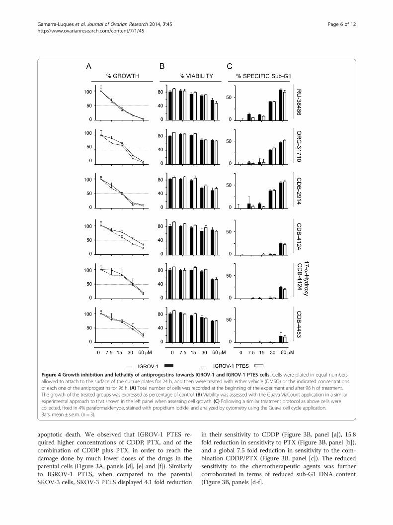
Figure 4 Growth inhibition and lethality of antiprogestins towards IGROV-1 and IGROV-1 PTES cells. Cells were plated in equal numbers,allowed to attach to the surface of the culture plates for 24 h, and then were treated with either vehicle (DMSO) or the indicated concentrationsof each one of the antiprogestins for 96 h. (A) Total number of cells was recorded at the beginning of the experiment and after 96 h of treatment.The growth of the treated groups was expressed as percentage of control. (B) Viability was assessed with the Guava ViaCount application in a similarexperimental approach to that shown in the left panel when assessing cell growth. (C) Following a similar treatment protocol as above cells werecollected, fixed in 4% paraformaldehyde, stained with propidium iodide, and analyzed by cytometry using the Guava cell cycle application.Bars, mean ± s.e.m. (n = 3).
Gamarra-Luques et al. Journal of Ovarian Research 2014, 7:45 Page 6 of 12http://www.ovarianresearch.com/content/7/1/45
apoptotic death. We observed that IGROV-1 PTES re-quired higher concentrations of CDDP, PTX, and of thecombination of CDDP plus PTX, in order to reach thedamage done by much lower doses of the drugs in theparental cells (Figure 3A, panels [d], [e] and [f]). Similarlyto IGROV-1 PTES, when compared to the parentalSKOV-3 cells, SKOV-3 PTES displayed 4.1 fold reduction
in their sensitivity to CDDP (Figure 3B, panel [a]), 15.8fold reduction in sensitivity to PTX (Figure 3B, panel [b]),and a global 7.5 fold reduction in sensitivity to the com-bination CDDP/PTX (Figure 3B, panel [c]). The reducedsensitivity to the chemotherapeutic agents was furthercorroborated in terms of reduced sub-G1 DNA content(Figure 3B, panels [d-f].
Gamarra-Luques et al. Journal of Ovarian Research 2014, 7:45 Page 7 of 12http://www.ovarianresearch.com/content/7/1/45
Although with different potencies, antiprogestins blockgrowth of ovarian cancer cells regardless of theirsensitivities to the combination CDDP-PTXWe next studied the responses of IGROV-1 and SKOV-3 cells and their respective, less chemosensitive siblingsIGROV-1 PTES and SKOV-3 PTES, to a panel of anti-progestin derivatives. Previously we have shown, usingdose-response studies, that antiprogestin RU-38486 (mife-pristone), ORG-31710, and CDB-2914 (ulipristal) havecytostatic effects at lower doses and lethal effects at higherconcentrations. Herein, in addition to those three antipro-gestins, we studied CDB-4124 (proellex) and two of itsderivatives, 17-α-hydroxy CDB-4124 and CDB-4453, thelatter carrying a de-methylation in position 11 (Figure 1).We assessed whether the panel of antiprogestins are ableto display their cytotoxic effect, either cytostasis orlethality, in cells that had been made simultaneouslyresistant (i.e., double resistant) to CDDP and PTX. Fourdays after treatment with the various antiprogestins, weevaluated the responses of the cells in terms of growth inculture by measuring cell number and of lethality byassessing cell viability and sub-G1 DNA content.Figure 4 shows that the six antiprogestins studied
inhibited the growth of both IGROV-1 and IGROV-1PTES cells in dose-related manners. The magnitude ofthe growth inhibition ranged with IC50s from ~11 to35 μM depending upon the compounds (Figure 4A, andTable 2). The growth inhibition effect did not change foreach compound in between the sibling cells, except for aslight, yet significant decline in the ORG-31710 andCDB-4124 IC50s for the PTES cells when compared tothe parental cells (Table 2). When we studied the lethal-ity of the antiprogestins towards IGROV-1 and IGROV-1 PTES we observed the six compounds impaired theviability of the cells when used at concentrations equalto or higher than 15 μM. The antiprogestins with higherlethality, as indicated by their manifestation at lowerconcentrations, were RU-38486, ORG-31710 and CDB-2914, when compared to the CDB-4124 derivatives thatdisplayed lethality only at concentrations over 30 μM(Figure 4B and C). Overall, the effect of the antiproges-tins was similar in IGROV-1 and IGROV-1 PTES, sug-gesting that their anti-cancer effect is independent of theintrinsic sensitivity to CDDP and PTX displayed by theovarian cancer cells.
Data shown are the IC50 (μM) expressed as the mean ± s.e.m (n = 3); OH-4124 indicate
In Figure 5 we show the response to antiprogestins ofthe ovarian cancer cells SKOV-3 and their derivativesSKOV-3 PTES less sensitive to CDDP and PTX. Simi-larly to what it was found with IGROV-1 cells, bothSKOV-3 and SKOV-3 PTES were impaired in theirgrowth and viability by antiprogestins in a dose-relatedmanner, without displaying major differences in theresponses among them (Figure 5A). The magnitude inthe IC50s for antiprogestins in the SKOV-3 cell linepair displayed a larger range when compared to theIGROV-1 pair, expanding from ~15 μM to 84 μM(Table 2). The most potent compounds in terms oflethality were RU-38486 and ORG-31710, with CDB-2914 and CDB-4124 derivatives having lesser effects(Figure 5B and C).To further study the effect of antiprogestins on cytos-
tasis and lethality towards ovarian cancer cells of simi-lar genetic backgrounds but different, double sensitivityto CDDP and PTX, we cultured IGROV-1 and IGROV-1 PTES in the presence of a fixed dose of 30 μM anti-progestins, which for these cells represents the limitingconcentration between the induction of cytostasis andlethality depending on the compound used (Figure 4C).Results in Figure 6 show that exposure to the saidconcentration of the compounds for 48 h caused anincrease in the abundance of the cell cycle inhibitorp27kip1, which was more notable for RU-38486, ORG-31710, CDB-2914 and 17-α-hydroxy CDB-4124 in bothparental and PTES cells. The cyclin dependent kinaseinhibitor p21cip1 also increased in response to the anti-progestins, yet this increase was more marked in PTEScells versus parental cells when comparing treatmentversus vehicle. The increases in p21cip1 and p27kip1 byantiprogestins are consistent with cell cycle arrest asso-ciated with growth inhibition. No major differenceswere observed in the expression of G1 regulatory pro-teins Cdk2 and cyclin E in response to antiprogestinsbetween parental and PTES cells. In terms of signs oflethality, the cleavage of PARP was observed in bothIGROV-1 and IGROV-1 PTES cells in response to thecompounds that had more potency in terms of reducingcellular viability (Figure 4C), with RU-38486, ORG-31710 and CDB-2914 displaying greater cleavage ofPARP when compared to the CDB-4124 derivatives(Figure 6).
P and PTX resistant cells
2914 CDB-4124 OH-4124 CDB-4453
1.10 35.5 ± 3.90 28.6 ± 3.40 30.6 ± 2.70
1.10 21.3 ± 1.80c 25.9 ± 2.40 25.6 ± 2.60
3.10 43.6 ± 5.10 84.2 ± 9.80 44.9 ± 4.60
1.80 47.4 ± 3.90 58.4 ± 8.30 52.9 ± 2.10
s 17-α-hydroxy CDB-4124. cp < 0.05 (Student’s t-test) compared to parental cells.
Figure 5 Growth inhibition and lethality of antiprogestins towards SKOV-3 and SKOV-3 PTES cells. Experiments were similar to thosedescribed in Figure 4, but with a different pair of cell lines. Percent cell growth (A), percent viability (B) and percent specific Sub-G1 (C) wereassessed for both cell lines in response to six different antiprogestins. Bars, mean ± s.e.m. (n = 3).
Gamarra-Luques et al. Journal of Ovarian Research 2014, 7:45 Page 8 of 12http://www.ovarianresearch.com/content/7/1/45
DiscussionMany studies with chemoresistant ovarian cancer cellsin vitro have been done using cells obtained from pa-tient’s ascites and that are not chemotherapy naïve. Forinstance, PEO4 cells were obtained from a patient thatreceived platinum-based therapy nine months earlier anddisplay a ~8 fold resistance to CDDP when compared totheir platinum sensitive counterparts PEO1 cells [16,17].Another example is the SKOV-3 cell line, which is consid-ered semi-resistant to platinum in vivo as was obtained
from a patient that did not respond to the maximal toler-ated dose of platinum [18]. The chemoresistance of thesecells developed within the in vivo environment of the pa-tient, can be considered clinically relevant and is usuallyreported to involve between 2- to 5-fold increases in theirIC50 values when compared to the parental cells (reviewedin [19]). However, there are also various cell line pairs thatwere made resistant to platinum-therapy by stepwiseexposure to CDDP in vitro, and, because they are highlystable, such as the OV2008 and OV2008/C13 or the
Figure 6 Effect of antiprogestins on the expression of p21cip1, p27kip1, Cdk2, cyclin E, and PARP in IGROV-1 (A) and IGROV-1 PTES (B)cells. Cells were treated with 30 μM antiprogestins for 48 h, whole cell extracts were isolated, electrophoresed, transferred to a PVDF membrane,and exposed to the indicated antibodies. HSC-70, a highly conserved protein that belongs to the HSP70 family of molecular chaperones, wasused as control for protein loading.
Gamarra-Luques et al. Journal of Ovarian Research 2014, 7:45 Page 9 of 12http://www.ovarianresearch.com/content/7/1/45
A2780 and A2780/CP70 siblings, they have been very use-ful to study mechanisms of chemoresistance in the labora-tory [20]. Yet, because these cells exhibit over 8-foldincrease in resistance, they should be considered less rele-vant from a clinical standpoint [12,19]. Furthermore, thereis significant heterogeneity in the type of resistance pa-tients develop after front line platinum-taxane chemother-apy, including patients that show high sensitivity, othersthat show sensitivity to one agent but resistance to theother, or patients that show resistance to both drugs [21].We decided to develop cell lines with resistance to
both platinum and taxanes, within the range of clinicalrelevance. We utilized the IGROV-1 and SKOV-3 celllines as IGROV-1 cells were previously reported to becapable of acquiring cross-resistance to PTX when maderesistant to CDDP [22], whereas SKOV-3 cells, with clin-ically relevant endogenous resistance to CDDP, weremade PTX resistant upon pulse selection [23]. We gen-erated clinically relevant IGROV-1 and SKOV-3 cells re-sistant to CDDP and PTX by exposing them to bothdrugs in six pulse-selection challenges. We termed theIGROV-1 and SKOV-3 derivatives IGROV-1 PlatinumTaxane EScape or PTES and SKOV-3 PTES, respectively,which showed double resistances in the range of 2-7folds compared to their parental cells. We then asked ifthe sibling cell lines depict cross-resistance to the anti-growth effect of antiprogestins. Indeed we confirmedthat all antiprogestins utilized in the study (i.e., mifepris-tone, ORG-31710, CDB-2914, CDB-4124, 17-α-hydroxyCDB-4124 and CDB-4453) blocked the growth of theparental and resistant derivatives cells with overall simi-lar potency, with cytostasis manifested at concentrationslower than 15 μM, and lethality manifested at doses
higher than 15 μM. These results are in agreement witha previous study we performed using the antiprogestinmifepristone in OV2008 cells and compared its effectagainst that observed in the highly resistant OV2008/C13siblings. Mifepristone killed cells when used at doseshigher than 20 μM, whereas at lower doses, it causedcytostasis that was reversed when the drug was removedfrom the culture [8]. The anti-growth effect of mifepris-tone was also independent of the presence of the tumorsuppressor p53, because it did not discriminate betweenA2780 wt 53 platinum sensitive cells and A2780/CP70platinum resistant cells carrying a mutant version of p53[24-26], IGROV-1 cells with p53 wt expression [27], orSKOV-3 reported to carry a single nucleotide deletion inthe p53 gene leading to no expression of p53 [28-31].Mifepristone is one of the most popular antiprogestins
ever developed. It was synthesized in the early 1980’s asan antiglucocorticoid but soon afterward was found toblock the transcriptional activity of progesterone recep-tors (PR). Because of such an antiglucocorticoid effect,new generation antiprogestins were developed aiming atreducing antiglucocorticoid activity while maintaining orenhancing antiprogesterone activity. Such compounds in-clude ORG-31710 and the CDB family members studiedhere, CDB-2914 and CDB-4124. The differences betweenthe compounds are in the substitutions localized at thepositions 11β and 17α. Whereas the dimethylaminophenylsubstitution at the 11β-position seems to confer antipro-gestin activity [32-34], modifications in position C-17 aremostly geared at modifying the antiglucocorticoid receptoractivity of the compounds. Thus, ORG-31710 and the CDBderivatives are considered to have potent antiprogestinactivity with less antiglucocorticoid activity when compared
Gamarra-Luques et al. Journal of Ovarian Research 2014, 7:45 Page 10 of 12http://www.ovarianresearch.com/content/7/1/45
to mifepristone [35,36]. Of these compounds, mifepristonehas been approved in the US to terminate early pregnancy(working as an antiprogestin) or ameliorating the hypergly-cemia in patients with endogenous Cushing’s (working asan antiglucocorticoid) (reviewed in [37]). CDB-2914(ulipristal) and CDB-4124 (proellex) are currently underintense investigation to assess their capacity to mitigatesigns and symptoms associated with increased cell growthin endometriosis and uterine fibroids (reviewed in [38]).We have found that mifepristone, the compound with
the highest antiglucocorticoid effect, is the most potentagainst the growth of ovarian cancer cells either sensitiveor resistant to the combination CDDP/PTX. The new gen-eration antiprogestins represented by the CDB compoundsare effective, but with a higher IC50. It is interesting tonote that either the 17-α-hydroxylated or demethylatedforms of CDB-4124 did not show superior potency overCDB-4124 in terms of growth inhibition, suggesting thatthe anti-growth effect of the molecules resides in a yet tobe identified functional group.CDB-4124 and the putative mono-demethylated metab-
olite CDB-4453 are all potent antiprogestins but havelimited antiglucocorticoid activity when compared againstmifepristone or CDB-2914 [35,39]. Because the anti-growth potency of the CDB derivatives was slightlyreduced when compared to that of mifepristone or ORG-31710, these results suggest that the antiprogestin functionof the molecule may be unrelated to its anti-growthcapacity. Indeed we have shown that cancer cells of differ-ent tissues of origin and different degrees of hormone-dependency, such as MCF-7 breast cancer cells carryingPR, MDA-MB-231 breast cancer cells with no PR expres-sion, PR negative and androgen receptor positive LNCaPprostate cancer cells, and PR negative androgen receptorpositive PC3 prostate cancer cells are all inhibited by mife-pristone with similar potency [40], strongly suggesting thatthe presence of the PR is not required for the inhibition ofcancer growth triggered by antiprogestins. Further sup-porting this hypothesis it was shown that mifepristoneblocked the growth of estrogen receptor negative and PRnegative MDA-MB-231 breast cancer cells [41].In another line of reasoning, it is possible that the anti-
glucocorticoid effect of the molecules may have some rolein the antigrowth effect, as all cell lines being studiedexpress glucocorticoid receptors (GR) [40]. The humanNR3C1 gene undergoes alternative splicing generating twomain isoforms, GRα and GRβ. Considerable evidence indi-cates that the GRα isoform drives GR-mediated transactiva-tion activity, whereas GRβ is a natural dominant negativeinhibitor of GRα; however, GRβ can directly regulate genesnot controlled by GRα (reviewed in [42]). We have shownthat mifepristone blocked the growth of cancer cells withvery low expression of GRα, such as OVCAR-3 ovariancancer cells, MCF-7 breast cancer cells, and U-2OS
osteosarcoma cells [40], suggesting that the presence ofGRα may not be required for the antigrowth effect of anti-progestins. It remains to be determined, however, whetherGRβ, which was reported capable of binding mifepristone[43], plays a role in the anti-growth effect of antiprogestins,as this receptor isoform seems to be present in all cell lineswe studied so far [40].
ConclusionsAntiprogestins of different generations with higher orlesser antiglucocorticoid activity can block the growth ofovarian cancer cells that have been made resistant toCDDP and PTX in a clinically relevant manner. Althoughthe molecular mechanisms driving the growth inhibitionby antiprogestins requires more detail, it is clear that thedrugs could be developed further for anti-ovarian cancertherapy, in particular for those cases that show upfrontresistance to standard of care, or for recurrent patientswith platinum and/or taxane resistant disease. Due to thelow toxicity of these drugs, their potential use as mainten-ance therapy or antirepopulation therapy following stand-ard of care is anticipated. In this regard we havedemonstrated that mifepristone was capable of abrogatingthe regrowth of cancer cells that escaped CDDP [9] or thecombination CDDP/PTX therapy [10]. The results pre-sented herein highlight the fact that other antiprogestins inaddition to mifepristone can be efficacious against plat-inum/taxane double-resistant ovarian cancer cells. Whetheror not ovarian cancer cells may develop resistance to anti-progestin therapy after exposure to the drugs for longperiods of time, remains to be investigated.
Competing interestsThe authors declare that they have no competing interests.
Authors’ contributionsCGL and CMT conceived and designed the experiments. AAG performedinitial validation experiments with CDB-4124 derivatives. CGL developed thedrug-resistant cell lines and performed a comprehensive study on theirresponses to a panel of antiprogestins. MBH contributed with the westernblot assays. CMT contributed with the reagents, materials and analysis tools.CGL and CMT wrote the paper. All authors approved the final version of themanuscript.
AcknowledgementsThis research was supported by award number R15 CA164622 from theNational Cancer Institute, the National Institutes of Health (NIH). We thankMr. Nahuel Telleria for the edition of the manuscript.
Author details1Division of Basic Biomedical Sciences, Sanford School of Medicine, TheUniversity of South, Dakota, 414 East Clark Street, Vermillion, SD 57069, USA.2Present Address: Institute of Medicine and Experimental Biology of Cuyo,National Council for Scientific and Technical Research (CONICET), Mendoza,Argentina.
Gamarra-Luques et al. Journal of Ovarian Research 2014, 7:45 Page 11 of 12http://www.ovarianresearch.com/content/7/1/45
Received: 12 March 2014 Accepted: 21 April 2014Published: 27 April 2014
References1. Romero I, Bast RC Jr: Minireview: human ovarian cancer: biology, current
management, and paths to personalizing therapy. Endocrinology 2012,153(4):1593–1602.
2. Vaughan S, Coward JI, Bast RC Jr, Berchuck A, Berek JS, Brenton JD, CoukosG, Crum CC, Drapkin R, Etemadmoghadam D, Friedlander M, Gabra H, KayeSB, Lord CJ, Lengyel E, Levine DA, McNeish IA, Menon U, Mills GB, NephewKP, Oza AM, Sood AK, Stronach EA, Walczak H, Bowtell DD, Balkwill FR:Rethinking ovarian cancer: recommendations for improving outcomes.Nat Rev Cancer 2011, 11(10):719–725.
3. Bast RC Jr, Hennessy B, Mills GB: The biology of ovarian cancer: newopportunities for translation. Nat Rev Cancer 2009, 9(6):415–428.
4. Kurman RJ, Shih Ie M: Molecular pathogenesis and extraovarian origin ofepithelial ovarian cancer–shifting the paradigm. Hum Pathol 2011,42(7):918–931.
6. Bast RC Jr: Molecular approaches to personalizing management ofovarian cancer. Ann Oncol 2011, 22(Suppl 8):viii5.
7. Telleria CM, Goyeneche AA: Antiprogestins in Ovarian Cancer. In OvarianCancer -Clinical and Therapeutic Perspectives. Chapter 11. Edited by FarghalyS. Rijeka, Croatia: InTechopen; 2012.
8. Goyeneche AA, Caron RW, Telleria CM: Mifepristone inhibits ovariancancer cell growth in vitro and in vivo. Clin Cancer Res 2007,13(11):3370–3379.
9. Freeburg EM, Goyeneche AA, Telleria CM: Mifepristone abrogatesrepopulation of ovarian cancer cells in between courses of cisplatintreatment. Int J Oncol 2009, 34(3):743–755.
10. Gamarra-Luques CD, Goyeneche AA, Hapon MB, Telleria CM: Mifepristoneprevents repopulation of ovarian cancer cells escaping cisplatin-paclitaxeltherapy. BMC Cancer 2012, 12:200.
11. Goyeneche AA, Seidel EE, Telleria CM: Growth inhibition induced byantiprogestins RU-38486, ORG-31710, and CDB-2914 in ovarian cancercells involves inhibition of cyclin dependent kinase 2. Invest New Drugs2012, 30(3):967–980.
12. Freeburg EM, Goyeneche AA, Seidel EE, Telleria CM: Resistance to cisplatindoes not affect sensitivity of human ovarian cancer cell lines tomifepristone cytotoxicity. Cancer Cell Int 2009, 9:4.
13. Kurata T, Tamura T, Shinkai T, Ohe Y, Kunitoh H, Kodama T, KakinumaR, Matsumoto T, Kubota K, Omatsu H, Nishiwaki Y, Saijo N: Phase I andpharmacological study of paclitaxel given over 3 h with cisplatin foradvanced non-small cell lung cancer. Jpn J Clin Oncol 2001,31(3):93–99.
14. Himmelstein KJ, Patton TF, Belt RJ, Taylor S, Repta AJ, Sternson LA: Clinicalkinetics on intact cisplatin and some related species. Clin Pharmacol Ther1981, 29(5):658–664.
15. du Bois A, Luck HJ, Buser K, Meerpohl HG, Sessa C, Klaassen U, Meden H,Bochtler H, Diergarten K: Extended phase II study of paclitaxel as a 3-hinfusion in patients with ovarian cancer previously treated with platinum.Eur J Cancer 1997, 33(3):379–384.
16. Langdon SP, Lawrie SS, Hay FG, Hawkes MM, McDonald A, Hayward IP,Schol DJ, Hilgers J, Leonard RC, Smyth JF: Characterization and propertiesof nine human ovarian adenocarcinoma cell lines. Cancer Res 1988,48(21):6166–6172.
17. Cooke SL, Ng CK, Melnyk N, Garcia MJ, Hardcastle T, Temple J, Langdon S,Huntsman D, Brenton JD: Genomic analysis of genetic heterogeneity andevolution in high-grade serous ovarian carcinoma. Oncogene 2010,29(35):4905–4913.
18. Ormerod MG, O'Neill C, Robertson D, Kelland LR, Harrap KR: cis-Diamminedichloroplatinum(II)-induced cell death through apoptosis insensitive and resistant human ovarian carcinoma cell lines. CancerChemother Pharmacol 1996, 37(5):463–471.
19. MdDermott M, Eustace AJ, Busschots S, Breen L, Crown J, Clynes M,O'Donovan N, Stordal B: In vitro development of chemotherapy andtargeted therapy drug-resistant cancer cell lines: a practical guide withcase studies. Frontiers Oncol 2014, 4:40.
20. Katano K, Kondo A, Safaei R, Holzer A, Samimi G, Mishima M, Kuo YM,Rochdi M, Howell SB: Acquisition of resistance to cisplatin isaccompanied by changes in the cellular pharmacology of copper.Cancer Res 2002, 62(22):6559–6565.
21. Ledermann JA, Kristeleit RS: Optimal treatment for relapsing ovariancancer. Ann Oncol 2010, 21(Suppl 7):vii218–vii222.
22. Stordal B, Hamon M, McEneaney V, Roche S, Gillet JP, O'Leary JJ, GottesmanM, Clynes M: Resistance to paclitaxel in a cisplatin-resistant ovariancancer cell line is mediated by P-glycoprotein. PLoS One 2012,7(7):e40717.
23. Yan XD, Li M, Yuan Y, Mao N, Pan LY: Biological comparison of ovariancancer resistant cell lines to cisplatin and Taxol by two differentadministrations. Oncol Rep 2007, 17(5):1163–1169.
24. Lu X, Errington J, Curtin NJ, Lunec J, Newell DR: The impact of p53 statuson cellular sensitivity to antifolate drugs. Clin Cancer Res 2001,7(7):2114–2123.
25. Siddik ZH, Mims B, Lozano G, Thai G: Independent pathways of p53induction by cisplatin and X-rays in a cisplatin-resistant ovarian tumorcell line. Cancer Res 1998, 58(4):698–703.
26. Debernardis D, Sire EG, De Feudis P, Vikhanskaya F, Valenti M, Russo P, Parodi S,D'Incalci M, Broggini M: p53 status does not affect sensitivity of humanovarian cancer cell lines to paclitaxel. Cancer Res 1997, 57(5):870–874.
27. Righetti SC, Perego P, Corna E, Pierotti MA, Zunino F: Emergence of p53mutant cisplatin-resistant ovarian carcinoma cells following drugexposure: spontaneously mutant selection. Cell Growth Differ 1999,10(7):473–478.
28. Yaginuma Y, Westphal H: Abnormal structure and expression of the p53gene in human ovarian carcinoma cell lines. Cancer Res 1992,52(15):4196–4199.
29. O'Connor PM, Jackman J, Bae I, Myers TG, Fan S, Mutoh M, Scudiero DA,Monks A, Sausville EA, Weinstein JN, Friend S, Fornace AJ Jr, Kohn KW:Characterization of the p53 tumor suppressor pathway in cell lines ofthe National Cancer Institute anticancer drug screen and correlationswith the growth-inhibitory potency of 123 anticancer agents. Cancer Res1997, 57(19):4285–4300.
30. Berglind H, Pawitan Y, Kato S, Ishioka C, Soussi T: Analysis of p53 mutationstatus in human cancer cell lines: a paradigm for cell line cross-contamination. Cancer Biol Ther 2008, 7(5):699–708.
31. Hamroun D, Kato S, Ishioka C, Claustres M, Beroud C, Soussi T: The UMDTP53 database and website: update and revisions. Hum Mutat 2006,27(1):14–20.
32. Belanger A, Philibert D, Teutsch G: Regio and stereospecific synthesis of11 beta- substituted 19-norsteroids. Influence of 11 beta-substitution onprogesterone receptor affinity - (1). Steroids 1981, 37(4):361–382.
33. Benagiano G, Bastianelli C, Farris M: Selective progesterone receptormodulators 1: use during pregnancy. Expert Opin Pharmacother 2008,9(14):2459–2472.
34. Moller C, Hoffmann J, Kirkland TA, Schwede W: Investigationaldevelopments for the treatment of progesterone-dependent diseases.Expert Opin Investig Drugs 2008, 17(4):469–479.
35. Attardi BJ, Burgenson J, Hild SA, Reel JR, Blye RP: CDB-4124 and itsputative monodemethylated metabolite, CDB-4453, are potent antipro-gestins with reduced antiglucocorticoid activity: in vitro comparison tomifepristone and CDB-2914. Mol Cell Endocrinol 2002, 188(1–2):111–123.
36. Leonhardt SA, Edwards DP: Mechanism of action of progesteroneantagonists. Exp Biol Med (Maywood) 2002, 227(11):969–980.
37. Telleria CM: Drug Repurposing for Cancer Therapy. J Cancer Sci Ther 2012,4(7):ix–xi.
38. Spitz IM: Clinical utility of progesterone receptor modulators and theireffect on the endometrium. Curr Opin Obstet Gynecol 2009, 21(4):318–324.
39. Attardi BJ, Burgenson J, Hild SA, Reel JR: In vitro antiprogestational/antiglucocorticoid activity and progestin and glucocorticoid receptorbinding of the putative metabolites and synthetic derivatives of CDB-2914, CDB-4124, and mifepristone. J Steroid Biochem Mol Biol 2004,88(3):277–288.
40. Tieszen CR, Goyeneche AA, Brandhagen BN, Ortbahn CT, Telleria CM:Antiprogestin mifepristone inhibits the growth of cancer cells ofreproductive and non- reproductive origin regardless of progesteronereceptor expression. BMC Cancer 2011, 11:207.
41. Liang Y, Hou M, Kallab AM, Barrett JT, El Etreby F, Schoenlein PV: Inductionof antiproliferation and apoptosis in estrogen receptor negative MDA-
Gamarra-Luques et al. Journal of Ovarian Research 2014, 7:45 Page 12 of 12http://www.ovarianresearch.com/content/7/1/45
231 human breast cancer cells by mifepristone and 4-hydroxytamoxifencombination therapy: a role for TGFbeta1. Int J Oncol 2003, 23(2):369–380.
42. Kadmiel M, Cidlowski JA: Glucocorticoid receptor signaling in health anddisease. Trends Pharmacol Sci 2013, 34(9):518–530.
43. Lewis-Tuffin LJ, Jewell CM, Bienstock RJ, Collins JB, Cidlowski JA: Humanglucocorticoid receptor beta binds RU-486 and is transcriptionally active.Mol Cell Biol 2007, 27(6):2266–2282.
doi:10.1186/1757-2215-7-45Cite this article as: Gamarra-Luques et al.: Resistance to cisplatin andpaclitaxel does not affect the sensitivity of human ovarian cancer cellsto antiprogestin-induced cytotoxicity. Journal of Ovarian Research2014 7:45.
Submit your next manuscript to BioMed Centraland take full advantage of:
• Convenient online submission
• Thorough peer review
• No space constraints or color figure charges
• Immediate publication on acceptance
• Inclusion in PubMed, CAS, Scopus and Google Scholar
• Research which is freely available for redistribution
Submit your manuscript at www.biomedcentral.com/submit