Interleukin-6 receptor specific RNA aptamersfor cargo delivery into target cells
Cindy Meyer,1 Katja Eydeler,1 Eileen Magbanua,1 Tijana Zivkovic,1 Nicolas Piganeau,1 Inken Lorenzen,2 Joachim Grötzinger,2
Günter Mayer,3 Stefan Rose-John2 and Ulrich Hahn1,*
1Institute for Biochemistry and Molecular Biology; Chemistry Department; MIN-Faculty; Hamburg University; Hamburg, Germany; 2Institute of Biochemistry; Medical Faculty;
Christian-Albrechts-University; Kiel, Germany; 3Life and Medical Sciences Institute; University of Bonn; Bonn, Germany
Aptamers represent an emerging strategy to deliver cargo molecules, including dyes, drugs, proteins or even genes, intospecific target cells. Upon binding to specific cell surface receptors aptamers can be internalized, for example bymacropinocytosis or receptor mediated endocytosis. Here we report the in vitro selection and characterization of RNAaptamers with high affinity (Kd = 20 nM) and specificity for the human IL-6 receptor (IL-6R). Importantly, these aptamerstrigger uptake without compromising the interaction of IL-6R with its natural ligands the cytokine IL-6 and glycoprotein130 (gp130). We further optimized the aptamers to obtain a shortened, only 19-nt RNA oligonucleotide retaining allnecessary characteristics for high affinity and selective recognition of IL-6R on cell surfaces. Upon incubation with IL-6Rpresenting cells this aptamer was rapidly internalized. Importantly, we could use our aptamer, to deliver bulky cargos,exemplified by fluorescently labeled streptavidin, into IL-6R presenting cells, thereby setting the stage for an aptamer-mediated escort of drug molecules to diseased cell populations or tissues.
Introduction
Interleukin-6 (IL-6) is a multifunctional cytokine that is involvedin many immune and inflammatory responses. It belongs to thefamily of the four-helix bundle cytokines.1 IL-6 interacts with itsnatural receptor, namely IL-6R, and two molecules of theglycoprotein 130 (gp130), the transducer of the IL-6 signal.2
IL-6R appearance is restricted to a few cell types, includinghepatocytes, monocytes, macrophages and some lymphocytes.2 Incontrast, gp130 can be found on nearly all cell types. Cells that donot produce the membrane bound IL-6R (mIL-6R) can bestimulated by IL-6 via a soluble form of the IL-6R (sIL-6R). ThesIL-6R is produced by cells positive for mIL-6R via alternativesplicing or proteolytic cleavage of the membrane bound form.3,4
The sIL-6R in complex with IL-6 enables IL-6R lacking cells torespond to IL-6 mediated signaling.2
IL-6 and its receptor are involved in the progression of variousinflammatory diseases, such as Crohn’s disease and rheumatoidarthritis (RA), and certain cancers, for example multiple myelomaor hepatocellular carcinoma.5 One promising therapeutic strategyto tackle the mentioned diseases could be the specific delivery ofdrugs into cells by targeting IL-6R.
Aptamers are nucleic acids (DNA or RNA) consisting of about15–100 nt, which are able to bind target molecules with highaffinity and specificity, exhibiting Kd values in the picomolarrange. Aptamers can be obtained by an in vitro selection process,termed SELEX (systematic evolution of ligands by exponentialenrichment).6,7 Starting with a combinatorial oligonucleotidelibrary containing up to 1015 different nucleic acid species, theSELEX process leads to the enrichment of an oligonucleotidepopulation (polyclonal aptamers) that interacts with the targetmolecule due to defined three-dimensional structures. In respectof recognition properties aptamers are comparable to antibodieseven though they reveal remarkable advantages, such as lowimmunogenicity and toxicity, longer shelf-life, and lower produc-tion costs. Up to now, aptamers were evolved for a wide variety oftarget molecules, including ions,8 fluorescent dyes,9 antibiotics,10
peptides,11 and proteins.12 Furthermore aptamers targetingviruses13 as well as whole cells14 have previously been selectedand characterized for potential therapeutic applications. Next totheir appliance in diagnostics, aptamers can serve as stimulating15
or inhibiting16 ligands, respectively. One very promising approachis the application of cell-specific aptamers as delivery vehicles fordrug molecules into target cells or tissues. One auspicious example
is the successful selection of an RNA aptamer binding to PSMA(prostate-specific membrane antigen) presented on the surfaceof prostate cancer cells.17 Upon binding, anti-PSMA aptamersundergo receptor-mediated internalization. Coupling those nuc-leic acids to cargo molecules, such as toxins,18 siRNAs,19 chemo-therapeutics, and nanoparticles20 improves delivery to the cells ofinterest. This can allow to lower the dose required for effectivetreatment and is therefore considered a promising therapeuticapproach which had been validated in animal studies.21
Thus, aptamers are very promising tools for cell-specifictargeting. However, there is only a limited number of appropriaterepresentatives to date. Therefore, new aptamers specific for cellsurface proteins or whole cells are urgently needed.
We set out to enhance the toolbox of cell-targeting and drugdelivering molecules. Thus, we decided to select aptamers specificfor the human IL-6R, a cell surface receptor presented on a varietyof cells often connected to numerous diseases, like the alreadymentioned multiple myeloma. Our studies yielded a 19-nt RNAaptamer, namely AIR-3A, which specifically recognizes cells thatpresent IL-6R. This aptamer is able to transport cargo moleculesof different sizes, exemplified by fluorescent dyes and streptavidin,into living IL-6R presenting cells. In view that IL-6 activelyparticipates in inflammation associated cancer,22,23 selectivetargeting of IL-6R presenting tumor cells with toxic substancescoupled to aptamers might be a valuable strategy to broadenestablished IL-6 or IL-6R directed treatment regimens.
Results
In vitro selection of sIL-6R-specific RNA aptamers. Aiming forRNA aptamers binding to cells that present IL-6R on their sur-face, we used the extracellular soluble part of the receptor (sIL-6R)as target molecule during the in vitro selection experiment. In thefirst selection cycle the RNA library R1 containing 1013 individualRNA molecules was incubated with sIL-6R, immobilized onmagnetic beads. RNA molecules binding to sIL-6R were capturedby magnetic separation and amplified by RT-PCR. After in vitro
transcription the enriched RNA library was used as startingmaterial for the subsequent selection cycle. The stringency ofthe selection process was enhanced gradually by increasing thenumber of washing steps with each additional selection cycle.After 16 selection rounds the enriched RNA library was analyzedfor sIL-6R binding by filter retention analysis (FRA), whichrevealed a significantly increased affinity of the cycle 16 RNA forsIL-6R in comparison to the initial RNA library R1 (Fig. S1).
Subsequently, the corresponding cycle 16 dsDNA library wascloned and the sequences of 20 clones were determined (Fig. 1).We identified six individual sIL-6R-binding RNA aptamers allsharing a G-rich consensus motif.
Affinity and specificity of the RNA aptamer AIR-3 for sIL-6R.Initial FRAs revealed that among other tested aptamers theRNA aptamer AIR-3 exhibited the highest affinity for sIL-6R(Table S1 and Fig. S2). We further determined the dissociationconstant (Kd) for AIR-3 binding to sIL-6R more precisely byrepeating the FRA for ten times. Thereby, the initial RNA libraryR1 served as a control. After quantification resulting curveswere fitted assuming a 1:1 binding stoichiometry between bothinteracting partners. AIR-3 revealed a high affinity for sIL-6Rwith a calculated Kd-value of 19.7 ± 4.2 nM (Table 1 and Fig. 2).No binding of the RNA library R1 was detectable. Further FRAsrevealed that AIR-3 did not bind to the control proteins IL-6 andlysozyme (data not shown).
Influence of AIR-3 on the interactions between sIL-6R and itsligands IL-6 and gp130. To determine whether aptamer AIR-3competed with the cytokine IL-6 for binding to sIL-6R, filter
Figure 1. Sequences of RNA aptamers binding the soluble part of the IL-6 receptor (sIL-6R). Sequences are printed in 5’–3’ direction omitting flankingprimer-binding sites. Bold letters indicate conserved positions. F, frequency; consensus sequence is underlined and additionally given below. H: A, C or U;W: A or U.
Do not distribute.retention assays were employed. Increasing amounts of sIL-6Rwere pre-incubated with or without its ligand IL-6. Constantamounts of radioactively labeled aptamer AIR-3 were added.Figure 3A displays the results of the FRAs revealing nocompetition between IL-6 and the RNA aptamer AIR-3 forbinding to sIL-6R.
Knowing that the aptamer AIR-3 specifically interacts withsIL-6R even in the presence of its natural ligand IL-6, we inves-tigated the interaction of aptamer AIR-3 with the designercytokine Hyper-IL-6, a fusion protein consisting of human IL-6and the human sIL-6R connected by a flexible polypeptidechain.24 First, we performed FRAs to proof the ability of AIR-3 tointeract with Hyper-IL-6 (Fig. S3).
The ability of aptamer AIR-3 to disrupt the interactionbetween Hyper-IL-6 and the second receptor subunit gp130 wasexamined by electrophoretic mobility shift assay (EMSA). ThisEMSA confirmed that AIR-3 did bind to Hyper-IL-6 asillustrated by a retarded migration rate in the native PAGE ifcompared with sole RNA (Fig. 3B, lanes 1 and 2). Additionally, asupershift confirmed the interaction between AIR-3 and Hyper-IL-6 complexed with gp130 (Fig. 3B, lane 4). The complexformation between Hyper-IL-6 and gp130 was not disrupted byAIR-3. No unspecific binding of AIR-3 to gp130 could beobserved (Fig. 3B, lane 3).
Minimization of the RNA aptamer AIR-3. All identified sIL-6R specific aptamers contained a conserved G-rich motif (Fig. 1).According to this observation the aptamer AIR-3 was truncatedfinally yielding a 19 nucleotides short aptamer, termed AIR-3A(5'-GGGGAGGCUGUGGUGAG17G18G-3'). Three variants ofAIR-3A namely G17U, G18U and G17U/G18U were generatedto serve as controls. These variants bear G to U nucleotideexchanges at position 17, position 18 or at both positions,
respectively. The abilities of these four truncated RNA moleculesto bind Hyper-IL-6 were subsequently determined by FRA(Fig. S4, Table 1). The truncated aptamer AIR-3A exhibited ahigh affinity for Hyper-IL-6, whereas the variants G17U, G18Uand G17U/G18U did not show any significant binding.
Structural analyses of AIR-3A and its derived variants. In afirst CD spectroscopic experiment the spectrum of the RNAaptamer AIR-3A, solved in 1 � PBS (containing Na+ or K+ ions),revealed two main peaks, a maximum at 265 nm and a minimumat 240 nm (Fig. 4A), suggesting the formation of G-quadruplexeswith parallel strand orientation.25,26 The variants G17U, G18U,and G17U/G18U, each solved in PBS as well, did clearly show a
Figure 2. Aptamer AIR-3 binds the soluble IL-6 receptor (sIL-6R) withhigh affinity. Filter retention assays. Constant amounts (,1 nM) of32P-radioactively labeled aptamer AIR-3 (red diamonds) and RNA startinglibrary R1 (black circles) were incubated with increasing amounts ofsIL-6R (0–300 nM). Protein-bound RNA was visualized by autoradio-graphy. Fractions of bound RNA molecules were plotted against theconcentration of sIL-6R (logarithmic scale). Data points represent meanvalues of 10 independent measurements.
Figure 3. Aptamer AIR-3 does not compete with IL-6 or gp130 for bindingto IL-6R. (A) Filter retention assays. Constant amounts of IL-6 (1 mM) werepre-incubated with increasing amounts of sIL-6R (0–300 nM) beforeradiolabeled aptamer AIR-3 (,1 nM) was added to the complex. Fordata evaluation see legend to Figure2. (B) Interaction of aptamer AIR-3(,1 nM) with Hyper-IL-6 (500 nM) or with Hyper-IL-6/gp130 complex(1:2 stoichiometry) analyzed by gel-electrophoretic mobility shift assay.Lanes 1 and 5, free aptamer; remaining lanes contained aptamer + 500 nMHyper-IL-6 (2), + 1 mM gp130 (3) or both proteins (4).
decrease in signal intensity at the characteristic wavelength. The Gto U nucleotide exchanges seemed to destabilize or destruct theG-quadruplexes in all variants.
The formation of stable G-quadruplexes requires the presenceof special metal ions like K+ or Na+.27 Therefore we measured theCD spectra of AIR-3A in T-HCl buffer omitting monovalentsodium and potassium cations, respectively (Fig. 4B). Thequadruplex formation was obviously reduced as the resultingpeak amplitudes of the spectra decreased. After addition of potas-sium or sodium chloride both peaks increased again indicatingthat these ions are prerequisites for structural stability.
To confirm the potential G-quadruplex folding of AIR-3A weperformed UV melting transitions at 295 nm.28 First, we solvedAIR-3A in 10 mM Tris buffer (pH 7.5) including 5 mM KCl.We could show that the UV melting profile (Tm value 48.4°C)was characterized by a hypochromic shift as typically observedfor nucleic acids containing G-quadruplex structures (Fig. 4C andTable S2). We evaluated the stability of the AIR-3A G-quadruplexby determining its melting temperatures at different potassiumconcentrations (0 mM, 1 mM, and 10 mM, respectively). Themelting temperature decreased as a result of the reduction of theKCl concentration (Fig. 4C and Table S2). In absence of K+
cations melting curves did not show any hypochromic shift.Therefore it was not possible to determine the corresponding Tm
value. Thus folding was significantly dependent on K+ cations.The melting temperature of AIR-3A did not dependent on
RNA concentration over a range from 1 to 10 mM since the Tm
corresponding values did not change significantly (Fig. S5A andTable S2).
All three variants G17U, G18U, and G17U/G18U did notexhibit a hypochromic shift within the corresponding UV meltingprofiles neither in absence nor in presence of 5 mM KCl(Fig. S5B, Table S2). Therefore we could not calculate any Tm
values.Taken together, these results indicated that only AIR-3A
exhibited a stable intramolecular G-quadruplex under physio-logical conditions.
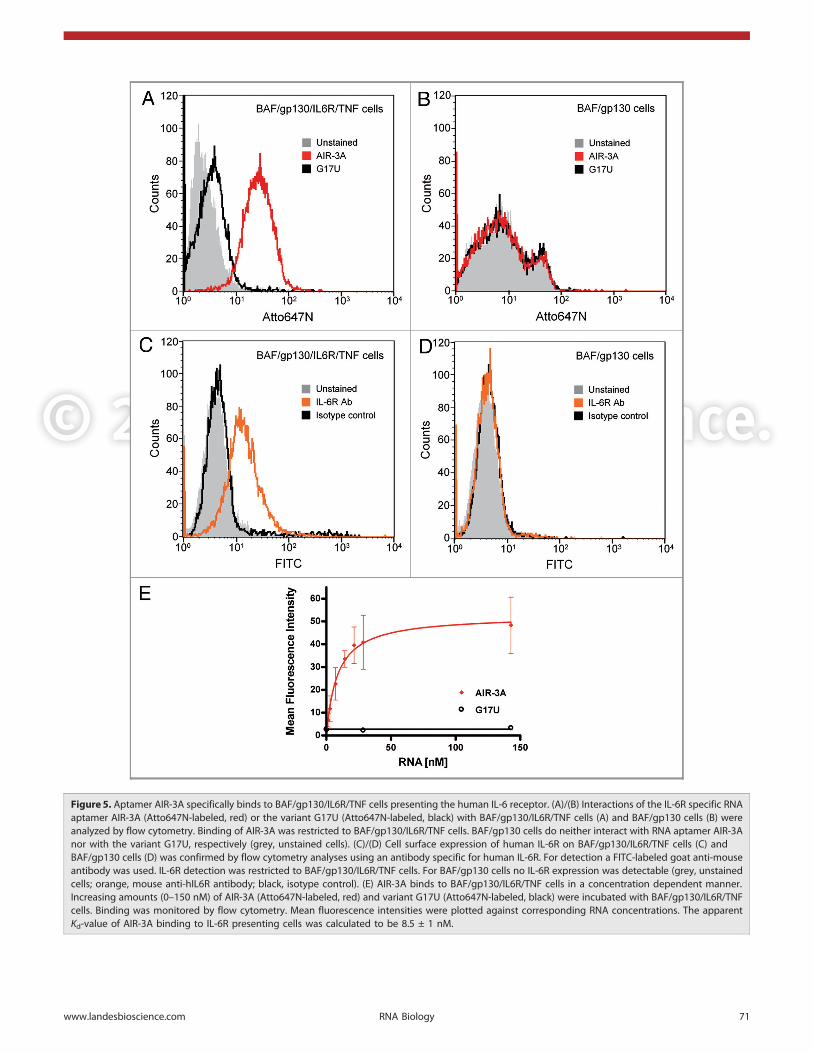
Interaction of AIR-3A with IL-6R presenting cells. Binding ofAIR-3A to BAF/gp130/IL6R/TNF cells. We subsequently deter-mined whether AIR-3A was able to bind IL-6R presenting cells.Therefore, we used two murine cell lines, BAF/gp130/IL6R/TNF-cells, a parental pro-B cell line transfected with the cDNA ofhuman gp130, human IL-6R, and TNF, as well as BAF/gp130cells, that were transfected solely with the cDNA of humangp130, the latter serving as control.
Atto647N-labeled aptamer AIR-3A and the control variantG17U were assayed for their binding capacities on IL-6Rpresenting BAF/gp130/IL6R/TNF cells by flow cytometry.AIR-3A but not G17U did bind to those cells (Fig. 5A). BothRNAs failed to bind to cells lacking IL-6R (Fig. 5B). IL-6Rexpression on BAF/gp130/IL6R/TNF cells but not on BAF/gp130 cells was confirmed using a monoclonal human IL-6Rspecific antibody. This antibody did bind to BAF/gp130/IL6R/TNF cells but not to IL-6R-negative BAF/gp130 cells (Fig. 5Cand D, respectively).
To analyze whether the binding capacity of the RNA aptamerin serum containing medium might be disturbed by contaminat-ing RNases we performed binding assays in selection buffer, cellculture medium and serum containing medium at 4°C or 37°C,
Figure 4. Structural analyses of AIR-3A and its variants. (A) CDspectroscopic analyses of AIR-3A, G17U, G18U and G17U/G18U (5 mM inPBS). (B) CD spectrum of AIR-3A (5 mM) in PBS or Tris buffer (50 mM)without or in the presence of potassium or sodium ions (100 mM each),respectively. (C) Melting profile of AIR-3A (2.5 mM; taken at 295 nm) in10 mM Tris buffer containing 1 mM, 5 mM or 10 mM KCl, respectively.
Figure 5. Aptamer AIR-3A specifically binds to BAF/gp130/IL6R/TNF cells presenting the human IL-6 receptor. (A)/(B) Interactions of the IL-6R specific RNAaptamer AIR-3A (Atto647N-labeled, red) or the variant G17U (Atto647N-labeled, black) with BAF/gp130/IL6R/TNF cells (A) and BAF/gp130 cells (B) wereanalyzed by flow cytometry. Binding of AIR-3A was restricted to BAF/gp130/IL6R/TNF cells. BAF/gp130 cells do neither interact with RNA aptamer AIR-3Anor with the variant G17U, respectively (grey, unstained cells). (C)/(D) Cell surface expression of human IL-6R on BAF/gp130/IL6R/TNF cells (C) andBAF/gp130 cells (D) was confirmed by flow cytometry analyses using an antibody specific for human IL-6R. For detection a FITC-labeled goat anti-mouseantibody was used. IL-6R detection was restricted to BAF/gp130/IL6R/TNF cells. For BAF/gp130 cells no IL-6R expression was detectable (grey, unstainedcells; orange, mouse anti-hIL6R antibody; black, isotype control). (E) AIR-3A binds to BAF/gp130/IL6R/TNF cells in a concentration dependent manner.Increasing amounts (0–150 nM) of AIR-3A (Atto647N-labeled, red) and variant G17U (Atto647N-labeled, black) were incubated with BAF/gp130/IL6R/TNFcells. Binding was monitored by flow cytometry. Mean fluorescence intensities were plotted against corresponding RNA concentrations. The apparentKd-value of AIR-3A binding to IL-6R presenting cells was calculated to be 8.5 ± 1 nM.
respectively (Fig. S6). In all cases no interference with aptamerbinding could be detected within one hour of incubation.
We next determined the apparent Kd-value for the interactionof AIR-3A with IL-6R on corresponding cells. Therefore weincubated 500,000 BAF/gp130/IL6R/TNF cells with increasingamounts of fluorescently labeled aptamer AIR-3A or the non-binding control G17U. Flow cytometry analysis revealed anapparent affinity (Kapp) of 8.5 ± 1.0 nM for AIR-3A (Fig. 5E andTable 1).
Influence of PMA-stimulated IL-6R-shedding on AIR-3A binding.To further analyze if the human IL-6R is indeed the target mole-cule of AIR-3A on BAF/gp130/IL6R/TNF cells we pretreatedthe cells with PMA (phorbol-12-myristate-13-acetate). PMA isknown to stimulate the shedding of the IL-6R ectodomain fromcells expressing the membrane bound IL-6R by activating theprotease ADAM17. Subsequently, this leads to the release ofsIL-6R from the cell surface.29
In accordance with our interaction data, PMA treatment, led toa significantly reduced binding of aptamer AIR-3A to target cells(Fig. 6A). To confirm the shedding of the IL-6R on BAF/gp130/IL6R/TNF cells treated with PMA we performed flow cytometryanalyses using antibodies against human IL-6R and gp130, as acontrol, respectively. Thus we could show that PMA stimulationdid only influence the expression pattern of IL-6R on the cellsurface and not of gp130 (Fig. 6B and C).
Binding of AIR-3A to further cell lines. We broadened theanalysis of AIR-3A binding to further cell lines presenting orlacking IL-6R (Fig. S7). Flow cytometric profiles indicated thatAIR-3A as well as the IL-6R-binding mAb recognized their targeton IL-6R-presenting U937 cells. In contrast the same probesfailed to bind HeLa and HEK293 cells, respectively.
IL-6 receptor mediated internalization of derivatized aptamerAIR-3A. Internalization of fluorescently labeled AIR-3A. In order totest the applicability of aptamer AIR-3A to serve as deliveryvehicle, we analyzed whether the AIR-3A is being taken upthrough an active process by BAF/gp130/IL6R/TNF cells.
To achieve this we first incubated fluorescently labeled AIR-3Awith target cells. Potential internalization was visualized by con-focal fluorescence microscopy (Fig. 7). Intracellular fluorescence
Figure 6. AIR-3A specifically binds to IL-6R on BAF/gp130/IL6R/TNF cells.(A) BAF/gp130/IL6R/TNF cells treated without (red) or with PMA (black)were analyzed for binding of AIR-3A (Atto647N-labeled). UntreatedBAF/gp130/IL6R/TNF cells revealed a higher binding of IL-6R specificaptamer AIR-3A in comparison to PMA-treated cells (grey, unstainedcells). (B) BAF/gp130/IL6R/TNF cells without (orange) or with (black) PMA(phorbol-12-myristate-13-acetate) treatment were analyzed for IL-6Rpresentation using a mouse antibody specific for human IL-6R. Foruntreated BAF/gp130/IL6R/TNF cells a higher IL-6R presentation on thecell surface was detectable in comparison to PMA-stimulated cells.(C) PMA-treated (black) or untreated (green) BAF/gp130/IL6R/TNF cellswere analyzed for gp130 presentation using a mouse antibody specificfor human gp130. Flow cytometry analyses revealed the samepresentation pattern of gp130 on both cell types. Consequently, theproduction of gp130 was not influenced by PMA. For detection aFITC-labeled goat anti-mouse antibody was used.
signals were detected only when incubated with cells presentingIL-6R. Binding of fluorescently labeled G17U to BAF/gp130/IL6R/TNF cells was not detectable (data not shown).
To reveal time depending binding of AIR-3A to IL-6R pre-senting cells fluorescently labeled RNAs were incubated withBAF/gp130/IL6R/TNF or BAF/gp130 cells for up to 8 h inserum containing medium. If incubated at 37°C AIR-3A bindingto receptor presenting cells was detectable within less than 2 minreaching the maximal signal intensity after around 4 h (Fig. 8A).The interaction of G17U with these cells as well as the interactionof both RNAs with BAF/gp130 cells were much lower andshowed different kinetics. At 4°C, however, only AIR-3A was ableto bind to IL-6R presenting cells (Fig. 8B). The maximal signalintensity peaked earlier (30 min) but remained significantly lowerif compared with 37°C.
IL-6R-mediated internalization of aptamer-streptavidin complexes.After having shown that the aptamer is able to mediate membranetranslocalization of a fluorescence dye, we next investigated whetherlarger cargos, such as streptavidin, could also be translocated byAIR-3A. We therefore built-up a ternary complex of AIR-3A, afluorescence dye (Atto635), and streptavidin (Fig. 9A). The formertwo components, both biotinylated, were mixed in a 3:1 ratio andsubsequently combined with one part of streptavidin. This mixturewas then incubated with IL-6R presenting cells. Complex formationwas monitored by native PAGE (Fig. S8). The correspondingternary complex in which AIR-3A was replaced by the inactivevariant G17U was used as negative control. As expected the G17U-streptavidin-Atto635 control did not interact with the cells whereasthe AIR-3A-streptavidin-Atto635 complex bound to BAF/gp130/IL6R/TNF cells (Fig. 9B).
Figure 7. Internalization of AIR-3A bound to BAF/gp130/IL6R/TNF cells. Cells were incubated with Atto645N-labeled AIR-3A for 30 min at 37°C andvisualized via confocal laser scanning microscopy. All panels show fluorescence micrographs at various depths within the cells (z-stack starting fromupper left to lower right; stack size: 15.6 mm; scanning step depths: 1.3 mm; scale bars: 20 mm).
Furthermore, confocal microscopy revealed that even the largecomplex was internalized upon target cell recognition (Fig. 10).However, internalization was found to be less pronounced com-pared with the dye-construct and the residual portion still boundto the cell surface could be removed by trypsin treatment(Fig. 10A and B). Incubation on ice did not result in inter-nalization clearly indicating that the process is receptor-mediatedrather than related to simple passive diffusion (Fig. 10C and D).
Discussion
In vitro selections of aptamers have been performed for more thantwo decades. During this period a large variety of aptamers hasbeen successfully selected and applied for numerous purposesboth in vitro and in vivo. Aptamers have been used in vitro for
protein purification30 and quantification31 or for enzyme-linkedoligonucleotide assays.32 The in vivo usage included inhibition ofenzymes,33 interference with ligand-receptor interactions16 orimaging purposes.34 More recently, aptamers, that bind to cellsurface proteins, became attractive tools to regulate the function oftheir protein targets in an agonistic15 or antagonistic manner.16,35
Since most cell-surface proteins undergo recycling processes,such as ligand-induced internalization, aptamers that recognizeand bind to those cell-surface proteins might be infiltrated intocorresponding cells and thus be used for a cell specific delivery ofcargo molecules. This would open new strategies for aptamer-based drugs.
Currently, delivery approaches of cargo molecules into targetcells comprise both covalently and non-covalently connectedconstructs. Non-covalent assemblies include the encapsulation ofsiRNAs into lipid particles.36 Covalent conjugation was alsosuccessfully performed for siRNAs and lipids, like cholesterol.37
One major drawback in most cases, however, is the non-specificcellular uptake of these conjugates. To gain cell-specific uptakeand to reduce off-target effects, strategies for selective drugdeliveries are desirable.
So far, only few aptamers have been used to specifically escortcargo molecules, like siRNAs,19 protein toxins,18 chemotherapeu-tics,38 nanoparticles,39 and enzymes,40 to or into specific cells ortissues of interest.
As already mentioned in the introduction, one of the best-studied aptamers for cell-specific delivery is the aptamer forprostate specific membrane antigen (PSMA), a well-known tumormarker of prostate cancer.17 Another promising delivery approachdeals with the gp120 aptamer that was coupled to siRNAs specificfor HIV-mRNAs and subsequently used to interfere with virusinfectivity and reproduction12 (for review see ref. 41).
Thus, aptamers that target cell surface proteins comprise apromising and emerging class of molecules that might be used forthe targeted delivery of drug-like agents to special cells or tissues,going along with a high therapeutic potential and a relatively lowcytotoxicity. The development of further aptamers to specificallybe internalized by cells would be a very attractive strategy for cell-specific escort purposes and therapies.
In this study, we describe the selection and characterization ofRNA aptamers targeting the human Interleukin-6 receptor(IL-6R). This receptor is primarily produced by hepatocytes,monocytes, macrophages and some lymphocytes.2 Its nativeligand is the cytokine IL-6. This cytokine plays a pivotal role in avariety of medical conditions, for example in multiple myelomaIL-6 was shown to be responsible for the progression of this typeof cancer.42 Additionally, in the case of IL-6 it was recently shownthat F4/80 tumor infiltrating macrophages secreted high amountsof IL-6, which increased STAT3 phosphorylation and cellulargrowth in the tumor cells.22,23 Therefore, targeting IL-6Rexpressing tumor cells with an IL-6R specific aptamer carrying acellular toxin or other interfering molecules seems to be a noveland promising strategy.
To facilitate in vitro selection of IL-6R-specific aptamers weused the recombinantly produced soluble extracellular portion ofthe receptor, sIL-6R. Sixteen rounds of in vitro selection yielded
Figure 8. Time course of AIR-3A binding to IL-6R presenting cells. Cellswere incubated with Atto647N-labeled oligonucleotides (25 nM) at37°C (A) or 4°C (B) for up to 8 h in serum containing medium andsubsequently analyzed by flow cytometry. Median fluorescenceintensities (MFI) resulting from two independent measurements wereplotted against the incubation time.
Do not distribute.six sIL-6R-binding RNA aptamers (Fig. 1). Aptamer AIR-3revealed best binding properties with high affinity and selectivity.Competition experiments (Fig. 3) revealed that AIR-3 did neitherinterfere with cytokine IL-6 nor with gp130 recognition of IL-6R.Thus, the aptamer AIR-3 seemed not to interfere with the IL-6mediated formation of the active signaling IL-6R complex.
For minimization of the IL-6R aptamers we took into accountthat the initially obtained six individual aptamers shared onecommon G-rich consensus motif of 19 nucleotides (Fig. 1).Consequently, aptamer AIR-3 was truncated from 106 to 19 nt,the latter corresponding to a molecular weight of only 6 kDa. Thisshortened version, named AIR-3A, still bound IL-6R with highaffinity (Table 1). The variants G17U, G18U and G17U/G18Uderived from AIR-3A by replacing a G to U in position 17, 18 orin both positions, lacked any binding activities.
As it is known that G-rich regions in nucleic acids can adoptG-quadruplex conformations we wanted to inquire if structuralchanges in G17U, G18U and G17U/G18U might have causedthe loss of their ability to bind IL-6R and therefore performed CDspectroscopic and UV-melting analyses. AIR-3A as well as G17Uin turn fulfilled the criteria for the formation of G-quadruplexes asboth possessed a minimum of four interspersed GG dinucleotides.CD analyses and UV-melting studies showed that AIR-3A butnot G17U adopted a parallel G-quadruplex structure (Fig. 4,Table S2). Thus a structural distortion of the G-quadruplex wasprobably the reason why G17U lost its affinity for IL-6R. Thevariants G18U and G17U/G18U did not fulfill the criteria forG-quadruplex formation. Indeed they did not adopt G-quadruplexstructures as revealed by biophysical structural analyses.
Next to these structural investigations we wanted to deepen ourknowledge about the functional properties of AIR-3A. Using flow
cytometry we revealed that AIR-3A, but not the control G17U,did bind to Baf/gp130/IL-6R/TNF cells that were stably trans-fected with the cDNA encoding human IL-6R (Fig. 5). AIR-3Aas well as G17U did not bind to Baf/gp130 cells lacking IL-6R.However, in case of U937 cells that endogenously present about2.800 IL-6R molecules on their surface,43 AIR-3A did bind(Fig. S7). Contrarily, cells that do not present IL-6R endo-genously, like HEK293 cells, or only a few IL-6R molecules, likeHeLa cells,44 were neither bound by AIR-3A nor by an IL-6R-specific monoclonal antibody.
We further analyzed whether the medium components,especially serum, could interfere with the interaction betweenIL-6R-presenting cells and AIR-3A. We could not detect anydifference between selection buffer, medium and serum contain-ing medium (Fig. S6). Advantageously, G-quadruplex structurescan reveal enhanced stabilities to serum and cellular nucleasescompared with differently structured nucleic acid molecules.45
However, for future in vivo applications, additional modificationof the aptamer might be a prerequisite to further improve stabilityand shelf-life of AIR-3A.
Next to cell-specific binding we could show that fluorescentlylabeled AIR-3A was additionally internalized by IL-6R-presentingcells at 37°C and not at 4°C (Figs. 7 and 8). Due to the fact thatIL-6R endocytosis also only occurs at 37°C,46,47 we conclude atemperature- and receptor dependent endocytotic process forAIR-3A uptake. Alternative forms of endocytosis like macro-pinocytosis48 presumably can by excluded in case of AIR-3A, atleast within the first minutes. However, the exact mechanism ofthe AIR-3A uptake remains to be verified. There are only fewstudies that propose a model for aptamer uptake. In cancer cells,Reyes-Reyes et al. for example proved macropinocytosis as basic
Figure 9. Binding of AIR-3A-streptavidin complexes to BAF/gp130/IL6R/TNF cells. (A) Schematic illustration of aptamer-streptavidin-Atto635 conjugates.Aptamers and Atto635 were covalently modified with a biotin group and non-covalently assembled with streptavidin in a 3:1:1 ratio to gain ternarycomplexes. (B) Ternary complexes consisting of streptavidin, Atto635 and AIR-3A or G17U, respectively, were analyzed for binding to BAF/gp130/IL6R/TNF cells. Flow cytometric analyses revealed clearly binding of AIR-3A-streptavidin-Atto635 complexes (blue), but not any binding of the G17U-streptavidin-Atto635 complexes (black). Interaction of the IL-6R specific RNA aptamer AIR-3A (Atto647N-labeled, red) with BAF/gp130/IL6R/TNF cellsserved as positive control (grey, unstained cells).
process for the uptake of AS1411, a nucleolin binding G-richDNA aptamer serving as anticancer agent.45 For successful cargodelivery by aptamers into target cells sole uptake is not the onlyhurdle to overcome. Furthermore the endosomal escape—thetransfer of cargo molecules via the endosomal membrane into
the cytoplasm—is an additional prerequisite. Currently, allconceivable steps involved are poorly understood.49
In the study described here the IL-6R specific RNA aptamerAIR-3A was proven to serve as specific carrier for the inter-nalization of larger cargos, exemplified by the protein streptavidin
Figure 10. IL-6R-mediated internalization of AIR-3A-streptavidin complexes by BAF/gp130/IL6R/TNF cells. (A) and (B) BAF/gp130/IL6R/TNF cells wereincubated with Atto645N-labeled AIR-3A-streptavidin complexes for 30 min at 37°C without (A) or (B) with subsequent trypsin digestion. After incubationat 37°C internalized complexes could be detected. (C) and (D) BAF/gp130/IL6R/TNF cells were incubated with Atto645N-labeled AIR-3A-streptavidincomplexes for 30 min at 4°C without (C) and with (D) subsequent trypsin treatment (degradation of surface proteins) before microscopy. After incubationat 4°C no internalized complexes could be detected. For each experiment panels of fluorescence micrographs, transmitted light micrographs andoverlays of both (from left to right) are shown. (scale bars: 20 mm)
(MW about 67,2 kDa). In other words AIR-3A—only 19 ntshort—was capable of specifically carrying a cargo protein with amolecular weight 10 times higher than its own into cells. Due tothe high potential of AIR-3A to additionally serve as deliveryvehicle for siRNAs, toxins, or nanoparticles, this aptamer mightbecome interesting for the treatment of various diseases connectedto IL-6R presenting cells. AIR-3A may also serve as a tool to studycellular uptake of nucleic acids in greater detail on its own.Further structural investigations of AIR-3A and its derivatives,including crystallization of free aptamer or in complex withsIL-6R as well as the selection of aptamers capable of blocking theactive receptor are under way.
Materials and Methods
Chemicals. Unless otherwise noted, all chemicals were purchasedfrom Sigma-Aldrich. Buffers were prepared using de-ionized waterobtained from a water purification system (Millipore). Selectionbuffer for SELEX consisted of 137 mM NaCl; 2.7 mM KCl;6.5 mM Na2HPO4; 1.5 mM KH2PO4 and 3 mM MgCl2 atpH 7.5.
Oligonucleotides. See Table 2. (1) All RNAs were synthesized,optionally modified (5'-Biotin, 5'-Atto647N, or 5'-Cy5), andpurified by IBA. (2) DNA library R1, containing 60 randomizednucleotides (N60), and corresponding primers were purchasedfrom Metabion. The T7 promoter region is underlined.
Cell lines. BAF/gp130 cells and BAF/gp130/IL6R/TNF cellswere kindly provided by Dr. Athena Chalaris (University Kiel,Germany). Those and all other cell lines used were cultured at37°C and 5% CO2 in Dulbecco’s Modified Eagle’s Medium(DMEM, PAA, E15–810) supplemented with 10% fetal bovineserum (FBS, PAA, K41–001), penicillin (60 mg/L, PAA, P11–010) and streptomycin (100 mg/L, PAA, P11–010). Culturemedium for BAF/gp130 cells was further supplemented withHyper-IL-6 (10 ng/mL).24 Culture medium for BAF/gp130/IL-6R/TNF cells was further supplemented with Interleukin-6(10 ng/mL). For stimulation of BAF/gp130/IL6R/TNF cells withphorbol-12-myristate-13-acetate (PMA, AppliChem, A0903), cells
were incubated for 2 h at 37°C in presence of 100 nM PMA.The PMA stock solution (10 mM) was prepared immediatelybefore usage.
Protein preparation/cloning and expression of IL-6,sgp130Fc, sIL-6R and Hyper-IL-6. The proteins IL-6,50 sIL-6R,51 sgp130Fc,52 and Hyper-IL-624 were produced as previouslydescribed.
Fill in reaction and in vitro transcription. The initial singlestranded DNA library R1 contained 60 randomized nucleotides(N60) flanked by two constant primer-binding sites. The forwardprimer (T7 primer R1) contained the sequence for the T7 RNApolymerase promoter region. The DNA library R1 was convertedinto dsDNA by a two-step fill in reaction. First the ssDNA librarywas hybridized with the reverse primer in a hybridization reaction:equal amounts (1 mM) of DNA library R1 and reverse primer(RT primer R1) were mixed in 1 � PCR buffer B (SolisBioDyne), heated to 80°C for 5 min and slowly cooled downto RT. The second strand synthesis was completed using theKlenow fragment (Thermo Fisher Scientific, EP0051) under thefollowing conditions: 1.25 U Klenow fragment per 100 mL;0.5 mM hybridization product; 500 mM of each dNTP inKlenow buffer (Thermo Fisher Scientific); temperature profile:1 h 37°C, 10 min 75°C. Double stranded nucleic acids (0.1 mM)were directly used for in vitro transcription by T7 RNA poly-merase for 3 h at 37°C in transcription buffer (40 mM TRIS-HCl, pH 7.9) containing T7 RNA polymerase (0.25 U/mL),NTPs (2.5 mM each) and MgCl2 (15 mM). The derived RNAlibrary R1 was purified on 8% denaturing polyacrylamide gels.
Biotinylation of sIL-6R and immobilization on Streptavidin-coated Dynabeads. For immobilization of a biotinylated targetprotein on Streptavidin-coated magnetic beads (Dynabeads1 M-280 Streptavidin, Invitrogen, 112.06D) 100 mg of sIL-6R weremixed with a 3-fold molar excess of Sulfo-NHS-LC-Biotin(Thermo Fisher Scientific, 10538723) in a final volume of100 mL selection buffer followed by incubation on ice for 15 minand further 15 min at RT.53 The excess of non-reacted andhydrolyzed biotin reagent was removed by dialysis against selec-tion buffer using a Slide-A-Lyzer1 dialysis cassette (MWCO 10K;Thermo Fisher Scientific). The biotinylated protein was immo-bilized on 5 mg Dynabeads and suspended in selection buffer(including 1.25 mg BSA/mL).
In vitro selection procedure. In the first round of the in vitroselection process 500 pmol of the RNA library (~1013 molecules)were incubated with 100 pmol sIL-6R immobilized on magneticbeads in selection buffer (containing 1 mg BSA/mL) for 30 min at37°C. Unbound RNA molecules were removed by magneticseparation. After washing with 200 mL selection buffer, boundRNA molecules were eluted in 50 mL water by heating the sampleto 80°C for 3 min and amplified by reverse transcription andpolymerase chain reaction (RT-PCR). Therefore the followingRT-PCR reaction was prepared: 1 � PCR buffer B; 0.2� First-Strand Buffer (Invitrogen); forward and reverse primer (1 mMeach); 1.5 mM MgCl2; 0.3 mM dNTPs; 2 mM DTT. For reverseprimer hybridization the mixture was heated to 65°C for 5 minand cooled down on ice. The RT-PCR was started after additionof 15 U SuperScriptTM III Reverse Transcriptase (Invitrogen,
Table 2. Oligonucleotides
Oligonucleotide Sequence (printed in 5’–3’ direction) Note
AIR-3A GGGGAGGCUGUGGUGAGGG (1)
G17U GGGGAGGCUGUGGUGAUGG (1)
G18U GGGGAGGCUGUGGUGAGUG (1)
G17U/G18U GGGGAGGCUGUGGUGAUUG (1)
DNA library R1 AATGCTAATACGACTCACTATAGG-AAGAAAGAGGTCTGAGACATTCT–N60–CTTCTGGAGTTGACGTTGCTT
(2)
T7 primer R1 AATGCTAATACGACTCACTATA-GGAAGAAAGAGGTCTGAGACATT
(2)
RT primer R1 AAGCAACGTCAACTCCAGAAG (2)
(1) All RNAs were synthesized, optionally modified (5’-Biotin, 5’-Atto647N,or 5’-Cy5), and purified by IBA. (2) DNA library R1, containing 60 rando-mized nucleotides (N60), and corresponding primers were purchasedfrom Metabion. The T7 promoter region is underlined.
18080–044) and 5 U FIREPol1 (Solis BioDyne, 01–01–02000)per 100 mL reaction. Following settings were used: 10 min at54°C for the reverse transcription; for PCR amplification: 30 secat 95°C; 30 sec at 60°C and 30 sec at 72°C for an appropriatenumber of PCR cycles. For the subsequent rounds of selection thederived dsDNAs were transcribed into a RNA library as describedabove. To increase the stringency during the following rounds ofselection the number of washing steps was raised by one eachround. After 16 rounds of this in vitro selection process thedsDNA library was cloned via TOPO TA Cloning (pCR2.1,Invitrogen, K456001) and individual clones were sequenced.
Filter retention assay (FRA). To investigate the binding ofRNA molecules to the target protein, filter retention assays wereperformed. Nucleic acids were radioactively labeled during T7transcription by incorporation of [a-32P]-ATP (3,000 Ci/mmol,Hartmann Analytic, SCP-207). After gel purification, constantamounts of labeled RNA (,1 nM) were incubated with increas-ing amounts of the target protein (0–500 nM) in 1� selectionbuffer. After incubation the samples were filtered through a pre-equilibrated nitrocellulose membrane (0.45 mm, Carl Roth) on avacuum manifold (Minifold1 I Dot-Blot-System; Schleicher &Schuell) and washed four times with selection buffer. Thenitrocellulose membrane was dried and exposed to a phosphor-imaging screen (Bio-Rad). The amount of radioactively labeledRNA on the filter was quantified using Quantity One1 software(Version 4.6.6, Bio-Rad) and used for the calculation of thebound RNA fraction. For the determination of characteristic Kd-values (dissociation constants) and Bmax-values (maximal binding)data were fitted using a one site-binding model with the aid of theprogram GraphPad Prism. The following equation was used:
RNAbound = (Bmax � cProtein) / (Kd + cProtein)
Electrophoretic mobility shift assay (EMSA). RNA-protein-interactions were investigated using a native electrophoreticmobility shift assay. Radioactively labeled RNAs (,1 nM) wereincubated with proteins of interest in selection buffer for 30 minat room temperature. 6� DNA Loading Dye (Thermo FisherScientific, R0611) was added and samples were loaded on 5%non-denaturing polyacrylamide gels (acrylamide/bisacrylamide37.5:1) and electrophoresed at 60 V for 2–3 h in 1� TBEbuffer. The gel was dried on a vacuum dryer at 70°C for 2 h,exposed to a phosphor imager screen over night, and detected asdescribed above.
Circular dicroism (CD) spectroscopy. For CD spectroscopyRNAs were dissolved in four different buffers (PBS, 50 mMTRIS-HCl (pH 7.5), and 50 mM TRIS-HCl (pH 7.5) optionallycontaining 100 mM KCl or NaCl; final RNA concentration5 mM). CD spectra were recorded using a Jasco J-815 CDspectrometer at 25°C. Each spectrum was accumulated for twotimes and corresponding values were averaged.
UV spectroscopy. For UV-melting experiments RNAs weredissolved in 10 mM TRIS-HCl (pH 7.5) optionally containing1 mM, 5 mM or 10 mM KCl. RNA concentrations ranged from1 mM to 10 mM. UV-melting studies of prepared RNAs were con-ducted on a Varian Cary Bio 300 UV-Visible Spectrophotometer
with a temperature controller. Each sample (1200 mL) was filledinto a quartz cuvette (1-cm path length), covered with a thin layerof mineral oil, transferred to the spectrophotometer, heated to80°C and cooled down to 20°C for two times with a heating orcooling rate of 0.5°C min21. Absorbance was recorded at 295 nmevery 30 sec. Each melting curve was analyzed using the methodof van’t Hoff to determine the Tm value.54,55
Flow cytometry. The presentation of human IL-6R and humangp130 on the surface of BAF/gp130/IL6R/TNF or BAF/gp130cells was determined by flow cytometry using antibodies specificfor human IL-6R and human gp130, respectively. Five hundred-thousand cells were washed two times in 1� selection buffer andsuspended in 350 mL 1 � selection buffer. After addition ofa murine primary antibody binding to human IL-6R (B-R6,antibodies-online, ABIN123898) or human gp130 (R&DSystems, MAB228), respectively, or an isotype-specific controlantibody (Tetra-His antibody, Qiagen, 34670), in final concen-trations of 0.3 ng/mL, cells were incubated for 30 min on ice.Three washing steps with 350 mL 1 � selection buffer followed.Cells were incubated with an APC- or FITC-labeled secondaryantibody (1:350 diluted; Th.Geyer, 550826, or Santa CruzBiotechnology, sc-2078, respectively) in 350 mL 1 � selectionbuffer for 30 min at 4°C. Cells were washed as previouslydescribed and resuspended in 350 mL 1 � selection buffer.Fluorescence intensities were determined by a FACSCaliburflow cytometer (BD Biosciences) counting 10.000 events andevaluated using the BD CellQuest software (Version 3.2.1).
Binding of the IL-6R-specific aptamer AIR-3A or its variantG17U to the surface of defined cells was determined by flowcytometry. Therefore 500,000 cells were washed twice in 1 �selection buffer, serum containing or lacking culture medium.Cy5- or Atto1647N-labeled RNAs (25 nM each) were incubatedwith 500,000 cells in corresponding solutions between 1 min and8 h at 37°C or 4°C. Afterwards cells were washed, suspended in350 mL of the same buffer and analyzed by flow cytometry asdescribed above.
To measure the relative affinities of Atto1647N-labeledaptamer AIR-3A and the variant G17U for BAF/gp130/IL6R/TNF-cells, increasing concentrations (0 nM–150 nM) ofcorresponding RNAs were mixed with 500,000 cells each. Afterincubation at 37°C for 20 min cells were treated as previouslydescribed and analyzed by flow cytometry. Median fluorescenceintensities (MFI) were plotted against RNA concentrations usingGraphPad Prism software.
Internalization studies of aptamer AIR-3A using laserscanning microscopy. To visualize the internalization of aptamerAIR-3A of into BAF/gp130/IL6R/TNF-cells 500,000 cells wereincubated with Atto1647N-labeled aptamer AIR-3A (25 nM) in350 mL 1 � selection buffer for 30 min at 37°C. Cells werewashed two times with 350 mL 1 � selection buffer andresuspended in 50 mL 1 � selection buffer only.
Suspensions were directly placed on a glass slide and covered by acoverslip. Samples were imaged with LSM 510 ConfoCor2 system(Carl Zeiss). Basic adjustments used: HeNe-Laser (633 nm), 5–15%laser power, 92–896 mm pinhole diameter, beam splitters: HFT514/633 nm and NFT 545 nm, LP 650 nm filter.
Binding and internalization studies of aptamer-streptavidin-complexes. For preparation of aptamer-streptavidin-complexes,biotinylated RNAs (30 mM) and biotinylated fluorescent dyeAtto635 (10 mM; ATTO-TEC, AD 635–71) were mixed in 1 �selection buffer. Streptavidin (New England Biolabs, N7021S)was added to a final tetrameric concentration of 10 mM and thereaction mixture was incubated for 10 min at room temperature.The formation of fluorescently labeled aptamer-streptavidin-complexes was analyzed by native PAGE (10% PAA; acryla-mide/bisacrylamide 37.5:1). Resulting bands were visualized byfluorescence detection (filter color: red; BP: 695 nm) using aVersaDoc Imaging System (Bio-Rad).
Binding of Alexa635-labeled complexes to BAF/gp130/IL6R/TNF-cells was tested by flow cytometry. Complexes (25 nM)were mixed with 500,000 cells in selection buffer, incubated for20 min at 37°C. Cells were analyzed as described above.
To visualize the internalization of aptamer-streptavidin-complexes into BAF/gp130/IL6R/TNF-cells 500,000 cells wereincubated with the Atto635-fluorescently labeled complexes(25 nM) in 350 mL 1 � selection buffer for 10 min at 4°C or37°C. In order to distinguish between cell surface boundcomplexes and intracellular complexes, cells were optionallytreated with trypsin (PAA, L11–004). This protease unspecifically
degrades proteins of the cell surface and protein componentsof the complexes outside the cells. Cells were washed two timeswith 350 mL 1 � selection buffer and resuspended in 50 mL 1 �selection buffer only.
Suspensions were placed on glass slides, covered by coverslipsand imaged using the LSM 510 ConfoCor2 system (Carl Zeiss) asdescribed above.
Disclosure of Potential Conflicts of Interest
No potential conflicts of interest were disclosed.
Acknowledgments
We are grateful to Elena Wasiljew and Viswatej Avutu forexcellent technical assistance and Andrea Rentmeister for criti-cally reading the manuscript. This work was supported by theDeutsche Forschungsgemeinschaft [SFB 877, projects A1, A6 toJ.G. and S.R.-J.; MA 3442/1–1 to G.M.], the cluster of excellence‘inflammation at interfaces’ (to J.G. and S.R.-J.), and theBundesministerium für Forschung und Bildung (to G.M.).
Supplemental Material
Supplemental materials may be found here:www.landesbioscience.com/journals/rnabiology/article/18062
References1. Kallen KJ, zum Buschenfelde KH, Rose-John S. The
2. Rose-John S, Scheller J, Elson G, Jones SA. Interleukin-6biology is coordinated by membrane-bound and solublereceptors: role in inflammation and cancer. J Leukoc Biol2006; 80:227-36; PMID:16707558; http://dx.doi.org/10.1189/jlb.1105674
3. Horiuchi S, Koyanagi Y, Zhou Y, Miyamoto H,Tanaka Y, Waki M, et al. Soluble interleukin-6 receptorsreleased from T cell or granulocyte/macrophage celllines and human peripheral blood mononuclear cells aregenerated through an alternative splicing mechanism. EurJ Immunol 1994; 24:1945-8; PMID:8056053; http://dx.doi.org/10.1002/eji.1830240837
4. Müllberg J, Schooltink H, Stoyan T, Gunther M,Graeve L, Buse G, et al. The soluble interleukin-6receptor is generated by shedding. Eur J Immunol1993; 23:473-80; PMID:8436181; http://dx.doi.org/10.1002/eji.1830230226
5. Febbraio MA, Rose-John S, Pedersen BK. Is interleukin-6receptor blockade the Holy Grail for inflammatorydiseases? Clin Pharmacol Ther 2010; 87:396-8; PMID:20305672; http://dx.doi.org/10.1038/clpt.2010.1
6. Tuerk C, Gold L. Systematic evolution of ligands byexponential enrichment: RNA ligands to bacteriophageT4DNA polymerase. Science 1990; 249:505-10; PMID:2200121; http://dx.doi.org/10.1126/science.2200121
7. Ellington AD, Szostak JW. In vitro selection of RNAmolecules that bind specific ligands. Nature 1990; 346:818-22; PMID:1697402; http://dx.doi.org/10.1038/346818a0
8. Rajendran M, Ellington AD. Selection of fluorescentaptamer beacons that light up in the presence of zinc. AnalBioanal Chem 2008; 390:1067-75; PMID:18049815;http://dx.doi.org/10.1007/s00216-007-1735-8
9. Holeman LA, Robinson SL, Szostak JW, Wilson C.Isolation and characterization of fluorophore-bindingRNA aptamers. Fold Des 1998; 3:423-31; PMID:9889155; http://dx.doi.org/10.1016/S1359-0278(98)00059-5
10. Schürer H, Stembera K, Knoll D, Mayer G, Blind M,Forster HH, et al. Aptamers that bind to the antibioticmoenomycin A. Bioorg Med Chem 2001; 9:2557-63;PMID:11557343; http://dx.doi.org/10.1016/S0968-0896(01)00030-X
11. Helmling S, Maasch C, Eulberg D, Buchner K, SchroderW, Lange C, et al. Inhibition of ghrelin action in vitroand in vivo by an RNA-Spiegelmer. Proc Natl Acad SciUSA 2004; 101:13174-9; PMID:15329412; http://dx.doi.org/10.1073/pnas.0404175101
12. Zhou J, Swiderski P, Li H, Zhang J, Neff CP, AkkinaR, et al. Selection, characterization and application ofnew RNA HIV gp 120 aptamers for facile delivery ofDicer substrate siRNAs into HIV infected cells. NucleicAcids Res 2009; 37:3094-109; PMID:19304999;http://dx.doi.org/10.1093/nar/gkp185
13. Gopinath SC, Sakamaki Y, Kawasaki K, Kumar PK. Anefficient RNA aptamer against human influenza B virushemagglutinin. J Biochem 2006; 139:837-46; PMID:16751591; http://dx.doi.org/10.1093/jb/mvj095
14. Raddatz MS, Dolf A, Endl E, Knolle P, Famulok M,Mayer G. Enrichment of cell-targeting and population-specific aptamers by fluorescence-activated cell sorting.Angew Chem Int Ed Engl 2008; 47:5190-3; PMID:18512861; http://dx.doi.org/10.1002/anie.200800216
15. Dollins CM, Nair S, Boczkowski D, Lee J, Layzer JM,Gilboa E, et al. Assembling OX40 aptamers on a mole-cular scaffold to create a receptor-activating aptamer.Chem Biol 2008; 15:675-82; PMID:18635004; http://dx.doi.org/10.1016/j.chembiol.2008.05.016
16. Mann AP, Somasunderam A, Nieves-Alicea R, Li X,Hu A, Sood AK, et al. Identification of thioaptamerligand against E-selectin: potential application forinflamed vasculature targeting. PLoS One 2010; 5:pii:e13050; PMID:20927342; http://dx.doi.org/10.1371/journal.pone.0013050
17. Lupold SE, Hicke BJ, Lin Y, Coffey DS. Identificationand characterization of nuclease-stabilized RNA mole-cules that bind human prostate cancer cells via theprostate-specific membrane antigen. Cancer Res 2002;62:4029-33; PMID:12124337
18. Chu TC, Marks JW, 3rd, Lavery LA, Faulkner S,Rosenblum MG, Ellington AD, et al. Aptamer:toxinconjugates that specifically target prostate tumor cells.Cancer Res 2006; 66:5989-92; PMID:16778167;http://dx.doi.org/10.1158/0008-5472.CAN-05-4583
19. McNamara JO, 2nd, Andrechek ER, Wang Y, VilesKD, Rempel RE, Gilboa E, et al. Cell type-specificdelivery of siRNAs with aptamer-siRNA chimeras.Nat Biotechnol 2006; 24:1005-15; PMID:16823371;http://dx.doi.org/10.1038/nbt1223
20. Dhar S, Kolishetti N, Lippard SJ, Farokhzad OC.Targeted delivery of a cisplatin prodrug for safer andmore effective prostate cancer therapy in vivo. Proc NatlAcad Sci USA 2011; 108:1850-5; PMID:21233423;http://dx.doi.org/10.1073/pnas.1011379108
21. Dassie JP, Liu XY, Thomas GS, Whitaker RM, ThielKW, Stockdale KR, et al. Systemic administration ofoptimized aptamer-siRNA chimeras promotes regressionof PSMA-expressing tumors. Nat Biotechnol 2009; 27:839-49; PMID:19701187; http://dx.doi.org/10.1038/nbt.1560
22. Schiechl G, Bauer B, Fuss I, Lang SA, Moser C,Ruemmele P, et al. Tumor development in murineulcerative colitis depends on MyD88 signaling of colonicF4/80+CD11bhighGr1low macrophages. J Clin Invest2011; 121:1692-708; PMID:21519141; http://dx.doi.org/10.1172/JCI42540
23. Lesina M, Kurkowski MU, Ludes K, Rose-John S,Treiber M, Kloppel G, et al. Stat3/Socs3 activation byIL-6 transsignaling promotes progression of pancreaticintraepithelial neoplasia and development of pancreaticcancer. Cancer Cell 2011; 19:456-69; PMID:21481788;http://dx.doi.org/10.1016/j.ccr.2011.03.009
24. Fischer M, Goldschmitt J, Peschel C, Brakenhoff JP,Kallen KJ, Wollmer A, et al. I. A bioactive designercytokine for human hematopoietic progenitor cell expan-sion. Nat Biotechnol 1997; 15:142-5; PMID:9035138;http://dx.doi.org/10.1038/nbt0297-142
25. Balagurumoorthy P, Brahmachari SK. Structure andstability of human telomeric sequence. J Biol Chem1994; 269:21858-69; PMID:8063830
26. Balagurumoorthy P, Brahmachari SK, Mohanty D,Bansal M, Sasisekharan V. Hairpin and parallel quartetstructures for telomeric sequences. Nucleic Acids Res1992; 20:4061-7; PMID:1508691; http://dx.doi.org/10.1093/nar/20.15.4061
27. Halder K. Hartig JS. RNA Quadruplexes. In: AstridSigel, Helmut Sigel, Sigel RKO, eds. Structural andCatalytic Roles of Metal Ions in RNA. Cambridge: TheRoyal Society of Chemistry, 2011.
28. Mergny JL, Phan AT, Lacroix L. Following G-quartetformation by UV-spectroscopy. FEBS Lett 1998;435:74-8; PMID:9755862; http://dx.doi.org/10.1016/S0014-5793(98)01043-6
29. Chalaris A, Gewiese J, Paliga K, Fleig L, Schneede A,Krieger K, Rose-John S, Scheller J. ADAM17-mediatedshedding of the IL6R induces cleavage of the membranestub by gamma-secretase. Biochim Biophys Acta 2010;1803:234-45; PMID:20026129; http://dx.doi.org/10.1016/j.bbamcr.2009.12.001
31. Kirby R, Cho EJ, Gehrke B, Bayer T, Park YS, NeikirkDP, et al. Aptamer-based sensor arrays for the detectionand quantitation of proteins. Anal Chem 2004; 76:4066-75; PMID:15253644; http://dx.doi.org/10.1021/ac049858n
32. Yan XR, Gao XW, Yao LH, Zhang ZQ. [Novelmethods to detect cytokines by enzyme-linked oligo-nucleotide assay. ]. Sheng Wu Gong Cheng Xue Bao2004; 20:679-82; PMID:15973989
33. Müller J, Freitag D, Mayer G, Potzsch B. Anticoagulantcharacteristics of HD1-22, a bivalent aptamer thatspecifically inhibits thrombin and prothrombinase. JThromb Haemost 2008; 6:2105-12; PMID:18826387;http://dx.doi.org/10.1111/j.1538-7836.2008.03162.x
34. Charlton J, Sennello J, Smith D. In vivo imaging ofinflammation using an aptamer inhibitor of humanneutrophil elastase. Chem Biol 1997; 4:809-16; PMID:9384527; http://dx.doi.org/10.1016/S1074-5521(97)90114-9
35. Chen L, Li DQ, Zhong J, Wu XL, Chen Q, Peng H,et al. IL-17RA aptamer-mediated repression of IL-6inhibits synovium inflammation in a murine modelof osteoarthritis. Osteoarthritis Cartilage 2011; 19:711-8; PMID:21310253; http://dx.doi.org/10.1016/j.joca.2011.01.018
36. Zimmermann TS, Lee AC, Akinc A, Bramlage B,Bumcrot D, Fedoruk MN, et al. RNAi-mediated genesilencing in non-human primates. Nature 2006; 441:111-4; PMID:16565705; http://dx.doi.org/10.1038/nature04688
37. Soutschek J, Akinc A, Bramlage B, Charisse K,Constien R, Donoghue M, et al. Therapeutic silencingof an endogenous gene by systemic administration ofmodified siRNAs. Nature 2004; 432:173-8; PMID:15538359; http://dx.doi.org/10.1038/nature03121
38. Dhar S, Gu FX, Langer R, Farokhzad OC, Lippard SJ.Targeted delivery of cisplatin to prostate cancer cells byaptamer functionalized Pt(IV) prodrug-PLGA-PEGnanoparticles. Proc Natl Acad Sci USA 2008; 105:17356-61; PMID:18978032; http://dx.doi.org/10.1073/pnas.0809154105
39. Kim D, Jeong YY, Jon S. A drug-loaded aptamer-goldnanoparticle bioconjugate for combined CT imagingand therapy of prostate cancer. ACS Nano 2010;4:3689-96; PMID:20550178; http://dx.doi.org/10.1021/nn901877h
40. Chen CH, Dellamaggiore KR, Ouellette CP, SedanoCD, Lizadjohry M, Chernis GA, et al. Aptamer-basedendocytosis of a lysosomal enzyme. Proc Natl Acad SciUSA 2008; 105:15908-13; PMID:18838694; http://dx.doi.org/10.1073/pnas.0808360105
41. Meyer C, Hahn U, Rentmeister A. Cell-specificaptamers as Emerging Therapeutics. J Nucleic Acids,In press; PMID:21904667
42. Lattanzio G, Libert C, Aquilina M, Cappelletti M,Ciliberto G, Musiani P, et al. Defective development ofpristane-oil-induced plasmacytomas in interleukin-6-deficient BALB/c mice. Am J Pathol 1997; 151:689-96; PMID:9284817
43. Taga T, Kawanishi Y, Hardy RR, Hirano T, KishimotoT. Receptors for B cell stimulatory factor 2. Quantita-tion, specificity, distribution, and regulation of theirexpression. J Exp Med 1987; 166:967-81; PMID:2821154; http://dx.doi.org/10.1084/jem.166.4.967
44. Snyers L, Fontaine V, Content J. Modulation ofinterleukin-6 receptors in human cells. Ann N Y AcadSci 1989; 557:388-93, discussion 94-5; PMID:2786701;http://dx.doi.org/10.1111/j.1749-6632.1989.tb24031.x
45. Reyes-Reyes EM, Teng Y, Bates PJ. A new paradigmfor aptamer therapeutic AS1411 action: uptake bymacropinocytosis and its stimulation by a nucleolin-dependent mechanism. Cancer Res 2010; 70:8617-29;PMID:20861190; http://dx.doi.org/10.1158/0008-5472.CAN-10-0920
46. Zohlnhöfer D, Graeve L, Rose-John S, Schooltink H,Dittrich E, Heinrich PC. The hepatic interleukin-6receptor. Down-regulation of the interleukin-6 bindingsubunit (gp80) by its ligand. FEBS Lett 1992; 306:219-22; PMID:1321736; http://dx.doi.org/10.1016/0014-5793(92)81004-6
47. Korolenko TA, Heinrich PK, Hemmann U,Weiergraber O, Dittrich E, Graeve L. [Cell endocytosisof the complex interleukin-6-soluble interleukin-6receptor and its intralysosomal degradation. ]. BiullEksp Biol Med 1997; 124:527-9; PMID:9471247;http://dx.doi.org/10.1007/BF02445668
48. Doherty GJ, McMahon HT. Mechanisms of endocy-tosis. Annu Rev Biochem 2009; 78:857-902; PMID:19317650; http://dx.doi.org/10.1146/annurev.biochem.78.081307.110540
50. van Dam M, Mullberg J, Schooltink H, Stoyan T,Brakenhoff JP, Graeve L, et al. Structure-functionanalysis of interleukin-6 utilizing human/murine chi-meric molecules. Involvement of two separate domainsin receptor binding. J Biol Chem 1993; 268:15285-90;PMID:8325898
51. Mackiewicz A, Schooltink H, Heinrich PC, Rose-JohnS. Complex of soluble human IL-6-receptor/IL-6 up-regulates expression of acute-phase proteins. J Immunol1992; 149:2021-7; PMID:1381393
52. Jostock T, Mullberg J, Ozbek S, Atreya R, Blinn G,Voltz N, et al. Soluble gp130 is the natural inhibitor ofsoluble interleukin-6 receptor transsignaling responses.Eur J Biochem 2001; 268:160-7; PMID:11121117;http://dx.doi.org/10.1046/j.1432-1327.2001.01867.x
53. Mayer G, Wulffen B, Huber C, Brockmann J, Flicke B,Neumann L, et al. An RNA molecule that specificallyinhibits G-protein-coupled receptor kinase 2 in vitro.RNA 2008; 14:524-34; PMID:18230760; http://dx.doi.org/10.1261/rna.821908
54. Bugaut A, Balasubramanian S. A sequence-independentstudy of the influence of short loop lengths on thestability and topology of intramolecular DNAG-quadruplexes. Biochemistry 2008; 47:689-97; PMID:18092816; http://dx.doi.org/10.1021/bi701873c
55. Marky LA, Breslauer KJ. Calculating thermodynamicdata for transitions of any molecularity from equilibriummelting curves. Biopolymers 1987; 26:1601-20; PMID:3663875; http://dx.doi.org/10.1002/bip.360260911