Title Vestibular receptors contribute to the cortical auditory evoked potentials Type Article URL http://ualresearchonline.arts.ac.uk/10435/ Date 2014 Citation Todd, Neil and Paillard, Aurora and Kluk, Karolina and Whittle, Elizabeth and Colebatch, James (2014) Vestibular receptors contribute to the cortical auditory evoked potentials. Hearing Research, 309. pp. 63-74. ISSN 0378- 5955 Creators Todd, Neil and Paillard, Aurora and Kluk, Karolina and Whittle, Elizabeth and Colebatch, James Usage Guidelines Please refer to usage guidelines at http://ualresearchonline.arts.ac.uk/policies.html or alternatively contact [email protected]. License: Creative Commons Attribution Non-commercial No Derivatives Unless otherwise stated, copyright owned by the author
Transcript
Title Vestibular receptors contribute to the cortical auditory evoked potentials
Type Article
URL http://ualresearchonline.arts.ac.uk/10435/
Date 2014
Citation Todd, Neil and Paillard, Aurora and Kluk, Karolina and Whittle, Elizabeth
and Colebatch, James (2014) Vestibular receptors contribute to the cortical
auditory evoked potentials. Hearing Research, 309. pp. 63-74. ISSN 0378-
5955
Creators Todd, Neil and Paillard, Aurora and Kluk, Karolina and Whittle, Elizabeth
and Colebatch, James
Usage Guidelines
Please refer to usage guidelines at http://ualresearchonline.arts.ac.uk/policies.html or
filtered (5 Hze1 kHz) and sampled using a Power 1401 interface (CED
Ltd., Cambridge, UK). The EMGwas sampled at a rate of 5 kHz, starting
10ms before to 80ms following stimulus onset, and averaged. Stimuli
were delivered by insert earphones (3A E-A-RTone Gold, Guymark UK
Limited). Up to 200 stimuli were presented at a rate of about 6 Hz.
VEMP thresholds (VT) were determined for each subject by
reducing the stimulus intensity in 5 dB steps over successive trials
and were defined as the smallest intensity at which a VEMP could
be produced in at least two trials. The procedure was performed for
left and right sides of stimulation independently.
2.6. VsEPs
VsEPs were recorded with subjects comfortably seated with
their gaze directed straight ahead to a screen displaying silent
movies at a viewing distance (about 70 cm). This recording position
was adopted in order to avoid significant eye movement and alpha-
wave artifact. AC pips were randomly presented between 600 and
1000 ms, up to a total of 400 stimuli per trial. Evoked potentials
(EPs) were recorded in two test sessions; sub- and supra-threshold
intensities were used in a first test session, i.e. �6, 0, þ6, þ12
and þ18 dB re VT, and only sub-threshold intensities were pre-
sented in a second part, i.e. �6, �12, �18 and �24 dB re VT.
EEG was recorded using a 64-channel EEG system (Biosemi, Inc.,
USA). Additional electrodes were also placed below each eye (i.e.
infra-ocular electrodes, IO1 and IO2), at deep frontal (F9 and F10)
and at ear-lobe locations (A1 and A2). Electrode offset (i.e. running
average of the voltage measured between CMS and each active
electrode) was maintained below 20 mV. Recordings were made
with a band-pass of between 0.16 Hz and 1 kHz. Artefact elimina-
tion, epoching and averaging of EPs were carried out using the BESA
5 software. Epochs were 350 ms in length, from 50 ms before to
300 ms following the stimulus onset. After collection, EPs were
filtered at 1e200 Hz and referenced either to the ear-lobe elec-
trodes or to an average reference using Scan software (v4.3, Neu-
roscan, USA). Amplitudes and latencies of mid and long-latency
AEPs were measured at response peaks.
2.7. Source analyses
BESA software (version 5.1 MEGIS Software GmbH, Germany)
was used for dipole modelling. The standard four-shell elliptical
head approximation was employed with the following parameters.
The radial thickness of the head, scalp, bone and CSF were 85, 6, 7
and 1 mm respectively with conductivities set to 0.33, 0.33, 0.0042
and 1.0, respectively. Prior to conducting the source analysis
changes in the global field power with intensity were also evalu-
ated in order to determine the appropriate fitting epoch.
3. Results
3.1. Auditory and vestibular thresholds
VEMP thresholds (VT) were recorded in all healthy subjects, with
a mean (SD) threshold of 108.7 (6.1) dB peak sound pressure level
(pkSPL) and 109.3 (6.5) dB pkSPL for left and right air-conducted
(AC) stimulation, respectively. Absolute auditory thresholds were
26.0 (5.3) dB pkSPL and 26.2 (4.1) dB pkSPL for left and right AC
stimulation, respectively. Combined together these are equivalent
to 82.7 and 83.1 dB sensation level (SL), similar values to that found
by Todd et al. (2008b). As noted in the methods section the
vestibular receptor threshold is likely to be below this, possibly by
as much as 10 dB or more, i.e. at around 70 dB SL (Todd et al., 2010).
3.2. Properties of the averaged electroencephalography (EEG)
Grand means for the major conditions are illustrated in Figs. 1
and 2. Sub-threshold conditions (�12, �18 and �24 dB re VT)
Fig. 1. Grand means of evoked potentials produced by 500-Hz 2-ms pips in standard 10e20 locations, plus infra-ocular (IO), F9, F10, P9 and P10. For each of the electrode locations
the two traces show the þ18 dB re VT (grey trace) vs. the �6 dB re VT (black trace) conditions.
N.P.M. Todd et al. / Hearing Research 309 (2014) 63e74 65
produced a typical auditory brainstem response (ABR) and middle/
sisting of the slowwave V followed by the Na, Pa, Nb, Pb/P50/P1, N1,
P2 waves. These were well illustrated in channel FCz (Fig. 2, AEPs
indicated with grey labels). In contrast, supra-threshold conditions
(þ6, þ12, þ18 dB re VT) produced responses with an altered
morphology.
The altered morphology includes short-latency waves (Todd
et al., 2008b). These waves are clearest in the infra-ocular (IO),
prefrontal (Fpz) and inion (Iz) leads (Fig. 2, VsEPs indicated with
Fig. 2. Grand means of evoked potentials produced by left ear presented 500-Hz 2-ms pips from the electrode positions IO2, Fpz, FCz, Pz and Iz for both (a) linked-ears reference
and (b) average reference. For each panel the three traces indicate sub-threshold (black), threshold (dark grey) and supra-threshold (light grey) intensities. AEPs are indicated in
light grey, VsEPs in black.
N.P.M. Todd et al. / Hearing Research 309 (2014) 63e7466
black labels). The IO responses consist of a series of alternating
waves, the earliest of which occurs at 10 ms and is referred to as an
N10 (Todd et al., 2008b). The polarity and latency of these waves
correspond with OVEMPs measured using a differential montage
(Todd et al., 2007). In the prefrontal lead (Fpz), an N15 (Todd et al.,
2008b) with similar latency to the latency of Na component of MLR
is apparent. In the parietal lead, a positivity at about 10 ms is pre-
sent. Such positivity has conventionally been referred to as P10
(Todd et al., 2008b), although this tends to merge with the wave V
of the ABR after low-pass filtering at 200 Hz. In the inion lead (Iz), a
series of waves analogous to the IO waves can be observed, the
earliest occurring at about 10 ms.
In addition to the above, some additional later waves not previ-
ously described were also observed. In particular a prominent pre-
frontal negativity (labelled “N*” in Fig. 2) with similar latency to the
Nb component of MLR was apparent, followed by a corresponding
positivity (labelled “P*”) with a similar latency to the Pb/P1 deflec-
tion of the MLR/LAER, but with a more definitely positive character
than for the sub-threshold condition. This N*eP* deflection could be
observed clearly in the midline frontal electrodes, Fpz and FCz. A
further change inmorphology was observed in the later waves with
enhancement of the N1 potential compared to the P2.
As the transition from Nb/Pb to N*/P* is quite a subtle one in the
grand mean we also illustrate in Fig. 3 the transition in the indi-
vidual responses at leads IO2 and FCz when passing through the
VEMP threshold. At �6 dB re VT there is little or no sign of the
OVEMP waveform in the individual traces, although there is a hint
of a small OVEMP in the grand mean. Similarly at FCz there is no
consistent Nb/Pb deflection, but a hint in the grand mean.
At þ18 dB re VT in contrast both OVEMP and N*/P* waveform are
consistently present in the individual traces, with the peakepeak
OVEMP and N*/P* amplitudes being approximately the same.
3.3. Changes in the averaged EEG with stimulus intensity
In order to investigate statistically the effects of stimulus intensity
on theamplitude of the responses,wemeasuredpeakepeakvalues of
the potentials at the latency of the PaeNb and NbePb components of
MLRs (including theN*P* components: Fig. 4a,b), and the peak values
of the N1 and P2 components of LAERs (Fig. 4c,d), for stimuli pre-
sented at�24,�18,�12,�6,0,þ6,þ12, andþ18dBreVT. As therewas
no significant difference in the �6 dB condition recorded at two
separate sessions, the average value of the two was used.
Fig. 4 shows amplitude vs. intensity functions for MLRs and
LAERs. The slopes show clear departures from linearity as they
pass through the vestibular threshold. In order to quantify this we
carried out a slope analysis by means of linear regression of both
individual amplitudes and grand mean amplitudes for the sub-
(�24, �18, �12 and �6 dB) vs. supra-threshold (0, þ6, þ12
and þ18 dB) conditions. A t-test (n ¼ 14, a ¼ .05, two-sided) to
compare the sub- vs. supra-threshold regression parameters for
each wave yielded essentially the same result, which was that the
MLR peakepeak amplitudes showed a significant increase in slope
(at the 5% level) when passing through the vestibular threshold.
For the PaeNb the slope changed from 12 to 42 nV per dB
(p ¼ .042) and for the NbePb (N*eP*) from �10e39 nV per dB
(p ¼ .003). For the N1 and P2 potentials there was no significant
change in slope.
In addition to the slope analysis ANOVAs were carried out
separately on the amplitudes for the MLR and LAER amplitudes
with within-subjects factors of “wave” (PaeNb and NbePb for the
MLR and N1/N74 and P2 for the LAER) and “intensity”
(þ18, þ12, þ6, 0, �6, �12, �18 dB re VT). For the MLR epoch the
ANOVA yielded a main effect of intensity (F(7, 91) ¼ 15.6, p < .001)
and a significant linear contrast (F(1, 13) ¼ 46.3, p < .001), but also a
the slope change detected above. For the LAER amplitudes the
ANOVA also yielded a main effect of intensity (F(7, 91) ¼ 12.5,
p < .001) and a linear contrast (F(1, 13) ¼ 21.5, p < .001), but in this
case a significant 5th order contrast (F(1, 13) ¼ 6.1, p< .05) indicating
a more complex slope pattern.
Changes in the latencies of Pa, Nb/N*, Pb/P*, N1 and P2 with
intensity are shown in Fig. 5. All waves show a general trend of
decreased latency with increase in intensity, with a shallowing
slope. ANOVAs carried out separately for each of the waves in the
range of �12 to þ18 dB confirmed that the Nb/N* and Pb/P* each
showed significant main effects of intensity, (F(5, 65) ¼ 6.7, p < .01)
and (F(5, 65)¼ 5.2, p< .05) respectively. Therewas a shift in latencies
with increase in stimulus intensity, from 50.3 to 42.1 ms for the Nb/
N* and from 60.6 to 52.8 ms for the Pb/P*, both by about 8 ms. In
contrast the MLR Pa and the LAER N1 and P2 latencies did not show
any significant effects, indicating that the intensity function had
reached its asymptote for these waves.
Fig. 3. Individual traces (light grey) of each of the 14 subjects compared with the grand mean (black) at IO2 and FCz electrodes for (a) �6 dB re VT and (b) þ18 dB re VT.
N.P.M. Todd et al. / Hearing Research 309 (2014) 63e74 67
3.4. AEPs in a patient with unilateral vestibular loss
Fig. 6. Grand means of evoked potentials produced by right-ear presented 500-Hz 2-
ms pips in 10 healthy subjects (black) vs. a unilaterally avestibular patient (grey).
Traces are shown from the electrode positions IO2, Fpz, FCz, Pz and Iz.
N.P.M. Todd et al. / Hearing Research 309 (2014) 63e74 69
4. Discussion
In the present study, we have provided evidence that for 500-Hz
AC-stimuli there are systematic changes that take place in LAEPs at
intensities above the VEMP threshold and have tentatively identi-
fied a new mid-latency potential which appears to have a signifi-
cant vestibular contribution. The changes that take place are of two
kinds: changes in the slope of the amplitude functions with in-
tensity of the AC stimulus and changes in the morphology and
distribution of the potentials above and below VT. We discuss each
of these below.
There are a number of prior studies that indicate AEPs tend to
plateau and saturate with increasing intensity. For the Pb compo-
nent saturation occurs at quite low intensity, from about 50 to
70 dB normal Hearing Level (nHL) (Thornton et al., 1977; Ozdamar
and Kraus, 1983), while for the N1/P2 components saturation oc-
curs at higher levels, i.e. at about 80e90 dB nHL (Picton et al., 1974;
Dierks et al., 1999; Picton, 2011). Our data also show evidence of
early saturation of the PaeNb and NbePb components in the form
of a plateau up to the vestibular threshold, consistent with the
earlier literature, but then this is followed by an increase in slope at
around the vestibular threshold where the N*eP* component is
recruited. Although the N1/P2 waves in our case do not show a
significant change in slope they do exhibit a plateau or inflexion
near the vestibular threshold (about 80 dB SL) followed by a
continued increase in amplitude.
Prior studies of changes in the latency of AEPs as a function of
intensity are consistent in revealing a general tendency of the la-
tency to reduce with intensity but with a decreasing rate of change
(slope) as intensity increases, possibly to a point of saturation, i.e.
where there is no further significant change. For the Na/Pa waves a
saturation may occur early at about 40e50 dB nHL (Maurizi et al.,
1984), but for the N1/P2 component, although the largest change
in slope takes place up to 40 dB nHL, the latency continues to
decrease up to 90 dB nHL, albeit at a slower rate (Picton, 2011). Prior
studies of the Pb component, however, indicate a more complex
non-monotonic latency-intensity function (Ozdamar and Kraus,
1983). In our results we detected a significant latency shift in the
Nb/N* and the Pb/P* components in passing through the vestibular
threshold, but not for the Na or the N1 and P2 components. This
result supports an interpretation that theNa,N1 andP2 components
of theAEPhad reachednear saturationbut anadditional processwas
occurring in the generators responsible for the N*eP* deflection.
Considering the changes in waveform morphology, the most
dramatic change around VT occurs in the infra-ocular and inion
leads with the appearance of OVEMP and inion response wave-
forms. However, in addition to these changes, there was also the
development of the N*eP* deflection. The avestibular patient had
absent OVEMP, N15, P10, and inion responses and also lacked the
N*eP* deflection. These two waves also showed a significant in-
crease in the peakepeak slope in the amplitude function of in-
tensity and a significant change in their latency in passing through
the vestibular threshold. Taken together with the latency changes,
our evidence supports the view that the N*eP* deflection is
vestibular in origin. For this reason we refer to these hereafter as
vestibular N42/P52 waves, in order to distinguish them from their
AEP counterparts the Nb/Pb waves. The Pb or P50 when considered
with the LAERs is referred to as a P1, preceding the N1 and P2. Thus
our data supports the case that vestibular receptors do indeed
contribute to the LAERs, which are believed to be cortical in origin,
+18 dB
+12 dB
+6 dB
0 dB
-6 dB
(ii)N*
N1
P2
P2
N1
Pa
N*
N1
P2
Pa
N1
P2
Vu
1
100 ms
(i)
b
-6 dB +18 dB+12 dB0 dB +6 dB
a
Fig. 7. Changes in the scalp potentials as a function of intensity from �6 dB to þ18 dB re VT. (a) (i) Voltage measured at FCz vs. (ii) associated global field power (GFP) and (b) scalp
map at 42 ms.
N.P.M. Todd et al. / Hearing Research 309 (2014) 63e7470
and thereby provides a new method of investigating vestibular-
cortical projections in intact human subjects.
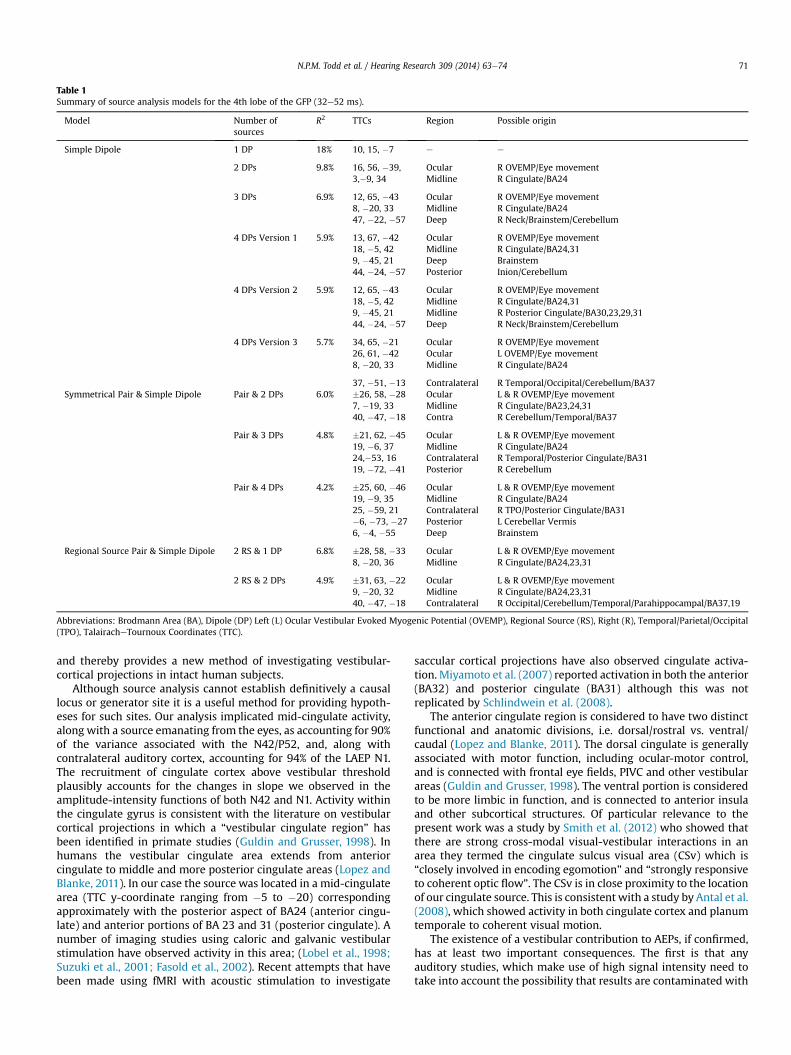
Although source analysis cannot establish definitively a causal
locus or generator site it is a useful method for providing hypoth-
eses for such sites. Our analysis implicated mid-cingulate activity,
along with a source emanating from the eyes, as accounting for 90%
of the variance associated with the N42/P52, and, along with
contralateral auditory cortex, accounting for 94% of the LAEP N1.
The recruitment of cingulate cortex above vestibular threshold
plausibly accounts for the changes in slope we observed in the
amplitude-intensity functions of both N42 and N1. Activity within
the cingulate gyrus is consistent with the literature on vestibular
cortical projections in which a “vestibular cingulate region” has
been identified in primate studies (Guldin and Grusser, 1998). In
humans the vestibular cingulate area extends from anterior
cingulate to middle and more posterior cingulate areas (Lopez and
Blanke, 2011). In our case the source was located in a mid-cingulate
area (TTC y-coordinate ranging from �5 to �20) corresponding
approximately with the posterior aspect of BA24 (anterior cingu-
late) and anterior portions of BA 23 and 31 (posterior cingulate). A
number of imaging studies using caloric and galvanic vestibular
stimulation have observed activity in this area; (Lobel et al., 1998;
Suzuki et al., 2001; Fasold et al., 2002). Recent attempts that have
been made using fMRI with acoustic stimulation to investigate
saccular cortical projections have also observed cingulate activa-
tion. Miyamoto et al. (2007) reported activation in both the anterior
(BA32) and posterior cingulate (BA31) although this was not
replicated by Schlindwein et al. (2008).
The anterior cingulate region is considered to have two distinct
functional and anatomic divisions, i.e. dorsal/rostral vs. ventral/
caudal (Lopez and Blanke, 2011). The dorsal cingulate is generally
associated with motor function, including ocular-motor control,
and is connected with frontal eye fields, PIVC and other vestibular
areas (Guldin and Grusser, 1998). The ventral portion is considered
to be more limbic in function, and is connected to anterior insula
and other subcortical structures. Of particular relevance to the
present work was a study by Smith et al. (2012) who showed that
there are strong cross-modal visual-vestibular interactions in an
area they termed the cingulate sulcus visual area (CSv) which is
“closely involved in encoding egomotion” and “strongly responsive
to coherent optic flow”. The CSv is in close proximity to the location
of our cingulate source. This is consistent with a study by Antal et al.
(2008), which showed activity in both cingulate cortex and planum
temporale to coherent visual motion.
The existence of a vestibular contribution to AEPs, if confirmed,
has at least two important consequences. The first is that any
auditory studies, which make use of high signal intensity need to
take into account the possibility that results are contaminated with
Table 1
Summary of source analysis models for the 4th lobe of the GFP (32e52 ms).
Model Number of
sources
R2 TTCs Region Possible origin
Simple Dipole 1 DP 18% 10, 15, �7 e e
2 DPs 9.8% 16, 56, �39, Ocular R OVEMP/Eye movement
3,�9, 34 Midline R Cingulate/BA24
3 DPs 6.9% 12, 65, �43 Ocular R OVEMP/Eye movement
8, �20, 33 Midline R Cingulate/BA24
47, �22, �57 Deep R Neck/Brainstem/Cerebellum
4 DPs Version 1 5.9% 13, 67, �42 Ocular R OVEMP/Eye movement
18, �5, 42 Midline R Cingulate/BA24,31
9, �45, 21 Deep Brainstem
44, �24, �57 Posterior Inion/Cerebellum
4 DPs Version 2 5.9% 12, 65, �43 Ocular R OVEMP/Eye movement
18, �5, 42 Midline R Cingulate/BA24,31
9, �45, 21 Midline R Posterior Cingulate/BA30,23,29,31
44, �24, �57 Deep R Neck/Brainstem/Cerebellum
4 DPs Version 3 5.7% 34, 65, �21 Ocular R OVEMP/Eye movement
26, 61, �42 Ocular L OVEMP/Eye movement
8, �20, 33 Midline R Cingulate/BA24
37, �51, �13 Contralateral R Temporal/Occipital/Cerebellum/BA37
Symmetrical Pair & Simple Dipole Pair & 2 DPs 6.0% �26, 58, �28 Ocular L & R OVEMP/Eye movement
7, �19, 33 Midline R Cingulate/BA23,24,31
40, �47, �18 Contra R Cerebellum/Temporal/BA37
Pair & 3 DPs 4.8% �21, 62, �45 Ocular L & R OVEMP/Eye movement
19, �6, 37 Midline R Cingulate/BA24
24,�53, 16 Contralateral R Temporal/Posterior Cingulate/BA31
19, �72, �41 Posterior R Cerebellum
Pair & 4 DPs 4.2% �25, 60, �46 Ocular L & R OVEMP/Eye movement
19, �9, 35 Midline R Cingulate/BA24
25, �59, 21 Contralateral R TPO/Posterior Cingulate/BA31
�6, �73, �27 Posterior L Cerebellar Vermis
6, �4, �55 Deep Brainstem
Regional Source Pair & Simple Dipole 2 RS & 1 DP 6.8% �28, 58, �33 Ocular L & R OVEMP/Eye movement
8, �20, 36 Midline R Cingulate/BA24,23,31
2 RS & 2 DPs 4.9% �31, 63, �22 Ocular L & R OVEMP/Eye movement
9, �20, 32 Midline R Cingulate/BA24,23,31
40, �47, �18 Contralateral R Occipital/Cerebellum/Temporal/Parahippocampal/BA37,19
Abbreviations: Brodmann Area (BA), Dipole (DP) Left (L) Ocular Vestibular Evoked Myogenic Potential (OVEMP), Regional Source (RS), Right (R), Temporal/Parietal/Occipital
(TPO), TalairacheTournoux Coordinates (TTC).
N.P.M. Todd et al. / Hearing Research 309 (2014) 63e74 71
Fig. 8. BESA solutions for the 4th and 5th lobes of the GFP corresponding with N*eP* and N1 deflection. Source waveforms (i) and source locations and orientations (ii) are shown
for (a) a 4th lobe 3 dipole model, (b) a 4th lobe model based on an ocular pair and 3 dipoles and (c) a 5th lobe 3 source model based on a pair of regional sources and a single dipole.
N.P.M. Todd et al. / Hearing Research 309 (2014) 63e7472
non-cochlear contributions. For example, a large number of studies
make use of the loudness dependence of AEPs (LDAEP) to make
inferences about the activity of certain catecholamine transmitters,
including serotonin and dopamine (Dierks et al., 1999). Such
methods have been applied to a number of psychiatric disorders,
including depression, schizophrenia and obsessive-compulsive
disorder. It is possible that the LDAEP results are influenced by
activation of the vestibular system, especially as it appears that the
highest intensity stimuli used in these studies, which may exceed
100 dB SPL, are the most critical (Gallinat et al., 2000). This is of
particular importance because the central projections of the
vestibular system include insular, cingulate and autonomic path-
ways, which employ transmitters of interest (Balaban and Yates,
2004; Lopez and Blanke, 2011).
A second potential consequence is that it is quite possible that
acoustically activated non-cochlear (vestibular) projections may
play a role in normal hearing, for example, in the vocal behaviour of
primate and amniote vertebrate species (Todd and Cody, 2000;
Todd and Merker, 2004; Todd, 2007), as well as the vocal behav-
iour of anamniotes for which the otolith organs are established as
auditory. In the case of humans many music environments, such as
at concerts, are of high-intensity and well above the threshold
levels we have considered (Todd and Cody, 2000). Similarly, the
singing voice is high-intensity and very likely produces self-
activation of the vestibular apparatus (Todd, 1993) as was sug-
gested as long ago as the 1930s by Tait (1932). Given a probable role
of cingulate cortex, such activation could contribute to the affective
responses to sound or in listening to music (Todd, 2001).
As noted above, the VEMP threshold overestimates the receptor
threshold. McCue and Guinan (1994) found a rate threshold in cat
vestibular afferents of 90 dB SPL for 50 ms tones, with a phase-
locking threshold 10 dB lower. Combined with the appropriate
psychophysical correction for short tone bursts, e.g. Meddis and
Lecluyse (2011), this places the receptor rate threshold at near 70
dB SL. It is likely, therefore, that vestibular receptors contribute at
intensities well below those experienced in the loud environments
described in the previous paragraphs. The presence at everyday
intensities of a vestibular component in cortical potentials from the
temporal lobe, hitherto considered purely cochlear in origin, raises
the possibility that acoustic activation of the otolith organs could
contribute directly to auditory discrimination, as well as to affective
processes. There is now a growing literature which provides evi-
dence of a central vestibulareauditory interaction which allows
vestibular inputs to improve temporal and spatial aspects of hear-
ing (Emami and Daneshi, 2012; Brimijoin and Akeroyd, 2012;
Probst and Wist, 1990), to contribute to speech perception and in
metrical aspects of musical perception (Emami et al., 2012; Phillips-
Silver and Trainor, 2008), and which indicates a general association
between hearing loss and vestibular dysfunction (Akin et al., 2012;
Zuniga et al., 2012; Kumar et al., 2010; Wang and Young, 2007).
Given the well-established cross-over from vestibular to auditory
pathways at the level of the brain-stem, (e.g. Barker et al., 2012),
and thalamus, (Roucoux-Hanus and Boisacq-Schepens, 1977; Blum
et al., 1979), vestibular effects at the level of temporal cortex should
be expected, especially as activation of superior temporal lobe is
consistently indicated in vestibular imaging studies (Lopez et al.,
2012).
5. Concluding remarks
We have in this paper presented evidence to support a vestib-
ular contribution to AEPs, and have tentatively identified a possible
new component, the N42/P52, which appears to be vestibular in
origin. Some of this evidence is quite subtle, but this probably ex-
plains why it has hitherto not been remarked upon. However, given
the potential implications of the vestibulareauditory interaction
for hearing research it is important that further work is conducted
to substantiate these. Among the most important of these future
studies should be work with a larger sample of avestibular patients
to investigate differences in AEPs. Given also the limitations of the
VEMP threshold in identifying a receptor threshold new methods
for establishing a N42/P52 threshold directly from AEPs should be
developed. Finally higher resolution imaging methods will be
required to substantiate the brain areas suggested by the BESA.
Acknowledgements
The research reported in this article was supported by a grant
from the Wellcome Trust (WT091961MA). We are grateful to
Sendhil Govender for assistance in recording EEG from the vestib-
ular patient and to DrMWelgampola and Professor M Halmagyi for
their cooperation in the recruitment of the patient. We would like
to thank Prof Chris Plack and Dr Selvino de Kort for their comments
on an earlier version of this manuscript. We would also like to
thank Aisha Mclean for assistance in the preparation of the
manuscript.
References
Akin, F.W., Murnane, O.D., Tampas, J.W., Clinard, C., Byrd, S., Kelly, J.K., 2012. Theeffect of noise exposure on the cervical vestibular evoked myogenic potential.
Ear Hear. 33, 458e465.
Antal, A., Baudewig, J., Paulus, W., Dechent, P., 2008. The posterior cingulate cortexand planum temporale/parietal operculum are activated by coherent visual
overexposure increases the expression of VGLUT-2 mediated projections fromthe lateral vestibular nucleus to the dorsal cochlear nucleus. PLoS One 7.
Bickford, R.G., Jacobson, J.L., Cody, D.T.R., 1964. Nature of average evoked potentialsto sound and other stimuli in man. Ann. N. Y. Acad. Sci. 194, 112e204.
Blum, P.S., Day, M.J., Carpenter, M.B., Gilman, S., 1979. Thalamic components of the
ascending vestibular system. Exp. Neurol. 64, 587e603.Brimijoin, W.O., Akeroyd, M.A., 2012. The role of head movements and signal
spectrum in an auditory front/back illusion. i-Perception 3, 179e182.Colebatch, J.G., Halmagyi, G.M., Skuse, N.F., 1994. Myogenic potentials generated by a
click-evoked vestibulocollic reflex. J. Neurol. Neurosurg. Psychiatry 57, 190e197.
Curthoys, I.S., Kim, J., McPhedran, S.K., Camp, A.J., 2006. Bone conducted vibrationselectively activates irregular primary otolithic vestibular neurons in the guinea
projections in the human cortex. Exp. Brain Res. 141, 541e551.Dierks, T., Barta, S., Demisch, L., Schmeck, K., Englert, E., Kewitz, A., Maurer, K.,
Poustka, F., 1999. Intensity dependence of auditory evoked potentials (AEPs) as
biological marker for cerebral serotonin levels: effects of tryptophan depletionin healthy subjects. Psychopharmacol 146, 101e107.
Emami, S.F., Daneshi, A., 2012. Vestibular hearing and neural synchronization. ISRNOtolaryngol.. http://dx.doi.org/10.5402/2012/246065.
Emami, S.F., Pourbakht, A., Sheykholeslami, K., Kamali, M., Behnoud, F., Daneshi, A.,
2012. Vestibular hearing and speech processing. ISRN Otolaryngol.. http://dx.doi.org/10.5402/2012/850629.
Fasold, O., von Brevern, M., Kuhberg, M., Ploner, C.J., Villringer, A., Lempert, T.,Wenzel, R., 2002. Human vestibular cortex as identified with caloric stimulation
in functional magnetic resonance imaging. Neuroimage 17, 1384e1393.Gallinat, J., Bottlender, R., Juckel, G., Munke-Puchner, A., Stotz, G., Kuss, H.J.,
Mavrogiorgou, P., Hegerl, U., 2000. The loudness dependency of the auditory
evoked N1/P2-component as a predictor of the acute SSRI response indepression. Psychopharmacol. 148, 404e411.
Guldin, W.O., Grusser, O.J., 1998. Is there a vestibular cortex? Trends Neurosci. 21,254e259.
Jones, T.A., Jones, S.M., Vijayakumar, S., Brugeaud, A., Bothwell, M., Chabbert, C.,
2011. The adequate stimulus for mammalian linear vestibular evoked potentials(VsEPs). Hear. Res. 280, 133e140.
Kumar, K., Vivarthini, C.J., Bhat, J.S., 2010. Vestibular evoked myogenic potential innoise-induced hearing loss. Noise Health 12, 191e194.
Lackner, J.R., Graybiel, A., 1974. Elicitation of vestibular side-effects by regional vi-bration of head. Aerosp. Med. 45, 1267e1272.
Levitt, H., 1971. Transformed up-down methods in psychoacoustics. J. Acoust. Soc.
Am. 49, 467e477.
N.P.M. Todd et al. / Hearing Research 309 (2014) 63e74 73
Lewis, E., Narins, P. (Eds.), 1999. Comparative Hearing: Fish and Amphibians.
Springer-Verlag, New York.Lobel, E., Kleine, J.F., Le Bihan, D., Leroy-Willig, A., Berthoz, A., 1998. Functional MRI
of galvanic vestibular stimulation. J. Neurophysiol. 80, 2699e2709.Lopez, C., Blanke, O., 2011. The thalamocortical vestibular system in animals and
humans. Brain Res. Rev. 67, 119e146.
Lopez, C., Blanke, O., Mast, F.W., 2012. The human vestibular cortex revealed bycoordinate-based activation likelihood estimation meta-analysis. Neurosci. 212,
156e179.Manley, G., Popper, A., Fay, R. (Eds.), 2004. Evolution of the Vertebrate Auditory
System. Springer-Verlag, New York.Maurizi, M., Ottaviani, F., Paludetti, G., Rosignoli, M., Almadori, G., Tassoni, A., 1984.
Middle-latency auditory components in response to clicks and low- and
middle-frequency tone pips (0.5e1 kHz). Audiology 23, 569e580.McCue, M.P., Guinan, J.J., 1994. Acoustically responsive fibres in the vestibular nerve
of the cat. J. Neurosci. 14, 6058e6070.McKnight, C.L., Doman, D.A., Brown, J.A., Bance, M., Adamson, R.B.A., 2013. Direct
measurement of the wavelength of sound waves in the human skull. J. Acoust.
Soc. Am. 133, 136e145.McNerney, K.M., Lockwood, A.H., Coad, M.L., Wack, D.S., Burkard, R.F., 2011. Use of
64-channel electroencephalography to study neural otolith-evoked responses.J. Am. Acad. Audiol. 22, 143e155.
Meddis, R., Lecluyse, W., 2011. The psychophysics of absolute threshold and signal
duration: A probabilistic approach. J. Acoust. Soc. Am. 129, 3153e3165.Miyamoto, T., Fukushima, K., Takada, T., de Waele, C., Vidal, P.-P., 2007. Saccular
stimulation of the human cortex: a functional magnetic resonance imagingstudy. Neurosci. Lett. 423, 68e72.
Naatanen, R., Picton, T., 1987. The N1 wave of the human electric and magneticresponse to sound e a review and analysis of the component structure. Psy-
Picton, T.W., Hillyard, S.A., Krausz, H.I., Galambos, R., 1974. Human auditory evokedpotentials. I. Evaluation of components. Electroencephalogr. Clin. Neurophysiol.
36, 179e190.Probst, T., Wist, E.R., 1990. Electrophysiological evidence for visual e vestibular
interaction in man. Neurosci. Lett. 108, 255e260.Rosengren, S.M., Colebatch, J.G., 2006. Vestibular evoked potentials (VsEPs) in pa-
tients with severe to profound bilateral hearing loss. Clin. Neurophysiol. 117,
potentials produced by stimulation with bone-conducted sound. Clin. Neuro-physiol. 116, 1938e1948.
Roucoux-Hanus, M., Boisacq-Schepens, N., 1977. Ascending vestibular projections e
further results at cortical and thalamic levels in cat. Exp. Brain Res. 29, 283e292.Scherg, M., Vajsar, J., Picton, T.W., 1989. A source analysis of the late human auditory
evoked potentials. J. Cog. Neurosci. 1, 336e355.Schlindwein, P., Mueller, M., Bauermann, T., Brandt, T., Stoeter, P., Dieterich, M.,
2008. Cortical representation of saccular vestibular stimulation: VEMPs in fMRI.Neuroimage 39, 19e31.
Smith, A.T., Wall, M.B., Thilo, K.V., 2012. Vestibular inputs to human motion-
Sohmer, H., Elidan, J., Plotnik, M., Freeman, S., Sockalingam, R., Berkowitz, Z.,
Mager, M., 1999. Effect of noise on the vestibular system-vestibular evokedpotential studies in rats. Noise Health 2, 41e51.
Stenfelt, S., Hakansson, B., Tjellstrom, A., 2000. Vibration characteristics of boneconducted sound in vitro. J. Acoust. Soc. Am. 107, 422e431.
Suzuki,M., Kitano, H., Ito, R., Kitanishi, T., Yazawa, Y., Ogawa, T., Shiino, A., Kitajima, K.,
2001. Cortical and subcortical vestibular response to caloric stimulation detectedby functional magnetic resonance imaging. Cogn. Brain Res. 12, 441e449.
Tait, J., 1932. Is all hearing cochlear? Ann. Otol. Rhinol. Laryngol. 41, 6812.Thornton, A.R., Mendel, M.I., Anderson, C.V., 1977. Effects of stimulus frequency and
intensity on the middle components of the averaged auditory electroencephalicresponse. J. Speech Lang. Hear. Res. 20, 81e94.
Todd, N.P.M., 2001. Evidence for a behavioral significance of saccular acoustic
sensitivity in humans. J. Acoust. Soc. Am. 110, 380e390.Todd, N.P.M., 2007. Estimated source intensity and active space of the American
Todd, N.P.M., Cody, F.W., 2000. Vestibular responses to loud dance music: a phys-
iological basis of the “rock and roll threshold”. J. Acoust. Soc. Am. 107, 496e500.Todd, N.P.M., Merker, B., 2004. Siamang gibbons exceed the saccular threshold:
intensity of the song of Hylobates syndactylus. J. Acoust. Soc. Am. 115, 3077e3080.
Todd, N.P.M., 1993. Vestibular feedback in musical performance e response to so-
evoked myogenic potentials (OVEMPs) produced by air- and bone-conductedsound. Clin. Neurophysiol. 118, 381e390.
Todd, N.P.M., Rosengren, S.M., Colebatch, J.G., 2003. A short latency vestibularevoked potential (VsEP) produced by bone-conducted acoustic stimulation.
J. Acoust. Soc. Am. 114, 3264e3272.
Todd, N.P.M., Rosengren, S.M., Colebatch, J.G., 2008a. A source analysis of short-latency vestibular evoked potentials produced by air- and bone-conducted
sound. Clin. Neurophysiol. 119, 1881e1894.Todd, N.P.M., Rosengren, S.M., Colebatch, J.G., 2008b. Tuning and sensitivity of the
human vestibular system to low-frequency vibration. Neurosci. Lett. 444, 36e41.
Todd, N.P.M., Rosengren, S.M., Colebatch, J.G., 2009. A utricular origin of frequencytuning to low-frequency vibration in the human vestibular system? Neurosci.
Lett. 451, 175e180.Todd, N.P.M., Rosengren, S.M., Govender, S., Colebatch, J.G., 2010. Single trial
detection of human vestibular evoked myogenic potentials is determined bysignal to noise ratio. J. Appl. Physiol. 109, 53e59.
Wang, Y.-P., Young, Y.-H., 2007. Vestibular-evoked myogenic potentials in chronic
noise-induced hearing loss. Otolaryngol. Head Neck Surg. 137, 607e611.Young, E.D., Fernandez, C., Goldberg, J.M., 1977. Responses of squirrel-monkey
vestibular neurones to audio-frequency sound and head vibration. Acta Oto-Laryngol. 84, 352e360.
Zhang, A.S., Govender, S., Colebatch, J.G., 2011. Tuning of the ocular vestibular
evoked myogenic potential (oVEMP) to AC sound shows two separate peaks.Exp. Brain Res. 213, 111e116.
Zhang, A.S., Govender, S., Colebatch, J.G., 2012. Tuning of the ocular vestibularevoked myogenic potential to bone-conducted sound stimulation. J. Appl.


![Nymphalis KLUK [1780] sensu lato · 2013. 2. 14. · The names proposed for taxa in the genus Nymphalis KLUK [1780] sensu lato Anglewing Butterflies Papiliones angulati. compiled](https://static.documents.pub/doc/80x56/61190bf0fafe5823d92ad33c/nymphalis-kluk-1780-sensu-lato-2013-2-14-the-names-proposed-for-taxa-in-the.jpg)


![Nymphalis KLUK [1780] sensu lato · 2013-02-14 · The names proposed for taxa in the genus Nymphalis KLUK [1780] sensu lato Anglewing Butterflies Papiliones angulati. compiled by](https://static.documents.pub/doc/80x56/5e604db63ae8eb558e521700/nymphalis-kluk-1780-sensu-2013-02-14-the-names-proposed-for-taxa-in-the-genus.jpg)


