+ All Categories
Home > Documents > research papers IUCrJ - (IUCr) Crystallography Journals...

research papers IUCrJ - (IUCr) Crystallography Journals...

Date post: 23-Feb-2018
Category:
Upload: duongdan
View: 220 times
Download: 0 times
Share this document with a friend
9
research papers 206 https://doi.org/10.1107/S2052252517002305 IUCrJ (2017). 4, 206–214 IUCrJ ISSN 2052-2525 CHEMISTRY j CRYSTENG Received 1 October 2016 Accepted 10 February 2017 Edited by M. Eddaoudi, King Abdullah University, Saudi Arabia Keywords: crystal engineering; co-crystals; molecular crystals. CCDC references: 1507491; 1507492; 1507493; 1507494; 1507495 Supporting information: this article has supporting information at www.iucrj.org Acemetacin cocrystal structures by powder X-ray diffraction Geetha Bolla, a Vladimir Chernyshev b,c * and Ashwini Nangia a,d * a School of Chemistry, University of Hyderabad, Prof. C. R. Rao Road, Central University PO, Hyderabad 500 046, India, b Department of Chemistry, M. V. Lomonosov Moscow State University, 1–3 Leninskie Gory, Moscow 119991, Russian Federation, c A. N. Frumkin Institute of Physical Chemistry and Electrochemistry RAS, 31 Leninsky Prospect, Moscow 119 071, Russian Federation, and d CSIR-National Chemical Laboratory, Dr Homi Bhabha Road, Pune 411 008, India. *Correspondence e-mail: [email protected], [email protected] Cocrystals of acemetacin drug (ACM) with nicotinamide (NAM), p-aminoben- zoic acid (PABA), valerolactam (VLM) and 2-pyridone (2HP) were prepared by melt crystallization and their X-ray crystal structures determined by high- resolution powder X-ray diffraction. The powerful technique of structure determination from powder data (SDPD) provided details of molecular packing and hydrogen bonding in pharmaceutical cocrystals of acemetacin. ACM–NAM occurs in anhydrate and hydrate forms, whereas the other structures crystallized in a single crystalline form. The carboxylic acid group of ACM forms theacid– amide dimer three-point synthon R 3 2 (9)R 2 2 (8)R 3 2 (9) with three different syn amides (VLM, 2HP and caprolactam). The conformations of the ACM molecule observed in the crystal structures differ mainly in the mutual orientation of chlorobenzene fragment and the neighboring methyl group, being anti (type I) or syn (type II). ACM hydrate, ACM—NAM, ACM–NAM-hydrate and the piperazine salt of ACM exhibit the type I conformation, whereas ACM polymorphs and other cocrystals adopt the ACM type II conformation. Hydrogen-bond interactions in all the crystal structures were quantified by calculating their molecular electrostatic potential (MEP) surfaces. Hirshfeld surface analysis of the cocrystal surfaces shows that about 50% of the contribution is due to a combination of strong and weak OH, NH, ClH and CH interactions. The physicochemical properties of these cocrystals are under study. 1. Introduction Cocrystallization is a standard strategy to tailor physico- chemical properties of drugs based on their chemical consti- tuents (Childs et al. , 2004; Duggirala et al., 2016; Bolla & Nangia, 2016) and supramolecular structure through crystal engineering (Desiraju et al., 2011; Desiraju, 2013). Pharma- ceutical cocrystals (Almarsson & Zaworotko, 2004; Schul- theiss & Newman, 2009; Thakuria et al., 2013) belong to a subclass of multicomponent systems in which one of the molecules must be an Active Pharmaceutical Ingredient (API) and the coformer is a Generally Regarded as Safe (GRAS) substance (http://www.fda.gov/Food/IngredientsPackaging- Labeling/GRAS/; accessed on 20/08/2016). Cocrystals incor- porate pharmaceutically acceptable coformers and the drug substance into the same crystal lattice to provide a new composition of the API (Aitipamula et al., 2012). Numerous cocrystal systems have been reported previously in more than a decade to modify the physicochemical and pharmacokinetic properties of drugs, notably solubility and bioavailability. The unique advantage of cocrystals is that they are amenable to those drugs which lack an ionizable functional group and thus present an alternative to the traditional salts for improving
Transcript

research papers

206 https://doi.org/10.1107/S2052252517002305 IUCrJ (2017). 4, 206–214

IUCrJISSN 2052-2525

CHEMISTRYjCRYSTENG

Received 1 October 2016

Accepted 10 February 2017

Edited by M. Eddaoudi, King Abdullah

University, Saudi Arabia

Keywords: crystal engineering; co-crystals;

molecular crystals.

CCDC references: 1507491; 1507492;

1507493; 1507494; 1507495

Supporting information: this article has

supporting information at www.iucrj.org

Acemetacin cocrystal structures by powder X-raydiffraction

Geetha Bolla,a Vladimir Chernyshevb,c* and Ashwini Nangiaa,d*

aSchool of Chemistry, University of Hyderabad, Prof. C. R. Rao Road, Central University PO, Hyderabad 500 046, India,bDepartment of Chemistry, M. V. Lomonosov Moscow State University, 1–3 Leninskie Gory, Moscow 119991, Russian

Federation, cA. N. Frumkin Institute of Physical Chemistry and Electrochemistry RAS, 31 Leninsky Prospect, Moscow 119

071, Russian Federation, and dCSIR-National Chemical Laboratory, Dr Homi Bhabha Road, Pune 411 008, India.

*Correspondence e-mail: [email protected], [email protected]

Cocrystals of acemetacin drug (ACM) with nicotinamide (NAM), p-aminoben-

zoic acid (PABA), valerolactam (VLM) and 2-pyridone (2HP) were prepared by

melt crystallization and their X-ray crystal structures determined by high-

resolution powder X-ray diffraction. The powerful technique of structure

determination from powder data (SDPD) provided details of molecular packing

and hydrogen bonding in pharmaceutical cocrystals of acemetacin. ACM–NAM

occurs in anhydrate and hydrate forms, whereas the other structures crystallized

in a single crystalline form. The carboxylic acid group of ACM forms theacid–

amide dimer three-point synthon R32(9)R2

2(8)R32(9) with three different syn

amides (VLM, 2HP and caprolactam). The conformations of the ACM molecule

observed in the crystal structures differ mainly in the mutual orientation of

chlorobenzene fragment and the neighboring methyl group, being anti (type I)

or syn (type II). ACM hydrate, ACM—NAM, ACM–NAM-hydrate and the

piperazine salt of ACM exhibit the type I conformation, whereas ACM

polymorphs and other cocrystals adopt the ACM type II conformation.

Hydrogen-bond interactions in all the crystal structures were quantified by

calculating their molecular electrostatic potential (MEP) surfaces. Hirshfeld

surface analysis of the cocrystal surfaces shows that about 50% of the

contribution is due to a combination of strong and weak O� � �H, N� � �H, Cl� � �H

and C� � �H interactions. The physicochemical properties of these cocrystals are

under study.

1. Introduction

Cocrystallization is a standard strategy to tailor physico-

chemical properties of drugs based on their chemical consti-

tuents (Childs et al., 2004; Duggirala et al., 2016; Bolla &

Nangia, 2016) and supramolecular structure through crystal

engineering (Desiraju et al., 2011; Desiraju, 2013). Pharma-

ceutical cocrystals (Almarsson & Zaworotko, 2004; Schul-

theiss & Newman, 2009; Thakuria et al., 2013) belong to a

subclass of multicomponent systems in which one of the

molecules must be an Active Pharmaceutical Ingredient (API)

and the coformer is a Generally Regarded as Safe (GRAS)

substance (http://www.fda.gov/Food/IngredientsPackaging-

Labeling/GRAS/; accessed on 20/08/2016). Cocrystals incor-

porate pharmaceutically acceptable coformers and the drug

substance into the same crystal lattice to provide a new

composition of the API (Aitipamula et al., 2012). Numerous

cocrystal systems have been reported previously in more than

a decade to modify the physicochemical and pharmacokinetic

properties of drugs, notably solubility and bioavailability. The

unique advantage of cocrystals is that they are amenable to

those drugs which lack an ionizable functional group and thus

present an alternative to the traditional salts for improving

solubility and dissolution rate (Childs et al., 2004; Bolla et al.,

2013), physical stability (Babu et al., 2012; Trask et al., 2006),

bioavailability (Weyna et al., 2012; Ganesh et al., 2015),

permeability (Sanphui, Devi et al., 2015) and mechanical

properties (Sun & Hou, 2008; Sanphui, Mishra et al., 2015).

However, certain drugs can be difficult to crystallize as single

crystals, and one such example in our experience is aceme-

tacin, whether it is the pure drug or its cocrystals. Structure

solution from powder diffraction data for acemetacin cocrys-

tals is reported in this paper as part of our continuing studies

on this system (Sanphui et al., 2013, 2014).

Acemetacin (ACM) is a glycolic acid ester prodrug of

indomethacin and belongs to the non-steroidal anti-inflam-

matory drug (NSAID) class. It is metabolized to indo-

methacin, which then acts as an inhibitor of cyclooxygenase to

produce the anti-inflammatory effects. ACM is sold under the

trade name Emflex as 60 mg capsules (Merck KGaA). Solid-

state forms of acemetacin have been studied by Chavez-Pina et

al. (2007), Yoneda et al. (1981), Burger & Lettenbichler (1993)

and Gelbrich et al. (2007). In our previous findings (Sanphui et

al., 2013, 2014), the crystal structures of ACM Form I and II

were identified as synthon polymorphs of carboxylic acid

dimer and catemer motifs. The binary adducts of cocrystals

with nicotinamide (NAM), isonicotinamide (INA), picolina-

mide (PAM) and caprolactam (CPR) are stabilized by acid–

amide hetero synthons and the p-aminobenzoic acid (PABA)

cocrystal has the hetero acid dimer synthon. We noted that

ACM tends to form a hydrate during any kind of solution-

based cocrystal preparation, and so its crystallization was

carried out in strictly anhydrous melt conditions (solventless).

The structures of ACM Form I, ACM–INA and ACM–PABA

were solved using single-crystal X-ray diffraction and those of

Form II polymorph and cocrystals ACM–PAM, ACM–CPR,

salt ACM–PPZ were solved by high-resolution powder X-ray

diffraction data [Scheme 1, where superscript a indicates

coformers reported in a previous study (Sanphui et al., 2014)

and superscript b coformers reported in this study].

Among the binary systems reported in our previous papers,

the ACM–NAM cocrystal was characterized by IR, powder X-

ray diffraction (PXRD) and differential scanning calorimetry

(DSC) but crystallization to obtain good diffraction quality

single crystals was unsuccessful (Sanphui et al., 2014), and

structure determination from powder data (SDPD) was also

not successful at that time. The cocrystal of ACM with p-

aminobenzoic acid showed poor quality diffraction data and

there was proton disorder in the ACM-COOH group. There-

fore, high-resolution powder diffraction data were collected to

solve the crystal structure of ACM–PABA and nicotinamide

cocrystals.

Single-crystal X-ray diffraction is undoubtedly the most

powerful tool to elucidate the molecular structure. However,

the requirement for single crystals of appropriate size and

quality limits the scope of this

technique, because many materials

crystallize as microcrystalline

powders. Fortunately, there have

been rapid advances during the

past two decades in structure

determination from powder

diffraction data (SDPD; Harris et

al., 1994; Chernyshev, 2001; Harris,

2003; Le Bail et al., 2009). SDPD is

the method of choice when there is

difficulty obtaining the optimum

size single crystals and also when

phase transformation, hydration or

solvate formation issues complicate

the isolation of good quality

single crystals for data

collection. Recently Ueto et al.

(2012) reported furosemide�

nicotinamide cocrystal polymorphs

and cocrystal hydrate crystal

structures solved from high-reso-

lution powder data. The crystal

structures of several API forms

research papers

IUCrJ (2017). 4, 206–214 Geetha Bolla et al. � Acemetacin cocrystal structures 207

Figure 1(a) O—H� � �O carboxylic acid catemer chain in ACM Form I. (b) Carboxylic acid dimer in ACM Form II.(c) to (g) The primary supramolecular synthons present in binary cocrystals ACM–INA, ACM–PABA,ACM–PAM, ACM–CPR and ACM–PPZ (Sanphui et al., 2014).

with three-dimensional coordinates determined have been

reported using high-resolution powder data (David &

Shankland, 2008; Braga et al., 2012; Chernyshev et al., 2013). In

this background, we report crystal structures of acemetacin

cocrystals listed in Scheme 1 (part b) from high-resolution

powder diffraction data.

2. Experimental

2.1. Preparation of acemetacin cocrystals/salts

Acemetacin was purchased from Dalian Hong Ri Dong

Sheng Import & Export Co. Ltd, China, http://dlhongridong-

sheng.guidechem.com/ and used as such without further

purification. All the coformers were purchased form Sigma-

Aldrich, India, and solvents are of analytically pure grade.

ACM and the appropriate coformer was weighed in a 1:1

stoichiometric ratio in a 25 ml beaker and melted at 160�C.

Cooling of the melt gave a glassy phase at room temperature

(30�C) after 1–2 h, which was crystallized from different

solvents, e.g. methyl isobutyl ketone (MIBK) and EtOAc. The

solvents must be anhydrous (dry) to avoid the formation of

ACM hydrates as by-products in crystallization. ACM–NAM-

I, ACM–NAM-H, ACM–VLM, ACM–2HP and ACM–PABA

cocrystals were prepared by melt crystallization. ACM–PABA

was crystallized from dry EtOAc solvent. The purity and

homogeneity of phases was confirmed by DSC (single endo-

therm).

2.2. Powder X-ray diffraction

Bulk samples were analyzed by powder X-ray diffraction

using a Bruker AXS D8 powder diffractometer (Bruker-AXS,

Karlsruhe, Germany). Experimental conditions: Cu K�radiation (� = 1.5418 A); 40 kV, 30 mA; scan range 5–50� 2�.

High-resolution X-ray powder diffraction data for ACM–

NAM-I, ACM–NAM-H, ACM–2HP, ACM–PABA and ACM–

VLM were collected at room temperature using a Huber G670

Guinier camera with an image plate detector and Cu K�1

radiation (� = 1.5406 A). The unit-cell dimensions were

determined using three indexing programs: TREOR90, ITO

and AUTOX (Werner et al., 1985; Visser, 1969; Zlokazov,

1992, 1995). The crystal structures were solved using the

simulated annealing technique (Zhukov et al., 2001) and

refined using MRIA (Zlokazov & Chernyshev, 1992) following

the procedure published earlier (Sanphui et al., 2014). The

initial molecular models for acemetacin and coformer mole-

cules were taken from the Cambridge Structural Database

(ConQuest, Version 1.18 with updates; Groom & Allen, 2014).

In simulated annealing runs (without H atoms), the total

number of degrees of freedom was either 20 or 21, i.e. 14

parameters for the acemetacin molecule (three translational,

three rotational and eight torsional) and six or seven para-

meters for the coformer were varied. For ACM–NAM-H, the

presence of solvent in the asymmetric part was approximated

by a water molecule disordered over two positions. The

occupancies were fixed to 0.5 s.o.f. and powder data collection,

refinement parameters, hydrogen-bonding values are given in

Tables 1 and 2. The diffraction profiles after the final bond-

restrained Rietveld refinement are shown later in the paper.

X-Seed (Barbour, 2001) was used to prepare the figures and

packing diagrams.

2.3. Thermal analysis

Differential Scanning Calorimetry (DSC) was performed on

a Mettler Toledo DSC 822e module. Samples were placed in

crimped but vented aluminium sample pans, with a typical

sample size of 2–5 mg. The temperature range was 30–200�C at

a heating rate of 5�C min�1. Samples were purged with a

stream of dry N2 flow at 80 ml min�1.

research papers

208 Geetha Bolla et al. � Acemetacin cocrystal structures IUCrJ (2017). 4, 206–214

Table 1Crystallographic details of ACM cocrystals.

ACM–NAM-I ACM–NAM-H ACM–VLM ACM–2HP ACM–PABA

CCDC No. 1507493 1507492 1507495 1507491 1507494Chemical formula C21H18ClNO6�C6H6N2O C21H18ClNO6�C6H6N2O�H2O C21H18ClNO6�C5H9NO C21H18ClNO6�C5H5NO C21H18ClNO6�C7H7NO2

Mr 537.94 555.96 514.95 510.91 552.95Crystal system Monoclinic Triclinic Triclinic Monoclinic MonoclinicSpace group P21/c P�11 P�11 P21/c P21

a (A) 4.8977 (11) 21.348 (2) 11.764 (12) 23.1400 (18) 17.294 (18)b (A) 40.914 (4) 4.1931 (12) 20.555 (19) 5.1900 (8) 4.819 (7)c (A) 12.8874 (19) 15.2174 (19) 5.1627 (9) 21.2642 (19) 16.955 (15)� (�) 90 90.567 (17) 89.543 (14) 90 90� (�) 100.328 (18) 101.40 (2) 93.300 (16) 111.714 (17) 113.529 (17)� (�) 90 89.473 (16) 96.276 (17) 90 90V (A3) 2540.6 (7) 1335.2 (4) 1238.8 (3) 2372.5 (5) 1295.5 (3)M20 36 24 21 21 21F30 61 (0.008, 64) 47 (0.010, 44) 39 (0.010, 53) 39 (0.010, 53) 39 (0.010, 53)Z 4 2 2 4 2�calc (g cm�3) 1.406 1.383 1.380 1.430 1.417� (mm�1) 1.784 1.743 1.787 1.866 1.7832�min–2�max, increment (�) 3.00–75.00, 0.01 3.00–75.00, 0.01 3.00–75.00, 0.01 3.00–85.00, 0.01 3.00–75.00, 0.01Number of parameters,

restraints195, 125 203/125 187/123 187/121 197/131

Rp/Rwp/Rexp 0.0160/0.0180/0.0167 0.0161/0.0191/0.0160 0.0208/0.0271/0.0175 0.0205/0.0265/0.0185 0.0183/0.0236/0.0173Goodness-of-fit 1.076 1.198 1.549 1.433 1.365

2.4. Solid-state NMR spectroscopy

Approximately 100 mg of fine crystalline sample was tightly

packed into a zirconia rotor with the help of Teflon stick up to

the cap Kel-F mark. A cross-polarization, magic angle spin-

ning (CP-MAS) pulse sequence was used for spectral acqui-

sition. Each sample was spun at a frequency of 5.0 � 0.01 kHz

and the magic angle setting was calibrated by the KBr method.

Each data set was subjected to a 5.0 Hz line-broadening factor

and subsequently Fourier transformed and phase corrected to

produce a frequency domain spectrum. Solid-state 13C NMR

spectra were obtained on a Bruker (Bruker BioSpin, Karls-

ruhe, Germany) Ultrashield 400 spectrometer utilizing a 13C

resonant frequency of 100 MHz (magnetic field strength of

9.39 T). The chemical shifts were referenced to trimethylsilyl

(TMS) using glycine (glycine = 43.3 p.p.m.) as an external

secondary standard. 15N CP-MAS spectra recorded at

400 MHz were referenced to glycine N and then the chemical

shifts were recalculated to nitromethane (glycine =

�347.6 p.p.m.).

3. Results and discussion

The chemical units present in the crystal structures of Form I,

II and cocrystals with INA, PAM, PABA are displayed in Fig.

1. Experimental conditions to yield single crystals in different

solvents always resulted in either ACM hydrate or a precipi-

tate. We therefore used the microcrystalline sample to solve

the crystal structures from powder X-ray data collected at high

resolution. The crystal structures of ACM–NAM were deter-

mined as two forms, an anhydrate ACM–NAM-I and a

cocrystal hydrate ACM–NAM-H. X-ray crystal structure

parameters are summarized in Table 1 and hydrogen-bond

metrics in Table 2. The binary adducts were prepared by melt

crystallization and characterized by their melting point,

PXRD and DSC. The bulk phase purity was checked by

PXRD Rietveld refinement and DSC.

3.1. Crystal structures of binary cocrystals

3.1.1. ACM–NAM-I (1:1). ACM–NAM-I (1:1) crystallized

in the monoclinic crystal system P21/c. The molecular packing

is stabilized by an acid–pyridine primary synthon and further

by an amide–acid hydrogen bond (Fig. 2a). NAM molecules

are bonded through amide catemer chains along the 21 screw

axis and also interact with ACM molecules to give a sandwich-

research papers

IUCrJ (2017). 4, 206–214 Geetha Bolla et al. � Acemetacin cocrystal structures 209

Figure 3ACM–NAM-H (1:1:1) is a cocrystal hydrate. (a) Robust acid–pyridinesynthon and NAM amide homodimers. (b) Two hydrogen-bonded layersextended through a water molecule in the crystal lattice. (c) The watermolecule stoichiometry and disorder in crystal structure are confirmed bySDPD. H atoms are removed for clarity.

Figure 2ACM–NAM-I. (a) Acid–pyridine and amide–acid synthons. (b) NAMcoformers extend through amide chains and also interact with ACM toresult in a sandwich-type packing. (c) NAM and ACM domains along the21 screw axis. H atoms are removed for clarity.

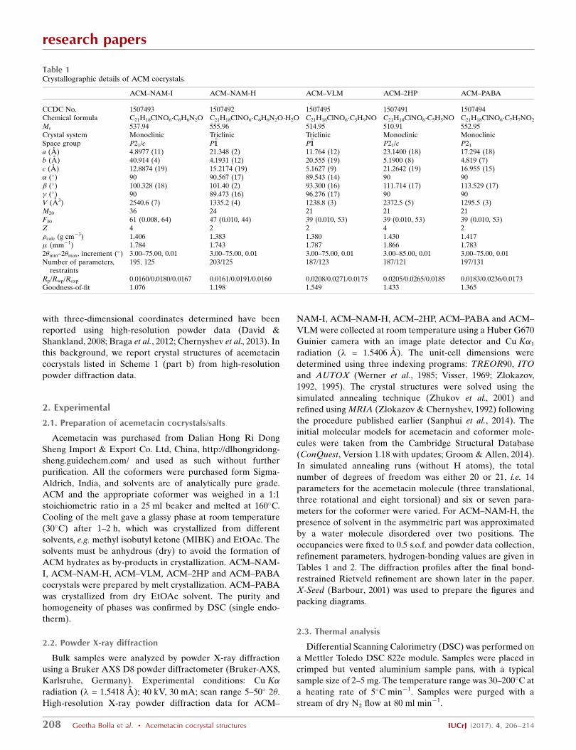
Table 2Hydrogen-bond geometry (A, �) in crystal structures.

D—H� � �A H� � �A D—H� � �A D—H� � �A

ACM–NAM-I O6–H6A� � �N3 1.71 2.532 (15) 175N2—H2A� � �O5i 2.13 2.866 (15) 144N2—H2B� � �O7ii 2.13 2.938 (15) 157

ACM–NAM-H O6—H6A� � �N3 1.72 2.530 (14) 168N2—H2A� � �O7iii 2.05 2.881 (14) 163N2—H2B� � �O1iv 2.15 2.999 (14) 167

ACM–VLM O6—H6A� � �O7 1.92 2.706 (12) 168N2—H2A� � �O7vii 2.12 2.921 (15) 156

ACM–2HP O6—H6A� � �O7 1.74 2.550 (10) 168N2—H2A� � �O7v 1.90 2.739 (12) 166

ACM–PABA O6—H6A� � �O8 1.79 2.581 (9) 161O7—H7� � �O5 2.00 2.811 (10) 172N2—H2A� � �O1ii 2.11 2.918 (15) 157N2—H2B� � �N2vi 2.33 3.168 (13) 166

Symmetry codes: (i) �x; 1� y;�z; (ii) 1þ x; y; z; (iii) 2� x; 2� y; 2� z; (iv)1� x; 1� y; 2� z; (v) 2� x; 2� y; 1� z; (vi) 3� x; 1

2þ y; 2� z; (vii)2� x; 1� y; 2� z.

type packing (Fig. 2b). The two-dimensional packing of the

asymmetric unit in Fig. 2(c) shows the separation of ACM and

NAM domains in the structure.

3.1.2. ACM�NAM-H (1:1:1). ACM–NAM-H is a hydrated

form of ACM–NAM, wherein ACM, NAM and H2O cocrys-

tallize in an equimolar ratio in the crystal structure of the

space group P�11. The acid–pyridine synthon, similar to that

observed in ACM–NAM, is observed (Fig. 3a) and further-

more NAM amide homodimers are present here, in contrast to

the amide catemer chain in the anhydrate. The anti-N—H of

NAM forms N—H� � �O hydrogen bonds with the amide group

of ACM (Fig. 3b). Water molecules are present in the (001)

plane in a disorder state split over two sites with s.o.f. of 0.6

and 0.4, which extend via the O—H� � �O C H bond with the

acid group of ACM. The water molecules act as spacers

between different layers along the c-axis (Fig. 3c).

3.1.3. ACM–VLM (1:1). The ACM–VLM (1:1) cocrystal in

the P�11 space group consists of an acid–amide three-point

synthon (Fig. 4a) R32(9)R2

2(8)R32(9) graph-set motif (Etter et al.,

1990; Bernstein et al., 1995) with VLM and is similar to the

caprolactam cocrystal from our previous report (Sanphui et

al., 2014) (see Fig. 1f). The R32(9)R2

2(8)R32(9) motif extends in

the (010) plane with C—H� � �O and C—H� � �Cl interactions

(Fig. 4b). VLM molecules form sandwiches with ACM mole-

cules in the crystal structure along the c-axis (Fig. 4c).

3.1.4. ACM–2HP (1:1). ACM–2HP (1:1) crystallized in the

P21/c space group via the acid–amide three-point synthon

R32(9)R2

2(8)R32(9) (Fig. 5a), similar to that in CPR and VLM

research papers

210 Geetha Bolla et al. � Acemetacin cocrystal structures IUCrJ (2017). 4, 206–214

Figure 4(a) Amide–amide homosynthon of VLM along with O—H� � �O H bondsin ACM–VLM results in an acid–amide three point synthon. (b) Thissynthon extends through weak C—H� � �Cl interactions. (c) Two-dimensional packing is displayed without H atoms for clarity.

Figure 5ACM–2HP (1:1). (a) Amide–amide homosynthon of 2HP along with O—H� � �O hydrogen bonds with ACM to give the acid–amide three-pointsynthon. (b) The extended hydrogen-bond network in the crystalstructure. (c) Two-dimensional sandwich packing. H atoms are removedfor clarity.

cocrystals. These synthons extend via C—H� � �O interactions

with adjacent ACM molecules via glycolate ester CH2 and

amide C O to result in a layered packing (Fig. 5b). 2HP

molecules are sandwiched with ACM molecules along the c-

axis (Fig. 5c).

3.1.5. ACM–PABA (1:1). The ACM–PABA (1:1) crystal

structure has been reported by us previously (Sanphui et al.,

2014). However, the diffraction quality of the tiny needle-

shape single crystals was not good enough and so proton

disorder in the COOH group and C O, C—O distances could

not be measured to a high enough precision (Fig. 6a). In order

to resolve this issue, we revisited the ACM–PABA structure by

SDPD. The bond distances of the COOH group in ACM and

PABA are now measured accurately and show that the COOH

group is present as a neutral group to confirm that the struc-

ture is a cocrystal (and not a salt or salt-cocrystal; Fig. 6b and

c). The significance of the SDPD technique is demonstrated in

this cocrystal structure.

All crystallographic parameters and hydrogen bond

distances are listed in Tables 1 and 2.

3.2. Conformational analysis

The alkyl chain, glycolic ester, p-Cl-benzoyl group and OMe

groups attached to the planar indole ring exhibit conforma-

tional flexibility. The rotations about C—C bonds (Fig. 7a) are

classified as Type I or II. The orientation of the p-Cl-benzoyl

and OMe group in ACM hydrate (Fig. 7b) match with that of

ACM–NAM-I, ACM–NAM-H in Type I conformation,

whereas the other cocrystals match with Form I ACM labeled

as Type II. The orientation of the OMe group of ACM–PPZ

adopts a parallel conformation with ACMH (Type I) and the

p-Cl-benzoyl group exhibits good similarity with ACM Form I

(Type II), and it resides in the middle of Type I and II. The

alkyl chain part such as glycolic acid is flexible (Fig. S2 of the

supporting information) and shows variable conformations in

the structures (torsion angles are listed in Table S2). ACMH,

ACM–NAM-I and ACM–NAM-H adopt the same confor-

mation (Type I), whereas the cocrystals ACM–PABA, ACM–

PAM, ACM–INA, ACM–CPR, ACM–VLM and ACM–2HP

are in parallel conformation with ACM Form I (Type II); the

PPZ salt is in between the two conformations. The strong

hydrogen-bonding synthons result in conformation changes to

research papers

IUCrJ (2017). 4, 206–214 Geetha Bolla et al. � Acemetacin cocrystal structures 211

Figure 7(a) Classification of the conformations present in ACM as Type I and II.Molecular overlay of ACMH Type I conformation in NAM, PPZ binarycocrystals (left) and ACM form I in Type II conformation (right) and thebinary adducts indicates torsional flexibility of the carboxamide and alkylchain in the glycolic acid ester. (b) The left side is the overlay of ACMForm I and cocrystals in the present study and the right side is the resultsfrom a previous study (Sanphui et al., 2014).

Figure 6(a) ACM–PABA. (a) Previously reported structure (Sanphui et al., 2014).(b) SDPD crystal structure with better precision C O and C—Odistance for the COOH group (this paper). (c) Bond lengths of the ACMcarboxylic acid group mean that the heterodimer of COOH groups andN—H� � �O H bonds are present in ACM–PABA. The unit-cell parametersare similar indicating no polymorphism.

guide the overall packing, but a detailed understanding of

conformation changes with packing forces (intra- and inter-

molecular) in crystal structures is still elusive.

3.3. PXRD and DSC analysis of binary cocrystals

The products of cocrystallization were characterized by

their powder XRD pattern and the overlay of experimental

line profile on the calculated lines from the crystal structure

(Fig. 8). Apart from ACM–NAM which is polymorphic, all

other cocrystals were crystallized in a single phase.

Crystallization of ACM–NAM melted solid from solvents

such as methyl isobutyl ketone and methyl ethyl ketone gave

Form I, whereas dry EtOAC, acetonitrile, resulted in a hydrate

(ACM–NAM-H). PXRD of ACM–NAM-I and ACM–NAM-

H are different. A broad endotherm was observed at 90–100�C

for ACM–NAM-H, whereas Form I starts to melt at 111�C

(Fig. 9). Since DSC shows melting below 100�C and a single

endotherm, our preliminary assumption was these two

products are polymorphs. After solving the crystal structure

from SDPD the same result was confirmed in that Form I is

anhydrate (ACM–NAM-I), whereas Form II is a hydrate

(ACM–NAM-H). The existence of the water in crystal lattice

was proven by SDPD to show that water loss from the hydrate

and melting occurs simultaneously in this compound. ACM–

VLM and ACM–2HP were similarly characterized by DSC in

the bulk phase (Fig. 9).

3.4. Solid-state NMR spectroscopy

Solid-state NMR (Tishmack et al., 2003; Widdifield et al.,

2013) is an informative tool to characterize cocrystals. The

purpose of the NMR experiments was twofold: to confirm the

molecular structure of the cocrystal and its stoichiometry, and

to confirm the proton state in terms of salt-cocrystal state.

Such questions are best answered by 15N NMR spectroscopy

because the chemical shift of neutral and ionic NH+ will be

very different. Three distinct carbonyl peaks exist for ACM

(carboxylic acid, ester and carboxamide). The coformers

NAM, VLM and 2HP have a C O bond group also, which

research papers

212 Geetha Bolla et al. � Acemetacin cocrystal structures IUCrJ (2017). 4, 206–214

Figure 9ACM cocrystals exhibit a single endotherm in DSC.

Figure 8PXRD plots by Rietveld refinement. (a) ACM–NAM-I, (b) ACM–NAM-H, (c) ACM–VLM, (d) ACM–2HP and (e) ACM–PABA showing theexperimental (black dots), calculated (blue) and difference (red) curves of powder XRD. The vertical bars denote calculated positions of the diffractionpeaks.

makes it extremely challenging to assign carbon peaks

unambiguously in 13C ss-NMR spectra (Fig. 10a; values are

listed in Table S1). The presence of four different carbonyl

peaks in the 150–180 p.p.m. region is characteristic of ACM–

VLM, whereas ACM–NAM-I and the hydrate exhibit a

difference of 51–62 p.p.m. in the aromatic region. 15N ss-NMR

spectra were recorded, but the peak intensities were extremely

low. There is a clear shift observed in 15N ss-NMR, e.g. NAM

peak at 102.5 shifted to 106.8 in Form I and 99.4 p.p.m. in the

hydrate form (Fig. 10b, Table S1).

3.5. Hirshfeld surface analysis

Hirshfeld surface analysis (Hirshfeld, 1977; Spackman &

Jayatilaka, 2009; Spackman & McKinnon, 2002) is related to

the proximity of near neighbor molecules and the inter-

molecular interactions. Hirshfeld analysis allows a pictorial

identification of the characteristic interactions throughout the

structure. The fingerprint plots and surface analysis of ACM

cocrystals are displayed in Fig. 11 and Fig. S1. Each crystal

structure exhibits a unique fingerprint plot of weak interac-

tions present in that particular system, and it is easy to

differentiate the percentage of H� � �X hydrogen bond to

hetero atom interactions. The Hirshfeld surface analysis shows

that O� � �H, C� � �H, N� � �H and Cl� � �H interactions vary from

one cocrystal structure to another (Fig. 12), and that their total

contribution is less than 50%. The isotropic van der Waals and

C—H� � �, H� � �H, � � � interaction wings appear at the top

of the fingerprint region. Among all the hetero interactions

observed in this study, H� � �O has a major contribution to the

two-dimensional fingerplots (Fig. 12).

4. Conclusions

The advantage and ease of SDPD is successfully demonstrated

in this study on acemetacin cocrystals. Cocrystals of ACM–

PABA, ACM–NAM-I, ACM–NAM-H, ACM�VLM and

ACM�2HP were prepared by melt crystallization and their

crystal structures solved using three-dimensional parameters

obtained from high-resolution powder X-ray data. ss-NMR

spectroscopy enabled the identification of cocrystals and

different forms of NAM based on the shift in 13C and 15N

resonance values. The novel binary phases of ACM–NAM

were prepared by solidification of the melt phase followed by

recrystallization from anhydrous solvents in dry conditions.

The observed proton disorder in PABA cocrystal, which was

previous solved as a less accurate crystal structure, is now

improved using high-resolution SDPD data. ACM–NAM-I,

research papers

IUCrJ (2017). 4, 206–214 Geetha Bolla et al. � Acemetacin cocrystal structures 213

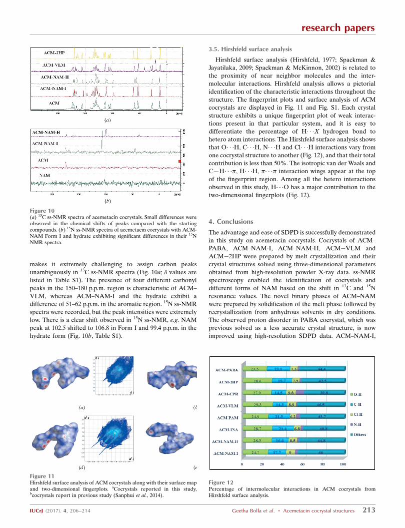
Figure 11Hirshfeld surface analysis of ACM cocrystals along with their surface mapand two-dimensional fingerplots. aCocrystals reported in this study,bcocrystals report in previous study (Sanphui et al., 2014).

Figure 12Percentage of intermolecular interactions in ACM cocrystals fromHirshfeld surface analysis.

Figure 10(a) 13C ss-NMR spectra of acemetacin cocrystals. Small differences wereobserved in the chemical shifts of peaks compared with the startingcompounds. (b) 15N ss-NMR spectra of acemetacin cocrystals with ACM-NAM Form I and hydrate exhibiting significant differences in their 15NNMR spectra.

ACM–NAM-H are confirmed as anhydrate and hydrate forms

by high-resolution powder data. DSC suggests single endo-

therms for both the forms and crystallization experiments for

single crystals resulting in ACM hydrate, showing that SDPD

is the method of choice to confirm the two forms. Hirshfeld

surface analysis exhibits unique fingerplots for different solid

phases and differences in wings and spikes for the novel

phases. The contribution of OH interactions in these crystal

structures is visually depicted in Hirshfeld plots.

Acknowledgements

G. Bolla thanks UGC for a Fellowship. We thank DST-SERB

scheme on Multi-component cocrystals (EMR/2015/002075)

and JC Bose Fellowship (SR/S2/JCB-06/2009) for funding and

University Grants Commission (UPE) and DST-PURSE and

DST-FIST for providing instrumentation facilities.

References

Aitipamula, S. et al. (2012). Cryst. Growth Des. 12, 2147–2152.Almarsson, O. & Zaworotko, M. J. (2004). Chem. Commun. 17, 1889–

1896.Babu, N. J., Sanphui, P. & Nangia, A. (2012). Chem. Asian J. 7, 2274–

2285.Barbour, L. J. (2001). J. Supramol. Chem. 1, 189–191.Bernstein, J., Davis, R. E., Shimoni, L. & Chang, N. L. (1995). Angew.

Chem. Int. Ed. Engl. 34, 1555–1573.Bolla, G. & Nangia, A. (2016). Chem. Commun. 52, 8342–8360.Bolla, G., Sanphui, P. & Nangia, A. (2013). Cryst. Growth Des. 13,

1988–2003.Braga, D., Grepioni, F., Maini, L., Lampronti, G. I., Capucci, D. &

Cuocci, C. (2012). CrystEngComm, 14, 3521–3527.Burger, A. & Lettenbichler, A. (1993). Pharmazie, 48, 262–272.Chavez-Pina, A. E., McKnight, W., Dicay, M., Castaneda-Hernandez,

G. & Wallace, J. L. (2007). Br. J. Pharmacol. 152, 930–938.Chernyshev, V. V. (2001). Russ. Chem. Bull. 50, 2273–2292.Chernyshev, V. V., Shkavrov, S. V., Paseshnichenko, K. A., Puryaeva,

T. P. & Velikodny, Y. A. (2013). Acta Cryst. C69, 263–266.Childs, S. L., Chyall, L. J., Dunlap, J. T., Smolenskaya, V. N., Stahly,

B. C. & Stahly, G. P. (2004). J. Am. Chem. Soc. 126, 13335–13342.David, W. I. F. & Shankland, K. (2008). Acta Cryst. A64, 52–64.Desiraju, G. R. (2013). J. Am. Chem. Soc. 135, 9952–9967.Desiraju, G. R., Vittal, J. & Ramanan, A. (2011). Crystal Engineering:

A Textbook. Singapore: World Scientific.Duggirala, N. K., Perry, M. L., Almarsson, O. & Zaworotko, M. J.

(2016). Chem. Commun. 52, 640–655.Etter, M. C., MacDonald, J. C. & Bernstein, J. (1990). Acta Cryst. B46,

256–262.

Ganesh, M., Jeon, U. J., Ubaidulla, U., Hemalatha, P., Saravanakumar,A., Peng, M. M. & Jang, H. T. (2015). Int. J. Biol. Macromol. 74,310–317.

Gelbrich, T., Haddow, M. F. & Griesser, U. J. (2007). Acta Cryst. C63,o451–o453.

Groom, C. R. & Allen, F. H. (2014). Angew. Chem. Int. Ed. 53, 662–671.

Harris, K. D. M., Tremayne, M., Lightfoot, P. & Bruce, P. G. (1994). J.Am. Chem. Soc. 116, 3543–3547.

Hirshfeld, F. L. (1977). Theor. Chim. Acta, 44, 129–138.Le Bail, A. et al. (2009). Powder Diffr. 24, 255–262.Sanphui, P., Bolla, G., Das, U., Mukherjee, A. K. & Nangia, A. (2013).

CrystEngComm, 15, 34–38.Sanphui, P., Bolla, G., Nangia, A. & Chernyshev, V. (2014). IUCrJ, 1,

136–150.Sanphui, P., Devi, V. K., Clara, D., Malviya, N., Ganguly, S. &

Desiraju, G. R. (2015). Mol. Pharm. 12, 1615–1622.Sanphui, P., Mishra, M. K., Ramamurty, U. & Desiraju, G. R. (2015).

Mol. Pharm. 12, 889–897.Schultheiss, N. & Newman, A. (2009). Cryst. Growth Des. 9, 2950–

2967.Spackman, M. A. & Jayatilaka, D. (2009). CrystEngComm, 11, 19–

32.Spackman, M. A. & McKinnon, J. J. (2002). CrystEngComm, 4, 378–

392.Sun, C. C. & Hou, H. (2008). Cryst. Growth Des. 8, 1575–

1579.Thakuria, R., Delori, A., Jones, W., Lipert, M. P., Roy, L. &

Rodrıguez-Hornedo, N. (2013). Int. J. Pharm. 453, 101–125.Tishmack, P. A., Bugay, D. E. & Byrn, S. R. (2003). J. Pharm. Sci. 92,

441–474.Trask, A. V., Motherwell, W. D. S. & Jones, W. (2006). Int. J. Pharm.

320, 114–123.Ueto, T., Takata, N., Muroyama, N., Nedu, A., Sasaki, A., Tanida, S. &

Terada, K. (2012). Cryst. Growth Des. 12, 485–494.Visser, J. W. (1969). J. Appl. Cryst. 2, 89–95.Werner, P.-E., Eriksson, L. & Westdahl, M. (1985). J. Appl. Cryst. 18,

367–370.Weyna, D. R., Cheney, M. L., Shan, N., Hanna, M., Zaworotko, M. J.,

Sava, V., Song, S. & Sanchez-Ramos, J. R. (2012). Mol. Pharm. 9,2094–2102.

Widdifield, C. M., Cavallo, G., Facey, G. A., Pilati, T., Lin, J.,Metrangolo, P., Resnati, G. & Bryce, D. L. (2013). Chem. Eur. J. 19,11949–11962.

Yoneda, M., Ohkawa, Y., Watanabe, Y., Ogawa, M. & Nagai, H.(1981). Yakugaku Zasshi, 101, 939–944.

Zhukov, S. G., Chernyshev, V., Babaev, E. V., Sonneveld, E. J. &Schenk, H. Z. (2001). Kristallogr. 216, 5–9.

Zlokazov, V. B. (1992). J. Appl. Cryst. 25, 69–72.Zlokazov, V. B. (1995). Comput. Phys. Commun. 85, 415–422.Zlokazov, V. B. & Chernyshev, V. V. (1992). J. Appl. Cryst. 25, 447–

451.

research papers

214 Geetha Bolla et al. � Acemetacin cocrystal structures IUCrJ (2017). 4, 206–214


Recommended